Image
Left Atrial Appendage: Real-Time Three-Dimensional Transthoracic
Echocardiographic Image
Marcelo Luiz Campos Vieira, Gláucia M. P. Tavares, Alexandre Cury, Edgar B. Lira Filho, Adriana Cordovil, Ana C. T.
Rodrigues, Cláudia G. Mônaco, Gustavo Naccarato, Cláudio H. Fischer, Samira S. Morhy
Hospital Israelita Albert Einstein – São Paulo, SP - Brazil
Mailing address: Marcelo Luiz Campos Vieira •
Rua Cardoso de Melo, 463/21 – 04548-002 – São Paulo, SP - Brazil E-mail: [email protected]
Article received on June 20, 2006, Article revised on September 21st, 2006
Accepted on October 21st, 2006
Introduction
The development of real-time three-dimensional transthoracic echocardiography enabled the identification of cardiac structures with baseon new observation planes1-4.
This fact becomes especially relevant when anatomical observation translates into prognostic clinical implications, such as in the identification of images of thrombi in the left atrium or left atrial appendage. In most clinical situations, the investigation of intra-atrial images of thrombi with the use of two-dimensional transthoracic echocardiography does not allow anatomical information reliable enough for a diagnostic definition. Use of multiplane bidimensional transesophagic echocardiography enabled additional diagnostic information for discrimination of intra-atrial masses caused by the use of transducers with a greater ultrasound emission frequency, at a greater proximity to the structure of interest, providing images with a better quality of structural definition. Nevertheless, this semi-invasive echocardiographic modality does not yet allow structural from all anatomic planes of observation (a primary limitation for structural identification based on frontal and transversal cardiac planes, with observation point in atrioventricular, pulmonary, and aortic valve rings).
Real-time three-dimensional transthoracic echocardiography is an advancement in anatomical analysis because it allows
a rotation of heart structures based on three primary planes of structural definition (infero-superior plane, mid-lateral plane, and depth or elevation plane), as well as a structural composition based on the analysis of composite planes or diagonal planes. Image acquisition is made in real-time, and the final image may be obtained by the projection of interest to the clinician or surgeon.
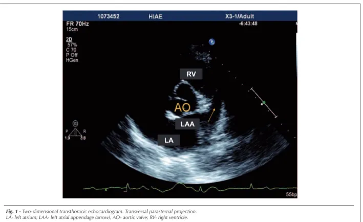
In the case shown, we observe the two-dimensional transthoracic echocardiographic analysis of the left atrial appendage (Figure 1), and the real-time three-dimensional transthoracic image of the left atrial appendage from different observation planes (Figures 2A, 2B and 2C), as from the frontal plane (en face). Currently, transesophagic echocardiography is still the most appropriate echocardiographic technique for visualization of the left atrial appendage and investigation of intracavitary thrombi, as it shows evidence based on analyses with a large number of patients. Further studies are needed for a definition of the diagnostic and prognostic impact of the anatomical findings obtained by the use of real-time three-dimensional transthoracic echocardiography.
Potential Conflict of Interest
No potential conflict of interest relevant to this article was reported.
Key words
Atrial appendage; three-dimensional echocardiography.
Image
Vieira et al
Left atrial appendage: real-time three-dimensional transthoracic echocardiographic image
Arq Bras Cardiol 2007; 88(4) : e92-e95
Fig. 1 - Two-dimensional transthoracic echocardiogram. Transversal parasternal projection. LA- left atrium; LAA- left atrial appendage (arrow); AO- aortic valve; RV- right ventricle.
LA RV
LAA
Fig. 2A - Real-time three-dimensional transthoracic echocardiogram. Longitudinal parasternal projection. Demonstration of extension of left atrial appendage (arrow). Observation of structural definition planes (infero-superior plane, mid-lateral plane, and depth or elevation plane), in yellow, purple, and red.
LAA- left atrial appendage; RV- right ventricle.
LAA RV
Imagem
Fig. 2B - Real-time three-dimensional transthoracic echocardiogram. Longitudinal parasternal projection in transversal rotation. Observation of left atrial appendage (arrow).
LA- left atrium; LAA- left atrial appendage; RV- right ventricle.
RV
LAA
LA
Fig. 2C - Real-time three-dimensional transthoracic echocardiogram. Frontal projection (en face). Demonstration of left atrial appendage (arrow). LA- left atrium; LAA- left atrial appendage; RV- right ventricle.
RV
LAA
LA
Vieira et al Left atrial appendage: real-time three-dimensional transthoracic echocardiographic image
Image
Vieira et al
Left atrial appendage: real-time three-dimensional transthoracic echocardiographic image
Arq Bras Cardiol 2007; 88(4) : e92-e95
References
1. Roelandt JRT, Yao J, Karsprazak JD. Three-dimensional echocardiography. Curr Opin Cardiol. 1998; 13: 386-98.
2. De Simone R, Glombitza G, Vahl CF, Meinzer HP, Hagl S. Three-dimensional Doppler: techniques and clinical applications. Eur Heart J. 1999; 20: 619-27.
3. Kisslo J, Firek B, Takahiro O, Kang DH, Fleishman CE, Stetten G, et al.
Real-time volumetric echocardiography: the technology and the possibilities. Echocardiography. 2000; 17: 773-9.
4. Kwan J, Shiota T, Agler DA, Popovic ZB, Qin JX, Gillinov MA, et al. Geometric differences of the mitral apparatus between ischemic and dilated cardyomyopathy with significant mitral regurgitation: real-time three-dimensional echocardiography study. Circulation. 2003; 107: 1135-40.