Rev Odonto Cienc 2011;26(2):187-190
187
Received: February 14, 2011 Accepted: May 20, 2011
Conflict of Interest Statement: The authors state that there are no financial and personal conflicts of interest that could have inappropriately influenced their work.
Copyright: © 2011 Yadav et al.; licensee EDIPUCRS. This is an Open Access article distributed under the terms of the Creative Commons Attribution-Noncommercial-No Derivative Works 3.0 Unported License.
Case Report
Concommitant occurance of
dens invaginatus
and talon cusp: A case report
Ocorrência simultânea de
dens invaginatus
e cúspide talon:
relato de caso
Monica Yadav a
Meghana S.M b
Sandip R. Kulkarni a
a Department of Oral Pathology and Microbiology, Terna Dental College and Hospital, Nerul, Navi Mumbai, India
b Department of Oral Pathology, Terna Dental College and Hospital, Nerul, Navi Mumbai, India
Correspondence:
Monica Yadav
Department of Oral Pathology and Microbiology, Terna Dental College and Hospital,
Sector no. 22,Nerul, Navi Mumbai-400706 India
E-mail: [email protected]
Abstract
Purpose: Morphological dental anomalies of the maxillary lateral incisors are relatively common. However, their simultaneous occurrence is a relatively rare event. We report a case of dens invaginatus and talon cusp concurrently affecting maxillary lateral incisors. The etiology, pathophysiology, association with other dental anomalies, as well as various treatment modalities of these anomalies are discussed.
Case description: An 18-year-old male patient reported with a complaint of crowding of maxillary front teeth. On intraoral examination, permanent dentition with Class I malocclusion with anterior crowding was observed. Tooth 12 showed a radiopaque invagination from a lingual pit but confined to the crown of the tooth. This invagination was approximately circular with a central core of radiolucency, which was consistent with the diagnosis of a dens invaginatus type I. Tooth 22 showed the talon cusp as a typical inverted cone with enamel and dentine layers and a pulp horn extending only into the base of the cusp. Talon cusp was treated by prophylactic enameloplasty to avoid plaque accumulation, the deep lingual pit was sealed using composite resin and regular clinical and radiographic follow-up was advised. Patient was scheduled for orthodontic treatment to correct crowding of maxillary anterior teeth.
Conclusion: We emphasize the fact that detailed clinical and radiographic examination of the maxillary lateral incisors is vital in avoiding complications.
Key words: Developmental anomalies; dens invaginatus; talon cusp
Resumo
Objetivo: Anomalias morfológicas dentárias dos incisivos laterais superiores são relativamente comuns. No entanto, a sua ocorrência simultânea é um evento relativamente raro. Relatamos um caso de dens invaginatus e cúspide talon simultaneamente afetando incisivos laterais superiores. A etiologia, fisiopatologia, associação com outras anomalias dentárias, bem como várias modalidades de tratamento destas anomalias são discutidas.
Descrição do caso: Um paciente de 18 anos, sexo masculino, relatou com queixa de apinhamento dos dentes anteriores superiores. Ao exame intraoral observou-se dentição permanente com má oclusão Classe I com apinhamento anterior. O dente 12 mostrou uma invaginação radiopaca lingual, confinada à coroa do dente. Esta invaginação era aproximadamente circular, com um núcleo central de radioluscência, que foi compatível com o diagnóstico de dens invaginatus tipo I. O dente 22 apresentou uma cúspide talon como um cone invertido típico com esmalte e dentina em camadas. A cúspide talon foi tratada por ameloplastia profilática para evitar acúmulo de placa, a fissura lingual foi selada com resina composta e foram aconselhados controles clínicos e radiográficos. O paciente foi encaminhado para tratamento ortodôntico para corrigir o apinhamento dos dentes anteriores superiores.
Conclusão: Enfatizamos o fato de que o exame clínico e radiográfico detalhado dos incisivos laterais superiores é vital para evitar complicações.
188
Rev Odonto Cienc 2011;26(2):187-190 Dens invaginatus and talon cuspIntroduction
Abnormalities affecting the teeth are commonly
encountered in the dental ofice. They are best understood
from the developmental point of view, since they occur
during morphogenesis, hence described as developmental
disturbances. Two such disturbances occurring in a rare
combination are:
dens invaginatus
and talon’s cusp.
Dens
invaginatus
(
dens in dente
, dilated composite odontome, etc.)
is a developmental anomaly resulting from an invagination
in the surface of the crown or root before calciication has
occurred (1). The invaginatus may vary from a slightly
accentuated lingual pit to deep foldings reaching to the
apical foramen (1). It can affect both primary and permanent
teeth and its prevalence is reported to be 1.7% – 10% (2).
All the published studies found that maxillary lateral
incisors were the most commonly affected teeth, followed
in descending order by permanent central incisors, canines
and molars (1,2). On the other hand, talon’s cusp, so named
because it resembles an eagle’s talon, is an uncommon
anomaly that occurs as a nodule or tubercle. This cusp is
composed of normal enamel, dentin and varying extension
of pulp tissue. It occurs with a frequency of 0.04-1.05% (3),
with the maxillary lateral incisors showing the maximum
predilection. Clinically it ranges from an enlarged cingulum
to a large well-delineated cusp extending beyond the incisal
edge of tooth. Based on the degree of their formation and
extension, the anomaly can be classiied as – talon,
semi-talon and trace semi-talon. This was later modiied as type 1:
major talon, type 2: minor talon and type 3: trace talon.
Majority of the cases reported in literature indicate talon’s
cusp as an isolated anomaly, however it may be associated
with other somatic (Rubinstein-Taybi syndrome, incontinenti
apigmentiachromians and Ellis-Van Creveld syndrome) and
odontogenic abnormalities (4).
Case Description
An 18-year-old male patient reported with a complaint of
crowding of maxillary anterior teeth. The patient appeared
healthy and of normal physical development for his age.
On intraoral examination, permanent dentition with Class
I malocclusion with anterior crowding was observed.
Interestingly, a deep pit was seen on the lingual surface
of tooth 12 (Fig. 1) and an accessory cusp was seen on the
lingual surface of tooth 22 (Fig. 2). These two developmental
anomalies form the subject of our case report. The oral
mucosa appeared normal with minimal inlammatory
changes.
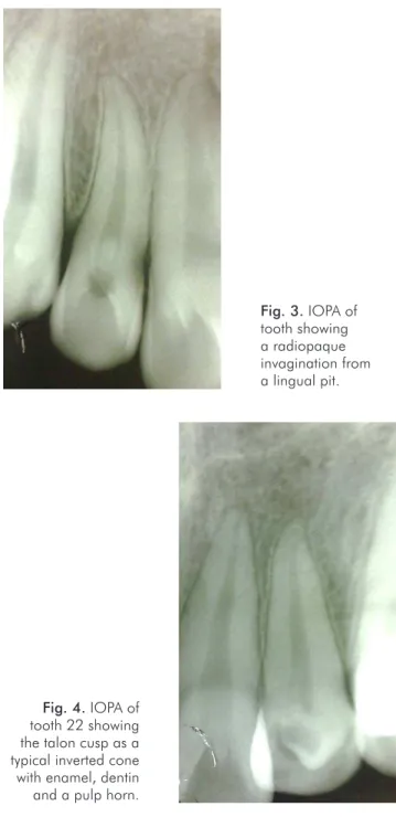
IOPA (intraoral) of tooth 12 showed a radiopaque
invagination from a lingual pit but conined to the crown
of the tooth. This invagination was approximately circular
with a central core of radiolucency, which was consistent
with the diagnosis of a
dens invaginatus
type I (Fig. 3).
On the other hand IOPA (intraoral) of tooth 22 showed
the talon cusp as a typical inverted cone with enamel and
dentine layers and a pulp horn extending only into the base
of the cusp (Fig. 4). Patient was clinically asymptomatic
and neither of these anomalies was associated with caries
or gingivitis and there was no evidence of loss of vitality of
the lateral incisors. Talon’s cusp was treated by prophylactic
enameloplasty to avoid plaque accumulation, the deep
lingual pit was sealed using composite resin and regular
clinical and radiographic follow-up was advised. Patient was
scheduled for orthodontic treatment to correct the crowding
of maxillary anterior teeth.
Fig. 1. Tooth 12 showing a deep pit on the lingual surface (arrow).
Rev Odonto Cienc 2011;26(2):187-190
189
Yadav et al.Discussion
Simultaneous occurrence of multiple dental
abnor-malities is relatively common. Anomalies of the talon’s cusp,
dens invaginatus
, and palato-gingival groove predominantly
affect the maxillary incisor region, which is also the most
frequent site for supernumerary teeth. Individually, the
develop-mental dental abnormalities affecting lateral incisors are well
characterized but their cause(s) remain unknown (5,6). It has
been proposed that the relatively small lateral incisor tooth
germ may be directly affected by forces generated by the
tooth germs of the central incisor and canine, which develop
seven months earlier. Localized pressure on a tooth germ
during morpho-differentiation might cause buckling, with
Fig. 4. IOPA of tooth 22 showing the talon cusp as a typical inverted cone with enamel, dentin and a pulp horn.
Fig. 3. IOPA of tooth showing a radiopaque invagination from a lingual pit.
either outfolding or infolding of the dental lamina. However,
it seems more likely that these malformations are genetically
determined because they are highly reproducible in shape,
show predilection for some racial groups and often occur
together (7). Moyer’s states that the most distal tooth within
each group displays the greatest variability in size, shape
and calciication timing (4). Studies have proved that the
prevalance of talon’s cusp varies considerably between ethnic
groups, ranging from 0.06% in Mexican children to 7.7% in
Indian children. The anomaly occurs with higher incidence
in Mongoloid populations than Caucasians and Negroes (4).
The genetic basis of tooth shape and size is being elucidated
and is very complex but tightly controlled. It seems likely that
these anomalies will turnout to be primarily under genetic
control even though not strongly heritable (7). While these
anomalies may sometimes compromise pulp vitality they are
often asymptomatic incidental indings during routine clinical
or radiographic examination, as in the current case. If this
condition is not recognized early, premature tooth loss may
result from communication with the pulp or predisposition to
caries, resulting in pulp necrosis and periapical pathosis.
Similar associations with other anomalies have been
reported occasionally: talon cusp with mesiodens (8), with
odontomes (9), with
dens invaginatus
, with supernumerary
teeth, with
dens evaginatus
of posterior teeth with palatal
invagination (10), with shovel-shaped incisors, with
congenitally missing teeth and large Carabelli’s cusps (5).
The presence of talon cusp and dens invaginatus in the same
tooth, as here, appears quite rare, if reported. Mader (1981)
and Acs, Pokala and Cozzi (1992) (11) have described
concomitant talon cusp and supernumerary mesiodens.
A possible association between dens invaginatus and
Carabelli’s cusps has also been reported previously (12).
Clinical management of these anomalies varies from
case to case. Treatment of dens invaginatus ranges from
conservative restoration of the opening to endodontic
treatment or extraction (13). Talon cusp may cause occlusal
interference and trap plaque, predisposing to caries,
periodontitis and trauma to the tongue. Attrition may expose
the central pulp horn, so that conservative management,
reduction, coverage and endodontic treatment may all play
a role (5). Also, gradual periodic reduction of the cusp with
luoride as a desensitizing agent along with regular clinical
and radiographic follow up could be beneicial.
The most signiicant clinical concern of
dens invaginatus
is the risk of developing pulpal pathology. The invagination
commonly communicates with the oral cavity, allowing
the entry of irritants and microorganisms directly into the
pulpal tissue. Sometimes ine canals extend between the
invagination and the pulp chamber, resulting in pulpal and
periapical pathology even in the absence of dental caries (14).
Pulpal involvement can occur at a young age, when the root
is immature and not completely formed. In these cases, the
dificulty is to facilitate the apical closure before obturation.
Other reported sequelae of undiagnosed and untreated
invaginated teeth included eruption delay, cysts and internal
190
Rev Odonto Cienc 2011;26(2):187-190 Dens invaginatus and talon cuspConclusions
To summarize, due to the high incidence of developmental
anomalies involving maxillary lateral incisors, these
teeth should be thoroughly investigated to avoid the risk
of developing pulpal and periapical pathologies. The
developmental anomalies thus established should be strictly
monitored from time to time.
References
1. Zengin AZ, Sumer AP, Celenk P. Double dens invaginatus: report of three cases. Eur J Dent 2009;3:67-70.Galindo-Moreno PA, Parra-Vázquez MJ, Sánchez-Fernández E, Avila-Ortiz GA.
2. Maxillary
cyst associated with an invaginated tooth: a case report and literature review. Quintessence Int 2003;34:509-14.
Abbot PV. Labial and palatal “talon cusp” on the same tooth-a case report. Oral Surg Oral 3.
Med Oral Pathol Oral Radiol Endod 1998,85:726-30.
Tiku A, Nadkarni U M, Damle S G. Management of two unusual cases of dens invaginatus 4.
and talon cusp associated with other dental anomalies. J Indian Soc Ped Prev Dent 2004;22:128-33.
Segura JJ, Jimenez-Rubio. A Talon cusp affecting permanent maxillary lateral incisors in 5.
2 family members. Oral Surg Oral Med Oral Pathol Oral Radiol Endod 1999;88:90-2. O’Sullivan EA. Multiple dental anomalies in a young patient: a case report. Int J Paediatr 6.
Dent 2000;10:63-6.
Lorena SC, Oliveira DT, Odell EW. Multiple dental anomalies in the maxillary incisor 7.
region. J Oral Sci 2003;45:47-50.
Davis PJ, Brook AH. The presentation of talon cusp: diagnosis, clinical features, associations 8.
and possible aetiology. Br Dent J 1985;160:84-8.
Natkin E, Pitts DL, Worthington P. A case of talon cusp associated with other odontogenic 9.
abnormalities. J Endod 1983;9:491-5.
Rusmah M R. Talon cusp in Malaysia. Aust Dent J 1991;36:11-4. 10.
Acs G, Pokala P, Cozzi E Shovel incisors, three-rooted molars, talon cusp, and supernumerary 11.
tooth in one patient. Pediatr Dent 1992;14:263-4.
Lewis R, Mountford D, Collins V, Miller J .Palatal invaginations in incisors and the presence 12.
of cusps of Carabelli. J Pedod 1984;8:285-92.
Chaniotis AM, Tzanetakis GN, Kontakiotis EG, Tosios KI.
13. Combined endodontic and
surgical management of a mandibular lateral incisor with a rare type of dens invaginatus. J Endod 2008;34:1255-60.
White SC, Pharoah MJ. Dental anomalies. Oral radiology: Principles and interpretation. 14.
5th ed. New York: Mosby; 2004.
Mupparapu M, Singer SR.