CASE REPORT
Life saving surgery for ruptured pseudo aneurysm of external
iliac artery: case report
Cirurgia para salvamento de vida após ruptura de pseudoaneurisma da artéria ilíaca:
relato de caso
Raafat Shalabi1, Wagih Ouda Ahmed2, Samer Mounir Soliman3, Ahmed Yousof Kandeel4, Khaled Mohammed A. Assiri5
Introdução
Vascular injury secondary to hip surgery is uncommon in that the reported incidence of major vascular injury ater surgical procedures on the hip is only 0.25%1. he
develo-pment of a pseudo aneurysm ater total hip arthroplasty (THA) is an extremely rare complication. Most reported cases are acute in onset and are usually due to direct trauma during the operative procedure1. We reported an unusual
case of ruptured pseudo aneurysm and control of life-thre-atening intra-operative hemorrhage of the external iliac ar-tery in a patient with displaced THA, planned for removal, occurring two years ater the last hip surgery.
Case report
A 68-year-old Saudi male patient was admitted for re-moval of infected total right hip replacement (THR), which was revised in 2000. A chronic hip sinus has developed follo-wing the revision surgery with continuous oozing, which, on occasions, was bloody. However, the culture swabs from the exudates were negative. Gradually, symptoms of pain and decreased hip motion have developed. He noticed right leg swelling prior to his admission to our hospital. He had other systemic medical diseases (diabetes mellitus and hyperten-sion). During examination, he had normal temperature and non-pitting edema in the right leg. Distal pulses were present
Abstract
he incidence of pseudo aneurysm after total hip arthroplasty is extremely rare. he most common mechanism of vascular injury is due to direct trauma during the operative procedure, and the most reported cases are acute in presentation. We reported an unusual case of ruptured pseudo aneurysm and control of life-threatening intra-operative hemorrhage of the external iliac artery in a male patient, 68 years old, with displaced total hip arthroplasty (THA), planned for removal, occurring 2 years after the last hip surgery, in Armed Forces hospital, Southern region, Saudi Arabia. his case highlights the importance of prompt recognition of life-threatening intra-operative hemorrhage to save the patient’s life and the limb.
Keywords: Aneurysm, false; arthroplasty; hemorrhage.
Resumo
A incidência de pseudoaneurisma após a artroplastia total de quadril é extremamente rara. O mecanismo mais comum de lesão vascular deve-se ao trauma direto durante o procedimento cirúrgico, e os casos mais relatados são de apresentação aguda. Relatamos um caso incomum de ruptura de pseudoaneurisma e controle de hemorragia intraoperatória com risco de morte da artéria ilíaca externa em um paciente do sexo masculino, de 68 anos, com artroplastia total do quadril deslocada, planejada para remoção, ocorrendo 2 anos depois da última cirurgia de quadril, no Hospital das Forças Armadas, região sul da Arábia Saudita. Este caso destaca a importância do pronto reconhecimento da hemorragia intraoperatória com risco de morte para salvar a vida e o membro do paciente.
Palavras-chave: Falso aneurisma; artroplastia; hemorragia.
Vascular Surgery Unit, Anaesthesia Department, Radiology and Orthopedic Surgery Department of Armed Forces Hospital, Southern Region, Saudi Arabia. 1 Consultant in Vascular Surgery, Armed Forces Hospitals, Southern Region (AFHSR), Saudi Arabia.
2 Consultant in Anaesthesia and Intensive Care Unit AFHSR, Saudi Arabia; Assistant Professor, Anaesthesia Zagazig University, Zagazig, Egypt. 3 Consultant in Anaesthesia and Intensive Care Unit, AFHSR, Saudi Arabia; Assistant professor, Anaesthesia Ain Shams University, Cairo, Egypt. 4 Professor of Diagnostic Radiology, Mansoura University, Mansoura, Egypt.
5 Specialist in Orthopedics, Germany; Consultant Orthopedic and Spinal Surgery, AFHSR, Saudi Arabia.
No conlicts of interest declared concerning the publication of this article. Received on Feb 18, 2009 Accepted on Jan 8, 2010
Ruptured pseudo aneurysm of external iliac artery - Shalabi R et al. J Vasc Bras 2010, Vol. 9, Nº 4
242
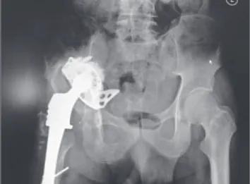
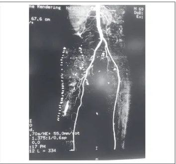
and there were no palpable swellings or bruit in the groin. Blood tests showed 9.5 g/dL of hemoglobin, white blood cell count of 7.7/mm and a normal coagulation screen. A radio-graph of the right hip showed pelvic bone with displacement of the prosthesis out of its cup into the medial pelvis (Figure 1). Doppler ultrasound scan of the right leg showed expan-ded, non-compressible femoral vein, suggestive of deep ve-nous thrombosis (DVT). he venogram conirmed iliofemo-ral DVT. A retrievable inferior vena cava ilter was inserted (Figure 2). he patient was started on low molecular heparin prior to the removal of the THR. hrough a standard lateral approach and ater opening the wound and the trailto take out the prosthetic femur from its socket in the acetabulum, uncontrollable bleeding was encountered. Immediate control of hemorrhage was achieved by packing. In the meantime, the vascular surgeon was contacted and transfusion of blood was started.
Bleeding was noticed from behind the acetabular prosthetic cup. A Foley’s urinary catheter (size 16) was inflated behind the cup and surrounded by gauze pa-cks. The pressure and the pulse rate began to improve and the patient became controlled. The orthopedic sur-geons started to take out the femoral prosthesis, which was toughly adhered to its surrounding. Infra-inguinal exploration of the proximal part of the common femoral artery and a retroperitoneal exploration of the right ex-ternal iliac artery were carried out. No apparent bleeding was seen. The clamping of the external iliac artery was carried out and the acetabular cup was removed from the acetabular space. A false capsule and granulation tissues were seen avulsed from the surroundings. After dissecting circumferentially the external iliac artery and the proximal part of the right common femoral artery, a posterior wall rounded hole was seen. After examining it and the lumen of the artery, there was an intimal dis-section with old subintimal disdis-section of 10-20 mm pro-ximal and distal to the arterial hole. An interposition of 6 mm Dacron graft was carried out by the use of 5/0 no absorbable prolene stitches for anastomosis.
he pulse regained palpable at the right posterior ti-bial and dorsalis pedis arteries. he patient’s postopera-tive course was uneventful until the ith postoperapostopera-tive day. he patient started to complain of lower abdominal pain. Abdominal and pelvic ultrasonography was carried out, which revealed massive retroperitoneal hematoma. Patient’s hemodynamics and hemoglobin level did not change. Computerized tomography with contrast showed a big retroperitoneal hematoma communicating with the la-teral wound of the removed prosthesis (Figure 3). he grat was intact and no signs of abnormality were seen associated
Figure 1 – Right total hip arthroplasty displaced.
Figure 2 – Computerized tomography with contrast showing inferior
Ruptured pseudo aneurysm of external iliac artery - Shalabi R et al. J Vasc Bras 2010, Vol. 9, Nº 4 243
with it (Figure 4). he patient was observed for two weeks until the skin stapler clips were removed. he follow-up was carried out monthly for over six months. he right pedal pulses were well palpable and there were no signs of grat infection or thrombosis.
Discussion
Vascular complications associated with THA are re-markably rare, making diagnosis and treatment of such sequelae extremely challenging for surgeons who are not familiar with their management (as evidenced by the high rate of limb loss, 70%). Pseudo aneurysms are usually asymptomatic and detected incidentally during surgery or radiographic study, unless infection, local compression on neurovascular structures or rupture oc-cur1. The common causes of pseudo aneurysms include
trauma, tumor, infection, vasculitis and inflammation, atherosclerosis, infarction, and various iatrogenic com-plications, such as those from surgery and angiography2.
The mechanism of vascular injury, in most cases, are due to direct trauma during the operative procedure, such as perforation of vessels by retractors, osteotomes, powered reamers, screws, cement or even maneuvers to dislocate hip1.
Injuries causing delayed symptoms are of three types and give rise to symptoms appearing between a few days and several years ater the operation:
a) pain in the hip caused by pressure of a pseudo aneurysm;
b) ischemic symptoms in the afected limb due to impai-red blood low or distal microembolization;
c) severe hemorrhage when extracting a hip prosthesis.
he etiology is either a too large volume of cement with intrapelvic spiculae causing thermal damage or erosion of the artery or an intrapelvic dislocation of the socket with pressure and angulations of the artery3.
Unlike most reported cases, our patient did not deve-loped symptoms of pseudo aneurysm since his last surgery. he sequence of the most likely events began with the screw in the acetablar cup at the time of the last surgery, two years ago. he patient developed iliofemoral DVT from the com-pression of the external iliac artery pseudo aneurysm.
Rengsen et al.1 reported an injury to the external iliac
artery from an acetablar cup, which resulted in formation of a pseudo aneurysm. hey believed that the threaded acetablar cup with sharp cutting lutes might have caused
direct lesion of the arterial wall. Although the acetablar component used in this patient did not have sharp cutting lutes, we believe that the arterial lesions might have been similarly caused by repetitive direct trauma from either the acetablar cup or the implanted femoral head.
Rupture of pseudo aneurysms of iliac artery usually demands immediate surgical repair, but surgery invol-ves high mortality and morbidity risks, especially for debilitated patients in the emergency setting4. In
mana-ging these aneurysms, there is a very high periopera-tive mortality rate (33 to 50% in emergency surgery; 7 to 11% in elective procedures)5,6. A systematic, planned
operative approach is necessary to reduce morbidity and mortality.
Conclusion
Rupture of pseudo aneurysms of iliac artery usually demands immediate surgical repair. Awareness of this rare complication, prompt diagnosis and immediate treatment are key factors in saving the lives of such patients.
his case demonstrated that a pseudo aneurysm can manifest as an acute presentation secondary to direct in-jury during a surgical procedure. It can appear late and be caused by repetitive trauma from arthroplasty components. If a pseudo aneurysm is suspected, an angiogram should be performed, followed by appropriate treatment as soon as possible.
Ruptured pseudo aneurysm of external iliac artery - Shalabi R et al. J Vasc Bras 2010, Vol. 9, Nº 4
244
References
1. Rengsen P, Abbas AA, Choon SK, Tai CC. Pseudoaneurysm of ex-ternal iliac artery following septic loosening of total hip arthroplas-ty. MOJ. 2007;1:42-4.
2. Huang WY, Huang CY, Chen CA, Hsieh CY, Cheng WF. Ruptured pseudoaneurysm of the external iliac artery in an advanced cer-vical cancer patient treated by endovascular covered stent place-ment. J Formos Med Assoc. 2008;107:348-51.
3. Bergqvist D, Carlsson AS, Ericsson BF. Vascular complications after total hip arthroplasty. Acta Orthop Scand. 1983;54:157-63.
4. Sanada J, Matsui O, Arakawa F, et al. Endovascular stent-grafting for infected iliac artery pseudoaneurysms. Cardiovasc Intervent Radiol. 2005;28:83-6.
5. Richardson JW, Greenield LJ. Natural history and management of iliac aneurysms. J Vasc Surg. 1988;8:165-71.
6. Brunkwall J, Hauksson H, Bengtsson H, Bergqvist D, Takolander R, Bergentz SE. Solitary aneurysms of the iliac arterial system: an esti-mate of their frequency of occurrence. J Vasc Surg. 1989;10:381-4.
Correspondence:
Raafat Shalabi, MD Armed Forces Hospital, Southern Region, Saudi Arabia Tel: +966-566874773, +966-7-2500001 ext.: 2552 Fax: +966-7-2571699 E-mail: [email protected]
Author contributions