Victoria Hatzelis1
Thomas Murry2
Descritores
Doenças da laringe/etiologia Doenças da laringe/terapia Cordas vocais Exercícios respiratórios Estudos longitudinais
Keywords
Laryngeal diseases/etiology Laryngeal diseases/therapy Vocal cords Breathing exercises Longitudinal studies
Endereço para correspondência:
Thomas Murry, PhD
Department of Otolaryngology-Head and Neck Surgery
1305 York Avenue, 5th floor, 10021, New York (NY), USA.
E-mail: [email protected]
Received: 11/30/2011
Accepted: 2/6/2012
Conflict of interests: None
(1) Department of Biobehavioral Sciences, Teachers College, Columbia University – New York (NY), United States.
(2) Department of Otolaryngology-Head and Neck Surgery, Weill Cornell Medical College, Cornell University – New York (NY), United States.
manage long-term symptoms
Movimento paradoxal de pregas vocais: reeducação
respiratória no monitoramento de sintomas de longo prazo
ABSTRACT
Paradoxical vocal fold motion (PVFM) is a laryngeal disorder characterized by abnormal adduction of the vocal folds during respiration. It is caused by a multitude of etiologies and their interactions. In the current study, a 24-year-old female patient with a 12-year complaint of shortness of breath was diagnosed with paradoxical vocal fold motion following nasal endoscopy, spirometry testing and detailed case history analysis. She had no history or indication of laryngopharyngeal reflux or chronic cough. She performed respiratory retraining exercises three to four times daily for a period of four weeks, and continued daily exercises for two additional months as needed. After four weeks of treatment, abnormal vocal fold adduction continued to be seen on en-doscopy and the patient was mildly symptomatic. One month post-treatment, there was no abnormal vocal fold adduction and the patient reported rare shortness of breath. At three months post-treatment, there was no abnor-mal vocal fold adduction and the patient no longer reported shortness of breath. She reported norabnor-mal breathing with no symptoms one year later. The results suggest that non-pulmonary related shortness of breath treated with respiratory retraining can effectively eliminate dyspnea in patients with long term breathing difficulties caused by paradoxical vocal fold motion. Resolution may require treatment over an extended period of time.
RESUMO
INTRODUCTION
Paradoxical vocal fold motion (PVFM) is a laryngeal dis-order in which excessive contraction of the adductor muscles causes the vocal folds to involuntarily adduct on inspiration(1) or less commonly, on expiration(2). Common indications of PVFM are shortness of breath and choking sensation(3).Patients may also complain of hoarseness. PVFM has also been associated with chronic cough and reflux disease(4).Although the majority of patients with PVFM are known to have normal pulmonary function, treatment of patients with PVFM often begins with pharmacological management of typical pulmonary symptoms such as cough and shortness of breath. This pharmacological treatment that often includes inhaled steroids usually does not resolve the patient’s symptoms(5).Triggers of PVFM may in-clude one, or a combination of, the following: stress, exercise, extreme temperatures, airway irritants, aromas, odors or food products(4,5).
Concomitant factors for PVFM in young active adults include significant social stressors, previous upper respiratory infection (URI) and a previous history of exercise induced asthma(6,7). While historically it was associated with psychia-tric illness and factitious symptoms, more recent evidence supports that PVFM is associated with multiple etiologies and their interactions, including psychological, physiological and neurogenic factors(2).
Diagnosis is based on case history and the visualization of the abnormal (paradoxical) movement of the vocal folds during flexible laryngoscopic examination and spirometry(3,4,8-10). This paradoxical movement may be seen following an exercise task such as running, or it may occur spontaneously during restful breathing(7).
Spirometry has been reported as an additional investi-gational tool in the diagnosis of PVFM. Spirometry offers information related to both inspiratory and expiratory events. The flow-volume loop pattern in PVFM is characterized by flattening or truncation of the inspiratory limb, compared to a U-shaped pattern in normal subjects(8).
The flow-volume loop is uniquely helpful in the evaluation of upper airway obstruction. It has been found that individuals with a definite diagnosis of PVFM tended to have normal ex-piratory phases with attenuation in the insex-piratory phase of the flow-volume loop, while those diagnosed with asthma presented in an opposite manner; that is, normal inspiratory phase and attenuated expiratory phase(11).
Recently, it has been suggested that the diagnosis of PVFM should include both laryngoscopy and pulmonary function testing, though the sensitivity of the flow-volume loop may be low(12).
Previous investigations have reported a range of incidence of PVFM of between 23-100% when the inspiratory portion of the flow-volume loop shows an abnormal inspiratory pattern in asymptomatic patients with documented PVFM(11).
Diagnosis of PVFM can be difficult when co morbidities such as asthma are present in the patient. However, with use of pulmonary function tests, pulmonary issues can be somewhat isolated from airway issues, especially if testing is done when
patients are not on medications. The likelihood of improving diagnostic specificity increases when proper testing is carried out. Typical symptomatic presentations of PVFM, such as change of voice and stridor, are rarely seen in patients suffering from asthma, or they are reported as incidental to their primary problem of shortness of breath and thus often excluded in the diagnosis(9).
Treatment options for PVFM generally begin with phar-macological management of commonly associated symptoms, including chronic cough and reflux disease. Various types of breathing exercises may be offered in conjunction with phar-macological management or separately. Often, no treatment options are offered secondary to inconclusive diagnosis. Occasionally, corticosteroids are erroneously offered as a treatment without resolution of symptoms.
The current investigation presents a case study of a 24-year--old female patient who initially presented with a history of shortness of breath and choking sensation unrelated to eating or drinking at approximately age 12. After living with the problem and trying various medications over a ten-year period, she was diagnosed with PVFM. The objective of this study was to determine if respiratory retraining exercises alone would eliminate her breathing difficulties.
CLINICAL CASE PRESENTATION
Subject
A 24-year-old female patient who presented with a twelve--year history of periodic shortness of breath and choking sensation was diagnosed with PVFM at a University Medical Center in 2009. Diagnosis was made on the basis of clinical presentation following analysis of detailed case history, spiro-metric testing, and nasal endoscopy, in which airway reduction due to vocal fold adduction was observed by both authors to be greater than 50% on inspiration during quiet breathing. The patient was a non-smoker with no history of psychiatric illness or voice disorders. She reported that episodes of shortness of breath were frequently triggered by periods of life stress and extremely hot and humid temperatures, including while swim-ming indoors. Swimswim-ming or running outdoors was not triggers to shortness of breath.
The patient reported no symptoms nor was she ever diag-nosed with chronic cough or laryngopharyngeal reflux, both commonly associated symptoms of PVFM(4). The presence of laryngopharyngeal reflux was investigated using a self--administered, valid questionnaire, the Reflux Symptom Index (RSI)(13). Of a possible 45 points, the patient obtained a score of zero on the RSI. Further, no clinical signs of reflux such as inflammation or redness were present upon nasal endoscopy.
after-school club. A multitude of examinations were performed, including allergy, immunology, pulmonary function, chemistry and hematology tests; all results were within normal limits and no specific treatments were offered. Table 1 describes the tests performed and the results obtained from her previous medical histories.
Following testing at age 13 and subsequent visits to various physicians, symptoms were ascribed to a combination of a ha-bit and a stress related problem. Through appropriate testing, asthma, allergies and reflux were ruled out. Nonetheless, a corticosteroid inhaler was prescribed by the physician follo-wing persuasion from the patient’s parent. It was ineffective in providing short- or long-term relief of symptoms and its use was eventually discontinued.
Procedures
The Free and Informed Consent Term was signed by the patient, thereby allowing the implementation and dissemina-tion of research results. Following a light nasal decongestant, nasal endoscopy was used to examine vocal fold movement. After insertion and initial acclimatization to the endoscope, the patient breathed quietly for ten seconds through her nose, for
ten seconds through her mouth, and then spoke the sentence “We see three green trees” two times. Observation of parado-xic motion was seen during both quiet breathing periods and following a spoken sentence. The patient then rhythmically tapped her feet for 15-30 seconds, and repeated the sequence of breathing quietly through her nose and mouth for ten secon-ds respectively. Rhythmic foot tapping was used to simulate activity without having to remove and re-insert the endoscope, as PVFM has been shown to be triggered by exercise(11).The paradoxic motion continued following the foot tapping and the patient reported feeling short of breath. The entire procedure was repeated immediately following treatment and one month following treatment.
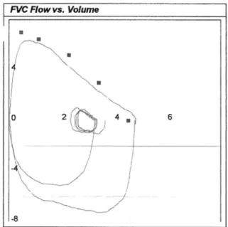
A diagnosis of paradoxical vocal fold motion was given after the video was reviewed. Significant airway closure was observed at rest, following sentence production and during and after the foot tapping. Spirometry was performed at initial diagnosis and at follow-up to objectively examine pulmonary function and airway obstruction. The expiratory phase was normal but a moderately flattened inspiratory loop was obser-ved (Figure 1).
Spirometry measures that were obtained and compared before and after treatment included FEV1/FVC, the proportion of forced vital capacity exhaled in the first second compared to the forced vital capacity and the FIV0.5/FIVC, the proportion of forced inspiratory capacity inhaled during the first 0.5 se-conds compared to the total forced inspiratory capacity. These measures were previously found to be abnormal in patients with PVFM(4,8).
Following diagnosis, the patient began respiratory retraining therapy. The second author (T.M.) conducted all therapy ses-sions and follow-ups. The patient performed the exercises daily for a one-month period, during which time she was seen for therapy once per week. At the end of one month she continued to perform the therapy techniques when she presented with Table 1. Previous medical tests and results conducted prior to diagnosis
of paradoxical vocal fold motion when investigating patient’s dyspnea symptoms
Test Results
Chemistry
Glucose serum fasting Triglycerides
Cholesterol
Cholesterol risk factor Vitamin B12
Ferritin sTSH Hematology Hemoglobin Hematocrit RBC RDW WBC
WNL WNL WNL
Below average risk Not B12 deficient Possibly iron deficient WNL
WNL WNL WNL WNL WNL Pulmonary function
FVC (L)
Percentage of predicted value FEV1/FVC (%)
Post-bronchodilation
3.77 107 90
No significant improvement Allergy/Immunology
Asthma
Mild seasonal allergies to tree pollen, grass, ragweed Negative
Echocardiogram and Color Doppler Normal cardiac structures and function
Normal valvular structures Note: WNL = within normal limits; RBC = red blood cells; RDW = red cell distribu-tion width; WBC = white blood cells; FVC = forced vital capacity; FEV1 = forced expiratory volume in one second
symptoms. She reported that her symptoms resolved entirely after two months. Nasal endoscopy was repeated immediately after the one month of intensive treatment and at one- and three-months post-therapy.
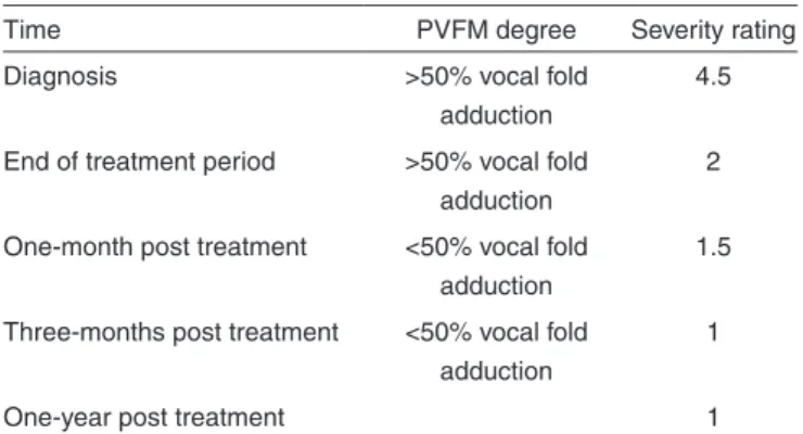
The patient’s symptoms were subjectively tracked using a severity rating scale from 1 to 5, 1 representing no breathing diffi-culty and 5 representing severe breathing diffidiffi-culty. She reported the number of training sessions she completed at each weekly treatment session. She also tracked her symptoms daily from time of initial diagnosis until three months following treatment.
Respiratory retraining exercises were based on the idea of augmenting the involvement of abdominal musculature during respiration, thus decreasing the demands and strain on the oropharyngeal muscles(10,14).The following exercises were performed:
- Rhythmic breathing and walking. The patient walked at a slow pace while purposely relaxing chest and shoulder muscles by swinging the arms gently. She steadily exhaled concurrently with every second right step that she took, extending the abdominal muscles after each breath and consciously focusing on exhalation. She performed forced oral exhalation and passive nasal inhalation. The exhalation to inspiration ratio was approximately 1:3 seconds. To increase breathing resistance and since the patient expe-rienced symptoms during exercise activities, slow walking was augmented to fast walking, followed by jogging and climbing stairs.
- Quiet rhythmic breathing and phonation. Patient force-fully exhaled for a count of one and inhaled for a count of three while sitting with shoulders and chest relaxed. She tapped her hand in order to keep a steady rhythm. Focus was on exhalation and abdominal extension so that passive inhalation could occur. To increase resistance, consonant phonation was added to exhalation phase for increasing lengths of time.
- Pulsed exhalation.Patient sniffed in rapidly through the nose and produced the sequence “ha” on a pulse of air. The sequence was produced with purposeful extension of abdominal muscles for as long as possible, followed by another nasal sniff.
- Abdominal focus in supine. Patient lay in supine position and placed a small book on her stomach. She consciously raised the book on inhalation and lowered the book on exhalation. After the breathing pattern was established, the book was removed and the patient continued abdominally-focused breathing. To increase resistance, the patient breathed through a straw while performing the exercise.
- Panting.Patient stood upright and took rapid, shallow inhalations mimicking panting. She focused on minimizing shoulder and upper body movement. She performed the exercise orally for 15 seconds, nasally for 15 seconds, and rested for 15 seconds, repeating the cycle four to five times. - Abdominal thrust. Patient produced a quick pulse of air
through the mouth for one second and slowly inhaled through the nose for three seconds. Her hands were placed on her abdomen initially to retain the focus on abdominal breathing.
Exercises were performed three to four times daily to in-crease motor learning based on the principle of neuroplasticity of intensity, which posits that brain plasticity requires high intensity training(15).As this was a long-standing problem for the patient, optimizing motor learning was an especially important consideration in the treatment procedures.
The patient participated in four sessions of respiratory retraining with the second author (T.M.) as described in the previous section. The duration of treatment was four weeks. Follow-up was conducted immediately following therapy, and one- and three-months post-therapy.
Results
Table 2 shows the results of the severity ratings. The patient’s symptoms were subjectively rated daily using a scale of 1 to 5. At initial diagnosis, the patient rated her symptoms at a severity of 4.5. She continued to track her symptoms until the last follow-up visit.
Immediately following treatment, airway closure was observed during nasal endoscopy during quiet breathing. The patient reported to be mildly symptomatic, providing a subjec-tive severity rating of 2. She continued to perform respiratory retraining exercises to manage her symptoms.
One month post-treatment, there was no paradoxical vocal fold motion seen on endoscopy. The patient reported to occasio-nally present with very mild symptoms, providing a subjective rating of 1.5. She continued the respiratory retraining therapy exercises when symptomatic.
Three months post-treatment, there was no vocal fold adductory motion was seen on endoscopy. The patient reported that she had not experienced any symptoms of either shortness of breath or choking sensation for over a month, providing a subjective rating of 1. She no longer continued respiratory retraining therapy as she was no longer symptomatic.
One year post-treatment, the patient communicated with second author (T.M.) that she had not presented with any PVFM symptoms since their resolution the previous year and she rated her symptoms as a 1.
Spirometric data before and immediately after treatment are Table 2. Severity of dyspnea symptoms associated with paradoxical vocal fold motion using a subjective rating between 1 (minimum) and 5 (maximum)
Time PVFM degree Severity rating Diagnosis >50% vocal fold
adduction
4.5
End of treatment period >50% vocal fold adduction
2
One-month post treatment <50% vocal fold adduction
1.5
Three-months post treatment <50% vocal fold adduction
1
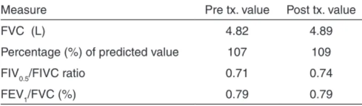
shown in Table 3. The data suggest an above normal forced vital capacity (FVC) for her age and weight. Of note is the FIV0.5/ FIVC ratio which showed a small positive improvement. The FEV1/FVC remained stable.
DISCUSSION
As evidenced by this case study, paradoxical vocal fold motion is an elusive diagnosis that can be easily overlooked, mis-diagnosed, or otherwise treated inappropriately. The multitude of symptoms, etiologies and their interactions associated with PVFM, including shortness of breath, chronic cough and reflux all exacerbate the difficulty of properly diagnosing the condition.
In cases in which the primary symptoms of PVFM are chro-nic cough and reflux, management is focused on elimination of reflux symptoms using medication such as proton pump inhibitors (PPIs)(4) followed by respiratory retraining. However, as in the current case of PVFM without chronic cough or reflux, a program of multiple daily exercises focused on respiratory retraining alone was sufficient to completely eradicate a 12-year history of symptoms of dyspnea.
The patient continued to report mild symptoms of dyspnea one-month after the initial treatment program even though vocal fold adduction was not observed on endoscopy. The reasons for this are unclear, but it is possible that the long-standing history of the problem resulted in a psychological component of inducing symptoms in moments of stress, for example, reverting to previous breathing patterns. As the patient conti-nued to practice the motor patterns learned in the one month of intensive treatment, she was able to entirely eliminate her breathing difficulties.
The inspiratory flow rate and ratios of inspiratory flow to vital capacity were not typical of data reported in previous studies(9,11). In this case study, the inspiratory flow loop was nearly normal and showed only minor flattening of the inspi-ratory segment. A possible explanation for this may be related to the subject who was an athlete and continued to be athletic has learned to control her inspiration when doing forceful tasks such as running or following the instructions during spirometry. Alternatively, as a result of numerous flow loop spirometry tests, the subject may have learned to control the inspiration during the test. Moreover, while many PVFM patients show a flattened inspiratory pattern with an abnormal FIV0.5/FIVC ratio, it has been reported in one study that as few as 28% of the PVFM patients had a truncated inspiratory flow loop(8).
The length of time between the patient’s initial evaluation of symptoms and actual diagnosis raises questions as to the general knowledge of PVFM in the greater medical community. Clearly, the use of scales such as the RSI, a thorough case history, proper nasal endoscopy and spirometry must be considered when the patient presents with episodic shortness of breath.
This case study of the patient’s non-diagnosis for greater than ten years serves as evidence that PVFM can and does get overlooked and/or misdiagnosed. Healthcare professionals must take responsibility to rule out other conditions before writing off a complaint as being only stress-related, as was the initial diagnosis at the first visit when the subject was 13 years old. Further, ineffective medication coupled with the inability to control one’s breathing can lead an affected psyche plagued by emotional distress and stress, as has been documented in the case of athletes(11).
PVFM has historically been associated with psychogenic disorders and factitious symptoms(3,9). It continues to pose challenges both in terms of diagnosis and treatment. Multiple possible contributions to a problem require careful diagnosis and treatment planning, as each case of PVFM is unique and deserves thorough consideration.
FINAL COMMENTS
Paradoxical vocal fold movement is a laryngeal disorder characterized by abnormal adduction of the vocal folds during respiration. It is caused by a multitude of etiologies and their interactions, and it is manifested by different symptoms, but primarily shortness of breath. The current case study presented a 24-year-old female patient with twelve year history of short-ness of breath and choking sensation without chronic cough or reflux that was eventually diagnosed as PVFM. We investigated whether her symptoms could be treated solely by an intense respiratory retraining program since no history of asthma or reflux was present. Both subjective and objective improvements were documented following a one month course of therapy, with complete resolution of symptoms and abnormal vocal fold movement by the three month follow-up visit.
REFERENCES
1. Cukier-Blaj S, Bewley A, Aviv JE, Murry T. Paradoxical vocal fold motion: a sensory-motor laryngeal disorder. Laryngoscope. 2008;118(2):367-70.
2. Hicks M, Brugman SM, Katail R. Vocal cord dysfunction/paradoxical vocal fold motion. Prim Care. 2008;35(1):81-103.
3. Rogers JH, Stell PM. Paradoxical movement of the vocal cords as a cause of stridor. J Laryngol Otol. 1978;92(2):157-8.
4. Murry T, Tabaee A, Aviv JE. Respiratory retraining of refractory cough and laryngopharyngeal reflux in patients with paradoxical vocal fold motion disorder. Laryngoscope. 2004;114(8):1341-5.
5. Sandage MJ, Zelazny SK. Paradoxical vocal fold motion in children and adolescents. Lang Speech Hear Serv Sch. 2004;35(4):353-62. 6. Powell DM, Karanfilov BI, Beechler KB, Treole K, Trudeau MD, Forrest
LA. Paradoxical vocal cord dysfunction in juveniles. Arch Otolaryngol Head Neck Surg. 2000;126(1):29-34.
7. Treole K, Trudeau MD, Forrest LA. Endoscopic and stroboscopic description of adults with paradoxical vocal fold dysfunction. J Voice. 1999;13(1):143-52.
Table 3. Spirometric data before and after respiratory retraining therapy
Measure Pre tx. value Post tx. value
FVC (L) 4.82 4.89
Percentage (%) of predicted value 107 109 FIV0.5/FIVC ratio 0.71 0.74 FEV1/FVC (%) 0.79 0.79
Change in FIV0.5/FIVC ratio = 0.03
8. Morris MJ, Deal LE, Bean DR, Grbach VX, Morgan JA. Vocal cord dysfunction in patients with exertional dyspnea. Chest. 1999;116(6):1676–82.
9. Vertigan AE, Gibson P, Theodoros DG, Winkworth AL. A review of voice and upper airway function in chronic cough and paradoxical vocal fold motion. Curr Opin Allergy Clin Immunol. 2007;7(1): 37-42.
10. Christopher KL, Wood, RP 2nd, Eckert RC, Blager FB, Raney RA, Souhrada JF. Vocal-cord dysfunction presenting as asthma. N Engl J Med. 1983;308(26):1566-70.
11. Newsham KR, Klaben BJ, Miller VJ, Saunders JE. Paradoxical vocal-cord dysfunction: management in athletes. J Athl Train. 2002;37(3):325-8.
12. Christopher, KL. Morris, MJ. Vocal cord dysfunction, paradoxic vocal fold motion or laryngomalacia? Our understanding requires an interdisciplinary approach. Otolaryngol Clin North Am. 2010;43(1):43-66.
13. Belafsky PC, Postma GN, Koufman JA. Validity and reliability of the reflux symptom index (RSI). J Voice. 2002;16(2):274-7.
14. Murry T, Sapienza C. The role of voice therapy in the management of paradoxical vocal fold motion, chronic cough, and laryngospasm. Otolaryngol Clin North Am. 2010;43(1):73-83, viii-ix.