Article
J. Braz. Chem. Soc., Vol. 27, No. 3, 510-514, 2016. Printed in Brazil - ©2016 Sociedade Brasileira de Química 0103 - 5053 $6.00+0.00
A
*e-mail: [email protected]
Meroterpenoid Hydroquinones from
Cordia globosa
Ana Karine O. Silva,a André Luis L. de Oliveira,a Francisco das Chagas L. Pinto,a Karisia S. B. de Lima,a Raimundo Braz-Filho,b Edilberto R. Silveiraa and Otilia Deusdênia L. Pessoa*,a
aDepartamento de Química Orgânica e Inorgânica, Centro de Ciências,
Universidade Federal do Ceará, CP 12.200, 60021-940 Fortaleza-CE, Brazil
bDepartamento de Química, Universidade Estadual do Norte Fluminense Darcy Ribeiro,
28013-602 Campos dos Goytacazes-RJ, Brazil
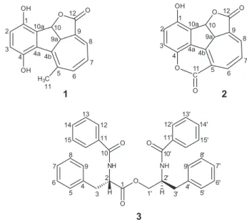
Two new meroterpenoid hydroquinones, rel-(4bE,6Z,8E,9aS,10S )-1,4-dihydroxy-9a,10-dihydro-10,12-epoxy-5-methylbenzo[a]azulen-12-one and rel-(4bZ,6Z,8E,9aS,10S )-1-hydroxy-9a,10-dihydro-4,11:10,12-diepoxy-benzo[a]azulen-11,12-dione, along with the known peptide derivative (S)-N-benzoylphenylalanine-(S)-2-benzamide-3-phenylpropyl ester, were isolated from the roots of Cordia globosa. Their structures were determined by 1D and 2D nuclear magnetic resonance (NMR) spectrometry, Fourier transform infrared (FTIR) spectroscopy and high resolution atmospheric pressure chemical ionization mass spectrometry (HRAPCIMS) data analysis. The new compounds were tested against three human cancer cell lines (colon adenocarcinoma, ovarian carcinoma and glioblastoma), but none of them exhibited any activity.
Keywords: Cordia globosa, Boraginaceae, hydroquinones
Introduction
The Cordia genus (Boraginaceae) comprises approximately 300 species widespread worldwide,1 many of
which used in traditional medicine for different purposes as cicatrizing, anti-inflammatory, anthelmintic, antimalarial, diuretic and to treat urinary infections.2-4 Cordia has
proved to be a prolific source of meroterpenoid quinones, chromenes, hydroquinones and hydrochromenes.5-9
Previously, we have investigated some plants belonging to the genus Cordia, including C. globosa and evaluated the antiproliferative properties of the isolated terpenoid quinones.10-12 In the present work the EtOH extract from
roots of C. globosa, an annual and aromatic shrub native to the northeast of Brazil, was investigated, which led to the isolation and characterization of two new terpenoid hydroquinones (1 and 2), and a known peptide derivative (3) (Figure 1).
Experimental
General experimental procedures
The Fourier transform infrared (FTIR) spectra were
recorded on a Perkin-Elmer spectrum 100 equipped with a universal attenuated total reflectance (UATR) accessory. Optical rotations were measured on a Perkin-Elmer 341 digital polarimeter. One-dimensional [1H, 13C, distortionless enhancement by polarization transfer
(DEPT)] and two-dimensional nuclear magnetic resonance (NMR) experiments [correlation spectroscopy (COSY), heteronuclear single quantum coherence (HSQC),
O
NH NH
O
O O
O O
C O OH
2
3 1
4
10
4b 12
8 9a
O
9
11 6 5 4a 10a
7
O O
H3C OH OH
2
3 1
4
10
4b 12
8 9a
4a 10a
5 6
7 9
11
1 2
1 2
3 4 5 6 7
8
9 10 11 12 13
14
15
1' 2'
3' 4'
5' 6' 7' 8'
9' 10' 11' 12'
13'
14'
15'
3
H H
heteronuclear multiple-bond correlation (HMBC) and nuclear Overhauser effect spectroscopy (NOESY)] were recorded on a Bruker DRX-500 spectrometer operating at 500 MHz for 1H and 125 MHz for 13C, using standard
pulse sequences supplied by the manufacturer. The high resolution mass spectrometry (HRMS) using atmospheric pressure chemical ionization (HRAPCIMS) was performed on a liquid chromatography-mass spectrometry ion trap and time-of-flight (LCMS-IT-TOF, Shimadzu) spectrometer. The positive ion mass spectra were recorded in the m/z 200-700 range, using a potential of 4.0 kV on the capillary and He as the collision gas. The high performance liquid chromatography (HPLC) analysis was carried out using an ultra-fast liquid chromatography (UFLC, Shimadzu) system equipped with a SPD-M20A diode array UV-Vis detector and a Phenomenex C-18 column, 5 µm (4.6 × 250 mm2).
The mobile phase consisted of H2O (trifluoroacetic acid
(TFA) 0.2% v/v) and MeCN with a 4.5 mL min-1 flow
rate, oven temperature of 40 oC and the chromatograms
were monitored at 210-350 nm. Low performance liquid chromatography was carried out in glass columns packed with silica gel 60 (70-230 mesh, Vetec or 230-400 mesh, Merck). Thin layer chromatography (TLC) was performed on silica gel precoated aluminum sheets (kieselgel 60 F254,
0.20 mm, Merck). Fractions and pure compounds were monitored by TLC, and the spots visualized by heating (at ca. 100 °C) the plates sprayed with a vanillin/perchloric acid/EtOH solution.
Plant material
Roots of C. globosa were collected at Pico Alto, located at an approximate altitude of 1000 m, in Guaramiranga County, Ceará State, northeast of Brazil. The plant material was identified by PhD Maria Iracema B. Loiola, botanist of the Departamento de Biologia, Universidade Federal do Ceará (UFC). A voucher specimen (No. 39.851) has been deposited at the Herbário Prisco Bezerra, UFC.
Extraction and isolation
The air-dried and powdered roots (7.5 kg) of C. globosa were extracted with hexane (3 × 10 L) followed by EtOH (3 × 10 L), at room temperature for 24 h, and the resulting solutions were concentrated under reduced pressure to yield 13.9 g (0.001%) and 67.8 g (0.001%) of the hexane and EtOH extracts, respectively. The EtOH extract (67.0 g) was fractionated over silica gel by elution with CH2Cl2,
followed by EtOAc, to yield two main fractions weighting 24.0 and 9.0 g, respectively. The CH2Cl2 fraction (24.0 g)
was subjected to a silica gel column chromatography using
hexane-EtOAc (2:8, 4:6, 6:4 and 8:2, v/v) and EtOAc as eluents, providing 28 fractions, which were monitored by TLC and then pooled to 8 subfractions. Subfraction 23-27 (hexane-EtOAc 8:2, 1.9 g) was subjected to repeated fractionation over silica gel eluted with hexane-EtOAc, to yield a main fraction of 129.0 mg [hexane-EtOAc (4:6, v/v)]. This material was subjected to semi-preparative HPLC using H2O (TFA 0.2% v/v)-MeCN 6.5:3.5 to yield
the pure compounds 1 (6.0 mg, tR 8.7 min) and 2 (8.0 mg,
tR 15.4 min). The EtOAc fraction (3.4 g) was subjected
to flash chromatography using an isocratic solution of CH2Cl2-EtOAc 8:2 (v/v) to yield 95 subfractions of 8 mL.
Sub-fraction 56-72 (80.0 mg) was further purified by HPLC using the mobile phase H2O (TFA 0.2% v/v)-MeCN 6:4 to
afford compound 3 (5.5 mg, tR 12.8 min).
rel-(4bE,6Z,8E,9aS,10S
)-1,4-Dihydroxy-9a,10-dihydro-10,12-epoxy-5-methylbenzo[a]azulen-12-one (1)
Yellow powder; m.p. 197-203 °C; [α]D20 –195.3° (c 0.01,
MeOH); IR (ATR) νmax / cm-1 3317, 1718, 1684, 1639,
1215, 1260; HRAPCIMS calcd. for C16H13O4 [M + H]+:
269.0808; found: 269.0808; 1H and 13C NMR spectral
data, see Table 1.
rel-(4bZ,6Z,8E,9aS,10S
)-1-Hydroxy-9a,10-dihydro-4,11:10,12-diepoxy-benzo[a]azulen-11,12-dione (2)
Yellow powder; m.p. 200-205 °C; [α]D20 –129.3° (c 0.9,
EtOAc); IR (ATR) νmax / cm-1 3466, 1750, 1628, 1465,
1188, 1209; HRAPCIMS calcd. for C16H9O5 [M + H]+:
281.0444; found: 281.0469; 1H and 13C NMR spectral
data, see Table 1.
Cytotoxicity evaluation
Cytotoxicity was evaluated against three human cancer cell lines provided by the National Cancer Institute (Bethesda, MD, USA): colon adenocarcinoma (HCT-116), ovarian carcinoma (OVCAR-8) and glioblastoma (SF-295). Cells were maintained in Roswell Park Memorial Institute (RPMI) 1640 medium supplemented with 10% (v/v) fetal bovine serum, 2 mmol L-1 glutamine, 100 U mL-1
penicillin, 100 µg mL-1 streptomycin at 37 °C under a 5%
CO2 atmosphere. For all experiments, cells were plated
in 96-well plates (105 cells per well for adherent cells or
0.3 × 10⁵ cells per well for suspended cells in 100 µL of
medium). After 24 h, all the compounds (0.048-5.0 µg mL-1)
of DMSO. Tumor cell growth was quantified by the ability of living cells to reduce the yellow dye 3-(4,5-dimethyl-2-thiazolyl)-2,5-diphenyl-2H-tetrazoliumbromide (MTT) to a purple formazan product as previously described.13
At the end of the incubation, the plates were centrifuged and the medium was replaced with fresh medium (150 µL) containing MTT (0.5 mg mL-1). Three hours later, the plates
were centrifuged, the MTT formazan product was dissolved in 150 µL DMSO, and the absorbance was measured using a multiplate reader (Spectra Count, Packard). The drug effect was quantified as the percentage of the control absorbance of the reduced dye at 550 nm. The concentration values that inhibit growth in 50% (IC50) were calculated, along with the
respective 95% of confidence interval (CI), by non-linear regression using the software GraphPad Prism 5.0.
Results and Discussion
Compound 1, a yellow powder, showed IR absorption bands for hydroxyl groups (3317 cm-1), conjugated carboxyl
of γ-lactone moieties (1718 cm-1), carbon-carbon double
bonds (1684 and 1639 cm-1) and carbon-oxygen bonds
(1215-1260 cm-1). The molecular formula of C
16H12O4 (11
degrees of unsaturation) was determined by HRAPCIMS through the molecule protonated peak [M + H]+ at m/z
269.0808 (calcd. m/z 269.0808). The 1H NMR spectrum
(Table 1) exhibited signals for an aromatic ring at dH 6.85 (d,
J 8.5 Hz, H-2) and 6.82 (d, J 8.5 Hz, H-3), indicating an AB system similar to those of a 1,4-hydroquinone moiety. In addition, signals at dH 7.03 (dd, J 5.8 and 1.6 Hz, H-8), 7.02 (d, J 11.4 Hz, H-6) and 6.77 (dd, J 11.4 and 5.8 Hz, H-7) were related to a coupling system of olefinic protons, while the signals at dH 6.17 (d, J 8.0 Hz, H-10) and 2.96 (d, J 8.0 Hz, H-9a) were associated with methines, one of which corresponding to an oxymethine proton. Finally, a singlet at dH 2.48 (s, Me-11), was compatible with a vinyl methyl. Besides the vicinal correlations for the protons H-2/H-3, H-6/H-7, and H-9a/H-10, the COSY spectrum exhibited the allylic coupling for H-9a and H-8, as well as the homoallylic coupling of H-9a and the Me-11. The 13C NMR
spectrum (Table 1) displayed signals for 16 carbon atoms, 13 of which corresponding to sp2 hybridized carbons. The
DEPT spectrum revealed a methyl group at dC 23.6 (C-11), an oxymethine at dC 79.8 (C-10) and another methine at
dC 47.7 (C-9a), in addition to five monohydrogenated sp2
carbon atoms at dC 118.8-144.2. Comparison of DEPT with 13C NMR spectra revealed eight non-hydrogenated
carbon atoms, one characteristic of a γ-lactone carboxyl
at dC 167.3 (C-12), as well as the signals at dC 149.4 (C-1) and 147.6 (C-4) related to the oxygenated carbons of the 1,4-hydroquinone moiety. In addition, signals for three olefinic double bonds were observed, which, after COSY and HMBC analyses (Figure 2), were shown to make part of an extensive conjugated system involving the lactone
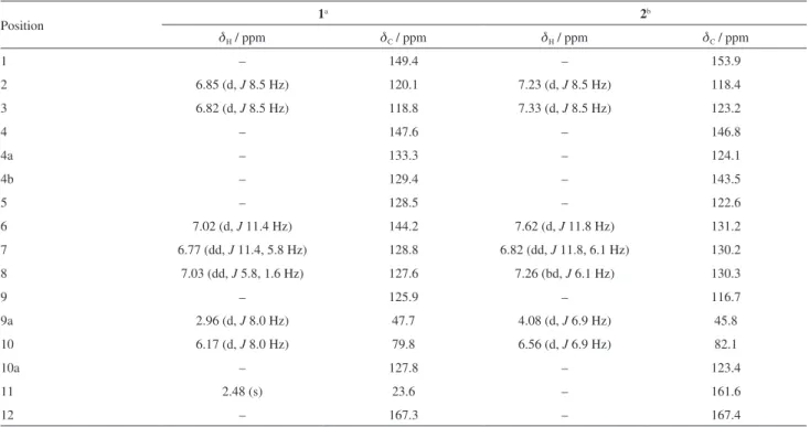
Table 1.1H and 13C NMR data for compounds 1 and 2
Position 1
a 2b
dH / ppm dC / ppm dH / ppm dC / ppm
1 – 149.4 – 153.9
2 6.85 (d, J 8.5 Hz) 120.1 7.23 (d, J 8.5 Hz) 118.4
3 6.82 (d, J 8.5 Hz) 118.8 7.33 (d, J 8.5 Hz) 123.2
4 – 147.6 – 146.8
4a – 133.3 – 124.1
4b – 129.4 – 143.5
5 – 128.5 – 122.6
6 7.02 (d, J 11.4 Hz) 144.2 7.62 (d, J 11.8 Hz) 131.2
7 6.77 (dd, J 11.4, 5.8 Hz) 128.8 6.82 (dd, J 11.8, 6.1 Hz) 130.2
8 7.03 (dd, J 5.8, 1.6 Hz) 127.6 7.26 (bd, J 6.1 Hz) 130.3
9 – 125.9 – 116.7
9a 2.96 (d, J 8.0 Hz) 47.7 4.08 (d, J 6.9 Hz) 45.8
10 6.17 (d, J 8.0 Hz) 79.8 6.56 (d, J 6.9 Hz) 82.1
10a – 127.8 – 123.4
11 2.48 (s) 23.6 – 161.6
12 – 167.3 – 167.4
a500/125 MHz, acetone-d
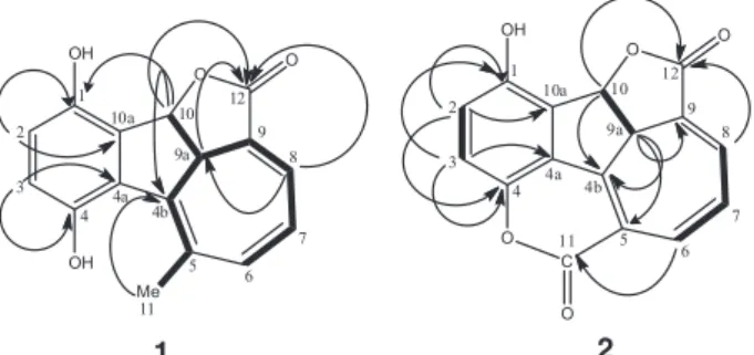
carboxyl and the phenyl moiety. The long range correlations displayed by the oxymethine proton at dH 6.17 (d, J 8.0 Hz, H-10) with the carbon atoms at dC 149.4 (C-1), 127.8 (C-10a) and 129.4 (C-4b), were fundamental to assign the structure of compound 1 as a 1,4-hydroquinone bearing a monoterpene side chain constituted of a γ-lactone fused
to a seven members ring. Unfortunately, the NOESY spectrum (Supplementary Information Figure S8) was not decisive to help defining the relative stereochemistry of 1. The only undoubtful NOE observed was that of CH3-11
with H-6, whose cross peaks have not shown-up on the COSY spectrum. All the other observed cross peaks can either be assigned to COSY breakthrough or chemical exchange. However, the C-9a and C-10 stereocenters were proposed to be trans, in agreement with the corresponding protons coupling constant of 8.0 Hz. Based on the above mentioned data, the structure of 1 was established as rel-(4bE,6Z,8E,9aS,10S )-1,4-dihydroxy-9a,10-dihydro-10,12-epoxy-5-methylbenzo[a]azulen-12-one.
Compound 2 was also isolated as a yellow powder. Its FTIR spectrum showed absorption bands for hydroxyl groups (3466 cm-1), conjugated carboxyl groups (1750 cm-1),
carbon-carbon double bonds (1628 and 1465 cm-1)
and carbon-oxygen bonds (1188 and 1209 cm-1). The
molecular formula of C16H8O5 (13 degrees of unsaturation)
was determined by HRAPCIMS analysis through the molecule protonated peak [M + H]+at m/z 281.0469 (calcd. m/z 281.0444). Despite the 1H NMR spectrum of 2 (Figure 1)
being run in a different solvent (C5D5N) than that used for 1 ((CD3)2CO), it showed the same number of protons and
splitting pattern, except for the disappearance of methyl group, revealing the same backbone structure of 1.
The 13C and DEPT NMR spectra of 2 were also
similar to those of 1 (Table 1). The main difference was the appearance of an additional d-lactone carboxyl group at dC 161.6 (C-11) in compound 2, in substitution of the Me-11 of 1. The HMBC correlation of the proton signal at
dH 7.62 (H-6) with the carboxyl at dC 161.6 supported the lactonization between C-4 and C-5 (Figure 2). Additional
long range correlations, depicted in Figure 2, corroborated the structure of 2.The structures of compounds 1 and 2 show a high degree of similarity, and one could then speculate on the biogenetic formation of 2 simply by the oxidation of Me-11 of 1 to the correspondent carboxyl acid followed by an intramolecular nucleophilic substitution reaction yielding the d-lactone moiety. Thus, the structure of 2 was established as rel-(4bZ,6Z,8E,9aS,10S )-1-hydroxy-9a,10-dihydro-4,11:10,12-diepoxy-benzo[a]azulen-11,12-dione. Additionally to the new compounds, the peptide
(S)-N-benzoylphenylalanine-(S
)-2-benzamide-3-phenylpropyl ester (3) was also isolated, currently designated as asperphenamate,14 anabellamide15 or
auranamide16 (Figure 1). This is the first report on the
occurrence of this compound in Cordia spp.
Compounds 1-3 were tested in vitro for their antiproliferative effects against cancer cell lines HCT-116, OVCAR-8 and SF-295, however they didn’t show cytotoxic activity (IC50 > 5.0 µg mL-1).
Conclusions
In this work two new meroterpenoid hydroquinones (1 and 2) were isolated from the EtOH extract of roots of Cordia globosa, in addition to a peptide derivative (3) not yet reported for this genus. Compounds 1 and 2
are 1,4-hydroquinones fused to a monoterpene moiety, a structural feature that is frequently found in Cordia species, particularly in roots.
Supplementary Information
Supplementary information (1H and 13C NMR, COSY,
HSQC, HMBC, NOESY, HRMS and FTIR spectra) is available free of charge at http://jbcs.sbq.org.br as PDF file.
Acknowledgements
The authors would like to thank the Brazilian agencies CNPq, CAPES and FUNCAP for the financial support.
References
1. Al-Musayeib, N.; Perveen, S.; Fatima, I.; Nasir, M.; Hussain, A.;
Molecules2011, 16, 10214.
2. Sertié, J. A. A.; Basile, A. C.; Panizza, S.; Matida, A. K.; Zelnik, R.; Planta Med.1990, 56, 36.
3. Marston, A.; Zagorski, M. G.; Hostettmann, K.; Helv. Chim. Acta1988, 71, 210.
4. Tiwari, R. D.; Srivastava, K. C.; Shukla, S.; Bajpai, R. K.; Planta Med.1967, 15, 144.
O O
C O OH
2 3
1 4
10 4b
12 8 9a
O
9 11
6 5 4a 10a
7
O O
Me OH OH
2 3
1 4
10 4b
12 8 9a 4a 10a
5 6
7 9 11
1 2
Figure 2. Selected COSY (H H) and HMBC (H→C) correlations
5. Bieber, L. W.; Messana, I.; Lins, S. C. N.; Da Silva-Filho, A. A.; Chiappeta, A. A.; De-Mello, J. F.; Phytochemistry 1990, 29, 1955.
6. Diniz, J. C.; Viana, F. A.; Oliveira, O. F.; Maciel, M. A. M.; Torres, M. C. M.; Braz-Filho, R.; Silveira, E. R.; Pessoa, O. D. L.; Magn. Reson. Chem. 2008, 47, 190.
7. Moir, M.; Thomson, R. H.; J. Chem. Soc.1973, 1, 1352. 8. Manners, G. D.; Jurd, L.; J. Chem. Soc.1977, 4, 405. 9. Dettrakul, S.; Surerum, S.; Rajviroongit, S.; Kittakoop, P.;
J. Nat. Prod. 2009, 72, 861.
10. De Menezes, J. E. S. A.; Lemos, T. L. G.; Pessoa, O. D. L.; Braz-Filho, R; Montenegro, R. C.; Wilke, D. V.; Costa-Lotufo, L. V.; Pessoa, C.; De Moraes, M. O.; Silveira, E. R.; Planta Med.2005, 71, 54.
11. Freitas, H. P. S.; Maia, A. I. V.; Silveira, E. R.; Marinho-Filho, J. D. B.; Moraes, M. O.; Pessoa, C.; Lotufo, L. V. C.; Pessoa, O. D. L.; J. Braz. Chem. Soc.2012, 23, 1558.
12. Marinho-Filho, J. D. B.; Bezerra, D. P.; Araújo, A. J.; Montenegro, R. C.; Pessoa, C.; Diniz, J. C.; Viana, F. A.; Pessoa, O. D. L.; Silveira, E. R.; Moraes, M. O.; Costa-Lotufo, L. V.;
Chem.-Biol. Interact. 2010, 183, 369.
13. Mosmann, T.; J. Immunol. Methods1983, 65, 55.
14. Pomini, A. M.; Ferreira, D. T.; Braz-Filho, R.; Saridakis, O. H.; Schmitz, W.; Ishikawa, N. K.; Faccione, M.; Nat. Prod. Res.
2006, 20, 537.
15. Macabeo, A. P. G.; Tudla, F. A.; Alejandro, G. J. D.; Kouam, S. F.; Hussain, H.; Krohn, K.; Biochem. Syst. Ecol. 2010,38, 857.
16. Boti, J. B.; Raphael, O. K.; Bighelli, A.; Eur. J. Sci. Res. 2010,
47,436.
Submitted: August 21, 2015