Article
J. Braz. Chem. Soc., Vol. 26, No. 1, 9-13, 2015. Printed in Brazil - ©2015 Sociedade Brasileira de Química 0103 - 5053 $6.00+0.00
A
*e-mail: [email protected], [email protected]
Chemical Constituents from Cultures of the Fungus
Marasmiellus ramealis
(Bull.) Singer
Ningning Yang,a,b Qingyun Ma,a Shengzhuo Huang,a Haofu Dai,a Zhikai Guo,a
Xuehua Lu,a Yuguang Wang,a Zhifang Yu*,b and Youxing Zhao*,a
aKey Laboratory of Biology and Genetic Resources of Tropical Crops, Institute of Tropical
Bioscience and Biotechnology, Chinese Academy of Tropical Agricultural Sciences, 571101 Haikou, People’s Republic of China
bCollege of Food Science and Technology, Nanjing Agriculture University,
210095 Nanjing, People’s Republic of China
A investigação química das culturas do fungo Marasmiellus ramealis resultou na primeira isolação de onze compostos incluindo dois novos sesquiterpenoides do tipo eudesmano 14(10→1)abeo-eudesmano-13-hidroxil-11-eno, 14(10→1)abeo-eudesmano-11,13-diol, e o novo derivado de meleína (R)-(–)-5-etoxicarbonil meleína. A atividade inibitória da aceticolinesterase (AChE) dos dois primeiros compostos foi avaliada, que mostraram ser ativos com uma porcentagem de inibição de 29 e 41%, respectivamente, em uma concentração de 100 µmol L-1.
Chemical investigation on the cultures of the fungus Marasmiellus ramealis resulted in the first isolation of eleven compounds including two new eudesmane-type sesquiterpenoids 14(10→1)abeo-eudesmane-13-hydroxyl-11-ene, 14(10→1)abeo-eudesmane-11,13-diol, and the new mellein derivative (R)-(–)-5-ethoxycarbonyl mellein. The first two compoundswere evaluated for their inhibitory activity against acetylcholinesterase (AChE), and proved to be active with a percentage inhibition of 29 and 41%, respectively, at a concentration of 100 µmol L-1.
Keywords: Marasmiellus ramealis (Bull.) Singer, eudesmane, sesquiterpenoids, mellein, AChE inhibition
Introduction
As a key enzyme of biological neural conduction, acetylcholinesterase (AChE) can degrade the level of acetylcholine,1 affecting the normal transmission of nerve
signals in vivo. The search for AChE inhibitors, which reduce the degradation of acetylcholine, is an effective approach to drug discovery in this field. As a common neurodegenerative disease, Alzheimer’s disease (AD) is caused by the absence of the brain neurotransmitter acetylcholine, so drug treatment for AD aims to improve the patient’s level of acetylcholine by inhibiting AChE.2,3 Many
AChE inhibitors, such as tacrine, donepezil, rivastigmine and galantamine, are approved by the US Food and Drug Administration,4 whereas huperzine A isolated from
traditional Chinese medicine Huperzia serrata is the most successful AChE inhibitor developed in China.5
The fungus Marasmiellus ramealis (Bull.) Singer belongs to the genus Marasmiellus in the family Marasmiaceae, and usually grows on deadwood. This fungus is a small, thin, white edible mushroom with a wide distribution in most parts of China, especially in Hainan, Hunan, Yunnan, and Tibet province.6 Previous
studies on chemical constituents of M. ramealis showed the presence of several natural products, such as marasin and isocoumarins.7-9 In order to make full use of M. ramealis, a thorough chemical investigation was thus undertaken to find bioactive metabolites from cultures of this fungus,leading to the isolation of eleven compounds: 14(10→1)abeo-eudesmane-13-hydroxyl-11-ene (1), 14(10→1)abeo-eudesmane-11,13-diol (2), (R )-(–)-5-ethoxycarbonyl mellein (3), (R)-(–)-5-carboxylic acid mellein (4),10 stachyline C (5),11 2-(4-hydroxyphenyl)
acetate (6),12 5α,8α-epidioxy-ergosta-6,22-dien-3β-ol (7),13
ergosta-7,22-dien-3β,5α,6β-triol (8),14 cytochalasin D (9),15
cytochalasin C (10),16 and 13,14-epoxycytochalasin D (11).17
nuclear magnetic resonance (NMR) and infrared (IR) spectroscopies, and comparison of spectroscopic data with those reported in literature. Compounds 1 and 2 are new eudesmane-type sesquiterpenoids and compound 3
isa new mellein derivative (Figure 1). Compounds 4-11
were isolated for the first time from M. ramealis. Two new eudesmane-type sesquiterpenoids (1 and 2) were assayed for AChE inhibitory activity and both showed moderate inhibition. Herein, the isolation and structural elucidation of these isolates, as well as their inhibitory activity against AChE, are described.
Experimental
General procedures
IR spectra were obtained on a Nicolet 380 FT-IR instrument, in KBr pellets (Thermo, Pittsburgh, PA, USA). Optical rotations were measured with a Rudolph Autopol III polarimeter (Rudolph Research Analytical, Hackettstown, NJ, USA). A Shimadzu UV-2550 spectrometer (Beckman, Brea, CA, USA) was used for scanning ultraviolet (UV) spectroscopy. High-resolution-electrospray ionization mass spectra (HRESIMS) were performed on an API QSTAR Pulsar mass spectrometer (Bruker, Bremen, Germany). 1D and 2D NMR spectra were recorded on AV-500 spectrophotometers (Bruker, Bremen, Germany) with tetramethylsilane (TMS) as the internal standard. Column chromatography (CC) was performed with Si gel (200-300 mesh; Qingdao Marine Chemical Inc., Qingdao, China) and Lichroprep RP-18 gel (40-63 µm; Merck, Darmastadt, Germany). The fractions were monitored by thin layer chromatography (TLC), and spots were visualized by heating Si gel plates sprayed with 5% H2SO4 in ethanol.
Fungus material
The fungus M. ramealis was collected in Jianfengling Mountain, Hainan province, China, in June 2012, and identified by Prof. Nian-kai Zeng, Hainan Medical College. The mycelium was isolated from the cap of M. ramealis and its strain was maintained on potato dextrose agar (PDA)
slant at 4 °C. A voucher specimen (No. HUANG 201201) was deposited at the Institute of Tropical Bioscience and Biotechnology, Chinese Academy of Tropical Agricultural Sciences.
Fermentation, extraction and isolation
The fungus was cultured on PDA at room temperature for a week. Two pieces of mycelial agar plugs (0.5 × 0.5 cm2)
were inoculated into 1 L Erlenmeyer flasks containing 500 mL potato dextrose broth (PDB). The fermentation was carried out on a shaker at 25 °C and 150 rpm for 7 days, and then kept intact at room temperature for 23 days. The culture broth (90 L) was filtered to give the filtrate and mycelia. The filtrate was evaporated in vacuo to a small volume and then suspended in H2O andpartitioned successively with
EtOAc and n-BuOH. The EtOAc solution was evaporated under reduced pressure to give a crude extract (15.6 g), which was separated into fractions 1-9 on silica gel CC using as gradient an eluent of petroleum ether-EtOAc (20:1-0:1, v/v, each 1 L). Fraction 2 (3.0 g) was separated by silica gel column using as gradient solvent petroleum ether-EtOAc (5:1-3:1, v/v, 500 mL) to afford compounds
5 (5.0 mg) and 6 (3.0 mg) according to their TLC pattern. Fraction 3 (2.0 g) was submitted to silica gel CC with petroleum ether-EtOAc (3:1, v/v, 600 mL) as eluent, and further purified by Sephadex LH-20 CC with CHCl3/MeOH
(1:1, v/v, 600 mL) as eluent, yielding compounds 9
(10.0 mg), 10 (15.0 mg) and 11 (10.0 mg). Compounds 3
(7.0 mg) and 4 (2.0 mg) were isolated from fraction 4 (3.5 g) by repeated silica gel CC eluted with petroleum ether-EtOAc (3:1, v/v, 800 mL). Compounds 1 (3.0 mg) and 2 (3.5 mg) were obtained from fraction 7 (2.0 g) by repeated silica gel CC with petroleum ether-EtOAc (2:1, v/v, 500 mL) and chromatographed over Sephadex LH-20 column, using CHCl3-MeOH (1:1, v/v, 600 mL) as eluent.
Fraction 8 (3.0 g) was purified by repeated silica gel CC eluted with petroleum ether-EtOAc (2:1, v/v, 600 mL) to yield compounds 7 (20.0 mg) and 8 (3.5 mg).
Compound 1:colorless oil; [α]D
32 –3.7 (c 0.035, CH 3OH);
UV (CH3OH) λmax/nm (log ε) 273 (3.36), 324 (3.30), 383
(3.24); IR (KBr)νmax/cm
-1 3350, 2920, 1643, 1540, 1427,
OH OH
H
H H
1 2 3
4 5 6
7 8 9 10
11
12 13
15 14
OH OH H
H
H OH
O
OH O
O O
1 3 4 4a 5 6 7
8 8a
9 10
11 12
1 2 3
1372, 1160, 1110, 1059, 611; HRESIMS m/z (%) 261.1826 (calcd. for C15H26O2Na, 261.1830); 1H NMR (500 MHz,
CDCl3) and
13C NMR (125 MHz, CDCl
3) see Table 1.
Compound 2:colorless oil; [α]D32 –1.8 (c 0.05, CH3OH);
UV (CH3OH) λmax/nm (log ε) 196 (3.98), 202 (3.79),
273 (3.57), 306 (3.36), 322 (3.25), 365 (3.19); IR (KBr)
νmax/cm
-1 3419, 2925, 1628, 1451, 1031; HRESIMS m/z (%)
279.1938 [M + Na]+ (calcd. for C
15H28O3Na, 279.1936); 1H NMR (500 MHz, CDCl
3) and
13C NMR (125 MHz,
CDCl3) see Table 1.
Compound 3: white powder; [α]D
32 –6.4 (c 0.02,
CH3OH); UV (CH3OH) λmax/nm (log ε) 225 (1.56); IR
(KBr)νmax/cm
-1 3419, 2925, 1609, 1420, 1110; HREIMS m/z (%) 250.0847 [M]+ (calcd. for C
13H14O5, 250.0841); 1H NMR (500 MHz, CDCl
3) and
13C NMR (125 MHz,
CDCl3) see Table 2.
Bioassay of AChE inhibitory activity
AChE inhibitory activity of these compounds was assayed by the spectrophotometric method developed by Ellman et al..18 Acetylthiocholine iodide (Sigma, St. Louis,
MO, USA) was used as substrate in the assay. Compounds were dissolved in dimethyl sulfoxide (DMSO). The reaction mixture, consisting of 110 µL phosphate buffer (pH 8.0), 10 µL of tested compounds solution (2000 µmol L-1), and
40 µL AChE solution (0.04 U per 100 µL), was mixed and incubated for 20 min (30 °C). The reaction was initiated
by the addition of 20 µL 5,5-dithiobis-2-nitrobenzoic acid (6.25 mmol L-1) and 20 µL acetylthiocholine. The
hydrolysis of acetylthiocholine was monitored at 405 nm after 30 min. Tacrine (Sigma-Aldrich 99%) was used as positive control. All reactions were done in triplicate. The percentage inhibition was calculated as follows: %inhibition = (E – S) / E × 100 (E is the activity of the enzyme without any test compound and S is the activity of enzyme with test compounds).
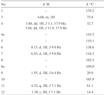
Table 2 1H NMR and 13C NMR data of 3 in CDCl
3 (d in ppm)
No. d1H d13C
1 – 170.2
3 4.66, m, 1H 75.8
4 3.88, dd, 1H, J 3.1, 17.9 Hz; 3.04, dd, 1H, J 11.9, 17.9 Hz
32.7
4a – 143.5
5 – 119.1
6 8.13, d, 1H, J 9.0 Hz 138.6
7 6.93, d, 1H, J 9.0 Hz 116.3
8 – 165.5
8a – 109.0
9 1.55, d, 3H, J 6.4 Hz 20.9
10 – 165.9
11 4.32, q, 2H, J 7.1 Hz 61.1
12 1.38, t, 3H, J 7.1 Hz 14.4
Table 1 1H NMR and 13C NMR data of 1 and 2 in CDCl
3 (d in ppm)
No. 1 2
d1H d13C d1H d13C
1 – 75.1 – 75.3
2 1.54 (β-H), 1.89 (α-H), m, 2H 36.9 1.23, 1.90, m, 2H 31.7
3 1.28, 1.68, m, 2H 31.7 1.23, 1.46, m, 2H 30.7
4 2.00, m, 1H 39.5 2.00, m, 1H 39.2
5 2.03, m, 1H 46.4 2.06, m, 1H 47.4
6 1.13, 1.45, m, 2H 30.1 1.45, 1.90, m, 2H 26.3
7 2.26, m, 1H 42.3 1.88, m, 1H 43.1
8 1.87, m, 2H 29.9 1.73 (β-H), 1.47 (α-H), m, 2H 24.5
9 1.53, 1.70, m, 2H 26.7 1.79, 1.73, m, 2H 21.1
10 2.11, m, 1H 55.4 2.08, m, 1H 55.8
11 – 156.9 – 76.2
12 4.93, dd, 1H, J 1.5, 2.1 Hz; 4.81, dd, 1H, J 1.5, 2.1 Hz
106.7 0.98, s, 3H 17.9
13 4.02, s, 2H 64.6 3.33, d, 1H, J 11.4 Hz;
3.58, d, 1H, J 11.4 Hz
68.4
14 1.16, s, 3H 29.2 1.15, s, 3H 32.2
Results and Discussion
Compound 1 was isolated as a colorless oil, and its molecular formula was assigned as C15H26O2 by the positive
HRESIMS at m/z 261.1826 [M + Na]+ (calcd. 261.1830)
and NMR data (Table 1), indicating three degrees of unsaturation. The IR spectrum displayed the presence of hydroxyl groups (3350 cm-1) and double bond (1643 cm-1).
Analysis of its 13C NMR and distortionless enhancement
by polarization transfer (DEPT) spectra (Table 1) showed the presence of 15 carbon resonances. These carbons were assigned to two methyl groups, seven methylenes (one olefinic and one oxygenated), four methines, and two quaternary carbons. Comparing the 13C NMR data of 1
with those of (1S,4S,5R,7R,10R )-10-desmethyl-1-methyl-11-eudesmene showed that both compounds had the same carbon skeleton.19 The main difference was that C-13
(dC 19.8) in (1S,4S,5R,7R,10R
)-10-desmethyl-1-methyl-11-eudesmene was shifted downfield to dC 64.6 in compound
1, which suggested the presence of a hydroxymethyl in the side chain of this compound. This was supported by its molecular formula and heteronuclear multiple-bond correlation (HMBC) spectrum correlations (Figure 2) from H-13 to C-11 [dC 156.9 (s)], C-12 [dC 106.7(t)], and
C-7 [dC 42.3 (d)]. The other HMBC correlations indicated
the atom connectivity in compound 1. The relative configurations of the chiral centers (C-1, C-4, C-5, C-7, and C-10) in 1 were assigned from rotational frame nuclear Overhauser effect spectroscopy (ROESY) experiments (Figure 3), which indicated β-orientations of CH3-14 and
H-10, and α-orientations of H-5, H-7 and CH3-15, as
well as from the similarity of its NMR data with those of (1S,4S,5R,7R,10R)-10-desmethyl-1-methyl-11-eudesmene. Thus, the structure of compound 1 was assigned as shown and named as 14(10→1)abeo -eudesmane-13-hydroxyl-11-ene.
Compound 2 was obtained as a colorless oil and had the molecular formula C15H28O3, based on the positive
HRESIMS at m/z 279.1938 [M + Na]+ (calcd. 279.1936),
indicating two degrees of unsaturation. The IR spectrum of 2 showed absorption bands at 3419 cm-1 ascribable
to hydroxyl groups. The 13C NMR and DEPT spectra
of 2 (Table 1) displayed a total of 15 carbon signals including three methyls, six methylenes, four methines and two oxygenated quaternary carbons, suggestive of a sesquiterpenoid skeleton. The 13C NMR spectrum of 2
(Table 1) was similar to that of compound 1 except for the presence of one methyl (dC 17.9) and one oxygenated
quaternary carbon (dC 76.2) in 2, replacing olefinic carbons
C-11 and C-12 in 1. This feature indicated that compound 2
is the∆1 hydrolyzed analogue of 1. This deduction was
confirmed by the HMBC (Figure 2) correlations from H-12 [dH 0.98 (s)] to C-11, C-13 [dC 68.4 (t)], and C-7 [dC 43.1
(d)]. Compound 2 had the same relative configurations as those of 1 according to its ROESY spectrum (Figure 3) and their similar NMR data. Thus, the structure of compound 2
was assigned as shown and named as 14(10→1)abeo -eudesmane-11,13-diol.
The molecular formula of compound 3 was determined to be C13H14O5 from its HREIMS at m/z 250.0847 [M]
+
O
OH O
O O
3
OH OH
H
H H
1
OH H
H H
OH OH
2
1 14
4
15 5 7 10
13
12 11 9
12
11 10
7
8a
1 3
9
4a 5
Figure 2. 1H,1H-COSY (
–
) and key HMBC ( ) correlations of compounds 1-3.(calcd. 250.0841). The 1H and 13C NMR spectra (Table 2)
displayed 13 carbon resonances comprising of two methyl groups, two methylenes, three methines, six quaternary carbons (including four olefinic carbons and two carbonyls at dC 165.9 and 170.2). The NMR data of 3 were similar to
those of (R)-(–)-5-methoxycarbonyl mellein except for the presence of ethoxyl signals (dC 61.1 and dC 14.4) attaching
to the carbonyl at C-10 in 3,replacing the methoxyl signal (dC 52.0) in (R)-(–)-5-methoxycarbonyl mellein,20 which
was confirmed by the HMBC (Figure 2) correlations from H-12 [dH 1.38 (t, 3H, J 7.1 Hz)] to C-11 (dC 61.1) and from
H-11 [dH 4.32 (q, 2H, J 7.1 Hz)] to C-10 (dC 165.9). The 1H,1H correlation spectroscopy (COSY) and other HMBC
correlations also supported the assignment of the mullein skeleton of 3. The configuration of the stereocenter at C-3 in compound 3 was determined to be R based on a comparison of similar NMR data and negative optical rotation (–6.4) with those of (R)-(–)-5-methoxycarbonyl mellein.20 Thus,
compound 3 was identified as (R)-(–)-5-ethoxycarbonyl mellein.
The AChE inhibitory activity of compounds 1 and 2
were determined by a previously described method.18 The
known AChE inhibitor tacrine was used as positive control in this assay and showed a percentage inhibition of 57%. Compounds 1 and 2 both exhibited moderate inhibitory activity with a percentage inhibition of 29 and 41%, respectively, at a concentration of 100 µmol L-1.
Conclusions
In this work, the new compounds 14(10→1)abeo -eudesmane-13-hydroxyl-11-ene (1), 14(10→1)abeo -eudesmane-11,13-diol (2), and (R)-(–)-5-ethoxycarbonyl mellein (3), together with eight known compounds were isolated from the cultures of the fungus Marasmiellus ramealis by column chromatography. The new eudesmane-type sesquiterpenoids 1 and 2 were evaluated for their inhibitory activity against acetylcholinesterase (AChE), and showed moderate inhibitory activity with a percentage inhibition of 29 and 41%, respectively, at a concentration of 100 µmol L-1.
Supplementary Information
Supplementary data are available free of charge at http://jbcs.sbq.org.br as a PDF file.
Acknowledgements
This work was supported by Special Fund for Agro-Scientific Research in the Public Interest (201303117),
National Support Science and Technology Subject (2013BAI11B04), the Natural Science Foundation of Hainan (214039) and the Fundamental Scientific Research Funds for CATAS (ITBB110301, ITBB140401).
References
1. Kamal, M. A.; Greig, N. H.; Alhomida, A. S.; Biochem. Pharmacol. 2000, 60, 561.
2. Terry, R. D.; Masliah, E.; Ann. Neurol. 1991, 30, 572. 3. Rösler, M.; Anand, R.; Cicin-Sain, A.; Gauthier, S.; Agid, Y.;
Dal-Bianco, P.; Stähelin, H. B.; Hartman, R.; Gharabawi, M.; BMJ 1999, 318, 633.
4. Giacobini, E.; Pharmacol. Res. 2004, 50, 433.
5. Wang, Y. E.; Yue, D. X.; Tang, X. C.; Acta Pharmacol. Sin. 1986, 7, 110.
6. Mao, X. L.; Macromycetes of China, 1st ed.; Science Press:
Beijing, China, 2009.
7. Jarrah, M. Y.; Thaller, V.; J. Chem. Soc., Perkin Trans. 1 1983, 1, 1719.
8. Holroyde, J. K.; Orr, A. F.; Thaller, V.; J. Chem. Soc., Chem. Commun. 1976, 7, 242.
9. Holroyde, J. K.; Orr, A. F.; Thaller, V.; J. Chem. Soc., Perkin Trans. 1 1978, 12, 1490.
10. Huang, Z. J.; Shao, C. L.; Chen, Y. G.; She, Z. G.; Lin, Y. C.; Zhou, S. N.; Chem. Nat. Compd. 2007, 43, 655.
11. Almeida, C.; Part, N.; Bouhired, S.; Kehrans, S.; König, G. M.; J. Nat. Prod. 2011, 74, 21.
12. Que, D. M.; Dai, H. F.; Zeng, Y. B.; Wu, J.; Mei, W. L.; Chin. J. Med. Chem. 2009, 19, 200.
13. Cai, H. H.; Liu, X. M.; Chen, Z. Y.; Liao, S. T.; Zou, Y. X.; Food Chem. 2013, 141, 2873.
14. Tang, J. G.; Shao, H. J.; Liu, J. K.; Chin. Tradit. Herb. Drugs 2008, 39, 1776.
15. Liu, J. K.; Tian, J. W.; Dong, Z. J.; Ding, Z. H.; Wang, X. H.; Liu, P. G.; Helv. Chim. Acta 2002, 85, 439.
16. Zhan, Z. J.; Sun, H. D.; Wu, H. M.; Yue, J. M.; Acta Bot. Sin. 2003, 45, 248.
17. Chappuis, G.; Tamm, C.; Helv. Chim. Acta 1982, 65, 521. 18. Ellman, G. L.; Courtney, K. D.; Featherstone, R. M.; Biochem.
Pharmacol. 1961, 7, 88.
19. Chavez, J. P.; Gottlieb, O. R.; Yoshida, M.; Phytochemistry 1995, 39, 849.
20. Klaiklay, S.; Rukachaisirikul, V.; Sukpondma, Y.; Arch. Pharmacal Res. 2012, 35, 1127.
Submitted: July 9, 2014