© 2012 Sociedade Brasileira de Hemodinâmica e Cardiologia Intervencionista. Published by Elsevier Editora Ltda. All rights reserved.
Temporal Evolution of Neointimal Proliferation
After Implantation of Two Types of Drug-eluting
Stents with Biodegradable Polymers in a Porcine Model:
Qualitative Assessment by Sequential Optical
Coherence Tomography
Micheli Zanotti Galon
1, Celso Kiyochi Takimura
2, Juliana Carvalho
3, Márcio José Figueira Chaves
4,
Silvia Lacchini
5, Vera Demarchi Aiello
6, Paulo Sampaio Gutierrez
7,
Francisco Rafael Martins Laurindo
8, Pedro Alves Lemos Neto
9ABSTRACT
Background: Based on the hypothesis that the neointima found
in drug-eluting stents (DES) with biodegradable polymers at 28 days is not a deinitive neointima and that optical coher-ence tomography (OCT) is an effective method for sequential neointimal evaluation, we aim, in this experimental study, to compare OCT indings at 28 and 90 days, in two different DES with biodegradable polymers: the sirolimus-eluting stent (Inspiron, Scitech) and the biolimus A9-eluting stent (Biomatrix, Biosensors International). Methods: Overall, 6 non-atherosclerotic pigs were submitted to the implantation of 6 Inspiron stents and 6 Biomatrix stents. Each pig received both stent types, one in each coronary artery (left anterior descending artery and circumlex artery) and after 28 and 90 days qualitative in-stent OCT analyses were performed at 1-millimeter inter-vals. Results: Qualitative assessment was performed in-stent pairing millimeter by millimeter. Heterogeneous neointimal tissue was evidenced in 39% at 28 days and in 0% at 90 days, the presence of intraluminal tissue in 18% at 28 days and in 0% at 90 days, luminal irregularity in 62% at 28 days and in 2% at 90 days (P < 0.005). There was no difference between groups regarding the quality of the neointima over time (P > 0.05). Conclusions: The OCT indings corroborate the hypothesis that the neointima found in DES with
biode-1 Master. Interventionist Cardiologist Physician-researcher at Instituto
do Coração do Hospital das Clínicas da Faculdade de Medicina da Universidade de São Paulo. São Paulo, SP, Brazil.
2 Doctor. Physician-researcher at Instituto do Coração do Hospital das
Clínicas da Faculdade de Medicina da Universidade de São Paulo. São Paulo, SP, Brazil.
3 Biomedical physician at Instituto do Coração do Hospital das Clínicas da
Faculdade de Medicina da Universidade de São Paulo. São Paulo, SP, Brazil.
4 Biologist at Instituto do Coração do Hospital das Clínicas da
Faculda-de Faculda-de Medicina da UniversidaFaculda-de Faculda-de São Paulo. São Paulo, SP, Brazil.
5 Doctor. Biologist at Instituto do Coração do Hospital das Clínicas da
Faculdade de Medicina da Universidade de São Paulo. São Paulo, SP, Brazil.
6 Full Professor. Physician at Instituto do Coração do Hospital das
Clínicas da Faculdade de Medicina da Universidade de São Paulo. São Paulo, SP, Brazil.
7 Full Professor. Physician at Instituto do Coração do Hospital das
Clínicas da Faculdade de Medicina da Universidade de São Paulo. São Paulo, SP, Brazil.
8 Full Professor. Physician at Instituto do Coração do Hospital das
Clínicas da Faculdade de Medicina da Universidade de São Paulo. São Paulo, SP, Brazil.
9 Full Professor. Physician at Instituto do Coração do Hospital das
Clínicas da Faculdade de Medicina da Universidade de São Paulo. São Paulo, SP, Brazil.
Correspondence to: Pedro Alves Lemos Neto. Av. Dr. Enéas Carvalho de Aguiar, 44 – Cerqueira César – São Paulo, SP, Brazil – CEP 05403-900 E-mail: [email protected]
Received on: 8/31/2012 • Accepted on: 11/26/2012
Original Article
RESUMO
Evolução Temporal da Proliferação Neointimal após Implante de Dois Tipos de Stent Farmacológico com Polímeros Biodegradáveis em Modelo Porcino:
Avaliação Qualitativa por Tomografia de Coerência Óptica Sequencial
gradable polymers at 28 days is not a deinitive neointima. The most signiicant experimental evidence is the change in the neointimal characteristics observed at sequential OCT.
DESCRIPTORS: Tomography, optical coherence. Drug-eluting
stents. Angioplasty. Polymers. Neointima.
METHODS
All procedures followed the standards for labora-tory animal protection and care established by the Ethical Principles in Animal Experimentation of the Research Support and Animal Experimentation Service of the Instituto do Coração da Faculdade de Medicina da Universidade de São Paulo, as well as the Guide for the Care and Use of Laboratory Animals (Institute of Laboratory Animal Resources, Commission on Life Sciences and National Research Council, National Academy Press, Washington, DC, 1996) and the Ethical Principles in Animal Experimentation of the Brazilian College of Animal Experimentation (COBEA).
Subjects and stent implantation
Six domestic juvenile, non-atherosclerotic pigs, weighing between 18 kg and 23 kg, were obtained from a commercial breeding farm.
One day before the procedure, the pigs received 600 mg of acetylsalicylic acid and 150 mg of clopi-dogrel orally. The anaesthesia protocol consisted of premedication (ketamine 3 mg/kg and midazolam 0.5 mg/kg, intramuscular) followed by intravenous infusion of thiopental, endotracheal intubation, and the start of mechanical ventilation; the level of anaesthesia was maintained with isolurane.
A 6F vascular sheath was introduced under direct vision after dissection of the common femoral artery. A dose of 10,000 IU unfractionated heparin was ad-ministered and the left coronary artery was selectively catheterised under luoroscopy using a Judkins right (JR)4 6F guide catheter. Then, nitroglycerin was injected
through the catheter at a dose of 200 µg, and an Ins-piron or Biomatrix stent was randomly implanted in
each anterior descending or circumlex artery of each pig, trying to maintain a stent-releasing balloon/artery ratio of 1.1/1. The Inspiron stents were 2.5 mm or 3
mm in diameter and 16 mm in length. All implanted Biomatrix stents were 3 mm in diameter and 14 mm
in length.
C
urrent knowledge of the vascular response andthe resulting neointimal hyperplasia related to the implantation of drug-eluting stents (DES) is largely based on preclinical studies in porcine coro-nary arteries.1-4 In animals, peak neointimal growth
with bare-metal stents (BMS) occurs at 28 days; with DES, studies in animals have shown favourable results for re-endothelialization at 28 days, albeit incomplete.5-8
Vascular healing in response to DES implantation is dynamic and depends on the type of drug used, which in turn depends on its release kinetics from durable or biodegradable polymers.9-16 Evaluation through histology
does not allow for time-dependent analyses, and there are additional limitations, such as tissue loss related to the technique employed for material preparation (non-satisfactory cutting or colouring, thickness, and angle of the cut).
Due to its high resolution, optical coherence tomography (OCT) may be a promising intravascular imaging tool in the sequential evaluation of neointimal formation. Compared to histology, OCT allows for the evaluation of the vascular healing process in vivo at different stages, providing qualitative and quantitative information along the full length of the stent.17-20 By
enabling analysis at multiple time points, it is possible to compare the same intrastent region at different times and thereby assess temporal neointima matura-tion, as well as to evaluate each intrastent millimetre strut by strut.
Based on the hypothesis that the neointima found in DES with biodegradable polymers at 28 days are not deinitive, and that OCT is an effective method for the sequential evaluation of neointima, this experimental study aimed to compare the qualitative indings of OCT at 28 days and 90 days in two types of DES with biodegradable polymers: the sirolimus-eluting stent (Inspiron, Scitech Medical Products Ltda. – Goiânia,
Brazil) and the biolimus-A9 eluting stent (Biomatrix,
Biosensors International – Singapore).
0% aos 90 dias, presença de tecido intraluminal em 18% aos 28 dias e em 0% aos 90 dias, irregularidade luminal em 62% aos 28 dias e em 2% aos 90 dias (P < 0,005). Não houve diferença entre os grupos quanto à qualidade da neoíntima ao longo do tempo (P > 0,05). Conclusões: Os achados à TCO corroboram a hipótese de que a neoíntima encontrada em SFs com polímeros biodegradáveis aos 28 dias não é a neoíntima deinitiva. A evidência experimental mais signiica-tiva é a mudança das características da neoíntima observada à TCO sequencial.
DESCRITORES: Tomograia de coerência óptica. Stents
Optical coherence tomography
An M2 OCT Imaging System (Imaging LightLab, Inc., Westford, USA) was used with ImageWire automated
catheter pullback (LightLab Imaging, Inc.) at a speed of 1 mm/s and 2 mm/s after proximal occlusion of the arterial segment under study with a Helios balloon
(LightLab Imaging, Inc.). Speciic software from the OCT equipment was used to make measurements at 1 mm intervals starting from the most distal part of the stent and moving towards the proximal end.
Morphometric Analysis
The first stent image analysed was the one that allowed for the drawing of a complete circle using struts in the distal border. The stent and luminal areas were measured by manual tracing (mm2). The neointimal
area (mm2) was calculated as the lumen area minus
the stent area. The neointimal area percentage (%) was calculated as the ratio between the neointimal area and the stent area (neointimal area percentage = area of the stent – lumen area/stent area). For the analysis of neointimal thickness (mm), the distance from the luminal surface of the stent strut reflec-tion (called the “blooming artifact”) to the luminal contour was measured. Since the blooming artefact was systematically included in the measurements, to determine strut apposition it was necessary to take into account its total thickness (thickness of the metal strut + thickness of the polymer + drug) plus a cor-rection factor of 20 µm, which corrects for the actual location of the strut surface by discounting half of the blooming.18,19 The distance between the luminal surface
of blooming produced by the struts and the contour of the vascular lumen was analysed indivi dually for each strut. For the stents in this study, if the distance was greater than the sum of the thicknesses of the struts (Biomatrix, 112 µm; Inspiron, 75 µm) plus
the thicknesses of the polymers (Biomatrix, 10 µm;
Inspiron, 5 µm) plus 20 µm (correction factor), the
strut would be considered malpositioned.
Characterisation of neointimal tissue
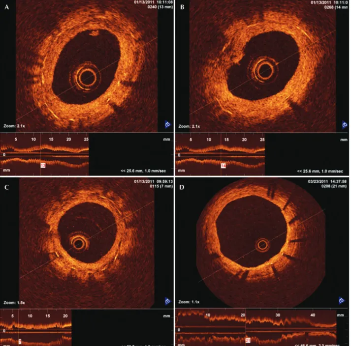
The qualitative analysis evaluated transverse sec-tions at each millimetre along the stent at 28 and 90 days. Features such as the presence of neointima with luminal irregularities, the presence of intraluminal tis-sue, and the heterogeneous aspects of neointima were detected and described.
Neointimal tissue with irregularities was deined by the presence of indistinct and irregular contours on the luminal surface of the vessel and by the presence or absence of discontinuous neointima. Intraluminal tissue was identiied as material protruding into the
vessel lumen, which may or may not adhere to the neointima. Heterogeneous aspects of neointimal tissue were identiied when an image with low light intensity was observed in the OCT of neointimal tissue without signal attenuation behind the area (Figure 1).
These events were compared in cross-sectional OCT images taken after 28 and 90 days and evaluated regarding their progression or resolution.
Statistical Analysis
Data are shown as the mean ± SD or as numbers and percentages. The comparison between days 28 and 90, as well as the interaction of the effects of the type of stent and the progression/regression of the temporal neointima over time were analysed with a general linear model with repeated measures. The signiicance level was set at 5%. Analyses were performed using SPSS 20.0 (IBM SPSS – Chicago, USA).
RESULTS
All six pigs survived the index procedure and underwent angiographic analysis and OCT at 28 days and at 90 days after implantation.
In total, 2,685 stent struts along 350 mm of total stent length were analysed (1,366 struts at 28 days and 1,319 struts at 90 days); 338 paired transverse sections were analysed at 28 days and at 90 days (Inspiron =
90 and Biomatrix = 79 transverse sections analysed
at each time point) (Figure 2). Twelve transverse sec-tions were excluded from the analysis due to image artifacts. No malpositioned struts were observed, as all struts analysed were covered by neointimal tissue.
Events such as the presence of luminal irregulari-ties, heterogeneous aspects of neointimal tissue, and the presence of intraluminal tissue were evaluated in the 12 stents at 28 days. The presence of heterogeneous neointimal tissue, irregular lumen, and luminal tissue in the transverse sections was higher at 28 days than at 90 days (38.8 ± 28.2% vs. 0%; P = 0.001; 62.4 ±
23.1% vs. 2.2 ± 5.1%; P < 0.001; 17.9 ± 16.3% vs. 0%; P = 0.005, respectively). This behaviour was observed for both stents analysed (Table 1).
The incidence of qualitative events in the trans-verse sections analysed was similar at 28 days for both stents, and each showed heterogeneous neointi-mal tissue (44.4% ± 31.1 and 33.2 ± 26.6% for the Inspiron and Biomatrix stents, respectively; P = 0.5),
intraluminal tissue (18.1 ± 20.7% and 17.7 ± 12.5% for Inspiron and Biomatrix stents, respectively; P > 0.9)
and luminal irregularities (67.9 ± 20% and 56.9 ±
26.4% for Biomatrix and Inspiron stents, respectively;
endothelialisation may play an important role in the pathogenesis of DES thrombosis.23,24 Additionally, autopsy
studies have identiied the presence of bare struts as a more robust morphological predictor for the occurrence of thrombosis in irst-generation DES.25
The present study with sequential OCT, despite not showing bare struts, did provide images that indicate
DISCUSSION
Several studies in animal models and previous clinical trials showed delayed endothelialisation of DES when compared to BMS,21-23 and observed the
frequent presence of struts covered by thin layers of neointimal tissue, with a thickness well below the limits of detection by intracoronary ultrasound. This delayed
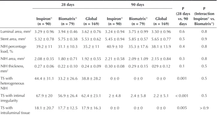
Table 1
Quantitative and qualitative findings from sequential optical coherence tomography images taken at 28 and 90 days after Inspiron and Biomatrix stent implantation in porcine coronary arteries
28 days 90 days
P (28 days
vs. 90 days
P (Interaction Inspiron vs.
Biomatrix)
Inspiron
(n = 90)
Biomatrix
(n = 79)
Global (n = 169)
Inspiron
(n = 90)
Biomatrix
(n = 79)
Global (n = 169)
Luminal area, mm2 3.29 ± 0.96 3.94 ± 0.46 3.62 ± 0.76 3.24 ± 0.94 3.75 ± 0.99 3.50 ± 0.96 0.6 0.8 Stent area, mm2 5.32 ± 0.78 5.75 ± 0.38 5.53 ± 0.62 5.45 ± 0.94 5.85 ± 0.57 5.65 ± 0.77 0.5 0.9 NIH percentage
load, %
39.2 ± 11 31.1 ± 10.3 35.2 ± 11 40.9 ± 10 35.3 ± 17.6 38.1 ± 13.9 0.4 0.8
NIH area, mm2 2.08 ± 0.35 1.80 ± 0.71 1.92 ± 0.55 2.21 ± 0.58 2.09 ± 1.09 2.15 ± 0.84 0.3 0.8 NIH thickness,
mm2
0.27 ± 0.06 0.22 ± 0.10 0.24 ± 0.09 0.30 ± 0.08 0.29 ± 0.15 029 ± 0.12 0.1 0.5
TS with heterogeneous NIH
44.4 ± 31.1 33.2 ± 26.6 38.8 ± 28.2 0 ± 0 0 ± 0 0 ± 0 0.001 0.5
TS with intimal irregularity
67.9 ± 20 56.9 ± 26.4 62.4 ± 23.1 2 ± 4.8 2.4 ± 5.8 2.2 ± 5.1 < 0.001 0.5
TS with
intraluminal tissue
18.1 ± 20.7 17.7 ± 12.5 17.9 ± 16.3 0 ± 0 0 ± 0 0 ± 0 0.005 > 0.9
NIH, neointima hyperplasia; TS, transverse sections.
heterogeneity in the healing process after 28 days. This result suggests that this is a step in the neointimal tis-sue maturation process for both stents studied, since, at 90 days, no heterogeneous neointimal tissue, luminal irregularity, and intraluminal tissue were observed.
It should be noted that at both 28 and 90 days, although the process of stent endothelialisation is faster in pigs than in humans,7 polymers from both Inspiron
and Biomatrix stents are still present and undergoing
degradation and their respective antiproliferative drugs are still present.12,26 A study with a later evaluation time
point (> six months) may be needed to analyse DES in order to evaluate the inlammatory and chronic biological responses after drug elution and polymer degradation.
It will be important to correlate the OCT indings with histological data to identify and understand the neointima maturation process. Teramoto et al.27 correlated
images with low light intensity signal visualised in OCT with histological indings, and observed areas of low cellularity and the presence of ibrin and proteoglycans.
Second-generation OCT systems, which are already available for both pre-clinical and clinical use (Fourier-domain optical coherence tomography [FDOCT]), are capable of acquiring images at higher speeds with fewer artefacts and without the need for proximal occlusion of the coronary artery. Recent FDOCT studies compared the optical density of neointimal tissue longitudinally after stent implantation to differentiate ibrin from neointimal tissue.28,29
These indings corroborate the results of the pres-ent study, as they demonstrated neointimal maturation over time and showed a progressive reduction in the amount of ibrin and its replacement by neointimal tissue, which was maintained after 28 days.
Further evaluations are needed to better under-stand the possible roles of these different regions in neointimal tissue in the arterial healing process after implantation of DES.
Study limitations
Qualitative analyses were performed by a single investigator; therefore, the assessment of the qualitative characteristics of the neointima could be biased despite the best efforts to remain objective.
The DES were implanted in porcine non-atheroscle-rotic coronary arteries and the vascular response was evaluated in the context of normal porcine coronary arteries. Preclinical studies with these stents in the arteries of animals with experimental atherosclerosis (minipigs, rabbits) can mora accurately answer the questions regarding the effectiveness of these stents.21
CONCLUSIONS
The OCT results corroborate the hypothesis that the neointima found in DES with biodegradable polymers
at 28 days is not the deinitive neointima. The most signiicant experimental evidence is the change in characteristics of the neointima observed in OCT images taken after 90 days. This fundamental tool is useful in the preclinical analysis of stents, allowing the sequential assessment of neointimal tissue in vivo.
FINANCIAL SUPPORT
The present study is part of the National Develop-ment Program of Vascular Stents (PDNS), initiated in 2004-2005. It is supported by the Secretariat of Science, Technology and Strategic Inputs (SCTIE)/Department of Science and Technology (DECIT), the Brazilian Minis-try of Health, the National Council for Scientiic and Technological Development (CNPq) and the Financier of Studies and Projects (FINEP) of the Brazilian Ministry of Science and Technology. The development of the laser cutting of stents was supported by the Foundation for Research Support of the State of São Paulo (FAPESP; Support type: Technological Innovation/Innovative Re-search in Small and Micro Enterprises [PIPE]).
CONFLICT OF INTEREST
Celso K. Takimura and Francisco R. M. Laurindo are scientiic consultants at Scitech Medical Products. Over the past 36 months, Pedro A. Lemos has been a scientiic consultant to Scitech Medical Products and has delivered national and international lectures sponsored by Boston Scientiic and Braun; he is also a member of Boston Scientiic Latin America SCIMAB (Scientiic Medical Advisory Board). The remaining authors declare to have no conlicts of interest.
REFERENCES
1. Suzuki Y, Yeung AC, Ikeno F. Importância dos estudos préclínicos em animais de experimentação para a cardiologia interven-cionista. Arq Bras Cardiol. 2008;91(5):348-60.
2. Schwartz RS, Huber KC, Murphy JG, Edwards WD, Camrud AR, Vliestra RE, et al. Restenosis and the proportional neointimal response to coronary artery injury: results in a porcine model. J Am Coll Cardiol. 1992;19(2):267-74.
3. Schwartz RS, Holder DJ, Holmes DR, Veinot JP, Camrud AR, Jorgenson MA, et al. Neointimal thickening after severe coro-nary artery injury is limited by a short-term administration of a factor Xa inhibitor: results in a porcine model. Circulation. 1996;93(8):1542-8.
4. Ikeno F, Buchbinder M, Yeung AC. Novel stent and delivery systems for the treatment of bifurcation lesions: porcine coro-nary artery model. Cardiovasc Revasc Med. 2007;8(1):38-42. 5. Carter AJ, Laird JR, Farb A, Kufs W, Wortham DC, Virmani R.
Morphologic characteristics of lesion formation and time course of smooth muscle cell proliferation in a porcine proliferative restenosis model. J Am Coll Cardiol. 1994;24(5):1398-405. 6. Edelman ER, Rogers C. Pathobiologic responses to stenting.
Am J Cardiol. 1998;81(7A):4E-6E.
8. Kotani J, Awata M, Nanto S, Uematsu M, Oshima F, Minamiguchi H, et al. Incomplete neointimal coverage of sirolimus-eluting stents: angioscopic indings. J Am Coll Car-diol. 2006;47(10):2108-11.
9. Nakazawa G, Finn AV, John MC, Kolodgie FD, Virmani R. The signiicance of preclinical evaluation of sirolimus-, pa-clitaxel-, and zotarolimus-eluting stents. Am J Cardiol. 2007; 100(8B):36M-44M.
10. Stone GW, Ellis SG, Cox DA, Hermiller J, O’Shaughnessy C, Mann JT, et al. One-year clinical results with slow-release, polymer-based, paclitaxel-eluting TAXUS stent: the TAXUS-IV trial. Circulation. 2004;109(16):1942-7.
11. Joner M, Finn AV, Farb A, Mont EK, Kolodgie FD, Ladich E, et al. Pathology of drug-eluting stents in humans: delayed healing and late thrombotic risk. J Am Coll Cardiol. 2006;48(1): 193-202. 12. Byrne RA, Mehilli J, Iijima R, Schulz S, Pache J, Seyfarth M,
et al. A polymer-free dual drug-eluting stent in patients with coronary artery disease: a randomized trial vs. polymer-based drug-eluting stents. Eur Heart J. 2009;30(8):923-31.
13. Chamié D, Costa Jr. JR, Abizaid A, Silva JFA, Feres F, Mattos LM, et al. Stents liberadores de sirolimus com e sem cobertura polimérica: análise seriada com angiograia e ultrassom in-tracoronariano tridimensional. Rev Bras Cardiol Invasiva. 2009;17(1):20-30.
14. Windecker S, Serruys PW, Wandel S, Buszman P, Trznadel S, Linke A, et al. Biolimus-eluting stent with biodegradable polymer versus sirolimus-eluting stent with durable polymer for coronary revascularization (LEADERS): a randomized non-inferiority trial. Lancet. 2008;372(9644):1163-73.
15. Takimura CK, Watanabe IS, Campos CAHM, Laurindo FRM, Ferreira CN, Gutierrez PS, et al. Avaliação por microscopia eletrônica de varredura da endotelização pós implante de stent farmacológico Scitech em artérias ilíacas de coelhos. Rev Bras Cardiol Invasiva. 2010;18 Supl 2:27.
16. Takimura CK, Galon MZ, Lopes Jr. ACA, Carvalho J, Ferreira SK, Chaves MJF, et al. Avaliação pela tomograia de coerência óptica de stent nacional recoberto com polímero biode-gradável eluidor de sirolimus vs. stent eluidor de biolimus A9 em artérias coronárias porcinas. Rev Bras Cardiol Invasiva. 2011;19(2):138-44.
17. Guagliumi G, Musumeci G, Sirbu V, Bezerra HG, Suzuki N, Fiocca L, et al. Optical coherence tomography assessment of
in vivo vascular response after implantation of overlapping
bare-metal and drug-eluting stents. JACC Cardiovasc Interv. 2010;3(5):531-9.
18. Mehanna EA, Attizzani GF, Kyono H, Hake M, Bezerra HG. Assessment of coronary stent by optical coherence tomography,
methodology and definitions. Int J Cardiovasc Imaging. 2011;27(2):259-69.
19. Bezerra HG, Costa MA, Guagliumi G, Rollins AM, Simon DI. Intracoronary optical coherence tomography: a comprehensive review clinical and research applications. JACC Cardiovasc Interv. 2009;2(11):1035-46.
20. Tahara S, Chamie D, Baibars M, Alraies C, Costa M. Optical coherence tomography endpoints in stent clinical investigations: strut coverage. Int J Cardiovasc Imaging. 2011;27(2):271-87. 21. van Beusekom HM, Saia F, Zindler JD, Lemos PA, Swager-Ten
Hoor SL, van Leeuwen MA, et al. Drug-eluting stents show delayed healing: paclitaxel more pronounced than sirolimus. Eur Heart J. 2007;28(8):974-9.
22. Lim SY, Jeong MH, Hong SJ, Lim do S, Moon JY, Hong YJ, et al. Inlammation and delayed endothelization with overlapping drug-eluting stents in a porcine model of in-stent restenosis. Circ J. 2008;72(3):463-8.
23. Finn AV, Joner M, Nakazawa G, Kolodgie F, Newell J, John MC, et al. Pathological correlates of late drug-eluting stent thrombosis: strut coverage as a marker of endothelialization. Circulation. 2007;115(18):2435-41.
24. Nakazawa G, Finn AV, Joner M, Ladich E, Kutys R, Mont EK, et al. Delayed arterial healing and increased late stent throm-bosis at culprit sites after drug-eluting stent placement for acute myocardial infarction patients: an autopsy study.Circulation. 2008;118(11):1138-45.
25. Finn AV, Joner M, Nakazawa G, Kolodgie F, Newell J, John MC, et al. Pathological correlates of late drug-eluting stent thrombosis: strut coverage as a marker of endothelialization. Circulation. 2007;115(18):2435-41.
26. Ribeiro EE. Estudo randomizado INSPIRON I: dois anos de follow-up. In: XXXIV Congresso da Sociedade Brasileira de Hemodinâmica e Cardiologia Intervencionista; 2012 jun. 20-22; Salvador, BA, Brasil.
27. Teramoto T, Ikeno F, Otake H, Lyons JK, van Beusekom HM, Fearon WF, et al. Intriguing peri-strut low-intensity area de-tected by optical coherence tomography after coronary stent deployment. Circ J. 2010;74(6):1257-9.
28. Templin C, Meyer M, Müller MF, Djonov V, Hlushchuk R. Coronary optical frequency domain imaging (OFDI) for in vivo evaluation of stent healing: comparison with light and
electron microscopy. Eur Heart J. 2010;31(14):1792-801. 29. Attizzani GF, Bezerra HG, Chamié D, Fujino Y, Spognardi AM,