© 2013 Sociedade Brasileira de Hemodinâmica e Cardiologia Intervencionista. Published by Elsevier Editora Ltda. All rights reserved.
Balloon Aortic Valvuloplasty in Degenerative Aortic
Stenosis: Therapeutic Impact on Patients
In Extremis
Vitor de Andrade Vahle
1, Fábio Augusto Pinton
2, Eduardo França Pessoa de Melo
3,
Cristiano Guedes Bezerra
4, Marco Antônio Perin
5, Santiago Raul Arrieta
6, Luiz Junya Kajita
7,
José Mariani Junior
8, Antônio Esteves Filho
9, Expedito Eustáquio Ribeiro da Silva
10,
Flávio Tarasoutchi
11, Max Grinberg
12, Pedro Alves Lemos Neto
13ABSTRACT
Background: Balloon aortic valvuloplasty (BAV) is used as a
palliative strategy in patients who are not eligible for valve replacement surgery, transcatheter aortic valve implantation, or as a bridge to these treatment modalities. The impact of BAV as a salvage procedure for patients in extreme clinical conditions (in extremis) is unknown. Methods: Patients with severe degenerative aortic stenosis undergoing BAV between July 2008 and January 2013 were evaluated. Patients were divided into the in-extremis group (deined by the presence of two or more of the following organ dysfunctions: mechanical ventilation, hemodynamic instability, dialysis, coagulopathy or severe hepatic dysfunction) and the control group, which included the remaining patients. Results: A total of 19 patients underwent BAV. The clinical condition in-extremis was present in 42.1% of them. Patients from the in-extremis group had a higher EUROSCORE II (41.1 ± 24.7 vs. 15.9 ± 14.0; P = 0.001) and LV ejection fraction lower than the control group (33.9 ± 17.3% vs. 49.0 ± 12.5; P = 0.04). None of the patients in the in-extremis group survived past the hospitalization period,
1 Resident Physician at the Hemodynamics and Interventional Cardiology Service of Instituto do Coração do Hospital das Clínicas da Faculdade de Medicina da Universidade de São Paulo. São Paulo, SP, Brazil. 2 Resident Physician at the Hemodynamics and Interventional Cardiology Service of Instituto do Coração do Hospital das Clínicas da Faculdade de Medicina da Universidade de São Paulo. São Paulo, SP, Brazil. 3 Resident Physician at the Hemodynamics and Interventional Cardiology Service of Instituto do Coração do Hospital das Clínicas da Faculdade de Medicina da Universidade de São Paulo. São Paulo, SP, Brazil. 4 Resident Physician at the Hemodynamics and Interventional Cardiology Service of Instituto do Coração do Hospital das Clínicas da Faculdade de Medicina da Universidade de São Paulo. São Paulo, SP, Brazil. 5 Full professor. Interventionist Cardiologist Physician at the Hemody-namics and Interventional Cardiology Service of Instituto do Coração do Hospital das Clínicas da Faculdade de Medicina da Universidade de São Paulo. São Paulo, SP, Brazil.
6 Interventionist Cardiologist Physician at the Hemodynamics and Interventional Cardiology Service of Instituto do Coração do Hospital das Clínicas da Faculdade de Medicina da Universidade de São Paulo. São Paulo, SP, Brazil.
7 Interventionist Cardiologist Physician at the Hemodynamics and Interventional Cardiology Service of Instituto do Coração do Hospital das Clínicas da Faculdade de Medicina da Universidade de São Paulo. São Paulo, SP, Brazil.
8 Interventionist Cardiologist Physician at the Hemodynamics and Interventional Cardiology Service of Instituto do Coração do Hospital
das Clínicas da Faculdade de Medicina da Universidade de São Paulo. São Paulo, SP, Brazil.
9 Interventionist Cardiologist Physician at the Hemodynamics and Interventional Cardiology Service of Instituto do Coração do Hospital das Clínicas da Faculdade de Medicina da Universidade de São Paulo. São Paulo, SP, Brazil.
10 Full professor. Interventionist Cardiologist Physician at the Hemody-namics and Interventional Cardiology Service of Instituto do Coração do Hospital das Clínicas da Faculdade de Medicina da Universidade de São Paulo. São Paulo, SP, Brazil.
11 Full professor. Cardiologist Physician at the Valve Disease Unit of Instituto do Coração do Hospital das Clínicas da Faculdade de Medicina da Universidade de São Paulo. São Paulo, SP, Brazil.
12 Full professor. Director of the Valve Disease Unit of Instituto do Coração do Hospital das Clínicas da Faculdade de Medicina da Uni--versidade de São Paulo. São Paulo, SP, Brazil.
13 Full professor. Director of the Hemodynamics and Interventional Cardiology Service of Instituto do Coração do Hospital das Clínicas da Faculdade de Medicina da Universidade de São Paulo. São Paulo, SP, Brazil.
Correspondence to: Pedro Alves Lemos Neto. Av. Dr. Enéas Carvalho de Aguiar, 44 – Jardim Paulista – São Paulo, SP, Brazil – CEP 05403-000 E-mail: [email protected]
Received on: 6/1/2013 • Accepted on: 8/18/2013
Original Article
RESUMO
Valvuloplastia Aórtica por Cateter Balão na Estenose Aórtica Degenerativa: Impacto Terapêutico em
Pacientes em Condição Clínica In Extremis
Introdução: A valvuloplastia aórtica por cateter balão (VAB)
whereas the control group mortality was 27.3% (P < 0.01).
Conclusions: BAV has an unfavorable result in patients with
severe degenerative aortic stenosis with two or more organ dysfunctions, that is, patients in extremis.
DESCRIPTORS: Aortic valve stenosis. Balloon valvuloplasty.
Heart valve prosthesis implantation.
extremis), in an attempt to avoid death, at the expense of an improvement in the cardiac output compromised by the aortic valve stenosis.
The aim of this study was to evaluate the thera-peutic impact of BAV in the treatment of degenerative aortic stenosis in patients with and without the clinical
condition in extremis.
METHODS
This is a retrospective study, conducted in a single quaternary care service of high complexity cardiology. The research was based on a database analysis and review of the electronic clinical record.
Study population
Between July 2008 and January 2013, all patients undergoing BAV for treatment of degenerative aortic stenosis at Instituto do Coração, Hospital das Clínicas da Faculdade de Medicina, Universidade de São Paulo (Incor-HCFMUSP), in São Paulo (SP) were reviewed. This study did not include procedures for treatment of congenital aortic stenosis.
Procedure
All procedures were performed by retrograde route, by puncturing the common femoral artery (right or left), and by the application of a long sheath with diameter 10F. Unfractionated heparin was administered to all patients. The size of the balloon catheter used in each procedure was chosen at the discretion of the surgeon. In some cases, the balloon inlation was preceded by a rapid stimulation (fast pacing), with a temporary pacemaker placed in the right ventricle.
The measurement of the aortic transvalvular gra-dient was performed before and after the BAV, through intracavitary manometry and transthoracic echocardio-graphy. The sheath was removed immediately after the
VAB no período. A condição clínica in extremis esteve pre-sente em 42,1%. Os pacientes do grupo in extremis tiveram EUROSCORE II mais elevado (41,1 ± 24,7 vs. 15,9 ± 14,0; P = 0,01) e fração de ejeção do VE mais baixa que o grupo controle (33,9 ± 17,3% vs. 49,0 ± 12,5%; P = 0,04). Nenhum paciente do grupo in extremis sobreviveu ao período intra-hospitalar, enquanto que, no grupo controle, a mortalidade foi de 27,3% (P < 0,01). Conclusões: Para o tratamento de pacientes com estenose aórtica grave de etiologia degenera-tiva, a VAB tem resultado desfavorável quando indicada para pacientes com duas ou mais disfunções orgânicas, ou seja, em condição clínica in extremis.
DESCRITORES: Estenose da valva aórtica. Valvuloplastia com
balão. Implante de prótese de valva cardíaca.
D
egenerative aortic stenosis is the valvulopathywhose incidence most increases with aging. Its prevalence in individuals over 75 years of age
is estimated at 4.6%.1 The prognosis after the onset
of symptoms is poor, with survival time between one
and three years.2
The treatment of choice for symptomatic patients with severe aortic stenosis secondary to valve
degen-eration is aortic valve replacement surgery (AoVR).3
However, approximately 30% of these patients do not receive surgical treatment due to the high
periopera-tive risk arising from multiple comorbidities.4 Thus, less
invasive modalities have emerged for the treatment of this valvulopathy, among which stand out the trans-catheter aortic valve implantation (TAVI) and balloon aortic valvuloplasty (BAV).
TAVI was safe and effective in patients with high surgical risk, providing a reduction in mortality in patients whose surgical procedure was refused by the
surgeon because of an excess of clinical comorbidities.5
However, the anatomical prerequisites required, the high cost related to the procedure, and the low number of trained professionals capable of its performance make this option unavailable for most patients.
BAV is a procedure used as a palliative strategy in patients unit for both surgical valve replacement and
TAVI, or as a bridge to these treatment modalities.2,6-8
Its low cost and wide availability in most cardiology centers justify its use for patients with prohibitive surgi-cal risk, despite the high recurrence rate of symptoms
and its ineffectiveness in reducing mortality.9,10
The best time to perform the BAV, in the context of symptomatic aortic stenosis, has not yet been deined. In most cases, the procedure is performed with urgency in patients refractory to optimized clinical treatment. In other times, however, BAV is performed as a meas- ure to rescue patients who already present multiple
procedure, with subsequent manual compression for hemostasis.
Procedural success was deined as the successful dilation of the aortic valve with the balloon catheter, in the absence of complications during the procedure.
Data collection
Clinical, echocardiographic, and hemodynamic data, and in-hospital outcomes were obtained retrospectively from the electronic case notes of each patient treated at Incor-HCFMUSP. After hospital discharge, the evaluation of cardiovascular events was performed by analyzing the electronic records, with supplementation by telephone contact, when needed.
The patients’ proile of disease severity was esti-mated by EuroSCORE II and STS Risk Score. Severe pulmonary hypertension was defined as a systolic
pressure in pulmonary artery (SPPA) ≥ 60 mmHg by
echocardiography. Renal failure was characterized as
the presence of creatinine clearance ≤ 60 mL/min.
The patients were divided in two groups: in extremis
(deined by the presence of two or more of the follow-ing dysfunctions: mechanical ventilation, hemodynamic instability, renal dialysis therapy, coagulopathy, or se-vere liver dysfunction) and control, which included the remaining patients.
Statistical analysis
The analysis of clinical, echocardiographic, and hemodynamic data was performed using the software SPSS (IBM Corp., New York, USA). Continuous variables were described as mean and standard deviation and
compared by Student’s t-test. Categorical variables were
described as frequency, and percentage, and compared by the chi-squared or Fisher’s exact test, when appropriate. The survival analysis was performed by Kaplan-Meier
method. A signiicance level of P < 0.05 was adopted.
RESULTS
A total of 19 patients with severe aortic stenosis with degenerative etiology were treated with BAV in the analyzed period. The study population was composed almost entirely by elderly people, and the mean age was
77.7 ± 11.1 years. The predominant symptom before the
procedure was dyspnoea, and all patients were classi-ied as heart failure functional class III or IV. Among the comorbidities, severe pulmonary hypertension was particularly prominent, with 31.6% of patients, and renal failure was present in 73.7%. The mean of STS
Risk Score was 36.6 ± 17.4 and that of EuroSCORE II
was 26.5 ± 22.6. Other clinical characteristics of the
patients are shown in Table 1.
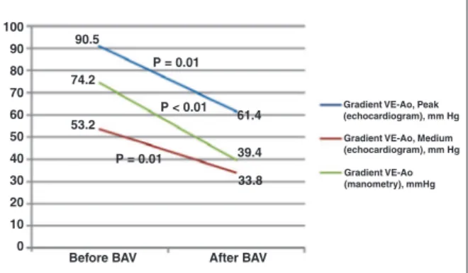
Procedural success was achieved in 100% of the cases. The temporary pacemaker was used to promote
rapid stimulation (fast pacing) in 68.4% of patients (Table 2). Signiicant reduction of the aortic transvalvular gradient was noted, both by echocardiography and by pressure measurements during the procedure (Figure 1).
The clinical condition in extremis was present in
42.1% of patients (Table 3). When compared to the
control group, patients in the in extremis group were
younger (70.1 ± 10.0 vs. 83.3 ± 8.5 years; P < 0.01)
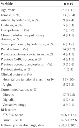
TABLE 1 Clinical characteristics
Variable n = 19
Age, years 77.7 ± 11.1
Female, n (%) 13 (68.4)
Arterial hypertension, n (%) 9 (47.4)
Diabetes, n (%) 5 (26.3)
Dyslipidemia, n (%) 7 (36.8)
Chronic obstructive pulmonary disease, n (%)
4 (21.1)
Severe pulmonary hypertension, n (%) 6 (31.6)
Renal failure, n (%) 14 (73.7)
Previous acute myocardial infarct, n (%) 4 (21.1)
Previous CABG surgery, n (%) 4 (21.1)
Previous coronary angioplasty, n (%) 3 (15.8)
Previous stroke, n (%) 1 (5.3)
Clinical picture, n (%)
Heart failure functional class III or IV 19 (100)
Angina 5 (26.3)
Current medication, n (%)
Diuretic 17 (89.5)
Digitalis 5 (26.3)
Vasoactive drugs 8 (42.1)
Risk scores
STS Risk Score 36.6 ± 17.4
EuroSCORE II 26.5 ± 22.6
Follow-up after discharge, days 268.3 ± 202.3
CABG = coronary artery bypass graft.
TABLE 2 Procedure characteristics
Variable n = 19
Success, n (%) 19 (100)
Use of pacemaker for rapid stimulation, n (%) 13 (68,4)
Vascular complications, n (%) 0
Stroke, n (%) 0
and had higher EuroSCORE II (41.1. ± 24.7 vs. 15.9
± 14.0l; P < 0.01). In addition, the in extremis group
had a lower left ventricle ejection fraction before the
procedure versus the control group (33.9 ± 17.3% vs.
49.0 ± 12.5%; P = 0.04). Hemodynamic and
echocar-diographic characteristics are shown in Table 4.
In both groups, there were no deaths during the
procedure. None of the eight patients in the in
extre-mis group survived the in-hospital period, while in
the control group the in-hospital mortality was 27.3%
(P < 0.01). Survival at 180 days is shown in Figure 2.
Three patients (15.8%) of the control group were submitted to BAV as a bridge to other deinitive treat-ment: one patient underwent TAVI by transapical ap-proach and died during the procedure, and two patients underwent AoVR (one of them was discharged and the other died in the immediate postoperative period). All patients who were discharged (42.1%) showed improve-ment in their heart failure functional class. The mean
follow-up was 268.3 ± 202.3 days.
DISCUSSION
This study was relevant in the analysis of the clini-cal, echocardiographic, and hemodynamic characteris-tics, as well as in the evolution of patients with severe degenerative aortic stenosis treated by BAV.
The high in-hospital mortality in the study relects the clinical proile of extreme severity of the patients, mainly due to the presence of multiple comorbidities. There are no other studies in the literature that include a group of patients as severe as those described in this work. The mean STS Risk Score in the PARTNER
study,5 which included only patients at high surgical
risk or considered inoperable, was 11.6 ± 6.0, i.e.,
much lower than that of the present study (36.6 ± 17,
4). Other studies involving only patients undergoing BAV also included patients with less severe conditions compared to the present study, with lower incidence of
comorbidities and with lower risk scores.6,7,11-14
Most patients (73.7%) showed renal failure at the time of the procedure, and most had pulmonary artery
pressure > 60 mmHg. Both conditions have been
as-sociated to an increase in mortality in earlier studys.9,15
In a study of 509 patients, Ben-Dor et al. de m- onstrated that the mortality after 5 months for patients
with SPPA > 60 mmHg was 49.1%, regardless of the
type of treatment received (BAV, TAVI, or AoVR).16
Several studies have correlated the presence of renal insuficiency before the procedure with increased
mortality.9,17,18 A study of 262 patients at high surgical
risk undergoing BAV showed that creatinine clearance ≤ 60 mL/min before the procedure is a strong
predic-tor of mortality.7
The absence of vascular complications in this study may be related to the use of sheaths with small calibre (10F) in all patients, as well as to the immediate with-drawal of the sheath. Despite being related to a shorter hospital stay and a lower rate of blood transfusions in
Figure 1 – LV-Ao gradient before and after balloon aortic valvuloplasty. LV, left ventricle, Ao, aorta; BAV, balloon aortic valvuloplasty.
TABLE 3
Clinical characteristics by group
Variable
In extremis
(n = 8)
Control (n = 11) P
Age, years 70.1 ± 10.0 83.3 ±
8.5
< 0.01
Female, n (%) 6 (75.0) 7 (63.6) 0.60
Systemic arterial hypertension, n (%)
3 (37.5) 6 (54.5) 0.46
Diabetes, n (%) 3 (37.5) 2 (18.2) 0.35
Dyslipidemia, n (%) 2 (25.0) 5 (45.5) 0.36
Chronic obstructive pulmonary disease, n (%)
3 (37.5) 1 (9.1) 0.13
Severe pulmonary hypertension, n (%)
3 (37.5) 3 (27.3) 0.64
Renal failure, n (%) 7 (87.5) 7 (63.6) 0.24
Acute myocardial infarction, n (%)
1 (12.5) 3 (27.3) 0.44
Previous CABG, n (%) 1 (12.5) 3 (27.3) 0.44
Previous coronary angioplasty, n (%)
0 (0) 3 (27.3) 0.11
Previous stroke, n (%) 0 (0) 1 (9.1) 0.38
Vasoactive drugs use 6 (75.0) 2 (18.2) 0.01
Risk scores
STS Risk Score 45.1 ± 15.9 30.4 ±
16.3
0.07
EuroSCORE II 41.1 ± 24.7 15.9 ±
14.0
0.01
CABG = coronary artery bypass graft.
1 0 0 9 0
8 0 7 0
6 0 5 0 4 0
3 0 2 0
1 0 0
9 0 .5
7 4 .2
5 3 .2
P = 0.01
P = 0.01 P < 0.01
61.4 39.4 33.8
After BAV Before BAV
Gradient VE-Ao, Peak (echocardiogram), mm Hg
Gradient VE-Ao, Medium (echocardiogram), mm Hg
these cases, mortality was 50%. Recently, some studies have addressed the use of BAV as a bridge to TAVI, showing excellent results in the short and long term,
when compared to BAV alone.6-8
The classification of patients according to the number of organ dysfunctions presented was of great importance to the understanding that there is a subgroup of more severe patients who evolve unfavourably, even after BAV. Patients with two or more organ dysfunc-tions had significantly higher mortality when compared to patients in the control group. The main reason for
the treatment of patients in the group in extremis
with BAV was the presence of a clinical condition of extreme gravity, whether or not with cardiac etiology, in the presence of severe aortic stenosis. It was hoped that, in these patients, the reduction of the gradient in the left ventricular (LV) outflow would provide an improvement of their LV function to the point that they could recover from the critical hemodynamic state present during their admission. However, even with the decrease of the aortic transvalvular gradient and the increase in LV ejection fraction after the pro-cedure, these results were not translated into clinical improvement, suggesting that these patients underwent the intervention too late.
Limitations of the study
The study had some limitations, such as the small number of patients included in the retrospective analysis of data and the fact that it was conducted in a single center.
TABLE 4
Hemodynamic and echocardiographic characteristics
Variable
In extremis
(n = 8)
Control
(n = 11) P
Before valvuloplasty
LV-Ao gradient (manometry), mmHg 70.6 ± 25.1 76.8 ± 23.1 0.59
LV-Ao gradient, peak (echocardiogram) mmHg 96.9 ± 35.6 85.9 ± 23.4 0.43
LV-Ao gradient, medium (echocardiogram) mmHg 55.9 ± 22.1 51.3 ± 15.8 0.60
Ejection fraction,% 33.9 ± 17.3 49.0 ± 12.5 0.04
Moderate or severe aortic regurgitation, n (%) 1 (12.5) 2 (18.2) 0.54
After valvuloplasty
LV-Ao gradient (manometry), mmHg 41.3 ± 21.5 38.0 ± 24.9 0.77
LV-Ao gradient, peak (echocardiogram) mmHg 66.8 ± 19.2 57.5 ± 20.5 0.34
LV-Ao gradient, medium (echocardiogram) mmHg 31.8 ± 18.9 35.3 ± 12.3 0.63
Ejection fraction,% 39.1 ± 15.9 52.7 ± 13.8 0.06
Moderate or severe aortic regurgitation, n (%) 2 (25.0) 3 (27.3) 0.36
LV = left ventricle; Ao = aorta.
Figure 2 – Survival after aortic balloon valvuloplasty.
patients undergoing AoBV,19 vascular occlusion devices
were not used in this study.
The division of patients according to the presence
or absence of the clinical condition in extremis was
extremely important for the understanding of the most appropriate time for performing BAV. The guidelines do not address BAV as a salvage therapy in patients in critical clinical condition with multiple organ dys-function, but do suggest that its performance can be
beneicial as a bridge to AoVR.2 In this study, only two
patients underwent BAV as a bridge to AoVR, and in 1.0
0,8
0.6
0.4
0.2
0.0
P < 0.01
0 30 60 90 120 150 180
Cum
ulative survi
va
l
Days after aortic valvuloplasty
Control
CONCLUSIONS
Balloon aortic valvuloplasty for treatment of severe aortic stenosis with degenerative etiology has unfavor- able outcome when indicated for patients with two or more organ dysfunctions, i.e., in a clinical condition in extremis.
CONFLICTS OF INTEREST
The authors declare no conlicts of interest.
REFERENCES
1. Nkomo VT, Gardin JM, Skelton TN, Gottdiener JS, Scott CG, Enriquez-Sarano M. Burden of valvular heart diseases: a population-based study. Lancet. 2006;368(9540):1005-11. 2. Bonow RO, Carabello BA, Chatterjee K, de Leon AC, Faxon
DP, Freed MD, et al. 2008 Focused update incorporated into the ACC/AHA 2006 guidelines for the management of patients with valvular heart disease: A report of the American College of Cardiology/American Heart Association Task Force on Practice Guidelines (Writing Committee to Revise the 1998 Guidelines for the Management of Patients With Valvular Heart Disease): endorsed by the Society of Cardiovascular Anesthesiologists, Society for Cardiovascular Angiography and Interventions, and Society of Thoracic Surgeons. Circulation. 2008;118(15):e523-661. 3. Lund O. Preoperative risk evaluation and stratiication of long-term survival after valve replacement for aortic stenosis: reasons for earlier operative intervention. Circulation. 1990;82(1):124-39. 4. Iung B, Baron G, Butchart EG, Gohlke-Bärwolf C, Levang OW,
Tornos P, et al. A prospective survey of patients with valvular heart disease in Europe: the Euro Heart Survey on Valvular Heart Disease. Eur Heart J. 2003;24(13):1231-43.
5. Leon MB, Smith CR, Mack M, Miller DC, Moses JW, Svensson LG, et al. Transcatheter aortic-valve implantation for aortic stenosis in patients who cannot undergo surgery. N Engl J Med. 2010;363(17):1597-607.
6. Ben-Dor I, Maluenda G, Dvir D, Barbash IM, Okubagzi P, Torguson R, et al. Balloon aortic valvuloplasty for severe aortic stenosis as a bridge to transcatheter/surgical aortic valve replacement. Catheter CardiovascInterv. 2012 Sep 27. [Epub ahead of print]
7. Ben-Dor I, Pichard AD, Satler LF, Goldstein SA, Syed AI, Gaglia MA, et al. Complications and outcome of balloon aortic valvuloplasty in high-risk or inoperable patients. JACC Cardiovasc Interv. 2010;3(11):1150-6.
8. Ussia GP, Capodanno D, Barbanti M, Scarabelli M, Imme S, Cammalleri V, et al. Balloon aortic valvuloplasty for severe
aortic stenosis as a bridge to high-risk transcatheter aortic valve implantation. J Invasive Cardiol. 2010;22(4):161-6. 9. Otto CM, Mickel MC, Kennedy JW, Alderman EL, Bashore
TM, Block PC, et al. Three-year outcome after balloon aortic valvuloplasty: insights into prognosis of valvular aortic stenosis. Circulation. 1994;89(2):642-50.
10. Lieberman EB, Bashore TM, Hermiller JB, Wilson JS, Pieper KS, Keeler GP, et al. Balloon aortic valvuloplasty in adults: Failure of procedure to improve long-term survival. J Am Coll Cardiol. 1995;26(6):1522-8.
11. Don C, Gupta PP, Witzke C, Kesarwani M, Cubeddu RJ, Ingles-sis I, et al. Patients with small left ventricular size undergo-ing balloon aortic valvuloplasty have worse intraprocedural outcomes. Catheter Cardiovasc Interv. 2012;80(6):946-54. 12. Dvir D, Sagie A, Porat E, Assali A, Shapira Y, Vaknin-Assa H,
et al. Clinical proile and outcome of patients with severe aortic stenosis at high surgical risk: single-center prospec-tive evaluation according to treatment assignment. Catheter Cardiovasc Interv. 2013;81(5):871-81.
13. Kapadia SR, Goel SS, Yuksel U, Agarwal S, Pettersson G, Svensson LG, et al. Lessons learned from balloon aortic val-vuloplasty experience from the pre-transcatheter aortic valve implantation era. J Interv Cardiol. 2010;23(5):499-508. 14. Tissot CM, Attias D, Himbert D, Ducrocq G, Iung B, Dilly MP,
et al. Reappraisal of percutaneous aortic balloon valvuloplasty as a preliminary treatment strategy in the transcatheter aortic valve implantation era. EuroIntervention. 2011;7(1):49-56. 15. Sherman W, Hershman R, Lazzam C, Cohen M, Ambrose J, Gorlin
R. Balloon valvuloplasty in adult aortic stenosis: determinants of clinical outcome. Ann Intern Med. 1989;110(6):421-5. 16. Ben-Dor I, Goldstein SA, Pichard AD, Satler LF, Maluenda
G, Li Y, et al. Clinical proile, prognostic implication, and response to treatment of pulmonary hypertension in patients with severe aortic stenosis. Am J Cardiol. 2011;107(7): 1046-51.
17. Ben-Dor I, Pichard AD, Gonzalez MA, Weissman G, Li Y, Goldstein SA, et al. Correlates and causes of death in patients with severe symptomatic aortic stenosis who are not eligible to participate in a clinical trial of transcatheter aortic valve implantation. Circulation. 2010;122(11 Suppl):S37-42. 18. Saia F, Marrozzini C, Ciuca C, Guastaroba P, Taglieri N,
Palm-erini T, et al. Emerging indications, in-hospital and long-term outcome of balloon aortic valvuloplasty in the transcatheter aortic valve implantation era. EuroIntervention.2013;8(12): 1388-97.