In vitro
evaluation of the effects of the interaction between
irrigating solutions, intracanal medication and Er:YAG laser in
dentin permeability of the endodontic system
Estudo
in vitro
dos efeitos da interação de substâncias irrigantes,
medicação intracanal e laser Er:YAG na permeabilidade dentinária
do sistema endodôntico
Denise Pontes Raldi* José Luiz Lage-Marques**
ABSTRACT:The purpose of this study was to evaluatein vitrothe effects of different associations between irrigating so-lutions (EDTA-T and citric acid), intracanal medicament (NDP), and Er:YAG laser irradiation on dentin permeability. Fifty-one extracted single-rooted teeth were instrumented and divided into seven groups. Groups GI and GII had final irrigation with a demineralizing solution only (EDTA-T and citric acid, respectively). Groups GIII and GIV had final irri-gation with EDTA-T and citric acid, respectively, plus an association of irrigating solution and Er:YAG laser. Groups GV and GVI had final irrigation with EDTA-T and citric acid, respectively, plus an association of intracanal medication and Er:YAG laser. Group GVII (control group) had final irrigation with distilled water. All root canals were filled with NDP associated with rhodamine B dye. After the experimental period, the samples were transversely cut into six 2.0 mm thick slices for subsequent reading using the ImageLab software. Analysis of the results allowed us to conclu-de that there were statistically significant differences (p < 0.05) between the groups as to the penetration of the dye-in-tracanal medication solution. Groups III and IV presented smaller values of dentinal permeability when compared to the other groups. The best results were obtained with the interaction between a demineralizing irrigating solution and the association of intracanal medicament and laser Er:YAG (groups V and VI). In these groups the observed penetra-tion of the intracanal medicament plus dye solupenetra-tion in the apical third was, on average, 29% greater than in the other groups.
DESCRIPTORS:Dentin permeability; Root canal irrigants; Lasers.
RESUMO: Este experimento teve como objetivo avaliar in vitro os efeitos da interação entre soluções irrigantes desmineralizadoras (EDTA-T e ácido cítrico), medicação intracanal (NDP) e laser Er:YAG na permeabilidade dentinária. Foram utilizados 51 dentes unirradiculares extraídos que, após o preparo químico-cirúrgico, foram divididos em sete grupos experimentais: grupos I e II – irrigação final com solução de EDTA-T e ácido cítrico, respectivamente; grupos III e IV – irrigação final com EDTA-T e ácido cítrico, respectivamente, mais a associação entre solução irrigante e laser Er:YAG; grupos V e VI – irrigação final com EDTA-T e ácido cítrico, respectivamente, mais a associação entre medicação intracanal e laser Er:YAG, e grupo VII (controle) – irrigação final com água destilada. Os canais radiculares foram preenchidos com o corante rodamina B solubilizado na medicação de uso intracanal NDP. Após o período experimental, as amostras foram cortadas transversalmente para posterior leitura com o software ImageLab. A análise dos resultados permitiu concluir que existiram diferenças estatisticamente significantes (p < 0,05) quanto à penetração da solução corante-medicação intracanal nos diferentes grupos. Os grupos III e IV apresentaram menores valores de permeabilidade dentinária quando comparados aos outros e, finalmente, os melhores resultados foram obtidos quando da interação entre a solução irrigante desmineralizadora e a associação medicação intracanal/laser Er:YAG (grupos V e VI). Nesses, constatou-se que a diferença de penetração da solução corante-medicação intracanal no terço apical foi, em média, 29% maior do que nos demais grupos.
DESCRITORES:Permeabilidade da dentina; Irrigantes do canal radicular; Lasers.
INTRODUCTION
The formation of a smear layer during chemi-cal-surgical preparation is an undisputed fact. It is deposited on the dentinal walls and obliterates the canaliculae, thus reducing dentinal permeability.
*Assistant Professor; **PhD, Professor – Discipline of Endodontics, School of Dentistry, Ibirapuera University.
It thus hinders penetration of chemical substan-ces and intracanal medicaments into the dentinal mass, thereby preventing adequate cleaning and disinfection of the root canal.
solu-tions and demineralizing substances (EDTA, citric acid) has been suggested. Both the organic and inorganic portions of magma would thus be acted upon. However, some studies have shown that these associations do not remove it completely, particularly when it is formed on the apical third of the root canal1
.
Since the 1980s, several studies analyzing the effect of Er:YAG laser irradiation on dental tissues have been presented. As this laser emits a 2.94mm wavelength, it is highly absorbed by water and hydroxyapatite, and consequently by enamel and dentin.
When applied to dentin, this irradiation is highly absorbed and produces vaporization follo-wed by micro explosions, causing ablation of the mineralized tissue12
. Because of this property, stu-dies have demonstrated the efficiency of the Er:YAG laser for cleaning root canal walls, vapori-zing pulpal tissues, removing magma and increa-sing dentin permeability21
. It is also noteworthy that these results have proven satisfactory even at the apical third, a less permeable root area13.
The aim of the present experiment is to evaluate
in vitrothe effects of the interaction of irrigating
so-lutions employed after chemical-surgical prepara-tion (EDTA-T and citric acid), intracanal medica-tion and Er:YAG laser on dentin permeability.
MATERIAL AND METHODS
In our experiment, 51 single-rooted, extracted teeth were used. They were stored in 1% thymol solution until commencement of the experimental phase. Their crowns were sectioned at the ena-mel-cementum limit, and then hydrated in saline solution for a period of at least 72 hours.
Chemical-surgical preparation was carried out 1 mm short of the radiographic vertex with type-K files (up to #40), auxiliary chemical substance Endo-PTC* and irrigation with 1% sodium hypoch-lorite, according to the technique described by Pai-va & Antoniazzi16
(1988). Immediately after prepa-ration, the root canals were irrigated with 5 ml of 1% sodium hypochlorite.
Final irrigation was then carried out with 15 ml of 17% EDTA** solution or 15% citric acid, accor-ding to the experimental groups described below (Table 1). Irrigation time was standardized at 4 mi-nutes. The control group was irrigated with 15 ml of distilled water. Drying of the root canals was carried out with aspiration cannulae and absor-bent paper cones.
After prior sealing of the apical foramina with utility wax, the roots were externally made imper-meable with ethyl cyanoacrylate.
The salt of the dye rhodamine B was used as the disclosing solution for dentin permeability, dissol-ved in the solution of intracanal medication NDP*** at a concentration of approximately 20%, following a pilot study performed prior to this expe-riment. The choice of the disclosing solution was based on studies5,7, in which rhodamine B was
found to be effective with regard to degree of pene-tration and coloring of the dentin when used in as-sociation with drugs used as intracanal medica-tion: NDP, chlorhexidine.
The root canals were filled with the dye-intraca-nal medication solution. After 60 s, the solution was removed by means of suction cannulae and absorbent paper cones until there was no further trace observed. This time duration was determined on the basis of the pilot study carried out prior to this experiment. Over 24 hours – a time commonly described in the literature11
– total penetration of the dye into all the tested samples occurred. The same result was obtained over 12, 6 and 2 hours. The 60 s duration could be explained by the fact that the concentration of rhodamine B powder (@20%) used in this study is considerably greater than in the abovementioned studies (1% to 2%).
The specimens of four groups (Table 1) were ir-radiated by Er:YAG laser, with a wavelength of 2.94mm,input pulse energy of 200 mJ, output of 84 mJ, repetition rate (frequency) of 6 Hz and ave-rage power of 1.2 W. Total energy was 24 J. The de-livery system was a fiber optic cable (0.375 mm thickness, 40 x 28 diameter, with two rings and a transmission factor of 0.42) coupled to a Kavo n. 2055 contra-angle handpiece (Kavo E 2055,
Bi-* Endo-PTC – ureia peroxide (10%)/tween 80 (15%)/carbowax (75%) – Fórmula & Ação Pharmacy, São Paulo, Brazil. ** EDTA-T – disodium salt of ethylenediaminetetraacetic 17 g/tergentol (sodium sulfate lauryldietyleneglycol ether), q. suff.
100 ml – Fórmula & Ação Pharmacy, São Paulo, Brazil.
berach, Germany). Irradiation was delivered by the contact method, introducing the fiber-optic cable along the entire length of the root canal, irradiating the dentin wall from apical to cervical along its en-tire length, with helicoidal movements during ap-proximately 5 s following Gutknecht et al.10(1996).
This procedure was repeated four times over ap-proximately 20 s, making up a total of 120 pulses.
Two groups (GIII and GIV) were irradiated with canals filled by the irrigating substance (EDTA-T or citric acid), and two further groups were irradia-ted after filling of the root canals with the intraca-nal medication-dye solution (GV and GVI). The root canals were thus filled either with an irriga-ting chemical substance or intracanal medication during irradiation.
Of the 51 specimens, four were used for analy-sis under the scanning electron microscope, and the remaining 47 specimens were divided up ac-cording to Table 1.
Using a Labcut Microtome (Extec®Labcut 1010
Low Speed Diamond Saw, Enfield, USA) seven transversal cuts were made in each specimen, of a standard 2.0 mm thickness, which resulted in six slices: two from the cervical third (C1 and C2), two from the medial third (C3 and C4), and two from the apical third (C5 and C6). The first two slices corresponding to the cervical and apical thirds were ignored.
The images of the dyed slices were digitalized using the ImageLab 2.3 software* to assess the pe-netration of the dye-intracanal medication soluti-on by measurement of the dyed area. The
percen-TABLE 1 -Distribution of the 47 specimens according to treatment performed.
Group I (7 specimens) Final irrigation with EDTA-T + solution of rhodamine B-intracanal medication NDP Group II (7 specimens) Final irrigation with citric acid + solution of rhodamine B-intracanal medication NDP
Group III (7 specimens) Final irrigation with EDTA-T + association of EDTA-T/Er:YAG laser + solution ofrhodamine B-intracanal medication NDP
Group IV (7 specimens) Final irrigation with citric acid + association of citric acid/Er:YAG laser + solution ofrhodamine B-intracanal medication NDP
Group V (7 specimens) Final irrigation with EDTA-T + association of solution of rhodamine B-intracanal me-dication NDP/Er:YAG laser
Group VI (7 specimens) Final irrigation with citric acid + association of solution of rhodamine B-intracanalmedication NDP/Er:YAG laser
Group VII (5 specimens) Control – final irrigation with distilled water + solution of rhodamine B-intracanal me-dication NDP
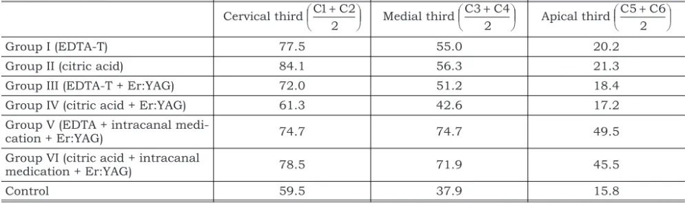
TABLE 2 -Percentual averages of penetration of the dye-intracanal medication solution in the cervical, medial and api-cal thirds of the experimental groups.
Cervical third C1 C2 2 + æ è
ç öø÷ Medial third C3 C4 2 + æ è
ç öø÷ Apical third C5 C6 2 + æ è
ç öø÷
Group I (EDTA-T) 77.5 55.0 20.2
Group II (citric acid) 84.1 56.3 21.3
Group III (EDTA-T + Er:YAG) 72.0 51.2 18.4
Group IV (citric acid + Er:YAG) 61.3 42.6 17.2
Group V (EDTA + intracanal
medi-cation + Er:YAG) 74.7 74.7 49.5
Group VI (citric acid + intracanal
medication + Er:YAG) 78.5 71.9 45.5
Control 59.5 37.9 15.8
tage of the dyed area in relation to the total radicular area was calculated for each cut.
RESULTS
The results obtained are set out in Table 2 and Graph 1 and represent the average penetration, as a percentage, of the rhodamine B dye - NDP intra-canal medication solution, respectively in the cer-vical, medial and apical thirds.
The results of the statistical analysis by the Kruskal-Wallis test, at a level of significance of 5%, are presented on Table 3.
Results of scanning electron microscopy for specimens of Group III and IV (500 X magnifica-tion) are presented in Figures 1, 2 and 3.
DISCUSSION
Groups I and II (specimens treated with irriga-ting substances EDTA-T or citric acid) presented superior results in comparison with the control group, with a statistically significant difference for the penetration of the dye-intracanal medication solution in the cervical and medial thirds (Table 3). These results are borne out in present literature8-9
, in which the use of demineralizing irrigating solu-tions is reported as helping in the cleaning of root canals since they act on the inorganic portion of
the canal’s contents increasing dentin permeabi-lity and thus enabling a freer passage of medica-tions and solumedica-tions through the dentinal tubules6
. It is noteworthy that these differences, in most studies, are minor and not statistically significant. They are justified by possible variations both in the evaluation methodology and in the concentrations of chelating substances and pH of the solutions in question.
The fact that there is no difference in penetra-tion of the dye-intracanal medicapenetra-tion solupenetra-tion in the apical third between groups I, II and the con-trol group might be explained by the fact that the region is the least permeable owing to its anatomi-cal conditions, as well as the most refractory to the action of instruments and therefore the most criti-cal in terms of cleanliness and the formation of dentinal magma.
In groups III and IV specimens, penetration of the dye-intracanal medication solution was grea-ter than in the control group. However, it was less than in the non-irradiated specimens, groups I and II (Table 1). These differences were unexpected in that several studies have shown greater clea-ning of the walls of root canals when irradiated with Er:YAG laser (Key IIâ, Kavo, Biberach,
Ger-many – Laboratório Experimental de Laser em
Odontologia do Departamento de Dentística da Fa-culdade de Odontologia da Universidade de Sao Paulo. This is because the laser causes ablation of the dentinal tissue, removal of dentinal magma and opening of the dentinal tubules20,21
. Better re-sults were therefore expected when associating de-mineralizing substances with laser irradiation.
In the present study, as with those by Pécora et al.18
(2000) and Brugnera Junior3
(2000), there may presumably have been interaction between the Er:YAG laser and the irrigating substances used, which interfered in the absorption of the laser energy by the dentinal tissue and in the values of dentin permeability to the dye-intracanal medica-tion solumedica-tion.
This interaction has been ascribed to the high ionic conductance of the substances in question, which, owing to their possessing a large quantity of free ions and different absorption peaks from the Er:YAG laser, prompted dispersion of laser irradia-tion, causing it to hit the target tissue with altered intensity, reducing its absorption and therefore its effects19.
One factor that might be questioned in this study is the thermal effect generated by laser irra-diation, since water jets were not used during ap-plication and studies have shown that cooling enhances the effectiveness of laser irradiation (de-gree of ablation of hard dental tissue) and mitiga-tes damaging effects15,17
. Two precautions were the-refore taken: at the moment of irradiation the
FIGURE 3 -View of the medial third, aspect of the dentin, after treatment with citric acid and Er:YAG (group IV).
White crystalline particles.
FIGURE 4 -View of the cervical third, with evident morp-hologic change of the dentin after irradiation with Er:YAG laser observed in specimen of group III (EDTA-T + Er:YAG). Melting and recrystallization zo-nes in dentin – “bulletscoring” aspect.
FIGURE 1 -View of cervical third, aspect of the dentin, af-ter treatment with citric acid + Er:YAG (Group IV). Note the entrances of unobstructed dentinal tubules and ab-sence of smear layer.
canals were filled with the irrigating substance or the dye-intracanal medication solution, and the rate of repetition (frequency) was reduced to 6 Hz, because it has been observed that the effect of an increased frequency rate of laser emission on the temperature curve is very pronounced in compari-son to an equivalent increase in incident energy12
. In order to better elucidate the results obtained with groups III and IV, it was decided that two spe-cimens from each group should be analyzed under scanning electron microscopy. In the tested sam-ples, areas of dentin that were totally free of debris and that had open dentinal tubules were found (Fi-gure 1); these findings are the most commonly seen in groups irradiated with Er:YAG laser13,20,21
. Areas with morphological changes (melting and re-solidification) indicative of laser irradiation were found (Figure 2).
Both in group III and in group IV, as with the studies of Miserendino et al.14
(1995), white crystalline particles were found deposited on the walls of the root canals, which had open dentinal tubules (Figure 3). The presence of these particles as well as nuclei of crystallization (Figure 4) in dif-ferent regions of the irradiated dentin may possibly account for the reduced penetration of the dye-in-tracanal medication solution.
Groups V and VI, when compared with the re-maining groups, showed much superior results, above all in the medial and apical thirds (Tables 2 and 3). Penetration of the dye-intracanal medica-tion solumedica-tion in the apical third was on average 29% higher than in the remaining groups.
These results are extremely important given the ceaseless pursuit of greater cleaning and disinfec-tion of the endodontic system, especially in this re-gion, which, as has been mentioned, is most
criti-TABLE 3 -Differences between penetration averages for the dye-intracanal medication solution in the cervical, medial and apical thirds of the experimental groups and the results of the Kruskal-Wallis test.
Cervical third
GII (84.1) GIII (72) GIV (61.3) GV (74.7) GVI (78.5) GVII (59.5)
GI (77.5) 6.6 5.5 16.2 2.8 1.0 18
GII (84.1) 12.1 22.8 9.4 5.6 24.6
GIII (72) 10.7 2.7 6.5 12.5
GIV (61.3) 13.4 17.2 1.8
GV (74.7) 3.8 15.2
GVI (78.5) 19
Medial third
GII (56.3) GIII (51.2) GIV (42.6) GV (74.7) GVI (71.9) GVII (37.9)
GI (55) 1.3 3.8 12.4 19.7 16.9 17.1
GII (56.3) 5.1 13.7 18.4 15.6 18.4
GIII (51.2) 8.6 23.5 20.7 13.3
GIV (42.6) 32.1 29.3 4.7
GV (74.7) 2.8 36.8
GVI (71.9) 34
Apical third
GII (21.3) GIII (18.4) GIV (17.2) GV (49.5) GVI (45.5) GVII (15.8)
GI (20.2) 1.1 1.8 3 29.3 25.3 4.4
GII (21.3) 2.9 4.1 28.2 24.2 5.5
GIII (18.4) 1.2 31.1 27.1 2.6
GIV (17.2) 32.3 28.3 1.4
GV (49.5) 4 33.7
GVI (45.5) 29.7
cal and less permeable to penetration by irrigating substances and intracanal medication1
.
This significant increase in penetration of intra-canal medication caused by irradiation by Er:YAG laser is, from a clinical point of view, extremely ap-plicable, especially in cases of resistant and refrac-tory infections in which the micro-organisms (fa-cultative aerobes and anaerobes) propagate into deeper regions of the endodontic system.
With regard to the superior results obtained in groups V and VI, this may be explained by the me-chanical effect stemming from the interaction of la-ser irradiation with the target tissue. This effect may result from the formation of shock waves from the production of photons in the irradiated tissue2
. In this case, irradiation with Er:YAG laser of root canals filled with the dye-intracanal medication solution may have provoked a “micro-explosion” of the solution into the dentinal tubules. During irra-diation of the specimens of these groups, this acti-on was visually cacti-onfirmed since the dye-intracanal medication solution could be seen being thrown in several directions.
The results obtained in this experiment allow us to state that Er:YAG laser proved effective as a coadjuvant in the medication phase of endodontic treatment, providing better penetration of the
in-tracanal medication into the root canal system. However, it should be pointed out that experi-ments evaluating the effects of Er:YAG laser irradi-ation on root canals are very recent, which is why varying and peculiar results have been obtained. Further studies should therefore be performed in order to better elucidate these interactions.
CONCLUSIONS
In light of what has been stated here, and of the results obtained, we feel it relevant to conclude that:
1. Final irrigation with demineralizing substances (EDTA-T and citric acid) enhanced dentinal per-meability.
2. Interaction between the demineralizing irriga-ting substances (EDTA-T and citric acid) and Er:YAG laser was unfavorable and inefficient in enhancing dentinal permeability.
3. The interaction between demineralizing subs-tance + association of intracanal medication and Er:YAG laser was efficient and promoted the greatest rates of permeability in all the root thirds, the permeability in the apical third being on average 29% greater than in the remaining groups.
REFERENCES
1. Aktener BO, Bilkay U. Smear layer removal with different concentrations of EDTA. J Endod 1993;49:228-31. 2. Bachmann L. Sistema de entrega de feixe para laser de
hól-mio e aplicações em Endodontia [Dissertação de Mestra-do]. São Paulo: Instituto de Pesquisas Energéticas e Nu-cleares da USP; 2000.
3. Brugnera Jr A. Estudo da ação dos lasers Er:YAG e Nd:YAG sobre a permeabilidade da dentina das paredes dos canais radiculares instrumentados [Tese de Doutorado]. Rio de Janeiro: Faculdade de Odontologia da UFRJ; 2000. 4. Cecchini SCM. Estudoin vitrodas aplicações do laser
Hól-mio:YLF em esmalte e dentina, visando a realização de ci-rurgia de acesso endodôntico e preparo cavitário [Disserta-ção de Mestrado]. São Paulo: Instituto de Pesquisas Energéticas e Nucleares da USP; 1995.
5. Fidel SR, Lage-Marques JL, Antoniazzi JH. Avaliação da capacidade de penetração dentinária radicular da chlorhe-xidine associada a três diferentes veículos. RPG Rev Pós Grad 1995;2:121-6.
6. Foster KH, Kulild JC, Weller RN. Effect of smear layer re-moval on the diffusion of calcium hydroxide through radi-cular dentin. J Endod 1993;19:136-40.
7. Fróis I. Análisein vitroda capacidade de penetração denti-nária de alguns fármacos utilizados na medicação intraca-nal [Dissertação de Mestrado]. São Paulo: Faculdade de Odontologia da USP; 1989.
8. Gaberoglio R, Becce C. Smear layer removal by root canal irrigants. A comparative scanning electron microscopic study. Oral Surg Oral Med Oral Pathol 1994;78:359-67. 9. Goldman M, Goldman LB, Cavaleri R, Lin PS. The efficacy
of several endodontic irrigating solutions: a scanning elec-tron microscopic study: part 2. J Endod 1982;8:487-92. 10. Gutknecht N, Kaiser F, Hassan A, Lampert F. Long-term
clinical evaluation of endodontically treated teeth by Nd:YAG lasers. J Clin Laser Med Surg 1996;14(1):7-11. 11. Hamaoka L, Moura AAM. Avaliaçãoin vitroda
permeabili-dade dentinária radicular, tendo como fonte de variação três diferentes tipos de corantes. Rev Odontol Univ São Paulo 1996;10:30-42.
12. Hibst R, Keller U. Heat effect of pulsed Er:YAG laser radia-tion (1990) apud Cecchini SCM. Estudoin vitrodas aplica-ções do laser Hólmio:YLF em esmalte e dentina, visando a realização de cirurgia de acesso endodôntico e preparo ca-vitário [Dissertaçao de Mestrado]. São Paulo: Instituto de Pesquisas Energéticas e Nucleares da USP; 1995. 13. Matsuoka E, Kimura Y, Matsumoto K. Studies on the
re-moval of debris near the apical seats by Er:YAG laser and assessment with a fiberscope. J Clin Laser Med Surg, 1998;18:255-61.
15. Paghdiwala AF. Root resection of endodontically treated te-eth by Erbium:YAG laser radiation. J Endod 1993;19:91-4. 16. Paiva JG, Antoniazzi JH. Fase do preparo do canal radicu-lar. In: Endodontia: bases para a prática clínica. 2ª ed. São Paulo: Artes Médicas; 1988. p. 531-629.
17. Pécora JD, Brugnera Jr A, Cussioli AL, Zanin F, Marche-san MA, Daghastnli NS, et al. Effect of energy (J) on the temperature changes at the apical root surface when using Er:YAG laser to enlarge root canal. Laser in Dentistry VI 2000;1:90-4.
18. Pécora JD, Brugnera Jr A, Cussioli AL, Zanin F, Silva R. Evaluation of dentin root canal permeability after instru-mentation and Er:YAG laser application. Lasers Surg Med 2000;26:277-81.
19. Ribeiro RG. Estudo da permeabilidade dentinária das pa-redes dos canais radiculares instrumentados com diferen-tes soluções irrigandiferen-tes, associadas ou não à irradiação de laser Er:YAG [Dissertação de Mestrado]. Ribeirão Preto: Faculdade de Odontologia da USP; 2001.
20. Takeda FH, Harashima T, Kimura Y, Matsumoto K. Effi-cacy of Er:YAG laser irradiation in removing debris and smear layer on root canal walls. J Endod 1998; 24:548-51. 21. Takeda FH, Harashima T, Kimura Y, Matsumoto K. A
com-parative study of the removal of smear layer by three endo-dontic irrigants and two types of laser. Int Endod J 1999;32:32-9.
Recebido para publicação em 16/09/02 Enviado para reformulação em 21/02/03 Aceito para publicação em 07/04/03