v. 11 n. 4, 2007ISSN 1809-9246 Influence of different laser therapy wavelengths in tendon regeneration 247 Rev. bras. fisioter., São Carlos, v. 11, n. 4, p. 247-252, July/Aug. 2007
©Revista Brasileira de Fisioterapia
INFLUENCES OF DIFFERENT LOW LEVEL LASER THERAPY
WAVELENGTHS IN RAT TENDON REGENERATION AFTER TENOTOMY
A
RRUDAERB
1, R
ODRIGUESNC
2, T
ACIROC
3& P
ARIZOTTONA
41 Graduate Program in Bioengineering, University of São Paulo, São Carlos, SP - Brazil 2 Graduate Program in Biotechnology, Federal University of São Carlos - UFSCar, SP - Brazil
3 Graduate Program in Physical therapy, UFSCar 4 Physical therapy Department, UFSCar
Correspondence to: Nivaldo Antonio Parizotto, Departamento de Fisioterapia, Universidade Federal de São Carlos, Rod. Washington Luiz, km 235, CEP 13565-905, São Carlos, SP - Brasil
Received: 17/08/2006 - Revised: 24/04/2007 - Accepted: 28/06/2007
ABSTRACT
Objective: The aim of this study was to compare the effect of the low level laser therapy in the organization of the collagen fibers with 3 J/cm² of dose in two wavelengths (670nm and 904nm) and in the combination of both on the tissue repair of the calcaneus tendon. Methods: We used 37 female Wistar rats divided in 5 groups which had been submitted to the total tenotomy. Group GA (n= 8) was submitted to the laser irradiation in λ= 904nm, GaAS, pulsated emission, peak power 15W and dose of 3J/ cm², group GB (n= 8) was submitted to laser irradiation λ= 670nm AlGaInP diode, continuous emission, power 30 mW and dose 3J/cm², group GAB (n= 8) was submitted to association of both of lasers and group GCL (n= 7) which the calcaneum tendon was submitted to placebo treatment. The group GP (n= 6) was a standard group without lesion and treatment. The animals received 12 sessions of lasertherapy and submitted to euthanasia on 14th day after surgery. The tendons were submitted to histological
procedureand were analyzed to evaluate the optic retardation (in nm) for the birefringence measurement. Results: The statistic analysis showed that the irradiated groups have higher values of optic retardation statistically significant than placebo group (p= 0,00001). It indicates that all the treated groups show better organization of collagen fibers in the longitudinal axis of the tendon, evidencing better quality in the tissue repair. The group GAB was that shown best results (p= 0,00001) when compared to another groups.
Key words: low-intensity laser therapy; Achilles tendon; tissue repair; total tenotomy; birefringence.
INTRODUCTION
Tendinous tissue is a type of dense conjunctive tissue and has as function to transmit the forces produced by the muscle to the bone, making possible articular movement1.
Enwemeka et al.2 report that the tendon is constituted by
fibroblasts and extracelular matrices, in which are immerse fibrous collagen proteins as well as elastine, proteoglicanes, glycoproteins and muco-poli-saccharids. Collagen is the major extracelular matrix component, making up about 86% to 95% of the tendon’s humid weight. Collagen fibrils are long and crystalline. In tendons, fibrils and fibers are highly aligned, which results in a high mechanical resistance for traction forces. Birefringence analysis quantifies these fibers alignment due to its anisotropical properties, by means of which optic retardation is measured in nanometers. Therefore, the higher this measurement is, the greater is the degree of fiber organization and compacting1.
The tendinous lesions scarring process may take weeks or even months. During this period, the patient is generally immobilized to avoid ruptures, which causes innumerous functional complications, retarding the rehabilitation process3.
The initial phase of the repair process takes 7 to 10 days to consolidate, however, complete rehabilitation may take weeks or months4.
Due to the fact that the occurrence of these lesions is so common, development of studies with the objective to help tendinous repair becomes necessary, reducing recovery time, and the return to daily activities. The importance of these studies to be performed in rats is due to the difficulty of promoting some invasive procedures in human beings.
Over the last decades, an increase in clinical interest for the biological evidence of optimization of the tendinous repair process has been observed, which has influenced the strategies adopted for the treatment of tendon injuries5-11. In
modalities with the objective to accelerate the regenerative process, and thus have shown that photo-bio-modulation, through low intensity laser irradiation with the different wavelengths, interacts in different ways in tendinous repair3,7,11.
Since then, many studies with photo-biomodulation were performed only with male rat tendons, due to the females’ menstrual cycle, and its possible implications on tissue repair. However, incidence of this kind of lesion is increasing among women. This can be related to the search for sport practices that overload these women’s tendons12. Structural and
functional factors such as muscular weakness, limb discrepancies, which among others, influence the increase of the incidence of these injures for females.
Despite the possible interference of the menstrual cycle on the tendinous repair process, when this kind of injury occurs, it does not matter in which phase of the menstrual cycle the rat is at the moment, and, due to this factor, the estrogenic phase of the females was not pre-determined to simulate the different previous conditions in patients.
It was observed also that wavelength is the most important feature of laser radiation, since it determines which bio-molecules will interact14. Visible radiation has a
mitochondrial way, and an invisible, infrared action over the cell membrane chromophores. Therefore, the therapeutic effects are specific, which indicates that there is a possibility of using both stimulation methods with the objective of improving performance. However, because it is a relatively new technology, its real effects, effective applications and limitations are still in an innovative phase15,16.
METHODOLOGY
Experimental animals
Thirty-seven five-weeks-old Wistar female rats (Rattus norvegicus albinos), with corporal mass of 181 ± 12.3 g, obtained on the Central Biotherium of the Federal University of São Carlos (UFSCar) where used. They were kept on the biotherium of the Eletro-thermal-photo-therapy Laboratory, in cages of pattern polipropylene, in groups of four, kept in a controlled environment, with a light/dark cycle of 12 hours, receiving water and balanced food ad libitum. This study project was approved by the Commission of Ethics on Animal Experimentation of the São Carlos University – CEAE/UFSCar, protocol nº CEAE 009/2006. No sample loss was registered during the present experiment.
Surgical procedures
The model used on this study was based on the experiment by Enwemeka et al.3. The animals were initially
weighed and later intraperitoneally anesthetized with 10% Ketamine Chloridrate (95 mg/kg) and 2% Xilazine Chloridrate (12 mg/kg). The skin over the posterior region of the right posterior limb tibia, on the local correspondent to the calcaneus tendon, was manually trimmed. After local cleansing (with iodized alcohol), a longitudinal incision on the skin over the
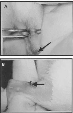
place correspondent to tenotomy was performed, on the middle third of the Achilles’ tendon, between the calcaneus insertion and muscular-tendinous junction, where it was performed total tenotomy by transversal shearing, using a number 11 scalpel blade (Figure 1A).
Following this, the skin was sutured with a 4.0 poliamide mono-filament non-absorbable thread, and was submitted to local cleansing (with iodized alcohol), no kind of segment immobilization occurred afterwards (Figure 1B).
Figure 1. A. Calcaneus tendon exposed after incision in the skin, ready to suffer tenotomy for transversal shearing, the arrow evidences the calcaneus. B. Suture accomplished in the skin (arrow) after tenotomy of tendo calcaneus and cleaning with iodized alcohol.
Experimental groups
The animals were randomly divided into five groups: GA group (n= 8): Achilles tendon was injured and submitted to infrared laser irradiation with a wavelength of 904 nm, pulsed, peak power of 15W and 3J/cm2, and an application
time of 9 seconds; GB group (n= 8): the calcaneus tendon was injured and submitted to red laser irradiation with a wavelength of 670 nm, continuous, power peak of 30mW and dose 3J/cm2, and a time of application of 6 seconds; GAB
v. 11 n. 4, 2007 Influence of different laser therapy wavelengths in tendon regeneration 249
Equipment
The utilized equipment was calibrated at the Optic Group of the University of São Paulo-USP, São Carlos Campus, in order to obtain a high reliability for the effective intensity of the laser emission. An IBRAMED class 3B laser was chosen with a dose of 3J/cm2 at the wavelengths of 904 nm and 670
nm.
The equipment has a GaAs diode, which works only at a pulsed regime with wavelength of 904 nm and peak power of 15W (the diode’s potency is fixed), pulsed at the frequency of 2000Hz, 3J/cm2 dose, sheaf diameter of 0.07 cm2, pulse
duration of 180 ns, resulting in a time of application of nine seconds. It also possesses an AlGaInP diode with a wavelength of 670 nm and power of 30mW (diode potency is fixed), continuous, 3J/cm2, sheaf area of 0.02 cm2, resulting in a
time of application of 6 seconds. The combination of both wavelengths applied one after the other resulted on a time sum of 15 seconds. The way of emission differentiated between both wavelengths, due to the fact that GaAs diode only emits pulsed radiation, and by the fact that the diodes potency on this equipment is fixed, there was no standardization of the same in order to reproduce with greater efficiency their clinical practice use.
Laser therapy started 24 hours after tenotomy. Treatment was made up of 12 consecutive uninterrupted sessions. All experimental procedures were made at the same circadian period. For laser applications, animals were immobilized by means of manual apprehension, and were irradiated by trans-skin photo-stimulation on the region of the injury, with the contact technique, at an angle of 90º in relation to the injured area surface.
On the 14th day after tenotomy (24 hours after the last
session), animals suffered euthanasia and their tendons were surgically removed by dissection from the heel insertion until the muscular-tendinous junction. The tendons were washed immediately in saline solution at 0.9% for plate confection in order to perform the birefringence analysis.
Slide preparation
For a qualitative and quantitative assessment of concentration, aggregate state and collagen fiber orientation, the tendons were dissected, washed and submitted to slide confection. Each slide was made with a series of three consecutive cuts of each tendon and each group of animals, being represented by 111 slides confectioned with the tendons of each one of the experimental groups. These slides did not receive any kind of staining, and remained uncovered in order to be re-hydrated at the moment of analysis.
Birefringence measurements
The collagen fiber analysis was performed using one of its optic anisotropic properties: total birefringence, measured by means of polarization microscopy. The slides for each
group were immersed in distilled water (refraction index of n= 1.333) for about 30 minutes, for analysis through birefringence, according to the studies by Vidal1.
After the immersion period, the slides were covered with laminules containing distilled water on the interfaces. For performance analysis, a LEICA polarized light microscope was used with a 10X/0.22 inches objective, 0.9 condenser, Sénarmont λ/4 compensator, monochromatic light λ= 546 nm, obtained through a LEICA interference filter from the LAMAV (UFSCAR Department of Material Engineering). This kind of analysis has been used in order to measure the degree of collagen fiber organization in a quantitative way, as in several other studies1,17,18. Measurements were taken with the tendon’s
axis at a 45º inclination in relation to the polarizer and the analyzer, allowing greater birefringence brightness and, therefore, an analysis of the collagen fibers alignment throughout the tendon’s axis.
RESULTS
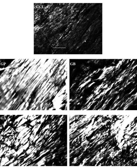
In Figure 2, a representation of the normally observed images through polarization microscopy may be verified. It may be noticed that there is qualitative evidence on the appearance of the tendon’s collagen fiber organization, showing that the groups treated with laser are in better conditions of structural organization. Application of both kinds of lasers on the same tendon has shown to be more effective on the collagen fibers organization.
For quantitative analysis of optic retardation, the results were analyzed using one-way ANOVA with Dunnett’s post hoc analyses. The GAB group showed the best collagen fiber organization when compared to the other groups, except for GP. The GB group obtained a higher significance index in comparison to the GA and GCL, and finally the GA obtained the best results when compared to the GCL (Figure 3). The differences were statistically significant (p< 0.01) in the comparisons of all groups.
DISCUSSION
The benefits of the low intensity laser therapy (LILT) on the collagen fibers organization occurs with certain wavelengths during the repair process according to several experiments performed9,10,11. These properties seem to result
from the fact that LILT projects a signal on the extracellular matrix, promoting changes of the fiber structures. This occurs after the electromagnetic energy interacts with the collagen molecules. The azimuth organization of the collagen on the longitudinal axis of the tendon shows strong evidence of structural changes that occur with the fibers after having been laser-irradiated10,19,20.
O P T I C A L R E T A R D (nm)
0.00 20.00 40.00 60.00 80.00
GP GCL GAB GB GA
Experimental groups 0.00
20.00 40.00 60.00 80.00
GP GCL GAB GB GA
Experimental groups
measurements after total calcaneus tenotomy treatment are observed1,16,17. Such data corroborate the results from other
authors who have used LILT during 12 days of tissue repair processes and verified responses from the fibroblastic cells and collagen molecules to the treatment7,8,19-28.
The types of activation ways proposed for the laser, take into account its action over the chromophores situated at the mitochondrias and cell membranes. Gigo-Benato et al.29 have described a possible synergic effect while using
two distinct wavelengths in a peripheral nervous injury, which indicates that this synergy may occur on the same way with the tendinous tissue. The chromophores seem to be different for the distinct wavelengths. Lievens29 has verified an increase
in the fibroblastic proliferation of injuries, by using a combination of He-Ne laser (632.8 nm) and As-Ga (904nm), 68.8mW, suggesting that the combination of lasers in different
λ may bring better results on the conjunctive tissue recovery.
GCL
____
GP GB
GA GAB
Figure 2. The image shows the string collagens birefringency shine of the mice groups analyzed in polarization microscopy. The GCL group \shows the disposition of the collagen bundle slightly aligned with the longitudinal axis of the tendon (45°). The GP group reveals a high degree of azimuthal order alignment, while the GB group exhibited some bundles along the longitudinal axis were more organized than the GCL. The GA group shows better alignment quality when compared with GCL and GB. And finally the GAB group showed the best bundle organization in comparison to the other injured groups, except by the GP, which did not suffer injury. The photomicrograph amplification was 20Xpol. The bar corresponds to 200μm.
v. 11 n. 4, 2007 Influence of different laser therapy wavelengths in tendon regeneration 251
The present results provide evidence for a synergy of action of the red and infrared lasers for the repair of conjunctive tissue in the tendons of rats. Due to the divergences found in the literature and the lack of research, there is still necessity of further more detailed studies in order to elucidate the action mechanisms of the LILT and the combination of different λ to standardize the parameters for its use in clinical
practice.
CONCLUSIONS
Based on the results obtained on the birefringence measurements from this experiment, it is possible to conclude that LILT was efficient in promoting a better level of collagen fiber organization throughout the longitudinal axis, thus suggesting a better tendinous repair, after total calcaneus tendon tenotomy, at the dose of 3J/cm2, with 904 nm and
670 nm λ, and with the association of both. The best results
were obtained with the association of both 670 nm and 904 nm wavelengths.
REFERENCES
1. Vidal BC. Image analysis of tendon helical superstructure using interference and polarized light microscopy. Micron. 2003; 34:423-32.
2. Enwemeka CS. Inflammation, cellularity, and fibrillogenesis in regenerating tendon: implications for tendon rehabilitation. Phys Ther. 1989;69(10):816-26.
3. Enwemeka CS, Reddy K. The biological effects of laser therapy and other modalities on connective tissue repair process. Laser Therapy. 2000;12:22-30.
4. Soma CA, Mandelbaum BR. Repair of acute Achilles tendon ruptures. Orthop Clin North Am. 1995;26:239-47.
5. Palmes D, Spiegel HU, Schneider TO, Langer M, Stratmann U, Budny T, et al. Achilles tendon healing: long-term biomechanical effects postoperative immobilization and mobilization in a new mouse model. J Orthop Res. 2002;20(5):939-46.
6. Koeke PU, Salate ACB, Parizotto NA, Barbosa G, Gaspar P, Benze BG, et al. Effect of In-Ga-Al-P diode laser irradiation on angiogenesis in partial ruptures of Achilles tendon in rats. Photomed Laser Surg. 2005;23(5):470-5.
7. Vinck EM, Cagnie B, Cornelissen MJ, Declercq HA, Cambier DC. Increased fibroblast proliferation induced by light emitting diode and low power laser irradiation. Lasers Med Sci. 2003;18(2):95-9.
8. Azevedo LH, Eduardo FP, Moreira MS, Eduardo CP, Marques MM. Influence of different power densities of LILT on cultured human fibroblast growth: A pilot study. Lasers Med Sci. 2006;21(2):86-9.
9. Reddy GK, Stehno-Bittel L, Enwemeka CS. Laser photostimu-lation of collagen production in healing rabbit Achilles tendons. Lasers Surg Med. 1998;22:281-7.
10. Enwemeka CS, Cohen-Korneberg GE, Duswalt EP, Weber DM, Rodriguez IM. Biomechanical effects of three different periods of GaAs laser photostimulation on tenotomized tendons. Laser Therapy. 1994;6:181-8.
11. Enwemeka CS, Rodrigues OO, Gall NG, Walsh NE. Morphome-trics of collagen fibril populations in He-Ne laser photostimu-lated tendons. J Clin Laser Med Surg. 1990;8:47-62.
12. Kannus P, Niitymaki S, Jarvinen M. Recent trends in women’s sports injuries. A three-year prospective, controlled study. Journal Sports Trauma. 1990;12:161-7.
13. Vladimirov YA, Osipov AN, Klebanov GI. Photobiological prin-ciples of therapeutic applications of laser radiation. Biochemistry. 2004;69(1):81-90.
14. Amat A, Rigau J, Waynant RW, Ilev IK, Anders JJ. The electric field induced by light can explain cellular responses to electromagnetic energy: A hypothesis of mechanism. J Photochem Photobiol B. 2006;82:152-60.
15. Ortiz MCS, Carrinho PM, Santos AAS, Gonçalves RC, Pari-zotto NA. Laser de baixa intensidade: princípios e generalidades – parte1. Fisioter Bras. 2001;2(4):221-40.
16. Carrinho PM, Koeke PU, Rennó AC, Vidal BC, Parizotto NA. Comparative study using 685 nm and 830 nm lasers in the tissue repair of tenotomized tendons in mices. Photomed Laser Surg. 2006;24(6):754-8.
17. Koeke PU, Parizotto NA, Carrinho PM, Salate AC. Compara-tive study of the efficacy of the topical application of hydrocortisone, therapeutic ultrasound and phonophoresis on the tissue repair process in rat tendons. Ultrasound Med Biol. 2005;31(3):345-50.
18. Parizotto NA, Baranauskas V. Hidrogen bonding of collagen molecule stimulated by He-Ne laser in regenerating of tendon. Proceeding 2º Congress World Association for Laser Therapy; 1998 Sept 2-5; Kansas City; 1998. p. 64-5.
19. Parizotto NA, Baranauskas V. Structural analysis of collagen fibrils after He-Ne laser photostimulated regenerating rat tendon. Proceeding 2º Congress World Association for Laser Therapy; 1998 Sept 2-5; Kansas City; 1998. p. 66-7.
20. Tavares MR, Mazer N, Pastorello M. Efeito do laser terapêutico na cicatrização tendinosa: estudo experimental em ratos. Fisioter Bras. 2005;6(2):96-100.
21. Salate AC, Barbosa G, Gaspar P, Koeke PU, Parizotto NA, Benze BG, et al. Effect of In-Ga-AL-P diode laser irradiation on angiogenesis in partial ruptures of Achilles tendon in rats. Photomed Laser Surg. 2005;23(5):470-5.
22. Mester E, Mester AF, Mester A. The biomedical effects of laser application. Lasers Surg Med. 1985;5:31-9.
23. Simunovic Z, Ivankovich AD, Depolo A. Wound healing of animal and human body sport and traffic accident injuries using low-level laser therapy treatment: a randomized clinical study of seventy-four patients with control group. J Clin Laser Med Surg. 2000;18(2):67-73.
25. Gum SL, Reddy GK, Stehno-Bittel L, Enwemeka CS. Combied ultrasound, electrical stimulation, and laser promote collagen syntesis with moderate changes in tendon biomechanics. Am J Phys Med Rehabil. 1997;76(4):288-96.
26. Ortiz MCS, Carrinho PM, Santos AAS, Gonçalves RC, Pari-zotto NA. Laser de baixa intensidade: efeitos sobre os tecidos biológicos – parte 2. Fisioter Bras. 2001;2(6):337-52.
27. Longo L, Evangelista S, Tinacci, Sesti AG. Efects of diode laser silver arsenide-aluminium (Ga-Al-As) 904nm on healling of experimental wounds. Lasers Surg Med. 1987;7:444-7.
28. Gigo-Benato, Geuna S, de Castro Rodrigues A, Tos P, Fornaro M, Boux E, et al. Low power laser biostimulation enhances nerve repairs after end-to-side neurorrhaphy: a double-blind randomized study in the rat median nerve model. Lasers Med Sci. 2004;19(1):57-65.