online | memorias.ioc.fiocruz.br
High-density lipoprotein (HDL) particles are mul-tifunctional lipoprotein complexes that transport lipids and they have anti-inflammatory properties that are as-sociated with protection against atherosclerosis and oth-er inflammatory diseases (Bartoth-er et al. 2004, Wu et al. 2004). The anti-inflammatory properties of HDL include the inhibition of adhesion molecule expression (Cocker-ill et al. 1995), the stimulation of endothelial nitric oxide synthase (eNOS) production (Yuhanna et al. 2001) and the protection of low-density lipoprotein (LDL) against peroxidative damage (Mackness et al. 2000). HDL and other plasma lipoproteins can also bind and neutralise the activity of Gram-negative bacterial lipopolysac-charide (LPS) (Munford et al. 1982) and Gram-positive bacterial lipoteichoic acid (Grunfeld et al. 1999), which elicit strong proinflammatory responses.
The knowledge regarding the biological roles of the acute-phase protein serum amyloid A (SAA) has greatly increased in the past few years. For example, its involve-ment in many human diseases, such as obesity and its metabolic complications (Yang et al. 2006) and chronic inflammatory diseases (Yamada et al. 1996, Gutfeld et al. 2006), is much better understood. In previous stud-ies, we demonstrated the following immunomodulatory activities of SAA on human leukocytes: the induction of the expression and release of tumour necrosis
factor-alpha (TNF-α) (Hatanaka et al. 2004), interleukin-1 beta (IL-1β) (Furlaneto & Campa 2000), interleukin-8 (IL-8) (Ribeiro et al. 2003) and CCL20 (Sandri et al. 2008a), the priming activity for opsonised particles (Hatanaka et al. 2003) and the production of nitric oxide (Sandri et al. 2008b). Recently, SAA was proposed to control the plas-ticity of neutrophil differentiation (De Santo et al. 2010). The production of SAA is usually triggered by proin-flammatory cytokines and SAA is mainly secreted by the liver (Uhlar & Whitehead 1999). SAA reaches the bloodstream and associates with HDL, thereby becom-ing the major HDL apolipoprotein durbecom-ing acute-phase immune responses (Malle & De Beer 1996). Many re-ports have described the extrahepatic production of SAA by various tissues and cells such as atherosclerotic lesions (Meek et al. 1994), synovial tissue (O’Hara et al. 2004), endothelial cells (Meek et al. 1994), activated macrophages (Urieli-Shoval et al. 1994) and adipocytes (Poitou et al. 2005). Delipidated SAA is expected to be present at the aforementioned sites.
The biological activities of SAA are dependent on its association with lipids. In vitro studies have described the inhibitory effect of HDL on SAA-mediated activi-ties, including the induction of chemotaxis (Badolato et al. 1994), the release of tissue factor (Cai et al. 2007) and IL-8 (Baranova et al. 2010) and TNF-α mRNA expres -sion (Song et al. 2009a, b). Previously, we demonstrated that the proinflammatory effects of SAA are related to the free form of SAA. HDL from acute-phase patients did not trigger the release of cytokines (Furlaneto & Campa 2000). In the current study, we extended our pre-vious findings by showing that HDL prevented SAA-in-duced TNF-α production in THP-1 monocytic cells and peripheral blood mononuclear cells (PBMC).
Financial support: FAPESP, CNPq, CAPES + Corresponding author: [email protected] Received 25 March 2011
Accepted 16 August 2011
High-density lipoprotein prevents SAA-induced production of TNF-α
in THP-1 monocytic cells and peripheral blood mononuclear cells
Andressa Grecco Franco, Silvana Sandri/+, Ana Campa
Departamento de Análises Clínicas e Toxicológicas, Faculdade de Ciências Farmacêuticas, Universidade de São Paulo, São Paulo, SP, Brasil
In this study, we evaluated whether human serum and lipoproteins, especially high-density lipoprotein (HDL), af-fected serum amyloid A (SAA)-induced cytokine release. We verified the effects of SAA on THP-1 cells in serum-free medium compared to medium containing human serum or lipoprotein-deficient serum. SAA-induced tumour necrosis
factor-alpha (TNF-α) production was higher in the medium containing lipoprotein-deficient serum than in the medium containing normal human serum. The addition of HDL inhibited the SAA-induced TNF-α release in a dose-dependent
manner. This inhibitory effect was specific for HDL and was not affected by low-density lipoprotein or very low-density
lipoprotein. In human peripheral blood mononuclear cells, the inhibitory effect of HDL on TNF-α production induced
by SAA was less pronounced. However, this effect was significant when HDL was added to lipoprotein-deficient me-dium. In addition, a similar inhibitory effect was observed for interleukin-1 beta release. These findings confirm the important role of HDL and support our previous hypothesis that HDL inhibits the effects of SAA during SAA transport in the bloodstream. Moreover, the HDL-induced reduction in the proinflammatory activity of SAA emphasizes the in-volvement of SAA in diseases, such as atherosclerosis, that are characterized by low levels of HDL.
MATERIALS AND METHODS
Reagents - Histopaque®, Percoll and Roswell Park Memorial Institute (RPMI)-1640 that was supple-mented with glutamine and sodium bicarbonate were purchased from Sigma-Aldrich (St. Louis, MO, USA). Foetal bovine serum (FBS), penicillin, phosphate-buff-ered saline (10 x) and streptomycin were acquired from Gibco (Grand Island, NY, USA). Recombinant human SAA was purchased from PeproTech Inc (Rocky Hill, NJ, USA) and the amount of endotoxin was lower than 0.1 ng/µg of protein according to the supplier. Human lipoprotein-deficient serum (LDS) was purchased from Sigma-Aldrich and the content of lipoprotein was less than 5% of normal according to the supplier.
Human serum - Human serum was obtained from total blood that was collected from healthy volunteers. The serum was separated by centrifugation at 1,500 g
for 10 min at 4ºC and inactivated at 56ºC for 30 min. The blood collection was performed according to a pro-tocol that was approved by the Ethical Committee of the School of Pharmaceutical Sciences at São Paulo Univer-sity (CEP/FCF 465).
Cell preparation and culture - THP-1 cells (human monocytic cell lineage) were kindly provided by Dr Hugo Pequeno Monteiro (UNIFESP, Brazil) and were cultured in RPMI-1640 medium that was supplemented with FBS (10%), penicillin (100 IU/mL) and streptomycin (100 µg/ mL). Human PBMC were isolated from fresh heparinised venous blood using density-gradient centrifugation and Histopaque® (d = 1.077) as previously described (Boyum 1974). For culturing, 2.0 x 10⁶ cells/mL were resuspended in RPMI-1640 medium that was supplemented with penicil- lin (100 U/mL) and streptomycin (100 µg/mL). The cells were plated into each well of a 96-well flat-bottom tissue-culture plate (Corning, Corning, NY, USA). The tissue-cultures were incubated at 37ºC in an atmosphere of 5% CO2.
Stimulation of cells - Before the cells were stimulat-ed, SAA (17 µg/mL) and different concentrations of lipo-proteins [HDL, LDL and very LDL (VLDL)] were pre-incubated with serum-free medium (SFM) or medium that was supplemented with 10% human serum or lipo-protein-deficient serum at 37ºC for 30 min under mild shaking and subsequently added to the cell cultures.
Isolation of lipoproteins - The VLDL, LDL and HDL lipoproteins were isolated from a pool of plasma that was obtained from the venous blood of healthy volun-teers and collected in tubes containing ethylenediamine tetraacetic acid (1 mg/mL) (Becton, Dickinson, Franklin Lakes NJ, USA). The plasma was immediately separated by centrifugation at 1,500 g for 10 min at 4ºC. VLDL (density, 1,006 g/mL), LDL (density, 1,065 g/mL) and HDL (density, 1.21 g/mL) were separated using a KBr solution and sequential flotation ultracentrifugation as previously described (Havel et al. 1955). The recovered lipoproteins were dialysed against cold 0.9% NaCl. The lipoproteins samples were concentrated using a Savant SC110 SpeedVac. The protein concentrations of the lipo-protein particles were determined using the Lowry
meth-od and bovine serum albumin as the standard (Lowry et al. 1951). The lipoprotein samples were filtered through sterile filters (Millipore®, 0.22 µm) and stored at 4ºC.
Measurement of cytokines - Cell-free supernatants were collected, centrifuged and assayed for TNF-α and IL-1β using the enzyme-linked immunosorbent assay (Duo-Set, R&D Systems, Minneapolis, MN, USA) according to the protocol that was recommended by the supplier.
Statistical analysis - Statistical analyses were based on one-way analysis of variance followed a the Student-Newman-Keuls multiple comparisons test using Prism v5.0 software from GraphPad (San Diego, CA, USA) for comparison among SFM, LDS-containing medium and serum-containing medium and for the analysis of lipoprotein-mediated effects. A t-test was used to com-pare SAA-stimulated and unstimulated cells.
RESULTS
Effects of human serum and HDL on SAA-induced
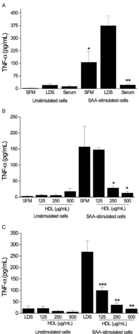
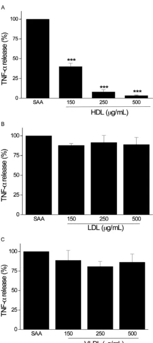
The inhibitory effects of lipoproteins on the action of SAA is limited to HDL - Given that HDL strongly inhibited SAA-induced TNF-α production, we investi-gated whether LDL or VLDL affected the SAA-induced TNF-α secretion in medium that was supplemented with LDS. Although concentrations of 125, 250 and 500 µg of protein/mL HDL inhibited SAA-induced TNF-α release by approximately 60%, 92% and 97%, respectively (Fig. 2A), LDL and VLDL at different concentrations did not inhibit SAA-induced TNF-α production (Fig. 2B, C).
Effects of human serum and HDL on SAA-induced
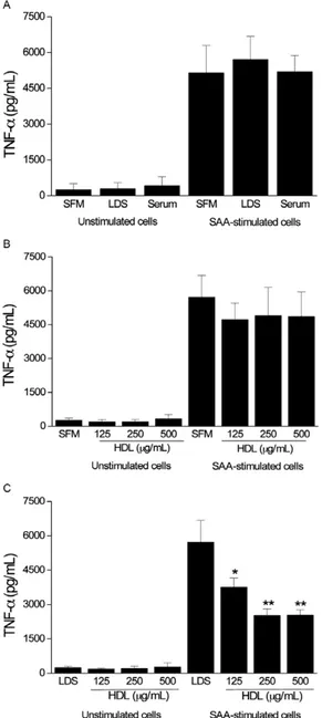
production of TNF-α and IL-1β in PBMCs -To further verify our results in an alternate cell type we evaluated SAA-induced TNF-α release from PBMCs under the same conditions as those used to assay THP-1 cells. We also assayed the levels of the proinflammatory cytokine IL-1β in PBMCs. As expected, SAA induced the release of TNF-α and IL-1β from PBMCs (p < 0.001 and p < 0.001 compared to unstimulated cells, respectively). Un-like THP-1 cells, PBMCs were not affected by the ab-sence of serum over the time interval that was assayed. SFM or medium that was supplemented with human se-rum or LDS did not differentially affect SAA-induced TNF-α production (Fig. 3A), even with the addition of HDL at different concentrations (Fig. 3B). The addition of HDL (250 µg of protein/mL) to the medium containing human serum did not affect TNF-α production induced by SAA (data not shown). However, the addition of HDL to LDS-containing medium promoted a significant de-crease in TNF-α release (Fig. 3C). We observed that the SAA-induced IL-1β release from PBMCs was affected by the presence of serum (Fig. 4A). Although no sig-nificant differences were detected, the presence of HDL decreased IL-1β production when PBMCs were cultured in SFM (Fig. 4B). Conversely, the inhibitory effect of HDL on IL-1β production was clear and pronounced in medium that was supplemented with LDS (Fig. 4C).
DISCUSSION
In the current paper, we reported the effects of whole serum, lipoprotein-depleted serum and medium that was reconstituted with HDL on SAA-induced release of TNF-α and IL-1β in monocytic cells. THP-1 cells and PBMCs released TNF-α and IL-1β following stimula -tion with SAA. PBMCs were more responsive to SAA than THP-1 cells. This result is likely associated with the differentiation state and amount of receptors that are expressed by these cells (Zarember & Godowski 2002).
In THP-1 cells, the SAA response was dramatically increased following incubation in lipoprotein-depleted serum and was abolished with incubation in whole hu-man serum. These results suggest that factorsthat are present in serummay modulate SAA-induced cytokine production. The presence of serum, even delipidated se-rum, may promote interactions between cells and differ-ent types of proteins, as well as other macromolecules and small molecules and mediate different effects on cell integrity and biological responses. The reconstitution of lipoprotein-deficient serum with HDL inhibited SAA-induced TNF-α release in a dose-dependent manner. Other studies have shown that HDL inhibits some SAA-supplemented with LDS. We verified that HDL inhibited
the release of TNF-α that was induced by SAA in a dose-dependent manner (Fig. 1C). The concentrations of HDL (125, 250 and 500 µg of protein/mL) that were assayed in the current study are close to the concentrations that are found in the sera of healthy individuals (Naiko 2003).
Fig. 1A: serum amyloid A(SAA)-induced tumour necrosis
factor-al-pha (TNF-α) production in THP-1 cells in the presence of serum-free
medium (SFM), medium supplemented with lipoprotein-deficient se-rum (LDS) or medium supplemented with human sese-rum (*: p < 0.05; **: p < 0.01 vs. cells incubated with LDS). Effects of high-density lipoprotein (HDL) (µg of protein/mL) at different concentrations on
mediated activities, such as chemotaxis (Badolato et al. 1994), the release of tissue factor (Cai et al. 2007) and IL-8 (Baranova et al. 2010) and TNF-α mRNA expres -sion (Song et al. 2009a, b). Furthermore, we previously demonstrated that acute-phase HDL (HDL-SAA) does not promote the production of TNF-α, IL-1β or IL-8 by human blood neutrophils (Furlaneto & Campa 2000).
In this study, we demonstrated the powerful effects of human serum on the inhibition of SAA activity in THP-1 cells. HDL appears to be one of the main factors that mediate this inhibitory effect. LDL or VLDL did not influence SAA-induced cytokine production by THP-1 cells. Although SAA forms complexes with phospho-lipids (Bausserman et al. 1983) and other lipoproteins (Marhaug et al. 1982), the absence of an inhibitory ef-fect for LDL and VLDL rules out the possibility that the SAA-induced cytokine production is due to the forma-tion of insoluble aggregates of SAA, which represent a possible structural rearrangement of delipidated SAA, as suggested by Kinkley et al. (2006).
Although the inhibitory effect of HDL on SAA-induced TNF-α production in PBMCs was not as dramatic as that in THP-1 cells, HDL did induce a significant inhibitory effect in PBMCs. In addition to TNF-α inhibition, we ob-served the inhibition of IL-1β in PBMCs. The reasons for the mitigated inhibitory effect in PBMCs may be related to the high variability in the cell preparations, which depends on the individual donor and the increased responsiveness of PBMCs to SAA compared to that of THP-1 cells.
The inhibitory effect of HDL on SAA-induced proinflammatory cytokine release is an additional anti-inflammatory property ascribed to HDL. The known anti-inflammatory actions of HDL include inhibition of SAA-mediated monocyte tissue factor production (Cai et al. 2007) and the inhibition of C-reactive protein-in-duced adhesion molecule production, which is another acute-phase protein (Wadham et al. 2004). These afore-mentioned studies and the results of our study compose the framework of the well-recognised anti-inflammatory effects of HDL. The best-characterised ability of HDL is to scavenge LPS (Munford et al. 1982). When HDL is added to the system, other interactions, including the cooperation between HDL and LPS-binding protein (LBP), are expected. Although the interaction between HDL and LBP has been proven (Massamiri et al. 1997), the complex interactions between HDL and LBP and cell membranes can be estimated by the range of responses that are dependent on the HDL: LBP ratio (Hamann et al. 2005). The anti-inflammatory properties of HDL are not restricted to its scavenging properties. HDL inhib-its the expression of adhesion molecules (Cockerill et al. 1995) and induces eNOS activity (Yuhanna et al. 2001). Although these direct effects might be considered in this study, the HDL-mediated inhibition of SAA-induced cy-tokine production may not be a direct effect of HDL on monocytic cells based on its small effect on the basal production of cytokine (Furlaneto & Campa 2000).
In summary, we previously demonstrated that the ability of SAA to induce the production of cytokines is restricted to its free form because acute-phase HDL (HDL-SAA) does not promote cellular activation (Fur-laneto & Campa 2000). At that time, we hypothesised that the HDL particle may serve as a reservoir or transporter of SAA which restricts SAA to specific sites (Furlaneto & Campa 2000, Okino et al. 2006). In this study, we con-firmed that HDL suppressed the in vitro proinflamma-tory activity of SAA and that this effect was not shared with other lipoproteins. It is important to consider that, as stressed by other researchers (Cai et al. 2007, Baranova
Fig. 2: percentage of inhibition by lipoproteins of serum amyloid A(SAA)-induced tumour necrosis factor-alpha (TNF-α) production by THP-1 cells in medium supplemented with lipoprotein-deficient serum. High-density lipoprotein (HDL) inhibited the production of
TNF-α induced by SAA (A). Low-density lipoprotein (LDL) (B) and
very LDL (VLDL) (C) (µg of protein/mL) did not affect the release
of TNF-α induced by SAA. THP-1 cells were stimulated with SAA
et al. 2010), SAA is expressed and synthesised by several extrahepatic sources and may exert biological effects in extravascular sites that are characterised by low concen-trations of lipoproteins (Nanjee et al. 2000). These sites include pleural and ascitic exudates (Okino et al. 2006) and inflamed synovial tissue from rheumatoid arthritis patients who exhibit elevated SAA levels (O'Hara et al. 2004). Furthermore, our data suggest that HDL is an ad-ditional factor that prevents an unbalanced release of
in-flammatory cytokines. This role of HDL seems especially important in diseases that are characterised by increased SAA levels and in which TNF-α and IL-1β have impor-tant roles, such as atherosclerosis (Libby 2002), obesity (Fantuzzi 2005) and sepsis (Zanotti & Kumar 2002). In all of these diseases, there is evidence suggesting that HDL confers protective effects (Murch et al. 2007).
Fig. 3A: serum amyloid A(SAA)-induced tumour necrosis factor-alpha
(TNF-α) production in peripheral blood mononuclear cells (PBMCs) in
the presence of serum-free medium (SFM), medium supplemented with lipoprotein-deficient serum (LDS) or medium supplemented with hu-man serum. Effects of high-density lipoprotein (HDL) (µg of protein/
mL) at different concentrations on SAA-induced TNF-α release in SFM
(B) and LDS-supplemented medium (C) (*:p < 0.05; **:p < 0.01 vs. cells incubated in the absence of HDL). PBMCs were stimulated with SAA (17 µg/mL) and incubated for 16 h. The data shown represent the aver-age ± standard error of mean of six independent experiments.
Fig. 4A: serum amyloid A(SAA)-induced interleukin-1 beta(IL-1β) production in peripheral blood mononuclear cells (PBMCs) in the presence of serum-free medium (SFM), medium supplemented with lipoprotein-deficient serum (LDS) or medium supplemented with hu-man serum (*: p < 0.05 vs. cells incubated in LDS supplemented me-dium). Effects of high-density lipoprotein (HDL) (µg of protein/mL)
at different concentrations on SAA-induced IL-1β release in SFM (B)
REFERENCES
Badolato R, Wang J, Murphy W, Lloyd A, Michiel D, Bausserman L, Kelvin D, Oppenheim J 1994. Serum amyloid A is a chemoat-tractant: induction of migration, adhesion, and tissue infiltra-tion of monocytes and polymorphonuclear leukocytes. J Exp Med 180: 203-209.
Baranova I, Bocharov A, Vishnyakova T, Kurlander R, Chen Z, Fu D, Arias I, Csako G, Patterson A, Eggerman T 2010. CD36 is a novel serum amyloid A (SAA) receptor mediating SAA binding and SAA-induced signalling in human and rodent cells. J Biol
Chem 285: 8492-8506.
Barter P, Nicholls S, Rye K, Anantharamaiah G, Navab M, Fogelman A 2004. Antiinflammatory properties of HDL. Circ Res 95: 764-772.
Bausserman LL, Herbert PN, Forte T, Klausner RD, McAdam KP, Osborne JC, Rosseneu M 1983. Interaction of the serum amyloid A proteins with phospholipid. J Biol Chem 258: 10681-10688.
Boyum A 1974. Separation of blood leucocytes, granulocytes and lymphocytes. Tissue Antigens 4: 269-274.
Cai H, Song C, Endoh I, Goyette J, Jessup W, Freedman S, McNeil H, Geczy C 2007. Serum amyloid A induces monocyte tissue factor.
J Immunol 178: 1852-1860.
Cockerill G, Rye K, Gamble J, Vadas M, Barter P 1995. High-density lipoproteins inhibit cytokine-induced expression of endothelial cell adhesion molecules. Arterioscler Thromb Vasc Biol 15: 1987-1994.
De Santo C, Arscott R, Booth S, Karydis I, Jones, Asher R, Salio M, Middleton M, Cerundolo V 2010. Invariant NKT cells modulate the suppressive activity of IL-10-secreting neutrophils differenti-ated with serum amyloid A. Nat Immunol 11: 1039-1046.
Fantuzzi G 2005. Adipose tissue, adipokines and inflammation. J Al-lergy Clin Immunol 115: 911-919.
Furlaneto C, Campa A 2000. A novel function of serum amyloid A: a potent stimulus for the release of tumour necrosis factor-alpha, interleukin-1 beta and interleukin-8 by human blood neutrophil.
Biochem Biophys Res Commun 268: 405-408.
Grunfeld C, Marshall M, Shigenaga J, Moser A, Tobias P, Feingold K 1999. Lipoproteins inhibit macrophage activation by lipoteichoic acid. J Lipid Res 40: 245-252.
Gutfeld O, Prus D, Ackerman Z, Dishon S, Linke R, Levin M, Urieli-Shoval S 2006. Expression of serum amyloid A, in normal, dys-plastic and neodys-plastic human colonic mucosa: implication for a role in colonic tumorigenesis. J Histochem Cytochem 54: 63-73.
Hamann L, Alexander C, Stamme C, Zähringer U, Schumann RR 2005. Acute-phase concentrations of lipopolysaccharide (LPS)-binding protein inhibit innate immune cell activation by different LPS chemotypes via different mechanisms. Infect Immun 73: 193-200.
Hatanaka E, Furlaneto C, Ribeiro F, Souza G, Campa A 2004. Serum amyloid A-induced mRNA expression and release of tumour ne-crosis factor-alpha (TNF-alpha) in human neutrophils. Immunol Lett 91: 33-37.
Hatanaka E, Ribeiro FP, Campa A 2003. The acute phase protein se-rum amyloid A primes neutrophils. FEMS Immunol Med Micro-biol 38: 81-84.
Havel RJ, Eder HA, Bragdon JH 1955. The distribution and chemical composition of ultracentrifugally separated lipoproteins in hu-man serum. J Clin Invest 34: 1345-1353.
Kinkley SM, Bagshaw WL, Tam SP, Kisilevsky R 2006. The path of murine serum amyloid A through peritoneal macrophages. Amy-loid 13: 123-134.
Libby P 2002. Inflammation in atherosclerosis. Nature 420: 868-874.
Lowry OH, Rosebrough NJ, Farr AL, Randall RJ 1951. Protein mea-surement with the Folin phenol reagent. J Biol Chem 193: 265-275.
Mackness M, Durrington P, Mackness B 2000. How high-density li-poprotein protects against the effects of lipid peroxidation. Curr Opin Lipidol 11: 383-388.
Malle E, De Beer FC 1996. Human serum amyloid A (SAA) protein: a prominent acute-phase reactant for clinical practice. Eur J Clin Invest 26: 427-435.
Marhaug G, Sletten K, Husby G 1982. Characterization of amyloid re-lated protein SAA complexed with serum lipoproteins (apoSAA).
Clin Exp Immunol 50: 382-389.
Massamiri T, Tobias PS, Curtiss LK 1997. Structural determinants for the interaction of lipopolysaccharide binding protein with puri-fied high density lipoproteins: role of apolipoprotein A-I. J Lipid Res 38: 516-525.
Meek R, Urieli-Shoval S, Benditt E 1994. Expression of apolipopro-tein serum amyloid A mRNA in human atherosclerotic lesions and cultured vascular cells: implications for serum amyloid A function. Proc Natl Acad Sci USA 91: 3186-3190.
Munford R, Hall C, Lipton J, Dietschy J 1982. Biological activity, lipoprotein-binding behaviour and in vivo disposition of extract-ed and native forms of Salmonella typhimurium lipopolysaccha-rides. J Clin Invest 70: 877-888.
Murch O, Collin M, Hinds C, Thiemermann C 2007. Lipoproteins in inflammation and sepsis. I. Basic science. Intensive Care Med 33: 13-24.
Naiko NK 2003. Coronary artery disease and disorders of lipid metabolism. In LA Kaplan, AJ Pesce, SC Kazmierczak (eds.),
Clinical chemistry: theory, analysis, correlation, Mosby, St. Louis, p. 603-638.
Nanjee MN, Cooke CJ, Olszewski WL, Miller NE 2000. Concentra-tions of electrophoretic and size subclasses of apolipoprotein A-I-containing particles in human peripheral lymph. Arterioscler Thromb Vasc Biol 20: 2148-2155.
O’Hara R, Murphy E, Whitehead A, Fitzgerald O, Bresnihan B 2004. Local expression of the serum amyloid A and formyl peptide receptor-like 1 genes in synovial tissue is associated with matrix metalloproteinase production in patients with inflammatory ar-thritis. Arthritis Rheum 50: 1788-1799.
Okino AM, Bürger C, Cardoso JR, Lavado EL, Lotufo PA, Campa A 2006. The acute-phase proteins serum amyloid A and C re-active protein in transudates and exudates. Mediators Inflamm 2006: 47297.
Poitou C, Viguerie N, Cancello R, De Matteis R, Cinti S, Stich V, Coussieu C, Gauthier E, Courtine M, Zucker J, Barsh G, Saris W, Bruneval P, Basdevant A, Langin D, Clément K 2005. Serum amyloid A: production by human white adipocyte and regulation by obesity and nutrition. Diabetologia 48: 519-528.
Ribeiro F, Furlaneto C, Hatanaka E, Ribeiro W, Souza G, Cassatella M, Campa A 2003. mRNA expression and release of interleu-kin-8 induced by serum amyloid A in neutrophils and monocytes.
Mediators Inflam 12: 173-178.
Sandri S, Hatanaka E, Franco A, Pedrosa A, Monteiro H, Campa A 2008a. Serum amyloid A induces CCL20 secretion in mononu-clear cells through MAPK (p38 and ERK1/2) signaling pathways.
Immunol Lett 121: 22-26.
Sandri S, Rodriguez D, Gomes E, Monteiro H, Russo M, Campa A 2008b. Is serum amyloid A an endogenous TLR4 agonist? J Leu-koc Biol 83: 1174-1180.
in monocytes/macrophages and lymphocytes. Atherosclerosis 207: 374-383.
Song C, Shen Y, Yamen E, Hsu K, Yan W, Witting PK, Geczy CL, Freedman SB 2009b. Serum amyloid A may potentiate prothrom-botic and proinflammatory events in acute coronary syndromes.
Atherosclerosis 202: 596-604.
Uhlar C, Whitehead A 1999. Serum amyloid A, the major vertebrate acute-phase reactant. Eur J Biochem 265: 501-523.
Urieli-Shoval S, Meek RL, Hanson RH, Eriksen N, Benditt EP 1994. Human serum amyloid A genes are expressed in monocyte/mac-rophage cell lines. Am J Pathol 145: 650-660.
Wadham C, Albanese N, Roberts J, Wang L, Bagley C, Gamble J, Rye K, Barter P, Vadas M, Xia P 2004. High-density lipoproteins neutralize C-reactive protein proinflammatory activity. Circula-tion 109: 2116-2122.
Wu A, Hinds CJ, Thiemermann C 2004. High-density lipoproteins in sepsis and septic shock: metabolism, actions and therapeutic applications. Shock 21: 210-221.
Yamada T, Kakihara T, Kamishima T, Fukuda T, Kawai T 1996. Both acute phase and constitutive serum amyloid A are present in ath-erosclerotic lesions. Pathol Int 46: 797-800.
Yang R, Lee M, Hu H, Pollin T, Ryan A, Nicklas B, Snitker S, Horen-stein R, Hull K, Goldberg N, Goldberg A, Shuldiner A, Fried S, Gong D 2006. Acute-phase serum amyloid A: an inflammatory adipokine and potential link between obesity and its metabolic complications. PLoS Med 3: e287.
Yuhanna I, Zhu Y, Cox B, Hahner L, Osborne-Lawrence S, Lu P, Marcel Y, Anderson R, Mendelsohn M, Hobbs H, Shaul P 2001. High-density lipoprotein binding to scavenger receptor-BI acti-vates endothelial nitric oxide synthase. Nat Med 7: 853-857.
Zanotti S, Kumar A 2002. Cytokine modulation in sepsis and septic shock. Expert Opin Investig Drugs 11: 1061-1075.