ABSTRACT
Obesity hypoventilation syndrome (OHS) is defi ned as the presence of obesity (body mass index ≥ 30 kg/m²) and daytime arterial hypercapnia (PaCO2 ≥ 45 mmHg) in the absence of other causes of hypoventilation. OHS is often overlooked and confused with other conditions associated with hypoventilation, particularly COPD. The recognition of OHS is important because of its high prevalence and the fact that, if left untreated, it is associated with high morbidity and mortality. In the present review, we address recent advances in the pathophysiology and management of OHS, the usefulness of determination of venous bicarbonate in screening for OHS, and diagnostic criteria for OHS that eliminate the need for polysomnography. In addition, we review advances in the treatment of OHS, including behavioral measures, and recent studies comparing the effi cacy of continuous positive airway pressure with that of noninvasive ventilation. Keywords: Obesity; Obesity hypoventilation syndrome; Noninvasive ventilation.
Obesity hypoventilation syndrome: a
current review
Rodolfo Augusto Bacelar de Athayde1,2,a, José Ricardo Bandeira de Oliveira Filho1,b,
Geraldo Lorenzi Filho2,c, Pedro Rodrigues Genta2,d
Correspondence to:
Rodolfo Augusto Bacelar de Athayde. Avenida Dr. Enéas de Carvalho Aguiar, 44, 8º andar, Pinheiros, CEP 05403-000, São Paulo, SP, Brasil. Tel.: 55 11 2661-5000. E-mail: [email protected]
Financial support: None. INTRODUCTION
Obesity hypoventilation syndrome (OHS) is defi ned as the presence of obesity and daytime hypoventilation (PaCO2 ≥ 45 mmHg) in patients without central, pulmonary, neuromuscular, metabolic, or chest wall disease that explains the hypercapnia.(1) Therefore, OHS is a diagnosis
of exclusion, and other causes of hypercapnia should be investigated. Obesity is the hallmark of the disease, there being a correlation between body mass index (BMI) and disease prevalence.(1-5) The identifi cation of OHS is important because of the possibility of clinical exacerbation leading to respiratory failure and the high mortality rate in untreated patients. OHS is accompanied by obstructive sleep apnea (OSA) in more than 90% of cases, and both share the same major risk factor, that is, obesity; however, the presence of OSA is not necessary for the diagnosis of OHS. This explains why polysomnography is not necessary for this diagnosis. Signs of right heart failure can be present in OHS and are secondary to chronic hypoxemia and pulmonary hypertension, both of which can accompany the clinical picture. In addition, arterial hypertension and insulin resistance are more prevalent in patients with OHS than in obese individuals without OHS.(1-3)
Since OHS is associated with high morbidity and mortality,(5-7) the objective of the present study was to conduct a current review of the epidemiology, pathophysiology, and treatment of OHS.
HISTORY
Obesity-related sleepiness was described in 1889, even prior to the recognition of OSA.(8) Bickelmann et al. published a case report in 1956(3) and popularized the
term “Pickwickian syndrome” (an eponym that has fallen into disuse) in a reference to the character Fat Boy Joe from Charles Dickens’s “The Posthumous Papers of the Pickwick Club,” who was always sleepy and hungry and would often fall asleep on the job any time during the day.(9) The patient reported by Bickelmann et al.(3) had
daytime hypoventilation, chronic hypoxemia, polycythemia, and pulmonary hypertension, with evidence of cor pulmonale. Several studies have since characterized the
epidemiology, clinical picture, and pathophysiology of OHS.(1,2,4,7) Since 1999, the American Academy of Sleep Medicine has defi ned the diagnostic criteria for OHS.(10,11)
EPIDEMIOLOGY
The prevalence of OHS is unknown because of the lack of population-based studies. The prevalence of OHS is estimated to be 10-20% in patients with OSA(7,12-16) and is estimated to be even higher in extremely obese patients.(7,14) Mokhlesi et al.(7) evaluated a population in the USA referred to a sleep medicine center for suspicion of OSA—180 patients were retrospectively selected, and 410 patients were prospectively selected. Of the patients diagnosed with OSA in the retrospective and prospective samples, 30% and 20%, respectively, met 1. Serviço de Pneumologia, Hospital
das Clínicas, Faculdade de Medicina, Universidade de São Paulo, São Paulo (SP) Brasil.
2. Laboratório do Sono, Disciplina de Pneumologia, Instituto do Coração, Hospital das Clínicas, Faculdade de Medicina, Universidade de São Paulo, São Paulo (SP) Brasil.
a. http://orcid.org/0000-0003-2482-3127 b. http://orcid.org/0000-0001-7976-2988 c. http://orcid.org/0000-0002-7011-7373 d. http://orcid.org/0000-0002-6764-165X
Submitted: 20 September 2017. Accepted: 11 February 2018.
the criteria for OHS, and those percentages increased with increasing BMI. Laaban et al.(14) retrospectively evaluated patients receiving home treatment for OSA in France. The sample included 1,114 adults, of whom approximately 10% met the diagnostic criteria for OHS, and a positive association was also found with increasing BMI.(14) Akashiba et al.(12) evaluated 611
patients in Japan referred to sleep medicine centers for OSA and diagnosed OHS in 9% of the patients. The patients with OHS were younger, were more obese, and had more severe OSA when compared with those without OHS. In a different approach, Kessler et al.(17) evaluated patients with OHS and detected OSA in most of the patients (90%); in addition, OHS patients with OSA were found to have poorer gas exchange and poorer pulmonary hemodynamics than did those without OSA.
Seeking to determine the prevalence and, consequently, the degree of underdiagnosis of OHS, Nowbar et al.(5) conducted a study involving obese patients admitted to internal medicine services for any cause. Of 29 obese inpatients with a BMI > 50 kg/m2, 14 (48%) were diagnosed with OHS. In the
same study, 31% of 150 obese inpatients did not have a previous diagnosis of OHS, although they met the criteria for this diagnosis.(5)
Because of the lack of studies on the prevalence of OHS in the general population, an exercise on epidemiological correlations has been repeatedly cited. Mokhlesi(18) infers that if approximately 3% of the general population in the USA are severely obese (BMI > 40 kg/m2), half of those individuals would have OSA.
Considering, therefore, the estimate that 10-20% of severely obese patients with OSA would have OHS, a conservative estimate indicates a prevalence of OHS of 0.15-0.30% in the general population in the USA (ranging approximately from 1:300 to 1:600 adults).(18)
MORBIDITY AND MORTALITY
Patients with OHS use more health care resources in the period prior to the diagnosis than do obese individuals without OHS or the general population.(19) Obesity per se leads to a greater likelihood of diseases
such as systemic arterial hypertension, diabetes, dyslipidemia, and hypothyroidism. Comorbidities such as heart failure, coronary artery disease, and
cor pulmonale are more common in patients with
OHS, and the likelihood that such patients will require invasive mechanical ventilation or ICU admission is also increased.(5,20) In addition, pulmonary hypertension is more common (50% vs. 15%) and more severe in patients with OHS than in patients with OSA.(16,21,22)
Berg et al.(19) conducted a study involving 20 patients with OHS, who were matched to control subjects by age, gender, and zip code (to try to equate socioeconomic factors). A comparison with controls revealed that the most common morbidities in patients with OHS were cardiovascular diseases: congestive heart failure (OR = 9.0; 95% CI: 2.3-35.0); angina pectoris (OR = 9.0; 95% CI: 1.4-57.1); and cor pulmonale (OR = 9.0;
95% CI: 1.4-57.1). In a retrospective study conducted by Basoglu & Tasbakan, having a BMI > 40 kg/m2
and obesity-related complications showed a strong association with an increased risk of premature death in hospitalized patients.(2) Nowbar et al.(5) reported that, at 18 months following hospital discharge, mortality was 23% in patients with obesity-related hypoventilation, which was almost twice as high as that among obese patients without hypoventilation.
CLINICAL PRESENTATION AND DIAGNOSIS
OHS occurs within a triad: obesity; daytime gas exchange abnormalities (hypercapnia); and the absence of other causes for the fi ndings (Chart 1).(23)
The American Academy of Sleep Medicine defi nes OHS as follows: the presence of awake daytime alveolar hypoventilation (PaCO2 > 45 mmHg as measured at sea level) in patients with a BMI ≥ 30 kg/m2 in the
absence of other causes of hypoventilation.(11)
The vast majority of patients with OHS have symptoms of OSA, including snoring, nighttime choking, witnessed apneas, nonrestorative sleep, excessive daytime sleepiness, and fatigue. In contrast to patients with OSA alone, patients with OHS complain of dyspnea, are often hypoxemic, and can have signs of cor
Chart 1. Diagnosis of obesity hypoventilation syndrome. Diagnostic criteria
• Presence of awake daytime alveolar hypoventilation (PaCO2 > 45 mmHg as measured at sea level) • BMI ≥ 30 kg/m2
• Absence of other causes of hypoventilation
Diagnoses of exclusion
• COPD or other severe obstructive lung diseases • Severe interstitial lung disease
• Mechanical respiratory limitation such as in severe chest wall disorders (such as kyphoscoliosis)
• Neuropathic and myopathic conditions (such as amyotrophic lateral sclerosis, Duchenne muscular dystrophy, myasthenia gravis, myositis, and diaphragmatic paralysis)
• Electrolyte disturbances (such as hypophosphatemia, hypomagnesemia, hypermagnesemia, hypokalemia, and hypocalcemia)
• Central causes (such as cerebrovascular disease and untreated hypothyroidism) • Congenital alveolar hypoventilation syndrome (Ondine’s syndrome)
pulmonale. Plethoric obese patients with hypoxemia,
an increased neck circumference, a decreased airway area, a prominent P2 (a loud second heart sound) on cardiac auscultation, and leg edema, as determined by physical examination, are at risk of having OHS.(1)
OHS is a diagnosis of exclusion. Other causes of hypoventilation, such as COPD; severe interstitial lung disease; mechanical respiratory limitation (for example, chest wall disorders such as kyphoscoliosis); myopathies (such as myasthenia gravis); neurological diseases; central causes (such as cerebrovascular disease and untreated hypothyroidism); and congenital causes (such as Ondine’s syndrome; Chart 1), should be ruled out.
Patients suspected of having OHS can initially be screened by pulse oximetry and by determination of serum levels of venous bicarbonate. Borderline oximetry values are common fi ndings. Patients with OHS undergoing arterial blood gas analysis rarely have PaO2 values > 70 mmHg. Consequently, SpO2 values < 93% on pulse oximetry would be suggestive of hypoventilation. However, higher values are not exclusionary, which explains why this is not a necessary criterion to establish the diagnosis, although it helps in screening. Nocturnal oximetry showing sustained hypoxemia and no associated apneas strengthens the suspicion for hypoventilation. A serum bicarbonate level ≥ 27 mEq/L had a sensitivity of 92% and a specifi city of 50%, justifying its use in screening.(7,24,25) After such screening, arterial blood gas analysis is mandatory. For excluding other causes of hypoventilation (Chart 1), pulmonary function testing and assessment of respiratory muscle strength (MIP and MEP), chest X-ray, electrocardiography, and thyroid function testing
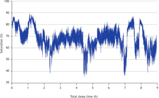
should be performed. In addition, the use of drugs and medications, such as sedatives, hypnotics, opiates, and alcohol (alcohol abuse), should be investigated. Polysomnography is not necessary for the diagnosis of OHS.(11) However, since it has been observed that
individuals with OHS have obstructive events, as well as lower saturation in REM sleep (Figure 1), polysomnography is requested with a view to treating comorbid sleep apnea and to justifying possible treatments.(6)
Unfortunately, despite being simple in concept, the diagnosis of OHS is delayed in most cases, occurring during acute events of respiratory failure or cardiac decompensation.(5,26)
PATHOPHYSIOLOGY
Several mechanisms are related to the pathogenesis of OHS (Figure 2), including an abnormal organic response of the respiratory system in certain obese individuals, as well as an inappropriate central response to hypercapnia and hypoxemia, in addition to neurohumoral changes. In comparison with other obese individuals, patients with OHS have decreased lung compliance, important reductions in functional residual capacity and chest wall compliance, and increased pulmonary resistance.(23,27)
Changes in pulmonary function
Obesity and the resulting greater chest wall thickness cause an excessive increase in the work of breathing. Breathing smaller volumes affects respiratory mechanics, reducing respiratory system compliance and increasing its resistance (which, in
0 1 2 3 4 5 6 7 8 9
100
90
80
70
60
50
40
30
Saturation (%)
Total sleep time (h)
Figure 1. Female patient with a body mass index of 45 kg/m2, PaCO
2 = 55.6 mmHg, obstructive sleep apnea, and
individuals with OHS, is approximately 20% higher than in other obese individuals and 60% higher than in normal-weight individuals).(23,27) Gas exchange is also affected, worsening the ventilation/perfusion ratio. Individuals with OHS tend to have lower tidal volume and higher RR, which increases the dead space effect. Consequently, hypoxemia is a common fi nding, which leads to an equally common outcome of pulmonary hypertension secondary to hypoxia.(16,17) In addition, abdominal fat deposition compromises the diaphragm’s infl uence on ventilation, compromising muscle function. Furthermore, there is thinning of the diaphragm and increased oxidative stress.(28)
Ventilatory control
Patients with OHS have arterial CO2 retention. A reduction in CO2 chemosensitivity was initially believed to
be the possible cause of this fi nding, which was proven untrue.(29-31) Unlike what occurs in chronic hypoxia, low daytime and nighttime saturation can be the cause of decreased ventilatory response.(32) Chemosensitivity
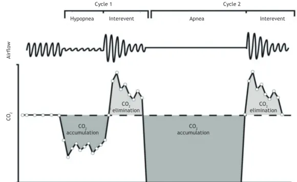
is progressively impaired by increased CO2 levels. Chronic hypercapnia is also believed to result from the inability to eliminate CO2, which accumulates at night during apnea and hypopnea episodes, during the day (Figure 3).(30) A secondary mechanism that also impairs chemosensitivity is elevated serum and cerebrospinal fl uid levels of bicarbonate.
Role of leptin
Leptin is a cytokine produced by adipocytes and may explain a causal relationship among obesity, ventilatory control, and chronic hypercapnia. Most data come from studies of mice. When obese, these animals, like humans, develop daytime hypercapnia and reduced ventilatory response to CO2. In mice, there is defi ciency of leptin. Leptin replacement reverses hypoventilation in mice with leptin defi ciency.(33)
Unlike in the animal model, there is no defi ciency but rather an increase in leptin levels in obese humans. Leptin is believed to initially have a protective effect, stimulating the ventilatory response. The persistence
Obesity OSA
Chronic hypercapnia Resistance
to leptin
Mechanical overload
Altered ventilatory response
Acute hypercapnia
Bicarbonate
Cycle 1 Cycle 2
Hypopnea Interevent Apnea Interevent
Airflow
CO
2
CO2 elimination
CO2 elimination
CO2 accumulation
CO2 accumulation
Figure 3. Infl uence of obstructive sleep events on hypercapnia. Adapted from Berger et al.(31)
of obesity would lead to leptin resistance (which is conceptually similar to insulin resistance), and thus, a consequent decrease in the ventilatory response to CO2.(25,32,34)
GENERAL TREATMENT MEASURES
Weight loss
Signifi cant weight loss promotes improvement in ventilatory parameters.(3,32) Bariatric surgery is the intervention resulting in the best outcome.(35) However, low-calorie diets may have satisfactory results. Bariatric surgery is the treatment of choice in the management of morbidly obese patients, but not every patient is a candidate for the procedure, given that the number of comorbidities that increase surgical risk is high. In fact, in some cases, the procedure will be contraindicated because of such comorbidities.
Although treatment improves ventilatory variables, it does not always resolve the problem. In a study conducted by Dixon et al.(36) involving 60 obese patients with a diagnosis of OSA who were divided into two groups—those undergoing calorie restriction and those undergoing bariatric surgery—weight loss was greater in the bariatric surgery group, but there was no statistically signifi cant difference regarding the apnea-hypopnea index. Greenburg et al.(37) published a meta-analysis that included 12 studies involving 342 patients who underwent polysomnography before bariatric surgery and after maximal weight loss. There was a 71% reduction in the apnea-hypopnea index, from 55 events/h (95% IC: 49-60 events/h) to 16 events/h (95% CI: 13-19 events/h). It is known that 7% to 20% of such patients are unable to maintain a BMI loss of at least 20% after 5-10 years,(38,39) which requires continued surveillance even after the procedure. Only one study evaluated the impact of bariatric surgery in patients with OHS. Sugerman et al.(40) evaluated 61 patients with OHS undergoing bariatric surgery. In 31 patients, there was improvement in PaO2 (from 53 mmHg to 73 mmHg) and in PaCO2 (from 53 mmHg to 44 mmHg)
at 1 year. At 5 years, only 12 patients underwent arterial blood gas analysis, which revealed marked worsening (mean PaO2 = 68 mmHg and mean PaCO2
= 47 mmHg); in addition, the mean BMI was found to have increased (from 38 kg/m2 to 40 kg/m2), having
been high since the fi rst postoperative year.
Oxygen therapy alone
Oxygen therapy alone is not appropriate, even in acute events, because it increases nocturnal CO2
retention (Haldane effect or “dead space” ventilation effect), which worsens sleep quality, and is considered a common error in the management of patients with OHS (this subject will be discussed below).(41)
Phlebotomy
There are no studies that examine the indications for phlebotomy in patients with OHS. Our group uses the
indications for phlebotomy for heart disease patients and lung disease patients (hematocrit > 56% or symptoms of hyperviscosity).(42)
Tracheostomy
Tracheostomy was the fi rst treatment instituted for OHS; however, today, tracheostomy is reserved only for patients who are refractory to noninvasive ventilation (NIV), because of risk and complications inherent in the procedure and in obese patients.(34)
Pharmacotherapy
Several medications (such as medroxyprogesterone and acetazolamide) have been tried to increase ventilatory response, without success, and are not recommended for the treatment of OHS.(25,32,34,43)
Positive pressure
Continuous positive airway pressure (CPAP) is the treatment of choice for stable OHS. CPAP improves alveolar ventilation by decreasing upper airway resistance, relieving the respiratory muscle load, and/ or increasing central respiratory activity.(6,19,24,41,44-52) Patients with OHS should be initially treated with CPAP if they are clinically stable and if PaCO2 is not severely altered (< 55 mmHg). If either of these conditions is not met, NIV should be used. In OHS patients without OSA, NIV should also be used. CPAP therapy is typically administered via a nasal mask. Some studies have shown that oronasal masks are less effi cient and are associated with poorer adherence and greater side effects than are nasal masks in patients with OSA. (53) Therefore, for long-term use, nasal masks are recommended. In critically ill patients with respiratory failure, oronasal masks are preferred.
mortality. In patients with refractory hypoventilation (PaCO2 > 45 mmHg despite proven adherence to treatment and use of PAP determined by titration and despite the elimination of obstructive events) or with persistent desaturation (SpO2 < 90% despite proven adherence to treatment and use of PAP determined by titration and despite the elimination of obstructive events), NIV should be used.(43,46,47,49,52,56,57)
Treatment objectives
The objective of therapy in OHS is to reverse the major abnormalities that give rise to the disease, that is, to normalize ventilation during sleep and to reduce body weight. The therapeutic goals for patients with OHS include normalization of PaCO2 during wakefulness and sleep; prevention of desaturations during sleep and wakefulness; control of erythrocytosis, pulmonary hypertension, and cor pulmonale; and relief of
hypersomnia. Poor adherence to PAP is associated with incomplete clinical improvement. Adherence can be assessed by reviewing the memory card of NIV and CPAP devices.
Management in the emergency room: common errors in caring for patients with OHS
Overuse of supplemental oxygen
Hypercapnia can be aggravated by hyperoxia via several mechanisms: an increase in FiO2 can result in a decrease in minute volume and, consequently, a decrease in tidal volume due to the activity of peripheral chemoreceptors; oxygenation of hypoxic areas causes vasodilation that changes blood fl ow to previously poorly ventilated areas, causing an increase in dead space; and the Haldane effect causes a reduction in hemoglobin affi nity for CO2 and decreases correction
of hypoxia, causing increased release of CO2 in plasma,
which increases hypercapnia.(29,41,58) Therefore, oxygen therapy alone is best indicated in hemodynamically stable patients with no excessive work of breathing (RR ≤ 30 breaths/min without use of accessory muscles or with other signs of risk of ventilatory failure), under clinical surveillance, with an SpO2 target of 89-92%.
(41)
Overuse of loop diuretics
Patients with OHS are commonly affected by conditions that cause edema due to cor pulmonale. Since decompensation of cor pulmonale can be the cause
for seeking medical care, a loop diuretic (furosemide) usually is used for the initial treatment of these patients in order to achieve a euvolemic state. However, overuse of diuretics can lead to acute prerenal renal failure. Contraction alkalosis secondary to the use of diuretics can worsen CO2 retention. In addition, overuse of furosemide can cause hypokalemia. Cautious use of diuretics is indicated in OHS, at the lowest dose possible to achieve a favorable clinical response and minimize the electrolytic and acid-metabolic impact. (41) The use of spironolactone for the prevention of hypokalemia is plausible.
Overuse of psychotropic drugs
The use of sedative/hypnotic drugs not only increases airway collapsibility but also decreases ventilatory response, which is harmful to patients with OHS.
Diagnostic confusion with COPD
Patients with chronic CO2 retention, such as patients
with OHS, are commonly diagnosed with COPD, despite the absence of documented obstructive ventilatory disorders. A retrospective study by Marik & Desai(59) showed that, of the morbidly obese patients admitted to the ICU for respiratory failure secondary to OHS, 75% had been erroneously treated for COPD and 86% had been treated for congestive heart failure (Chart 2).
PERIOPERATIVE PERIOD IN PATIENTS WITH OHS
Patients with OHS commonly have a consultation with a pulmonologist in the preoperative period. In addition to comorbidity care and the required cardiovascular evaluation in obese patients or in those who are known to have or are highly suspected of having OSA, specifi c perioperative care is required for these patients whatever the procedure. In addition to the already suggested screening with pulse oximetry and determination of serum bicarbonate, other measures are required. If screening is positive and OHS is confi rmed by arterial blood gas analysis, treatment should be started immediately, even a few days or weeks after the procedure; there is signifi cant evidence of improved gas exchange and improved ventilatory control, either with one-level positive pressure or with two-level positive pressure.(60)
Obesity is a risk factor for diffi cult mask ventilation.(61) A retrospective study by Rose & Cohen, involving 18,500 patients, showed that obesity is also an independent risk factor for diffi cult intubation.(62) ≥Kheterpal et al.(63) evaluated 22,660 procedures and identifi ed fi ve risk factors (limited mandibular protrusion, increased neck circumference, OSA, snoring alone, and BMI ≥ 30 kg/m2) as independent predictors of diffi cult mask
ventilation and diffi cult intubation during anesthesia induction. This suggests that patients with OHS are among those at highest risk for airway complications. (64) During anesthesia induction, patients with OHS should be placed in the ramp position with elevation of the torso and head (preferably at a 25° tilt). This has been shown to improve ventilation and the glottic view,(65) as well as oxygenation.(66)
Patients with OHS are more sensitive to the respiratory depressant effects of anesthetic agents and opioids because they are prone to airway collapse and
Chart 2. Common errors in the emergency care of patients
with obesity hypoventilation syndrome.
inappropriate physiological response to hypercapnia and hypoxemia. Regional block should be chosen, when possible. In addition, during the procedure if possible, patients with OHS should be monitored with a capnograph. At the end of the procedure, it is recommended that patients be placed in the ramp position or in the lateral decubitus position for improved oxygenation and maintenance of the airways, and tracheal extubation should be performed only after the patient is fully conscious.(64)
With regard to postoperative care, the use of CPAP for 24-48 h after extubation can reduce the risk of postoperative complications and extubation failure in severely obese patients admitted to the ICU (an absolute risk reduction of 16%), with a reduction in mortality in patients with hypercapnia.(67,68) In addition, pain
control has an impact on ventilatory status. Therefore, optimal analgesia is also required.
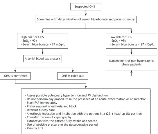
Figure 4 outlines a suggested algorithm for the screening and perioperative management of patients with suspected or confi rmed OHS.
FINAL CONSIDERATIONS
OHS is still a poorly recognized entity in Brazil. Delayed diagnosis of OHS is associated with an increase in morbidity, mortality, and costs of care of patients who are more severely ill. However, breaking free from myths and paradigms regarding diagnosis, such as that related to polysomnography, which is unnecessary, the possibility of screening for OHS with determination of venous bicarbonate, and the possibility of treatment with CPAP enable the diagnosis and treatment of OHS in a larger number of patients.
Suspected OHS
Screening with determination of serum bicarbonate and pulse oximetry
High risk for OHS - SpO2 < 93%
- Serum bicarbonate > 27 mEq/L
Low risk for OHS - SpO2 > 93%
- Serum bicarbonate < 27 mEq/L
Arterial blood gas analysis
Management of non-hypercapnic obese patients
OHS is confirmed OHS is ruled out
- Assess possible pulmonary hypertension and RV dysfunction
- Do not perform any procedure in the presence of an acute exacerbation or an infection - Start PAP immediately
- Prefer regional anesthesia and block - Difficult airway care
- Anesthesia induction and intubation with the patient in a (25°) head-up tilt position - Consider the use of capnography
- Extubation with the patient fully awake and seated - Use of positive pressure in the postoperative period - Pain control
Figure 4. Suggested algorithm for the screening and perioperative management of patients with suspected or confi rmed
obesity hypoventilation syndrome (OHS). PAP: positive airway pressure; and RV: right ventricle. Adapted from Chau et al.(64)
REFERENCES
1. Mokhlesi B, Kryger MH, Grunstein RR. Assessment and management of patients with obesity hypoventilation syndrome. Proc Am Thorac Soc. 2008;5(2):218–25. https://doi.org/10.1513/pats.200708-122MG
2. Basoglu OK, Tasbakan MS. Comparison of clinical characteristics in patients with obesity hypoventilation syndrome and obese obstructive sleep apnea syndrome: a case-control study. Clin Respir J. 2014;8(2):167-74. https://doi.org/10.1111/crj.12054
3. BICKELMANN AG, BURWELL CS, ROBIN ED, WHALEY RD. Extreme obesity associated with alveolar hypoventilation; a
Pickwickian syndrome. Am J Med. 1956;21(5):811-8. https://doi. org/10.1016/0002-9343(56)90094-8
4. Olson AL, Zwillich C. The obesity hypoventilation syndrome. Am J Med. 2005;118(9):948-56. https://doi.org/10.1016/j. amjmed.2005.03.042
6. Castro-Añón O, Pérez de Llano LA, De la Fuente Sánchez S, Golpe R, Méndez Marote L, Castro-Castro J, et al. Obesity-hypoventilation syndrome: increased risk of death over sleep apnea syndrome. PLoS One. 2015;10(2):e0117808. https://doi.org/10.1371/journal. pone.0117808
7. Mokhlesi B, Tulaimat A, Faibussowitsch I, Wang Y, Evans AT. Obesity hypoventilation syndrome: prevalence and predictors in patients with obstructive sleep apnea. Sleep Breath. 2007;11(2):117-24. https://doi.org/10.1007/s11325-006-0092-8
8. Lavie P. Who was the fi rst to use the term Pickwickian in connection with sleepy patients? History of sleep apnoea syndrome. Sleep Med Rev. 2008;12(1):5-17. https://doi.org/10.1016/j.smrv.2007.07.008
9. Dickens C. The posthumous papers of the Pickwick Club. Boston: Riverside Press; 1867.
10. American Academy of Sleep Medicine. International Classifi cation of Sleep Disorders. Darien, IL: the Academy; 1999.
11. American Academy of Sleep Medicine. Darien, IL: the Academy; 2014.
12. Akashiba T, Akahoshi T, Kawahara S, Uematsu A, Katsura K, Sakurai S, et al. Clinical characteristics of obesity-hypoventilation syndrome in Japan: a multi-center study. Intern Med. 2006;45(20):1121–5. https://doi.org/10.2169/internalmedicine.45.1747
13. Verin E, Tardif C, Pasquis P. Prevalence of daytime hypercapnia or hypoxia in patients with OSAS and normal lung function. Respir Med. 2001;95(8):693-6. https://doi.org/10.1053/rmed.2001.1120
14. Laaban JP, Chailleux E. Daytime hypercapnia in adult patients with obstructive sleep apnea syndrome in France, before initiating nocturnal nasal continuous positive airway pressure therapy. Chest. 2005;127(3):710-5. https://doi.org/10.1378/chest.127.3.710
15. Ayappa I, Berger KI, Norman RG, Oppenheimer BW, Rapoport DM, Goldring RM. Hypercapnia and ventilatory periodicity in obstructive sleep apnea syndrome. Am J Respir Crit Care Med. 2002;166(8):1112-5. https://doi.org/10.1164/rccm.200203-212OC
16. Kessler R, Chaouat A, Weitzenblum E, Oswald M, Ehrhart M, Apprill M, et al. Pulmonary hypertension in the obstructive sleep apnoea syndrome: prevalence, causes and therapeutic consequences. Eur Respir J. 1996;9(4):787-94. https://doi.org/10.1183/09031936.96.09 040787
17. Kessler R, Chaouat A, Schinkewitch P, Faller M, Casel S, Krieger J, et al. The obesity-hypoventilation syndrome revisited: a prospective study of 34 consecutive cases. Chest. 2001;120(2):369-76. https:// doi.org/10.1378/chest.120.2.369
18. Mokhlesi B. Obesity hypoventilation syndrome: a state-of-the-art review. Respir Care. 2010;55(10):1347-65; discussion 1363-5.
19. Berg G, Delaive K, Manfreda J, Walld R, Kryger MH. The use of health-care resources in obesity-hypoventilation syndrome. Chest. 2001;120(2):377-83. https://doi.org/10.1378/chest.120.2.377
20. Bender R, Trautner C, Spraul M, Berger M. Assessment of excess mortality in obesity. Am J Epidemiol. 1998;147(1):42-8. https://doi. org/10.1093/oxfordjournals.aje.a009365
21. Atwood CW Jr, McCrory D, Garcia JG, Abman SH, Ahearn GS; American College of Chest Physicians. Pulmonary artery hypertension and sleep-disordered breathing: ACCP evidence-based clinical practice guidelines. Chest. 2004;126(1 Suppl):72S-77S. https://doi.org/10.1378/chest.126.1_suppl.72S
22. Sugerman HJ, Baron PL, Fairman RP, Evans CR, Vetrovec GW. Hemodynamic dysfunction in obesity hypoventilation syndrome and the effects of treatment with surgically induced weight loss. Ann Surg. 1988;207(5):604-13. https://doi.org/10.1097/00000658-198805000-00015
23. Lopata M, Onal E. Mass loading, sleep apnea, and the pathogenesis of obesity hypoventilation. Am Rev Respir Dis. 1982;126(4):640-5.
24. Harada Y, Chihara Y, Azuma M, Murase K, Toyama Y, Yoshimura C, et al. Obesity hypoventilation syndrome in Japan and independent determinants of arterial carbon dioxide levels. Respirology. 2014;19(8):1233-40. https://doi.org/10.1111/resp.12367
25. Piper AJ, Grunstein RR. Obesity hypoventilation syndrome: mechanisms and management. Am J Respir Crit Care Med. 2011;183(3):292-8. https://doi.org/10.1164/rccm.201008-1280CI
26. Owens RL. A big problem in the ICU. Initiation of CPAP/bilevel PAP therapy. J Clin Sleep Med. 2014;10(10):1161-2.
27. Merkus PJ, van Pelt W, Quanjer PH. Effects of overweight on lung function. Arch Dis Child. 1991;66(2):273-4. https://doi.org/10.1136/ adc.66.2.273-c
28. Becker HF, Piper AJ, Flynn WE, McNamara SG, Grunstein RR, Peter JH, et al. Breathing during sleep inpatients with nocturnal
desaturation. Am J Respir Crit Care Med. 1999;159(1):112-8. https:// doi.org/10.1164/ajrccm.159.1.9803037
29. Hollier CA, Harmer AR, Maxwell LJ, Menadue C, Willson GN, Unger G, et al. Moderate concentrations of supplemental oxygen worsen hypercapnia in obesity hypoventilation syndrome: a randomised crossover study. Thorax. 2014;69(4):346-53. https://doi.org/10.1136/ thoraxjnl-2013-204389
30. Rapoport M, Garay SM, Epstein H, Goldring RM. Hypercapnia in the obstructive sleep apnea syndrome. A reevaluation of the “Pickwickian syndrome”. Chest. 1986;89(5):627-35. https://doi. org/10.1378/chest.89.5.627
31. Berger KI, Ayappa I, Sorkin IB, Norman RG, Rapoport DM, Goldring RM. CO(2) homeostasis during periodic breathing in obstructive sleep apnea. J Appl Physiol (1095). 2000;88(1):257-64. https://doi. org/10.1152/jappl.2000.88.1.257
32. Piper AJ. Obesity hypoventilation syndrome--the big and the breathless. Sleep Med Rev. 2011;15(2):79-89. https://doi. org/10.1016/j.smrv.2010.04.002
33. Tankersley CG, O’Donnell C, Daood MJ, Watchko JF, Mitzner W, Schwartz A, et al. Leptin attenuates respiratory complications associated with the obese phenotype. J Appl Physiol (1985). 1998;85(6):2261-9. https://doi.org/10.1152/jappl.1998.85.6.2261
34. Selim BJ, Junna MR, Morgenthaler TI. Therapy for sleep hypoventilation and central apnea syndromes. Curr Treat Options Neurol. 2012;14(5):427-37. https://doi.org/10.1007/s11940-012-0188-3
35. Sjöström L, Narbro K, Sjöström CD, Karason K, Larsson B, Wedel H, et al. Effects of bariatric surgery on mortality in Swedish obese subjects. N Engl J Med. 2007;357(8):741-52. https://doi.org/10.1056/ NEJMoa066254
36. Dixon JB, Schachter LM, O’Brien PE, Jones K, Grima M, Lambert G, et al. Surgical vs conventional therapy for weight loss treatment of obstructive sleep apnea: a randomized controlled trial. JAMA. 2012;308(11):1142-9. https://doi.org/10.1001/2012.jama.11580
37. Greenburg DL, Lettieri CJ, Eliasson AH. Effects of surgical weight loss on measures of obstructive sleep apnea: a meta-analysis. Am J Med. 2009;122(6):535-42. https://doi.org/10.1016/j.amjmed.2008.10.037
38. le Roux CW, Heneghan HM. Bariatric Surgery for Obesity. Med Clin N Am. 2018;(102):165-82.
39. Sjöström L, Lindroos AK, Peltonen M, Torgerson J, Bouchard C, Carlsson B, et al. Lifestyle, diabetes, and cardiovascular risk factors 10 years after bariatric surgery. N Engl J Med. 2004;(351):2683-93. https://doi.org/10.1056/NEJMoa035622
40. Sugerman HJ, Fairman RP, Sood R, Engle K, Wolfe L, Kellum J. Long-term effects of gastric surgery for treating respiratory insuffi ciency of obesity. Am J Clin Nutr. 1992;55(2 Suppl):597S-601S. https://doi. org/10.1093/ajcn/55.2.597s
41. Manthous CA, Mokhlesi B. Avoiding Management Errors in Patients with Obesity Hypoventilation Syndrome. Ann Am Thorac Soc. 2016;13(1):109-14. https://doi.org/10.1513/AnnalsATS.201508-562OT
42. McMullin MF, Bareford D, Campbell P, Green AR, Harrison C, Hunt B, et al. Guidelines for the diagnosis, investigation and management of polycythaemia/erythrocytosis. Br J Haematol. 2005;130(2):174-95. https://doi.org/10.1111/j.1365-2141.2005.05535.x
43. Piper A. Obesity Hypoventilation Syndrome Weighing in on Therapy Options. Chest. 2016;149(3):856-68. https://doi.org/10.1378/ chest.15-0681
44. Borel JC, Pepin JL, Pison C, Vesin A, Gonzalez-Bermejo J, Court-Fortune I, et al. Long-term adherence with non-invasive ventilation improves prognosis in obese COPD patients. Respirology. 2014;19(6):857-65. https://doi.org/10.1111/resp.12327
45. Bülbül Y, Ayik S, Ozlu T, Orem A. Frequency and predictors of obesity hypoventilation in hospitalized patients at a tertiary health care institution. Ann Thorac Med. 2014;9(2):87-91. https://doi. org/10.4103/1817-1737.128851
46. Combs D, Shetty S, Parthasarathy S. Advances in Positive Airway Pressure Treatment Modalities for Hypoventilation Syndromes. Sleep Med Clin. 2014;9(3):315-325. https://doi.org/10.1016/j. jsmc.2014.06.002
47. Esquinas AM, Petroianni A. Non-invasive mechanical ventilation in obesity hypoventilation syndrome: are multimodal therapeutic strategies disease essential? Respirology. 2013;18(2):385. https:// doi.org/10.1111/resp.12027
2014;192(2):251-8. https://doi.org/10.1007/s00408-014-9555-z
49. Lemyze M, Taufour P, Duhamel A, Temime J, Nigeon O, Vangrunderbeeck N, et al. Determinants of noninvasive ventilation success or failure in morbidly obese patients in acute respiratory failure. PLoS One. 2014;9(5):e97563. https://doi.org/10.1371/journal. pone.0097563
50. Palen BN, Kapur VK. Tailoring Therapy for Obesity Hypoventilation Syndrome. Am J Respir Crit Care Med. 2015;192(1):8-10. https://doi. org/10.1164/rccm.201504-0721ED
51. Salord N, Mayos M, Miralda RM, Farré A, Carreras M, Sust R, et al. Continuous positive airway pressure in clinically stable patients with mild-to-moderate obesity hypoventilation syndrome and obstructive sleep apnoea. Respirology. 2013;18(7):1135-42.
52. Storre JH, Seuthe B, Fiechter R, Milioglou S, Dreher M, Sorichter S, et al. Average volume-assured pressure support in obesity hypoventilation: A randomized crossover trial. Chest. 2006;130(3):815-21. https://doi.org/10.1378/chest.130.3.815
53. Andrade RG, Piccin VS, Nascimento JA, Viana FM, Genta PR, Lorenzi-Filho G. Impact of the type of mask on the effectiveness of and adherence to continuous positive airway pressure treatment for obstructive sleep apnea. J Bras Pneumol. 2014;40(6):658-68. https:// doi.org/10.1590/S1806-37132014000600010
54. Masa JF, Corral J, Alonso ML, Ordax E, Troncoso MF, Gonzalez M, et al. Effi cacy of Different Treatment Alternatives for Obesity Hypoventilation Syndrome: Pickwick Study. Am J Respir Crit Care Med. 2015;192(1):86-95. https://doi.org/10.1164/rccm.201410-1900OC
55. Howard ME, Piper AJ, Stevens B, Holland AE, Yee BJ, Dabscheck E, et al. A randomised controlled trial of CPAP vs non-invasive ventilation for initial treatment of obesity hypoventilation syndrome. Thorax. 2017;72(5):437-444. https://doi.org/10.1136/thoraxjnl-2016-208559
56. Ojeda Castillejo E, de Lucas Ramos P, López Martin S, Resano Barrios P, Rodriguez Rodríguez P, Morán Caicedo L, et al. Noninvasive mechanical ventilation in patients with obesity hypoventilation syndrome. long-term outcome and prognostic factors. Arch Bronconeumol. 2015;51(2):61-8. https://doi.org/10.1016/j. arbres.2014.02.015
57. Waldhorn RE. Nocturnal nasal intermittent positive pressure ventilation with bi-level positive airway pressure (BiPAP) in respiratory failure. Chest. 1992;101(2):516-21. https://doi.org/10.1378/ chest.101.2.516
58. Masa JF, Corral J, Romero A, Caballero C, Terán-Santos J, Alonso-Álvarez ML, et al. The effect of supplemental oxygen in obesity
hypoventilation syndrome. J Clin Sleep Med. 2016;12(10):1379-1388. https://doi.org/10.5664/jcsm.6194
59. Marik PE, Desai H. Characteristics of patients with the “malignant obesity hypoventilation syndrome” admitted to an ICU. J Intensive Care Med. 2012;28(2):124-30. https://doi. org/10.1177/0885066612444261
60. Chouri-Pontarollo N, Borel J, Tamisier R, Wuyam B, Levy P, Pepin J. Impaired objective daytime vigilance in obesity-hypoventilation syndrome: Impact of noninvasive ventilation. Chest. 2007;131(1):148-55. https://doi.org/10.1378/chest.06-1159
61. Langeron O, Masso E, Huraux C, Guggiari M, Bianchi A, Coriat P, et al. Prediction of diffi cult mask ventilation. Anesthesiology. 2000;92(5):1229-36. https://doi.org/10.1097/00000542-200005000-00009
62. Rose DK, Cohen MM. The airway: problems and predictions in 18,500 patients. Can J Anaesth. 1994;41(5 Pt 1):372-83. https://doi. org/10.1007/BF03009858
63. Kheterpal S, Han R, Tremper KK, Shanks A, Tait AR, O’Reilly M, et al. Incidence and predictors of diffi cult and impossible mask ventilation. Anesthesiology. 2006;105(5):885-91. https://doi. org/10.1097/00000542-200611000-00007
64. Chau EH, Lam D, Wong J, Mokhlesi B, Chung F. Obesity hypoventilation syndrome: a review of epidemiology, pathophysiology, and perioperative considerations. Anesthesiology. 2012;117(1):188-205. https://doi.org/10.1097/ALN.0b013e31825add60
65. Cattano D, Melnikov V, Khalil Y, Sridhar S, Hagberg CA. An evaluation of the rapid airway management positioner in obese patients undergoing gastric bypass or laparoscopic gastric banding surgery. Obes Surg. 2010;20(10):1436-41. https://doi.org/10.1007/s11695-009-9885-8
66. Dixon B, Dixon J, Carden J, Burn A, Schachter L, Playfair J, et al. Preoxygenation is more effective in the 25 degrees head-up position than in the supine position in severely obese patients: a randomized controlled study. Anesthesiology. 2005;102(6):1110-5; discussion 5A. https://doi.org/10.1097/00000542-200506000-00009
67. Rennotte MT, Baele P, Aubert G, Rodenstein DO. Nasal continuous positive airway pressure in the perioperative management of patients with obstructive sleep apnea submitted to surgery. Chest. 1995;107(2):367-74. https://doi.org/10.1378/chest.107.2.367