Activated Receptor-
c
in CHO Cells That Over-Express
Glycerol 3-Phosphate Acyltransferase-1
Cliona M. Stapleton1, Douglas G. Mashek1¤, Shuli Wang1, Cynthia A. Nagle1, Gary W. Cline2, Philippe Thuillier3, Lisa M. Leesnitzer4, Lei O. Li1, Julie B. Stimmel4, Gerald I. Shulman2, Rosalind A. Coleman1* 1Department of Nutrition, University of North Carolina, Chapel Hill, North Carolina, United States of America,2Departments of Internal Medicine and Cellular & Molecular Physiology, Yale University School of Medicine, New Haven, Connecticut, United States of America,3Oregon Cancer Institute, Oregon Health Sciences University, Portland, Oregon, United States of America,4Department of Screening and Compound Profiling, GlaxoSmithKline Inc., Research Triangle Park, North Carolina, United States of America
Abstract
Lysophosphatidic acid (LPA) is an agonist for peroxisome proliferator activated receptor-c(PPARc). Although glycerol-3-phosphate acyltransferase-1 (GPAT1) esterifies glycerol-3-glycerol-3-phosphate to form LPA, an intermediate in thede novosynthesis of glycerolipids, it has been assumed that LPA synthesized by this route does not have a signaling role. The availability of Chinese Hamster Ovary (CHO) cells that stably overexpress GPAT1, allowed us to analyze PPARcactivation in the presence of LPA produced as an intracellular intermediate. LPA levels in CHO-GPAT1 cells were 6-fold higher than in wild-type CHO cells, and the mRNA abundance of CD36, a PPARctarget, was 2-fold higher. Transactivation assays showed that PPARc
activity was higher in the cells that overexpressed GPAT1. PPARcactivity was enhanced further in CHO-GPAT1 cells treated with the PPARcligand troglitazone. Extracellular LPA, phosphatidic acid (PA) or a membrane-permeable diacylglycerol had no effect, showing that PPARchad been activated by LPA generated intracellularly. Transient transfection of a vector expressing 1-acylglycerol-3-phosphate acyltransferase-2, which converts endogenous LPA to PA, markedly reduced PPARc
activity, as did over-expressing diacylglycerol kinase, which converts DAG to PA, indicating that PA could be a potent inhibitor of PPARc. These data suggest that LPA synthesized via the glycerol-3-phosphate pathway can activate PPARcand that intermediates ofde novoglycerolipid synthesis regulate gene expression.
Citation:Stapleton CM, Mashek DG, Wang S, Nagle CA, Cline GW, et al. (2011) Lysophosphatidic Acid Activates Peroxisome Proliferator Activated Receptor-cin CHO Cells That Over-Express Glycerol 3-Phosphate Acyltransferase-1. PLoS ONE 6(4): e18932. doi:10.1371/journal.pone.0018932
Editor:Jeffrey M. Gimble, Pennington Biomedical Research Center, United States of America
ReceivedFebruary 1, 2011;AcceptedMarch 11, 2011;PublishedApril 20, 2011
Copyright:ß2011 Stapleton et al. This is an open-access article distributed under the terms of the Creative Commons Attribution License, which permits unrestricted use, distribution, and reproduction in any medium, provided the original author and source are credited.
Funding:NIH grants DK56598, DK59935, DK68993, DK40936, U24 DK59635, P30 DK34987; Marilyn Gentry Fellowship from AICR/WRF; Howard Hughes Medical Institute. The funders had no role in study design, data collection and analysis, decision to publish, or preparation of the manuscript.
Competing Interests:Two authors, Lisa M. Leesnitzer and Julie B. Stimmel are employees of GlaxoSmithKline. This does not alter our adherence to all the PLoS ONE policies on sharing data and materials.
* E-mail: rcoleman@unc.edu
¤ Current address: Department of Food Science & Nutrition, University of Minnesota, St. Paul, Minnesota, United States of America
Introduction
Peroxisome proliferator activated receptor-c (PPARc) is a nuclear receptor that is highly expressed when pre-adipocytes differentiate into adipocytes [1]. PPARcregulates gene expression by forming a heterodimer with the retinoid6receptor (RXR) and binding to PPAR response elements (PPRE) in the promoter region of target genes. Genes that are regulated by PPARc are primarily involved in adipocyte differentiation and fatty acid metabolism [1]. Ligands for PPARcinclude polyunsaturated fatty acids [2] and synthetically derived thiazolidinedione (TZD) ligands for PPARcthat improve insulin sensitivity in patients with type 2 diabetes [1].
Lysophosphatidic acid (LPA) derived from hydrolysis of plasma membrane phospholipids is well established as a ligand for specific G-coupled cell-surface LPA receptors that generate an intracellu-lar signal cascade that enhances cell proliferation [3]. Studies have suggested that exogenous LPA might also enter cells to activate PPARcbecause LPA can bind to and displace rosiglitazone from PPARc. Further, when LPA enters RAW264.7 monocytes, it
activates a PPRE reporter driven by a PPARcexpression vector [4]. Although the naturally occurring ether analog of LPA, 1-O-octadecenyl-2-hydroxy-sn-glycero-3-phosphate (AGP), can enter cells and is a ligand for purified PPARc(Kd,60 nM), the entry of LPA is more problematic [5]. Studies showing that LPA can activate the intracellular PPARc reporter required dimethyl sulfoxide (DMSO) and tridentate sulfonamid to enhance the transport of exogenously added LPA across the cell membrane [4,6]. Thus, although LPA can clearly activate PPARc[7], it is not certain that exogenous LPA would normally function as a major physiological activator of PPARc. Most LPA produced within cells is synthesized as an intermediate in the pathway of triacylglycerol and phospholipid biosynthesis, but it has been generally assumed that LPA formed byde novosynthesis from glycerol-3-phosphate (Fig. 1) has a role in lipid synthesis but not in signaling [8].
LPA content 4-fold [11], we used a CHO cell line that stably overexpresses GPAT1 [12] to determine whether endogenously synthesized LPA could act as a PPARc agonist. CHO-GPAT1 cells have a 3.8-fold increase in GPAT1 activity, contain a 2.7-fold higher triacylglycerol mass than control cells, and incorporate 3.4-fold more [14C]oleate into triacylglycerol, suggesting that the intermediates of glycerolipid synthesis might be higher than normal and that LPA levels might be elevated. We now provide evidence that LPA synthesized intracellularly via the pathway of triacylglycerol and phospholipid biosynthesis can activate PPARc and that PA may inhibit PPARcactivity.
Methods
Cell Culture
Chinese hamster ovary cells (CHO) (CHO-K1, #CCL-61, ATTC) were maintained in MEM with 10% heat-inactivated fetal bovine serum, 100 units/mL penicillin and 100mg/mL strepto-mycin (normal medium) at 37uC, 5% CO2. CHO cells that stably overexpress GPAT1 (CHO-GPAT1 cells) [12] were maintained in the same medium with 600mg/mL G418 until studied. Expression of GPAT1 in CHO-GPAT1 cells is controlled by the pTet-Off plasmid (CLONTECH), so GPAT1 expression is repressed when doxycycline is present.
Plasmids
pSG5 expression vectors encoding hPPARc and mRXRa [13,14] and the PPRE reporter plasmid ACOX2-tk-CAT [13,15] were described previously. The pShuttle2 expression vector encoding hAGPAT2 was a generous gift from Dr. A. K. Agarwal (University of Texas Southwestern Medical Center) [16]. pcDNA3-DGKa WT and pcDNA3-DGKa D196, encoding constitutively active DGKa, were generous gifts from Dr. J. P.
Walsh (Indiana University School of Medicine) [17]. The internal control reporter construct pRL-SV40 was from Promega. pcDNA3.1 was from Invitrogen.
Transactivation Assay
CHO cells and CHO-GPAT1 cells were plated at 46105cells/ well in 6-well dishes. After 24 h, cells were transfected in Opti-MEM (GIBCO) with equal concentrations (0.4mg) of expression vectors (PPARc and RXR) and a PPRE-CAT reporter vector using FuGENE 6 (Roche). Transfections of PPARc were in medium containing delipidated serum (HyClone). To monitor transfection efficiency, 0.1mg pRL-SV40 was transfected as an internal luciferase control. Equal concentrations of DNA were transfected into cells at all times. As a control, cells that were not transfected with either the AGPAT2 expression vector or the vector expressing constitutively active DGKa were transfected with equal amounts of a negative control, i.e., pcDNA3.1 or pcDNA3-DGKa WT, respectively. Twenty-four hours after transfection, cells were treated with troglitazone (Cayman Chemical Company) or dimethyl sulfoxide (vehicle control). After further incubation for 24 h, cells were assayed for Renilla luciferase (LUC) (Renilla Luciferase Assay System; Promega). Chloramphenicol acetyltransferase (CAT) activity, a measure of PPRE activity, was determined using the CAT enzyme-linked immunosorbent assay kit (Roche). CAT reporter activity relative to LUC activity was calculated and plotted. Experiments were performed in triplicate, and for the statistical analysis, either Student’s t-test, assuming equal variances, or ANOVA followed by multiple comparison of means with Bonferroni’s multiple comparison test was used (GraphPad Prism, version 4 statistical software; GraphPad Software). Representative figures from each experiment are shown. Variation of luciferase sensitivity between experiments was responsible for the difference in the scale of relative CAT activity.
Preparation of Fatty Acid:BSA Complex
Fatty acids were prepared in 20 mM stock solutions. Bovine serum albumin (BSA) (12.5%; essentially fatty acid-free) was prepared in MEM containing antibiotics and delipidated serum. Just before cell treatment, fatty acids were bound to BSA at a final concentration of 0.5 mM fatty acid and 1% BSA [18]. Cells were treated with a final concentration of 250mM fatty acid.
Preparation of Extracellular LPA and PA
1-Oleoyl-2-hydroxy-sn-glycero-3-phosphate (18:1 LPA) and 1,2-dioleoyl-sn-glycero-3-phosphate (18:1 PA) (Avanti Polar Lipids, Inc.) were dried under nitrogen and dissolved in 0.1% BSA immediately before use. Dioctanoyl glycerol (diC8:0) (Sigma) was dissolved in dimethyl sulfoxide before use.
Mass Spectrophotometry
CHO and CHO-GPAT1 cells grown to confluence in 150 mm dishes, washed with ice cold PBS, and scraped into 1 mL ice cold PBS. Cells were counted and stored at280uC, and acyl-CoAs were extracted [19,20]. To extract PA and LPA, cells were sonicated and homogenized in 1 mL of phosphate buffer (40 mM Na2HPO4, 30 mM citric acid, pH 4.0) plus an internal standard (heptadecanoyl-LPA). Two milliliters of butanol were added, and cells were rehomogenized and centrifuged at 4000 rpm for 15 min. Supernatants containing the LPA and PA were dried, redissolved in 1 mL water and applied to an Oasis HLB column. The column was washed once with water, and then LPA and PA were eluted with methanol. Authentic standards (acyl-CoA, TAG: Figure 1. Pathway of glycerolipid synthesis.AGPAT,
acyl-glycerol-3-phosphate acyltransferase; CL, cardiolipin; DGK, diacylglycerol kinase; GPAT, glycerol-3-phosphate acyltransferase; PC, phosphatidylcholine; PE, phosphatidylethanolamine; PG, phosphatidylglycerol; PI, phospha-tidylinositol; PS, phosphatidylserine.
Sigma Chemical Co. and 1,2-dodecanoin: IndoFine Chemical Co.) were used for quantification. Results are expressed as nanomoles analyte per gram protein extract. Analysis was performed with a bench-top tandem mass spectrometer API3000 (Perkin-Elmer Sciex) interfaced with TurboIonspray ionization source. The mobile phase was methanol and H2O with an isocratic gradient (50/50). Acyl-CoAs were ionized in negative ionspray mode [20]. Doubly charged ions and corresponding product ions were chosen as transition pairs for each acyl-CoA species for selective reaction monitoring quantitation. LPA and PA were ionized in TurboIonspray mode with MRM quantification of the parent ion (minus phosphate) and corresponding product ions of the fatty acid moiety.
PPARc LBD LEADseeker Scintillation Proximity Assay Storage buffer [50 mM Tris pH 8, 50 mM KCl (100 mL)] was added to 500 mg of streptavidin-coated LEADseeker SPA beads (Amersham). After mixing for 30 min on a Nutator platform, the beads were centrifuged at 1500 rpm.for 10 min at 22uC. The bead pellet was resuspended in 100 mL of storage buffer at 5 mg/mL. At room temperature, the bead stock (5 mL) was diluted with 7.5 mL of assay buffer (50 mM Tris pH 8, 50 mM KCl, 2 mM EDTA, 5 mM CHAPS, 0.1 mg/mL BSA, 10 mM DTT) and 120–150 nM of biotinylated PPARc LBD [21] was added to a final volume of 12.5 mL. The slurry was incubated for at least one hour with gentle agitation and then centrifuged at 1500 rpm for 10 min at 22uC. The bead pellet was gently washed with 12– 15 mL of assay buffer, centrifuged as described previously, resuspended (45 mL assay buffer, 5 mL 1 mM biotin), and incubated for 1.5 h with gentle agitation.
Radioligand ([3H]rosiglitazone) stock was diluted to 150 nM in assay buffer, mixed with an equal volume of receptor-coated beads, incubated for 5 min, and added (10mL) to 384-well assay plates (NUNC, 264675) containing 0.1mL of compound. Final assay concentrations were 75 nM radioligand and 0.25 mg/mL PPARc-LBD-coated beads. Fatty acids were diluted in ethanol: water (1:1) with a starting concentration ofmM and assayed as dose response curves. After a 2–15 h incubation at RT, covered and in the dark, the signal was determined at 613 nm using a Viewlux plate imager (Perkin Elmer). Non-specific binding was determined with 20mM unlabeled rosiglitazone.
Total RNA Extraction and Quantitative RT-PCR
CHO and CHO-GPAT1 cells were grown to 90% confluence, and RNA was extracted (RNeasy Midi Kit, Qiagen). Forward and reverse primers for mouse GPAT1 were 59 -ATGGACGCAAA-GACATTCTGT-39 and 59 -AAGATCTCCAGGAACTGCTG-39, respectively. The corresponding FAM probe was 59 -CGTTGCTCCATGGGCATGTAGTTG-39. The 18s primer and probe sequences were homologous for mouse, rat, and human (Forward: 59-AGAAACGGCTACCACATCCA-39; reverse: 59 -CTCGAAAGAGTCCTGTATTGT-39). The 18s TET probe was 59-AGGCAGCAGGCGCGCAAATTAC-39. Samples were ana-lyzed using the ABI Prism 7700 sequence detection system (Applied Biosystems). Data were analyzed using the relative standard curve method described in the product manual. Additionally, mRNA expression of hamster CD36 (F: 59 -TCAAGGGCATTGGGCAAACAGG-39 and R: 59 -ATGG-CACCGCTCTGCTCAAAC-39) was determined using Absolute SYBR Green Fluorescein (Thermo Scientific) PCR mix in an iCycler Thermal Cycler instrument (Bio-Rad) with hamster GAPDH as a control (F: 59 -ACGTGTCCGTTGTGGATCT-GAC-39 and R: 59 -CACCACCTTCTTGATGTCCTCATAC-39). Data were analyzed using the comparative CT method.
Results
CHO-GPAT1 cells contained higher intracellular levels of LPA than CHO cells
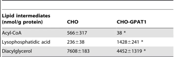
Rat GPAT1 expression was detected only in the overexpressing cell line (data not shown) with a cycle threshold average of 18.3 (n = 3). Mass spectrophotometry analysis showed that compared with the CHO cells, CHO-GPAT1 cells contained 93% less acyl-CoA, the substrate for GPAT1 (Table 1), and 6-times more LPA, the product of GPAT1. DAG in CHO-GPAT1 cells was 41.5% lower than in the CHO cells.
Overexpression of GPAT1 in CHO cells increased PPARc activation
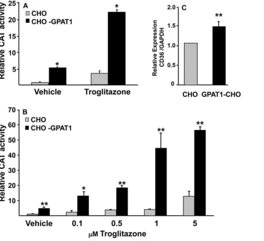
To determine whether the increased intracellular content of LPA in the CHO-GPAT1 cells activated PPARc, CHO cells and CHO-GPAT1 cells were transfected with a PPARcexpression vector and a PPRE-CAT reporter vector. PPARcactivity was 6-fold higher in CHO-GPAT1 cells than in the CHO control cells (Fig. 2A). The PPARc ligand troglitazone increased PPARc activity 4-fold in CHO cells and an additional 4-fold in the CHO-GPAT1 cells.
In both CHO and CHO-GPAT1 cells, PPARcactivity increased with increasing troglitazone concentrations up to 5mM (Fig. 2B). With all treatments, the CHO-GPAT1 overexpressing cells showed a 4- to 10-fold higher PPARc activity than their CHO cell counterparts. Because 1mM troglitazone elicited a near maximal response, it was used for all subsequent experiments. Additionally, mRNA expression of CD36, a PPARctarget, was elevated 2-fold in GPAT1-CHO cells (Fig. 2C), suggesting a functional increase in PPARc activity in GPAT1-overexpressing cells. Studies with a PPARb/dexpression vector showed very low relative CAT activity in both cell lines, so these isoforms were not investigated further. A PPARa expression vector showed no change in relative CAT activity in the two cell lines, and stimulation with linoleic acid or 1mM of the PPARa-specific activator Wy14643 did not show a differential effect (data not shown).
Adding AGPAT2 to CHO-GPAT1 cells decreased PPARc activity
In order to determine whether the increased PPARcactivity in CHO-GPAT1 cells was due to increased intracellular LPA, cells were co-transfected with a 1-acylglycerol-3-phosphate acyltrans-ferase-2 (AGPAT2) expression vector in order to convert LPA to PA and diminish the LPA signal (Fig. 1) [16]. Adding the AGPAT2 expression vector markedly lowered PPARcactivity in both CHO control and CHO-GPAT1 cells treated with either the
Table 1.Overexpression of GPAT1 in CHO cells increased intracellular levels of LPA, and lowered the content of acyl-CoA and DAG.
Lipid intermediates
(nmol/g protein) CHO CHO-GPAT1
Acyl-CoA 5666317 38 *
Lysophosphatidic acid 236638 14286241 *
Diacylglycerol 76086183 445261319 *
Lipid intermediates in CHO and CHO-GPAT1 cells were analyzed by mass spectrometry as described in Methods. Lysophosphatidic acid and
vehicle or troglitazone (Fig. 3A). The AGPAT2-mediated decrease in PPARc activity was consistent, although the extent of the decrease varied due to variation of luciferase sensitivity between experiments (compareFig. 3A and 3B).
Converting DAG to PA decreased PPARc activity Because DGKacatalyzes the phosphorylation of DAG to form PA, the transfection of constitutively active diacylglycerol kinasea (DGKa) either alone or together with AGPAT2, allowed us to analyze the effects of increasing cellular PA and of decreasing both LPA and DAG (Fig. 1). Co-transfecting AGPAT2 plus constitutively active DGKalowered PPARcactivation similar to AGPAT2 alone and decreased the effect of troglitazone in both cell lines (Fig. 3B). CHO-GPAT1 cells transfected with DGKaalone showed a PPARc response similar to cells co-transfected with both AGPAT2 and DGKa, a result which is consistent with inhibition of PPARcby PA. Of interest is the recent report that cyclic phosphatidic acid is an inhibitor of PPARc[22]. Regardless of treatment, PPARcactivity remained higher in CHO-GPAT1 cells than in CHO cells.
LPA and PA displace [3H]rosiglitazone
Radioactive competitive binding assays confirmed that LPA was a PPARc ligand. Palmitoyl-LPA, oleoyl-LPA, 1-palmitoyl, 2-oleoyl-PA, and 1,2 dioleoyl-PA competitively displaced between
20–30% of [3H]rosiglitazone with micromolar affinity from the PPARcligand binding domain (Table 2), as described previously [4,5]. PA appeared to be more potent than LPA, but LPA binding was similar to that described in previous reports [4,5].
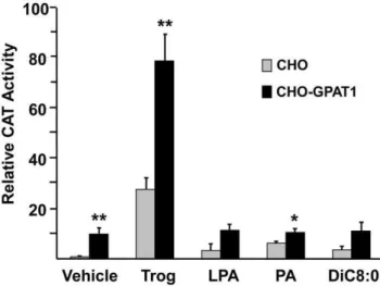
Extracellular addition of LPA, PA, or diC8:0 did not increase PPARc activity
LPA is formed from membrane phospholipids by phospholipases D and A2 and regulates internal signaling pathways via G protein-coupled cell surface receptors [3,23,24]. LPA entry into cells is problematic. Entry is inhibited by albumin [6] and by cell-specific enzymes like ecto-lipid phosphate phosphohydrolase (LPP) [25]. Exogenously provided LPA can activate a G protein-coupled signaling pathway that may affect PPARcactivity [3]. For example, extracellular LPA inhibits adipogenesis via the LPA1receptor [26], and the LPA1receptor is expressed by CHO cells [24]. In order to make certain that the effects on PPARcwere not due to effects of external LPA or PA on G protein-coupled receptors and to learn whether DAG might be playing a role in the observed effects, we added LPA, PA, or dioctanoyl glycerol to the culture medium (Fig. 4). Dioctanoylglycerol is a cell permeable DAG [27], and intact PA does not enter cells [28]. Compared with the vehicle control, these additions had no effect on PPARcactivity. Thus, neither DAG, LPA nor PA added externally were able to increase PPARcactivity. Figure 2. Overexpression of GPAT1 in CHO cells activated PPARc.(A) CHO and CHO-GPAT1 cells were transfected with 0.1mg of pRLSV40
(internal LUC control) and equal concentrations (0.4mg) of a PPARcexpression vector, an RXR expression vector, and a PPRE-CAT reporter vector for
24 h, then treated with either 5mM troglitazone or vehicle (dimethylsulfoxide). (B) CHO and CHO-GPAT1 cells were similarly transfected with the
Adding fatty acids to CHO-GPAT1 cells increased PPARc activity
Because the effects of GPAT1 and troglitazone were synergistic, we wondered whether PPARcligands like polyunsaturated fatty acids might act similarly. PPARcactivity is strongly activated by 12:0 and 18:2 and weakly activated by 18:1 and 16:0 [13]. In
CHO cells, both troglitazone and 18:2 increased PPARcactivity 2-fold compared to the vehicle control, but other fatty acids were less effective (Fig. 5). In the CHO-GPAT1 cells, 18:2 and troglitazone treatments were also equivalent; each increased PPARc activity 3- to 4-fold compared to the vehicle. None of the other fatty acids had an effect greater than treatment with the vehicle alone.
Discussion
LPA, an intermediate in the glycerol-3-phosphate pathway of glycerolipid synthesis had previously been identified as a PPARc ligand and agonist [4,5], but when added exogenously, it required tridentate sulfonamid or dimethyl sulfoxide to enhance its entry into RAW264.7 or CV-1 cells [4,6]. Evidence for functional activation of PPARcby exogenously added LPA was inferred by an increase in the PPARctarget CD36 on the surface of primary human monocytes [4]. However, because LPA does not readily enter cells, the physiological significance of these observations has remained uncertain.
Our study confirmed previous reports that showed that LPA can partially displace [3H]rosiglitazone [4,5] and suggested the possibility that the PPARc binding site might simultaneously Figure 3. Adding AGPAT2 and DGKato CHO-GPAT1 cells decreased PPARcactivity.(A) CHO and CHO-GPAT1 cells were transfected with 0.1mg
of pRLSV40 (internal LUC control), and equal concentrations (0.4mg) of expression and reporter vectors, PPARcRXR, PPRE-CAT, and either empty vector or
an AGPAT2 expression vector. (B) Cells were similarly transfected with the vectors described plus either a wild-type (inactive) DGKaexpression vector (2) or the constitutively active DGKa D196 expression vector (+). Twenty-four hours after transfection, cells were treated with either vehicle or 1mM troglitazone.
CAT activity was measured 24 h later and normalized to LUC activity. The results (mean+/2SEM; n = 3) are expressed as relative CAT activity. Results are representative of three and two independent experiments, respectively. *p,0.05 and **p#0.01 when comparing within treatment groups.
doi:10.1371/journal.pone.0018932.g003
Table 2.LPA and PA displace [3H]rosiglitazone from PPARc -LBD.
Glycerolipid species
Percent displacement of [3H]rosiglitazone
sn-1-18:0-LPA 25.961.5
sn-1-16:0-LPA 29.661.2
sn-1, 2-di18:1-PA 24.260.8
sn-1-16:0, 2-18:1-PA 21.760.9
Increasing concentrations of lipids were evaluated for the ability to displace [3H]rosiglitazone from PPARcLBD. LPA, lysophosphatidic acid; PA, phosphatidic
accommodate both thiazolidinedione and a second ligand. Dual binding provides a possible explanation for the synergistic effect of troglitazone and LPA as well as for the apparent inhibitory effect of PA on PPARc-mediated expression of CAT activity. We attribute the increase in PPARc activity to the increased LPA levels since it is known that LPA can compete with the selective PPARcligand rosiglitazone for binding to PPARc[4], and as little as 1 nM of the ether analog AGP is able to displace ,45% of bound [3H]rosiglitazone [5]. However, our results also showed that the overexpression of GPAT1 in CHO cells enhanced the effect of troglitazone on PPARc activity, suggesting that troglitazone and LPA worked synergistically to enhance PPARc activity. Synergistic enhancement is consistent with the suggestion that rosiglitazone and AGP bind tightly to PPARcand that their binding sites, although overlapping, are not in the same position, since the two ligands cannot fully displace each other [5]. A less likely possibility is that LPA might act indirectly by modifying the function of a PPARccoactivator.
Despite studies that show direct LPA binding to PPARc, LPA molecules that are produced within cells via a pathway ofde novo synthesis have not previously been considered to function as PPARcagonists [29]. In order to investigate this possibility, we used a CHO cell line that stably overexpresses GPAT1 [12]. These cells have a 3.8-fold increase in GPAT1 activity and contain a higher triacylglycerol mass than control cells, suggesting that the intermediates of glycerolipid synthesis, including LPA, might be elevated. This hypothesis was confirmed by a mass spectropho-tometric analysis that showed a 6-fold increase in LPA levels in the CHO-GPAT1 cells and acyl-CoA levels 93% lower than those in control CHO cells. These results are opposite those observed in Gpat12/2mice in which the liver LPA content is 50% lower and acyl-CoA content is 3-fold higher than in wild type controls [10]. Because CHO-GPAT1 cells have higher levels of LPA and TAG, we also expected to find increased DAG content, but DAG was
50% lower in the overexpressing cell line, perhaps because it was rapidly converted to TAG [12].
Decreased PPARcactivity has also been observed in another study in which cells were transiently transfected with an AGPAT2 expression vector and then treated with extracellular LPA; in that study, however, AGPAT expression did not inhibit PPARcin cells treated with rosiglitazone or two other PPARc agonists [4]. In contrast, our study showed that the AGPAT2 expression vector markedly lowered PPARc activity even in the presence of troglitazone, suggesting the possibility that PA, endogenously synthesized by AGPAT, may inhibit PPARc. On the other hand, PA may have had an inhibitory effect in our study, only because we used troglitazone, a relatively weak PPARcligand.
LPA can regulate a variety of signaling pathways within cells by binding to G protein-coupled receptors located on the cell surface [30], and it was possible that the effects on PPARc might be mediated by this type of pathway if the LPA synthesized by GPAT1 could cross the plasma membrane to leave the cell and then interact with LPA1 receptors present on the plasma membrane [24]. In order to ascertain whether the increased PPARcactivity we observed in CHO-GPAT1 cells was due to intracellular versus secreted LPA, we added LPA to the medium. Because externally added LPA had no effect on the PPARc reporter, endogenous and intracellular LPA must have activated PPARc.
In order to increase the LPA pool available to activate PPARc, CHO and CHO-GPAT1 cells were transfected with the PPARc expression vector and PPRE-CAT reporter and then treated with different fatty acids at 250mM. Although 18:2 strikingly increased PPARcactivity in the CHO-GPAT1 cells, the observed increase in PPARc activity in this and other studies [13] might be due either to direct binding of the fatty acid to PPARc or to an increase in the total amount of LPA available as an agonist. Figure 4. Treating CHO and CHO-GPAT1 cells with LPA, PA, or
DiC8:0 did not enhance PPARcactivity.CHO and CHO-GPAT1 cells were transfected with 0.1mg of pRLSV40 (internal LUC control), and equal concentrations (0.4mg) of a PPARc expression vector, an RXR expression vector, and a PPRE-CAT reporter vector. Twenty-four hours later, the transfected cells were treated with vehicle (0.1% BSA), 1mM troglitazone, 5mM oleoyl-LPA, 5mM dioleoyl-PA, or 10mM DiC8:0. CAT
activity was measured 24 h later and normalized to LUC activity. The results (mean +/2SEM; n = 3) are expressed as relative CAT activity. Results are representative of two independent experiments. *p,0.05 and **p#0.01 when comparing within treatment groups.
doi:10.1371/journal.pone.0018932.g004
Figure 5. Overexpression of GPAT1 in CHO cells enhanced the effects of fatty acid treatments on PPARcactivity.CHO and CHO-GPAT1 cells were transfected with 0.1mg of pRLSV40 (internal LUC
control), and equal concentrations (0.4mg) of a PPARc expression
vector, an RXR expression vector, and a PPRE-CAT reporter vector, as described. Twenty-four hours later, transfected cells were treated with either vehicle (0.1% BSA), 1mM troglitazone, or 250mM fatty acid (lauric
The major finding of this study is that LPA acts as a PPARc agonist when it is formed intracellularly as an intermediate in the glycerolipid biosynthetic pathway. This finding is important in light of the current focus on the association of obesity and hepatic steatosis, because stimulation of PPARc by thiazolidinedione drugs like troglitazone alleviates insulin resistance [31]. In hepatic steatosis, both PPARc and GPAT1 are normally upregulated [32,33], and adenovirus-mediated overexpression of GPAT1 in rat liver promotes the development of hepatic steatosis within 5–7 days [11]. Upregulation of GPAT1 increases its LPA product [11], which could physiologically synergize with other PPARcligands to enhance hepatic steatosis. In a related study in which Gpat12/2 mice were crossed with ob/ob mice, hepatic TAG content was reduced by 59% [34], suggesting that GPAT1 is responsible for most of SREBP-1 regulated hepatic TAG accumulation. In the current study, upregulation of PPARcis supported by an increase in the PPARctarget CD36. Although our study used cells that stably over-express GPAT1, GPAT specific activity in the CHO-GPAT1 cells is considerably lower than that normally measured in liver and adipocytes. Thus, the enhanced activity of GPAT that activated PPARcactivity in this study is relevant to GPAT specific activities that are normally present in the major lipogenic tissues. Our study showing synergism between LPA and troglitazone also suggests that different ligands activating a common nuclear
receptor may induce different effects, including different sets of genes and biological activities [1]. Future experiments to test the physiological effects of LPA generated by GPAT will require the use of mammalian cells that have both a high expression of PPARc and a low activity of GPAT that can be enhanced by transfection.
Acknowledgments
We thank Dr. James P. Walsh (Indiana University School of Medicine) for providing the DGKaexpression vectors, Dr. A. K. Agarwal (University of Texas Southwestern Medical Center, Dallas) for the pShuttle2 expression vector encoding hAGPAT2, and Dr. Anton M. Jetten (National Institute of Environmental Health Sciences, RTP) for the PPAR and RXR expression vectors and the PPRE reporter vector. Primers and probes used for qRT-PCR analysis were designed and synthesized by Dr. Hyung S. Kim at the UNC Animal Clinical Chemistry and Gene Expression Laboratories.
Author Contributions
Conceived and designed the experiments: CMS DGM RAC PT. Performed the experiments: CMS DGM SW CAN GWC LML LOL. Analyzed the data: CMS DGM CAN JBS GIS. Wrote the paper: CMS DGM CAN RAC.
References
1. Gervois P, Fruchart J-C, Staels B (2004) Inflammation, dyslipidaemia, diabetes and PPARs: pharmacological interest of dual PPARa and PPARg agonists. Int J Clin Pract 58: 22–29.
2. Berger J, Moller DE (2002) The mechanisms of action of PPARs. Annu Rev Med 53: 409–435.
3. Moolenaar WH, van Meeteren LA, Giepmans BN (2004) The ins and outs of lysophosphatidic acid signaling. Bioessays 26: 870–881.
4. McIntyre TM, Pontsler AV, Silva AR, St. Hilaire A, Xu Y, et al. (2003) Identification of an intracellular receptor for lysophosphatidic acid (LPA): LPA is a transcellular PPAR agonist. Proc Natl Acad Sci USA 100: 131–136. 5. Tsukahara T, Tsukahara R, Yasuda S, Makarova N, Valentine WJ, et al. (2006)
Different residues mediate recognition of 1-O-oleyl-lysophosphatidic acid and rosiglitazone in the ligand binding domain of PPARg. J Biol Chem 281: 3398–3407.
6. Zhang C, Baker DL, Yasuda S, Makarova N, Balazs L, et al. (2004) Lysophosphatidic acid induces neointima formation through PPARgamma activation. J Exp Med 199: 763–774.
7. Tigyi G, Parrill AL (2003) Molecular mechanisms of lysophosphatidic acid action. Prog Lipid Res 42: 498–526.
8. Aoki J (2004) Mechanisms of lysophosphatidic acid production. Semin Cell Devel Biol 15: 477–489.
9. Coleman RA, Lee DP (2004) Enzymes of triacylglycerol synthesis and their regulation. Prog Lipid Res 43: 134–176.
10. Hammond LE, Neschen S, Romanelli AJ, Cline GW, Ilkayeva OR, et al. (2005) Mitochondrial glycerol-3-phosphate acyltransferase-1 is essential in liver for the metabolism of excess acyl-CoAs. J Biol Chem 280: 25629–25636.
11. Nagle CA, An J, Shiota M, Torres TP, Cline GW, et al. (2007) Hepatic overexpression of glycerol-sn-3-phosphate acyltransferase 1 in rats causes insulin resistance. J Biol Chem 282: 14807–14815.
12. Igal RA, Wang S, Gonzalez-Baro´ M, Coleman RA (2001) Mitochondrial glycerol phosphate acyltransferase directs incorporation of exogenous fatty acids into triacylglycerol. J Biol Chem 276: 42205–42212.
13. Kliewer SA, Sundseth SS, Jones SA, Brown PJ, Wisely GB, et al. (1997) Fatty acids and eicosanoids regulate gene expression through direct interactions with peroxisome proliferator-activated receptors a and g. Proc Natl Acad Sci USA 94: 4318–4323.
14. Sher T, Yi HF, McBride OW, Gonzales FJ (1993) cDNA cloning, chromosomal mapping, and functional characterization of the human peroxisome proliferator activated receptor. Biochemistry 32: 5598–5604.
15. Yan ZH, Karam WG, Staudinger JL, Medvedev A, Ghanayem BI, et al. (1998) Regulation of peroxisome proliferator-activated receptor alpha-induced trans-activation by the nuclear orphan receptor TAK1/TR4. J Biol Chem 273: 10948–10957.
16. Haque W, Garg A, Agarwal AK (2005) Enzymatic activity of naturally occurring 1-acylglycerol-3-phosphate-O-acyltransferase 2 mutants associated with congen-ital generalized lipodystrophy. Biochem Biophys Res Commun 327: 446–453. 17. Jiang Y, Qian W, Hawes JW, Walsh JP (2000) A domain with homology to
neuronal calcium sensors is required for calcium-dependent activation of diacylglycerol kinase. J Biol Chem 275: 34092–34099.
18. Mashek DG, McKenzie MA, Van Horn CG, Coleman RA (2006) Rat long chain acyl-CoA synthetase 5 increases fatty acid uptake and partitioning to cellular triacylglycerol in McArdle -RH7777 cells. J Biol Chem 281: 945– 950.
19. Deutsch J, Grange E, Rapoport SI, Purdon AD (1994) Isolation and quantitation of long-chain acyl-coenzyme A esters in brain tissue by solid-phase extraction. Anal Biochem 220: 321–323.
20. Yu C, Chen Y, Cline GW, Zhang D, Zong H, et al. (2002) Mechanism by which fatty acids inhibit insulin activation of IRS-1 associated phosphatidylinositol 3-kinase activity in muscle. J Biol Chem 277: 50230–50236.
21. Nichols JS, Parks DJ, Consler TG, Blanchard SG (1998) Development of a scintillation proximity assay for peroxisome proliferator-activated receptor gamma ligand binding domain. Anal Biochem 257: 112–119.
22. Tsukahara T, Tsukahara R, Fujiwara Y, Yue J, Cheng Y, et al. (2010) Phospholipase D2-dependent inhibition of the nuclear hormone receptor PPARgamma by cyclic phosphatidic acid. Mol Cell 39: 421–432.
23. Wang X, Devaiah SP, Zhang W, Welti R (2006) Signaling functions of phosphatidic acid. Prog Lipid Res 45: 250–278.
24. Holdsworth G, Slocombe P, Hutchinson G, Milligan G (2005) Analysis of endogenous S1P and LPA receptor expression in CHO-K1 cells. Gene 350: 59–63.
25. Simon MF, Rey A, Castan-Laurel I, Gre´s S, Sibrac D, et al. (2002) Expression of ectolipid phosphate phosphohydrolases in 3T3F442A preadipocytes and adipocytes: Involvement in the control of lysophosphatidic acid production. J Biol Chem 277: 23131–23136.
26. Simons K, Ehehalt R (2002) Cholesterol, lipid rafts, and disease. J Clin Invest 110: 597–603.
27. Davis RJ, Ganong BR, Bell RM, Czech MP (1985)sn-1,2-Dioctanoylglycerol. A cell-permeable diacylglycerol that mimics phorbol diester action on the epidermal growth factor receptor and mitogenesis. J Biol Chem 260: 1562–1566. 28. Pagano RE, Longmuir KJ (1985) Phosphorylation, transbilayer movement, and facilitated intracellular transport of diacylglycerol are involved in the uptake of a fluorescent analog of phosphatidic acid by cultured fibroblasts. J Biol Chem 260: 1909–1916.
29. Aoki J, Taira A, Takanezawa Y, Kishi Y, Hama K, et al. (2002) Serum lysophosphatidic acid is produced through diverse phospholipase pathways. J Biol Chem 277: 48737–48744.
30. Anliker B, Chun J (2004) Lysophospholipid G protein-coupled receptors. J Biol Chem 279: 20555–20558.
31. Guo W, Huang N, Cai J, Xie W, Hamilton JA (2005) Fatty acid transport and metabolism in HepG2 cells. Am J Physiol Gastrointest Liver Physiol 290: G528–534.
33. Gimeno RE, Cao J (2008) Thematic review series: Glycerolipids. Mammalian glycerol-3-phosphate acyltransferases: new genes for an old activity. J Lipid Res 49: 2079–2088.

![Table 2. LPA and PA displace [ 3 H]rosiglitazone from PPARc- PPARc-LBD. Glycerolipid species Percent displacement of[3H]rosiglitazone sn -1-18:0-LPA 25.961.5 sn -1-16:0-LPA 29.661.2 sn-1, 2-di18:1-PA 24.2 6 0.8 sn -1-16:0, 2-18:1-PA 21.760.9](https://thumb-eu.123doks.com/thumbv2/123dok_br/17111694.237988/5.918.91.609.92.607/table-displace-rosiglitazone-glycerolipid-species-percent-displacement-rosiglitazone.webp)