Marta Viseu Rodrigues
Dissertation presented to obtain the Ph.D. degree in Biochemistry
Instituto de Tecnologia Química e Biológica | Universidade Nova de Lisboa
Biosynthesis of inositol-containing compatible solutes
IPCT
MDIPS
S
O O -‐
O P O
O-‐
O P
Marta Viseu Rodrigues
Dissertation presented to obtain the Ph.D. degree in Biochemistry
Instituto de Tecnologia Química e Biológica | Universidade Nova de Lisboa
Oeiras, July, 2011
Heat stress adaptation in hyperthermophiles
First of all, I thank Prof. Helena Santos, my supervisor, for accepting me in
her laboratory, for her guidance, support and motivation, and for all the effort
invested in providing all the conditions necessary to perform this work. I thank
for her patience during those long nights spent at the computer, but I must
say that for me, those were learning periods by excellence. I thank for her
persistence and for the confidence deposited in me. Prof. Helena Santos
extreme dedication to science, scientific knowledge and rigor, were a major
inspiration and driving force that greatly contributed to my education during
these years.
To Dr. Nuno Borges, for being the best colleague ever, and for introducing me
to the “bench” work by teaching me numerous experimental techniques. I
thank him for the effort invested in the “work marathons” that we
accomplished together, that made it much more easier to reach the end. His
extreme attention to details and perfectionism made me a more meticulous
scientist. I thank for the endless and unconditional support, for his drawing
skills and for the critical and careful corrections that were essential to put
together this dissertation.
To Dr. Pedro Lamosa, for his teaching and helpful discussions in many
scientific matters. For his important contribution to this work especially with
NMR related experiments.
To Dr. Luis (Gafeira) Gonçalves, for teaching me how to cultivate
work that were essential for its development. I thank him for the help with
NMR spectra, for teaching me many scientific matters and for the critical
reading of this thesis. And for listening.
To Dr. David Turner, for the important scientific discussions and for correcting
the abstract in this dissertation.
To Dr. Tiago Faria, for the experimental guidance and helpful discussions. For
his unconditional availability and for being a good colleague. To Ana Mingote,
the person with whom I shared the first days in this laboratory. For being a
good friend and for contributing to a good working atmosphere.
To Dr. Rute Castro, Ana Lúcia, Patricia Almeida, Sónia Neto, Laura Paixão, and
Teresa Maio, for their friendship, help, advices and just for being present. To
Tiago Pais, Carla Jorge, Cristiana Faria, Ana Esteves, Dr. Luis Fonseca, Pedro
Quintas, Dusica Rados, my other colleagues from the Cell Physiology & NMR
group, for their help and for creating a good and friendly working
environment. To Dr. Teresa Catarino, my girl guides “sister”, for interesting
discussions; to Dr. Paula Fareleira, for the support and scientific advices. To
former members of the Cell physiology & NMR group Dr. Clélia Neves, Dr.
Maria Manuel Sampaio, Dr. Margarida Santos, Dr. Melinda Noronha, Mafalda
Henriques, Filipa Cardoso, Dr. Cláudia Sanchez, João Cavalheiro, and specially
to Dr. Rasmus Larsen who taught me to work with lactic acid bacteria, and to
Dr. Tony Collins and Dr. César Fonseca for their help and advices, and for
contributing for a great working atmosphere on the 3rd floor. To Dr. Ana Rute
Neves, Dr. Paula Gaspar, and Sandra Carvalho from the Lactic Acid Bacteria &
Madeira, Clara Fonseca, Isabel Baía, Anabela Bernardo, and Cristina Amaral
for the help with practical issues.
To Fundação para a Ciência e Tecnologia for the financial support provided by
the Ph.D. grant, and to Instituto de Tecnologia Química e Biólogica, for
providing conditions to pursue scientific excellence.
Agradeço às minhas amigas de sempre Rita Carreira, Filipa Leão, Catarina
Rosa, Margarida Sancho, Aurora Costa, Ana Nunes, Marta Abrantes, Margarida
Matias e Lara Raquel e aos novos amigos para sempre Andreia Paiva, Rosário
Abrantes e Daniel Carmo, pela amizade e por todo o apoio ao longo dos anos.
Às minhas velhas amigas Guias Maria Rita, Ana Rita, Vera e Carminho, pela
amizade incondicional, pela constante disponibilidade e por todo o percurso
que fizémos juntas; à Ana Rute pela genuina amizade; às novas amigas Guias
Ana Catarina, Beatriz, Maria e Saskia, pela sua amizade, energia,
compreensão e apoio. Ao “Comi”, Carolina Abrantes, Mariana Fernandes, Inês
Morujo, Mariana Castro, Nina e Bárbara agradeço por terem apostado em mim
mesmo nesta altura.
Agradeço à minha família, sobretudo aos meus Pais, à Nhanhã, e ao
Rodrigo pelo verdadeiro amor, por acreditarem em mim e me
The accumulation of low-molecular mass organic compounds, named
compatible solutes, is an efficient, widespread strategy to counterbalance
increases in the external osmolarity, thereby preserving cell viability. The
intracellular accumulation of compatible solutes also occurs in response to
supra-optimal temperatures, and this observation led to the assumption that
they play a role in the thermoadaptation process. Hyperthermophiles,
organisms with optimal growth temperatures above 80ºC, have been isolated
from a variety of hot habitats. Many hyperthermophiles thrive in marine
geothermal areas and are slightly halophilic. As a result, they have to cope
with fluctuations in the salinity of the external medium and generally
accumulate compatible solutes as a defense strategy. Interestingly, these
hyperthermophilic organisms show a clear preference for negatively charged
solutes, such as diglycerol phosphate, di-myo-inositol 1,3’-phosphate and
mannosylglycerate, over neutral or zwitterionic solutes typically found in
mesophiles (glycerol, trehalose, myo-inositol, and ectoines). The question
then arises whether those charged solutes were selected by organisms
adapted to grow at high temperatures because they are more suitable to
protect proteins and other cell components against thermal denaturation.
Di-myo-inositol 1,3’-phosphate occurs in the most hyperthermophilic
organisms known, Pyrolobus fumarii and Pyrodictium occultum, and it is
widespread among hyperthermophilic archaea and bacteria; moreover, this
polyol phosphodiester has never been found in organisms with optimal growth
temperature below 60ºC. The accumulation of di-myo-inositol 1,3’-phosphate
usually increases in response to growth at supra-optimal temperatures, hence
of three inositol-containing solutes typically found in hyperthermophiles:
di-myo-inositol 1,3’-phosphate, glycerophospho-myo-inositol and 2-(O-β-D
-mannosyl)-di-myo-inositol 1,3’-phosphate. The genes involved in the synthesis
of these solutes were identified and the recombinant enzymes characterized.
Furthermore, the stereochemistry of these compounds was firmly established
by NMR by using substrates specifically labeled with 13C. The archaeon
Archaeoglobus fulgidus and the bacterium Thermotoga maritima were the two
target hyperthermophilic organisms used in this work.
Archaeoglobus fulgidus has an optimal growth temperature of about
83ºC and grows optimally in a slightly saline medium, containing 1.9 % NaCl
(wt/vol). The solute pool of this archaeon comprises diglycerol phosphate,
di-myo-inositol 1,3’-phosphate, glycerophospho-myo-inositol, and minor amounts
of glutamate. The accumulation of solutes is clearly dependent on the type of
stress imposed: diglycerol phosphate is the major solute under osmotic stress
conditions, while di-myo-inositol 1,3’-phosphate accumulates primarily under
heat stress conditions. The pathway for the synthesis of di-myo-inositol
1,3’-phosphate was established from the relevant enzyme activities in cell extracts
determined by using 31P-NMR to monitor substrate consumption and/or
product formation. The synthesis proceeds from glucose 6-phosphate via four
steps: (1) glucose 6-phosphate was converted into L-myo-inositol 1-phosphate
by L-myo-inositol 1-phosphate synthase; (2) L-myo-inositol 1-phosphate was activated to CDP-inositol at the expense of CTP; (3) CDP-inositol was coupled
with L-myo-inositol 1-phosphate to yield a phosphorylated intermediate,
di-myo-inosityl 1,3’-phosphate 1’-phosphate (DIPP); (4) finally, this product was
dephosphorylated into di-myo-inositol 1,3’-phosphate by the action of a
phosphatase. The identification of this novel pathway provided the first
condensation of CDP-glycerol with L-myo-inositol 1-phosphate yielding
glycerolphospho-myo-inositol 1’-phosphate, which is ultimately
dephosphorylated into the final product. The involvement of phosphorylated
intermediates in the synthesis of di-myo-inositol 1,3’-phosphate and
glycerolphospho-myo-inositol was firmly demonstrated by NMR analysis of the
pure metabolites.
A genomic approach was used to identify the genes implicated in the
synthesis of di-myo-inositol 1,3’-phosphate. The putative genes for
CTP:L-myo-inositol 1-phosphate cytidylyltransferase and DIPP synthase from several
(hyper)thermophiles (Archaeoglobus fulgidus, Pyrococcus furiosus,
Thermococcus kodakarensis, Aquifex aeolicus, and Rubrobacter xylanophilus)
were cloned in E. coli and the presence of those activities was confirmed in
the gene products. The cytidylyltransferase was absolutely specific for CTP
and L-myo-inositol 1-phosphate; the DIPP synthase used only L-myo-inositol
1-phosphate as alcohol acceptor, but CDP-glycerol as well as CDP-L-myo
-inositol, and CDP-D-myo-inositol were recognized as alcohol donors. Genome
analysis showed homologues in all organisms known to accumulate di-myo
-inositol 1,3’-phosphate and for which genome sequences were available. In
most cases, the two activities (L-myo-inositol 1-phosphate cytidylyltransferase
and DIPP synthase) were fused in a single gene product, but separate genes
were predicted in Aeropyrum pernix, Thermotoga maritima, and
Hyperthermus butylicus.
The solute pool of the hyperthermophilic bacterium Thermotoga
maritima was comprised α- and β-glutamate, di-myo-inositol 1,3’-phosphate, 2-(O-β-D-mannosyl)-di-myo-inositol 1,3’-phosphate (hereafter abbreviated as mannosyl-di-myo-inositol 1,3’-phosphate) and minor amounts of 2-(O-β-D
optimally at 80ºC with 2.7% NaCl (wt/vol); when cultivated at supra-optimal
temperatures such as 88ºC, it accumulated preferentially mannosyl-di-myo
-inositol 1,3’-phosphate and di-mannosyl-di-myo-inositol 1,3’-phosphate. The
synthesis of the mannosylated derivatives of di-myo-inositol 1,3’-phosphate
was investigated in this work. A putative gene for mannosyl-di-myo-inositol
phosphate synthase (MDIP synthase) was identified in the genome of
Thermotoga maritima and the activity was confirmed by functional expression
in E. coli. The recombinant enzyme used di-myo-inositol 1,3’-phosphate and
GDP-mannose for the synthesis of mannosyl-di-myo-inositol 1,3’-phosphate.
The enzyme exhibited maximal activity at 95°C and apparent km values of 16
mM and 0.7 mM for di-myo-inositol 1,3’-phosphate and GDP-mannose,
respectively. Moreover, this enzyme used mannosyl-di-myo-inositol
1,3’-phosphate as an acceptor of a second mannose residue, yielding the
di-mannosylated derivative of di-myo-inositol 1,3’-phosphate. MDIP synthase is a
β-1,2-mannosyltransferase, unrelated with known glycosyltransferases. Within
the domain Bacteria, it is restricted to members of the two deepest lineages,
i.e., the Thermotogales and the Aquificales. Homologues of MDIP synthase
were found only in the genomes of Archaeoglobus profundus and Ferroglobus
placidus.
The stereochemical configuration of all the metabolites involved in
di-myo-inositol 1,3’-phosphate synthesis was established by NMR analysis using
L-myo-inositol 1-phosphate labeled at C1 with carbon-13 as substrate for the
native and recombinant enzymes. This substrate was produced from D
-glucose labeled at C6 by coupling the activities of hexokinase from
Thermoproteus tenax and myo-inositol 1-phosphate synthase from
Archaeoglobus fulgidus. We concluded that the two myo-inositol moieties in
carbon C1 of one myo-inositol moiety with carbon C3 of the second myo
-inositol moiety. Thus, the use of the designation di-myo-inositol
1,3’-phosphate is recommended to facilitate tracing individual carbon atoms
through metabolic pathways. By using a similar methodology the
configurations of mannosyl-di-myo-inositol 1,3’-phosphate and
di-mannosyl-di-myo-inositol 1,3’-phosphate were determined; it was firmly established that
the mannosyl residue is linked at position 2 of the myo-inositol moiety whose
carbon C1 is bound to the phosphate group. The second mannosyl group in
di-mannosyl-di-myo-inositol 1,3’-phoshate is linked to the first mannose via a C1
-C2 glycosidic bond. The configuration of the myo-inositol moiety in
glycerophospho-myo-inositol was also established: the phosphate group is
bound to position 3 of myo-inositol. The stereochemistry of the glycerol
moiety was not determined.
Interestingly, different forms of glycerophospho-myo-inositol
accumulated in the bacterium Aquifex pyrophilus and in the archaeon
Archaeoglobus fulgidus. Moreover, the configuration of glycerophospho-myo
-inositol found in the bacterial representative (Aquifex pyrophilus) was identical
to that of the glycerophospho-myo-inositol group in membrane phospholipids
of eukaryotes. It is known that bacteria and eukarya use predominantly sn
-glycerol 3-phosphate, while archaea use sn-glycerol 1-phosphate, hence we
postulate that the different stereochemistry found in archaea and bacteria is
due to the different enantiomers of glycerol phosphate used for the synthesis
of CDP-glycerol. Finally, we obtained preliminary results on the heat-shock
response of Thermotoga maritima with respect to the accumulation of
compatible solutes and transcription profiles of genes encoding enzymes
involved in the synthesis of di-myo-inositol 1,3’-phosphate and mannosylated
important step towards the final goal of understanding the whole molecular
mechanism of heat stress adaptation, which goes from temperature sensing,
to the regulation of gene expression, and finally to the accumulation of
compatible solutes especially suited to protecting the cell machinery against
heat damage. HO OH HO OH O 1 2 3 4 5 6 OH OH OH OH HO O 3’ 2’ 1’ 6’ 5’ 4’ O- O P HO
CH2OH O
HO O
1 2 3 4 5 6 OH
2-(O-!-D-mannosyl)-di-myo-inositol 1,3’-phosphate Mannosyl-di-myo-inositol 1,3’-phosphate
HO OH HO OH O 1 2 3 4 5 6 OH OH OH OH HO O 3’ 2’ 1’ 6’ 5’ 4’ O- O P HO
CH2OH O
HO O
1 2 3 4 5 6 HO
CH2OH O HO OH O 1 2 3 4 5 6
2-(O-!-D-mannosyl-1,2-O-!-D-mannosy)-di-myo-inositol 1,3’-phosphate Di-mannosyl-di-myo-inositol 1,3’-phosphate
HO OH HO OH O 1 2 3 4 5 6 OH OH OH OH HO O 3’ 2’ 1’ 6’ 5’ 4’ O- O P OH
Di-myo-inositol 1,3’-phosphate O OH OH OH OH HO O 3’ 2’ 1’ 6’ 5’ 4’ O- O P OH HO
A acumulação de compostos orgânicos de baixa massa molecular,
denominados solutos compatíveis, é uma estratégia amplamente utilizada na
biosfera para contrabalançar flutuações na osmolaridade do meio exterior e
assim preservar a viabilidade celular. Curiosamente, a acumulação intracelular
de solutos compatíveis também ocorre em resposta a temperaturas de
crescimento supra-óptimas, o que fundamenta o pressuposto de que estes
compostos participem no processo de termo-adaptação. Organismos
hipertermofílicos, i.e., organismos cuja temperatura óptima de crescimento é
superior a 80ºC, têm sido isolados de diversos ambientes quentes, quer
naturais quer artificiais. Muitos hipertermófilos proliferam em áreas
geotermais marinhas e, por isso, são ligeiramente halofílicos. Portanto, estes
organismos têm que resistir a variações na salinidade do meio exterior,
geralmente acumulando solutos compatíveis como estratégia de defesa. É
interessante notar que organismos hipertermofílicos acumulam
preferencialmente solutos carregados negativamente, tais como fosfato de
diglicerol, fosfato de 1,3’-di-myo-inositol ou manosilglicerato, em vez de
solutos neutros ou “zwiteriónicos” (glicerol, trealose, myo-inositol ou
ectoínas), tipicamente encontrados em organismos adaptados a temperaturas
moderadas. Neste contexto, é pertinente questionar se estes solutos aniónicos
terão sido seleccionados durante o processo evolutivo pela sua eficácia
superior na protecção de proteínas e outros componentes celulares contra os
efeitos nocivos das altas temperaturas.
O fosfato de 1,3’-di-myo-inositol é acumulado por Pyrolobus fumarii e
Pyrodictium occultum, os dois organismos que detêm recordes de
hipertermofilia, exibindo temperaturas óptimas de crescimento de 105ºC.
temperatura óptima de crescimento inferior a 60ºC. O conteúdo intracelular
de fosfato de 1,3’-di-myo-inositol geralmente aumenta em resposta a
agressão térmica, pelo que frequentemente lhe seja atribuída uma função
termo-protectora. No entanto, ainda não existe evidência experimental
suficiente para fundamentar convincentemente esta hipótese.
Esta tese tem por objectivo principal a elucidação das vias
biossintéticas de três solutos derivados de inositol e tipicamente encontrados
em hipertermófilos: fosfato de 1,3’-di-myo-inositol, glicero-fosfo-myo-inositol e
fosfato de 2-(O-β-D-manosil)-1,3’-di-myo-inositol. Os genes envolvidos na
síntese destes solutos foram identificados e expressos em Escherichia coli, e
os respectivos produtos (enzimas biossintéticas) foram caracterizadas em
detalhe. Com base nesta informação, a estereoquímica destes compostos foi
firmemente estabelecida por NMR, usando substratos especificamente
marcados com carbono-13.
Archaeoglobus fulgidus e Thermotoga maritima, representantes
respectivamente dos Domínios Archaea e Bacteria na árvore filogenética,
foram os dois hipertermófilos seleccionados como alvos principais deste
estudo. Archaeoglobus fulgidus cresce optimamente a 83ºC, em meio
ligeiramente salino, contendo cerca de 1,9% (m/v) de NaCl. Este arqueão
acumula maioritariamente fosfato de diglicerol, fosfato de 1,3’-di-myo-inositol
e glicero-fosfo-myo-inositol, para além de quantidades menores de glutamato.
A acumulação de solutos foi claramente dependente do tipo de agressão
imposta: enquanto que fosfato de diglicerol é o soluto maioritário em resposta
a agressão salina, fosfato de 1,3’-di-myo-inositol é acumulado
preferencialmente em condições de agressão térmica. A via de síntese do
fosfato de 1,3’-di-myo-inositol foi estabelecida através da medição das
processa-se a partir de glucose 6-fosfato em quatro passos reaccionais: (1) a
glucose 6-fosfato é convertida em L-myo-inositol 1-fosfato na reacção
catalisada pela sintase do L-myo-inositol 1-fosfato; (2) o L-myo-inositol 1-fosfato é activado a CDP-inositol à custa da energia de ligação de uma
molécula de CTP; (3) CDP-inositol é ligado a L-myo-inositol 1-fosfato, dando origem ao intermediário fosforilado, fosfato de 1,3’-di-myo-inoitol 1’-fosfato
(DIPP); (4) finalmente, este produto é desfosforilado por acção de uma
fosfatase para produzir fosfato de 1,3’-di-myo-inositol. O presente trabalho
constitui a primeira demonstração da síntese de CDP-inositol em sistemas
biológicos.
A síntese de glicero-fosfo-myo-inositol processa-se através da
condensação de CDP-glicerol com L-myo-inositol 1-fosfato para formar glicero-fosfo-myo-inositol 1’-fosfato, que por sua vez é desfosforilado, originando o
produto final. O envolvimento de intermediários fosforilados na síntese de
fosfato de 1,3’-di-myo-inositol e de glicero-fosfo-myo-inositol ficou
definitivamente demonstrado através da caracterização estrutural dos
respectivos metabolitos por NMR multinuclear.
Uma abordagem genómica permitiu propor genes presumivelmente
envolvidos na síntese do fosfato de 1,3’-di-myo-inositol. Os melhores
candidatos para codificarem as enzimas citidililtransferase: L-myo-inositol
1-fosfato e sintase de DIPP em vários (hiper)termófilos (Archaeoglobus fulgidus,
Pyrococcus furiosus, Thermococcus kodakarensis, Aquifex aeolicus e
Rubrobacter xylanophilus) foram clonados em E. coli tendo-se confirmado a
presença das actividades enzimáticas suspeitadas. A citidililtransferase é
absolutamente específica para os substratos CTP e L-myo-inositol 1-fosfato; a
sintase do DIPP usa apenas L-myo-inositol 1-fosfato como aceitador do grupo
-duas actividades (citidililtransferase de L-myo-inositol 1-fosfato e sintase do
DIPP) encontram-se fundidas num único gene, no entanto em Aeropyrum
pernix, Thermotoga maritima e Hyperthermus butylicus existem em genes
isolados.
O conjunto de solutos acumulados pela bactéria hipertermofílica
Thermotoga maritima compreende α- e β-glutamato, fosfato de 1,3’-di-myo
-inositol, fosfato de 2-(O-β-D-manosil)-1,3’-di-myo-inositol (abreviado como
fosfato de manosil-1,3’-di-myo-inositol) e pequenas quantidades de fosfato de
2-(O-β-D-manosil-1,2-O-β-D-manosil)-1,3’-di-myo-inositol (abreviado como
fosfato de di-manosil-1,3’-di-myo-inositol). Thermotoga maritima cresce
optimamente a 80ºC com 2,7% (m/v) de NaCl; quando cultivada acima da
temperatura óptima, por exemplo a 88ºC, acumula preferencialmente fosfato
de manosil-1,3’-di-myo-inositol e fosfato de di-manosil-1,3’-di-myo-inositol.
Neste trabalho foi investigada a síntese destes derivados manosilados de
fosfato de 1,3’-di-myo-inositol. No genoma de Thermotoga maritima foi
identificado um candidato promissor para codificar a sintase do fosfato de
manosil-1,3’-di-myo-inositol (MDIP sintase) e a actividade foi confirmada
através da produção da proteína funcional em E. coli. A enzima recombinante
usa fosfato de 1,3’-di-myo-inositol e GDP-manose para a síntese de fosfato de
manosil-1,3’-di-myo-inositol, num único passo reaccional. A mesma enzima
tem actividade máxima a 95ºC e valores de km aparente de 16 mM e 0,7 mM
para fosfato de 1,3’-di-myo-inositol e GDP-manose, respectivamente. Além
disso, a enzima usa fosfato de manosil-1,3’-di-myo-inositol como aceitador de
um segundo resíduo de manose, originando fosfato de di-manosil-1,3’-di-myo
-inositol. A MDIP sintase é uma β-1,2-manosiltransferase totalmente distinta
das outras glicosiltransferases conhecidas, o que torna a sua descoberta
linhagens mais antigas, i.e., os Thermotogales e os Aquificales. Sequências
semelhantes à da MDIP sintase foram encontradas apenas nos genomas de
Archaeoglobus profundus e de Ferroglobus placidus.
A configuração estereoquímica de todos os metabolitos envolvidos na
síntese do fosfato de 1,3’-di-myo-inositol foi estabelecida por análise de NMR,
usando L-myo-inositol 1-fosfato marcado com carbono-13 no C1, como
substrato para enzimas nativas e também para enzimas recombinantes. Este
substrato foi obtido a partir de D-glucose 6-fosfato marcada no C6, acoplando
as actividades da hexocinase de Thermoproteus tenax e da sintase do L-myo
-inositol 1-fosfato de Archaeoglobus fulgidus. Concluiu-se que as duas
unidades de L-myo-inositol fosfato tinham configurações estereoquímicas
diferentes, em discordância com resultados encontrados na literatura. O grupo
fosfato está ligado ao C1 de uma molécula de myo-inositol e ao C3 da segunda
molécula de myo-inositol. Recomenda-se aqui a designação fosfato de
1,3’-di-myo-inositol para facilitar o seguimento de átomos de carbono específicos ao
longo das vias metabólicas. Metodologia semelhante foi usada para
determinar a configuração do fosfato de manosil-1,3’-di-myo-inositol e do
fosfato de di-manosil-1,3’-di-myo-inositol. Além disso, ficou firmemente
estabelecido que o grupo manosilo está ligado na posição 2 da molécula de
myo-inositol que por sua vez está ligada ao fosfato na posição C1. O segundo
grupo manosilo no fosfato de di-manosil-1,3’-di-myo-inositol está ligado ao
primeiro através de uma ligação glicosídica C1-C2. A configuração da unidade
myo-inositol no glicero-fosfo-myo-inositol foi também elucidada: o grupo
fosfato está ligado na posição 3 do myo-inositol. A estereoquímica da
molécula de glicerol permanece incerta.
É interessante notar que diferentes formas de glicero-fosfo-myo
inositol encontrado na bactéria Aquifex pyrophilus é idêntica à da unidade
glicero-fosfo-myo-inositol encontrada em fosfolípidos membranares de
eucariontes. Sabe-se que bactérias e eucariontes usam preferencialmente sn
-glicerol 3-fosfato, enquanto que os arqueões usam sn-glicerol 1-fosfato, pelo
que postulamos que a diferença na estereoquímica de glicero-fosfo-myo
-inositol proveniente de bactérias ou de arqueões é devida à utilização de
diferentes enantiómeros de glicerol fosfato na síntese de CDP-glicerol. Por fim,
obtivemos resultados preliminares para a resposta a agressão térmica em
Thermotoga maritima com respeito à acumulação de solutos compatíveis e
aos perfis de transcrição dos genes que codificam as enzimas envolvidas na
síntese de fosfato de 1,3’-di-myo-inositol e dos derivados manosilados.
Pensamos que os resultados apresentados nesta tese constituem um
passo importante para a compreensão cabal dos mecanismos moleculares
subjacentes à adaptação a agressão térmica, desde a percepção da condição
de agressão, à regulação da expressão génica, e finalmente à acumulação de
solutos protectores da maquinaria celular contra danos induzidos por
Fosfato de 2-(O-!-D-manosil)-1,3’-di-myo-inositol (Fosfato de manosil-1,3’-di-myo-inositol )
Fosfato de 2-(O-!-D-manosil-1,2-O-!-D-manosil)-1,3’-di-myo-inositol (Fosfato de di-manosil-1,3’-di-myo-inositol)
Fosfato de 1,3’-di-myo-inositol Glicero-fosfo-myo-inositol HO OH HO OH O 1 2 3 4 5 6 OH OH OH OH HO O 3’ 2’ 1’ 6’ 5’ 4’ O- O P HO
CH2OH O
HO O
1 2 3 4 5 6 OH HO OH HO OH O 1 2 3 4 5 6 OH OH OH OH HO O 3’ 2’ 1’ 6’ 5’ 4’ O- O P HO
CH2OH O
HO O
1 2 3 4 5 6 HO
Abbreviations xxv
Chapter 1, General introduction 1
Chapter 2, Pathways for the synthesis of two inositol
phosphodiesters, di-myo-inositol phosphate and glycero-phospho-myo
-inositol, used during stress adaptation by Archaeoglobus fulgidus 57
Chapter 3, A bifunctional enzyme catalyzes the synthesis of di-myo
-inositol phosphate in several (hyper)thermophiles 77
Chapter 4, Determination of the stereochemical configuration of
di-myo-inositol phosphate and glycero-phospho-myo-inositol; structural
characterization of intermediate phosphorylated metabolites 107
Chapter 5, A unique β-mannosyltransferase from Thermotoga
maritima that is involved in the synthesis of mannosyl-di-myo-inositol
phosphate 139
Chapter 6, Final discussion 175
References 205
Appendix 1, Transcriptional regulation of the genes involved in the
synthesis of di-myo-inositol phosphate and mannosyl-di-myo-inositol
ADP-glucose, adenosine 5′-diphosphoglucose
ATP, adenosine 5'-triphosphate
BnBr, benzyl bromide
CDP-glycerol, cytidine 5′-diphosphoglycerol CDP-inositol, cytidine 5′-diphospho-myo-inositol
CHAPS, 3-[(3-cholamidopropyl)dimethylammonio]-1-propanesulfonate
CMP, cytidine 5′-monophosphate
COSY, homonuclear correlation spectroscopy
CTP, cytidine 5’-triphosphate
DCM, dichloromethane
DDM, n-dodecyl beta-D-maltoside
DGP, di-glycerol phosphate
DGPP, X,X’-diglycerol phosphate X’-phosphate
DHAP, dihydroxyacetonephosphate
DIP or di-myo-inositol phosphate or di-myo-inositol 1,3’-phosphate, L,L
-di-myo-inositol 1,3’-phosphate
DIPP or di-myo-inositol phosphate phosphate or di-myo-inositol
1,3’-phosphate 1’-1,3’-phosphate, L,L-di-myo-inositol 1,3’-phosphate 1’-phosphate
DIPPS or DIPP synthase, CDP-inositol:L-myo-inositol 1-phosphate transferase
DMF, dimethylformamide
DSS, 2,2-dimethyl-2-silapentane-5-sulfonate or
3-(trimethylsilyl)-1-propanesulfonic acid or 4,4-dimethyl-4-silapentane-1-sulfonic acid
EDTA, ethylenediaminetetraacetic acid
EtOH, ethanol
Fos-choline-12, n-dodecylphosphocholine
GCT, CTP:L-glycerol 3-phosphate cytidylyltransferase
GDP, guanosine 5′-diphosphate
GDP-glucose, guanosine 5′-diphosphoglucose
GDP-mannose, guanosine 5′-diphospho-D-mannose
Glc-6-P, glucose 6-phosphate
sn-gly-1-P, sn-glycerol 1-phosphate
sn-gly-3-P, sn-glycerol 3-phosphate
Gly-P, rac-glycerol 1-phosphate
GPI or glycero-phospho-myo-inositol, glycero(X)-phospho(3’)-L-myo-inositol GPIP, glycero(X)-phospho(3’)-L-myo-inositol(1’)-phosphate
GTP, guanosine 5'-triphosphate
L-Inos-1-P, L-myo-inositol 1-phosphate LDAO, N,N-dimethyldodecylamine N-oxide
HMBC, heteronuclear multiple bond correlation spectroscopy
HMQC, heteronuclear multiple quantum coherence spectroscopy
HSQC, heteronuclear single quantum coherence spectroscopy
IMP, inositol monophosphatase (EC 3.1.3.25)
IPCT/DIPPS, CTP:L-myo-inositol 1-phosphate cytidylyltransferase/
CDP-inositol:L-myo-inositol 1-phosphate transferase
IPTG, isopropyl β-D-1-thiogalactopyranoside
MCPBA, m-chloroperbenzoic acid
MDIP or mannosyl-di-myo-inositol 1,3’-phosphate, 2-(O-β-D
-mannosyl)-di-myo-inositol 1,3'-phosphate
MDIP synthase, mannosyl-di-myo-inositol 1,3’-phosphate synthase
Mega-9, nonanoyl-N-methylglucamide
synthase (EC 5.5.1.4)
MMDIP or di-mannosyl-di-myo-inositol 1,3’-phosphate, 2-(O-β-D
-mannosyl-1,2-O-β-D-mannosyl)-di-myo-inositol 1,3'-phosphate
MOPS, 3-(N-morpholino)propanesulfonic acid
NAD+, nicotinamide adenine dinucleotide
NMR, nuclear magnetic resonance spectroscopy
Pi, inorganic phosphate
PPI, pyrophosphate
r.t., room temperature
SDS, sodium dodecyl sulfate
TBAF, tetrabutylammonium fluoride
TBDMSOTf, tert-butyldimethylsilyl trifluoromethanesulfonate
TDP-glucose, thymidine 5´-diphosphoglucose
TFA, trifluoroacetic acid
TOCSY, total correlation spectroscopy
tri-mannosyl-di-myo-inositol 1,3’-phosphate, 2-(O-β-D-mannosyl-1,2-O-β-D -mannosyl 1,2-O-β-D -mannosyl)-di-myo-inositol 1,3'-phosphate
p-TsOH, p-toluenesulfonic acid
Tris, 2-amino-2-(hydroxymethyl) 1,3-propanediol
Triton X-100, 4-(1,1,3,3-tetramethylbutyl)phenyl-polyethylene glycol TSPSA, 3-(trimethylsisyl) propane sulfonic acid (sodium salt)
CHAPTER 1
General introduction
Chapter 1 – Contents
Life in extreme environments 3
The three domains of Life 5
Thermophiles and hyperthermophiles 8
Extreme biocatalysts 12
Adaptation to extreme environments 14
Intrinsic factors for thermoadaptation 14
i) Lipids 15
ii) Nucleic acids 16
iii) Proteins 18
iv) Metabolites 19
The heat-shock response 21
Compatible solutes 26
Diversity and distribution of compatible solutes 27
Amino acids and derivatives 29
Sugars and derivatives 31
Polyols and derivatives 33
Compatible solutes of hyperthermophiles 35
Hexose derivatives 35
Polyol phosphodiesters 39
Other solutes 40
Accumulation, role and transport 40
Biosynthesis and regulation 42
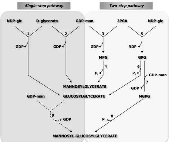
Biosynthesis of DIP 45
Life in extreme environments
Many organisms are adapted to thrive in habitats with non-conventional conditions. Such hostile environments include physical (temperature, pressure or radiation), and geochemical (pH, salinity, desiccation or oxygen availability) extremes. The harsh conditions of those habitats totally abolish the concept of “normal” life, as we know it. These inhospitable conditions can be found in the deep sea, where the pressure is very high and the environmental temperature may be as low as 1ºC, or as high as 400ºC in the vicinity of hydrothermal vents. Other sites include hot springs and geysers, characterized by hot water, steam and sometimes low pH. Hypersaline environments include salt lakes, salt ponds, and marine salterns, like the Dead Sea in Israel or the Soda Lake in Egypt (Falb et al. 2008). Places where liquid water is a limiting factor include deserts that are extremely dry, and hot (Atacama Desert) or cold (Antarctica valleys) (Rothschild and Mancinelli 2001). Furthermore, astrobiology (or exobiology), a rapidly emerging field dedicated to the study of the origin, evolution, distribution and future of the universe, also “labels” the Cosmos as an extreme environment (Cavicchioli 2002, Pikuta and Hoover 2007). The unique organisms that thrive in these extreme conditions are called “extremophiles” and they are classified according to the optimal growth conditions.
acidophiles (optimal growth requiring pH below 4); and piezophiles (optimal growth requiring more than 40 MPa) (Rothschild and Mancinelli 2001, Pikuta and Hoover 2007, Bowers and Wiegel 2011).
Some organisms can live in environments where more than one extreme condition exists. Acidophilic hyperthermophiles include species of the genus Sulfolobus whose physiology is characterized by growth at low pH and high temperature, or species of the genus Acidianus, which grow at pH 2 and 90ºC (Grogan 1989, Cavicchioli 2002). Haloalkaliphiles include several species of the genus Natronococcus that live optimally in habitats with 20% (wt/vol) NaCl and pH 9, or Natronomonas pharaonis which inhabits an environment with 35% (wt/vol) NaCl and a pH of 8.5 (Kanal et al. 1995, Xu et al. 1999, Falb et al. 2008). One of the most recent examples of an organism living in two extremes is Pyrococcus yayanosii, the first obligate piezophilic hyperthermophile isolated from a deep-sea hydrothermal vent, showing optimal pressure and temperature for growth of 52 MPa and 98ºC, respectively (Birrien et al. 2011). It is also possible to find organisms with extreme resistance to ionizing radiation like Deinococcus radiodurans, which was the first radiotolerant bacterium to be isolated, or the hyperthermophile
Thermococcus gammatolerans, which tolerates high doses of γ-radiation (Rothschild and Mancinelli 2001, Pikuta and Hoover 2007). As mentioned above, water limitation can also be considered as an extreme condition, and yet it is possible to find life in sites with extreme dryness. Organisms that thrive in extremely dry environments (desiccation conditions) are called “xerophiles”, and as examples we can find the classical cases of cacti and some fungi (Rothschild and Mancinelli 2001). Extremophiles have representatives in the three domains of Life, the Archaea, the Bacteria and the
The three domains of Life
The proposal for the division of Life on Earth into a higher order taxa, the “domain”, was put forward in 1990 by Carl R. Woese and colleagues. Based on molecular structures and sequences that evidenced the existence of profound differences particularly among prokaryotes, these authors proposed the division of Life into three domains: Bacteria, Eukarya, and Archaea
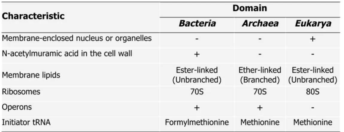
(Woese et al. 1990). The molecular information available at that time to fundament this division, namely the striking differences found in the small subunit of rRNA sequences and structures of several organisms, has been validated by comparative genomics and currently, the division of the biological world into three domains is widely accepted (Woese 1993, Gribaldo and Brochier-Armanet 2006). Some distinctive characteristics of the three domains are summarized in Table 1.1.
Table 1.1. Particular characteristics of the three domains of Life on Earth (adapted from Purves et al. 1998).
Domain Characteristic
Bacteria Archaea Eukarya
Membrane-enclosed nucleus or organelles - - +
N-acetylmuramic acid in the cell wall + - -
Membrane lipids (Unbranched) Ester-linked Ether-linked (Branched) (Unbranched) Ester-linked
Ribosomes 70S 70S 80S
Operons + + -
Initiator tRNA Formylmethionine Methionine Methionine
+, present; -, absent.
However, archaea have earned their position as a distinctive domain not only due to the distinct features of the small subunit rRNA sequence and structure, but also due to the presence of unique genomic signatures and biochemical properties. For example, archaeal organisms harbor a unique type of membrane polar lipids; in the Archaea, the phospholipid backbone is built on sn-glycerol 1-phosphate while in Bacteria and Eukarya the stereoisomer
sn-glycerol 3-phosphate takes place. Furthermore, archaeal lipids have methyl-branched isoprenoid side chains bound to glycerol phosphate by ether-linkages, while in Bacteria and Eukarya the side chains of membrane phospholipids are made of straight chain fatty-acids linked to the glycerol phosphate backbone by ester linkages (Koga and Morri 2007, Peretó et al. 2004).
Based on phylogenetic analyses of 16S rRNA sequences, originally the
Archaea were dividied into two major phyla, the Euryarchaeota and the
and halophiles. On the contrary, the Crenarchaeota include organisms isolated exclusively from thermophilic niches, and comprise hyperthermophiles, thermoacidophiles and sulfur-dependent organisms (Woese et al. 1990, Grabowski and Kelman 2003). Recent metagenomic analyses revealed that some archaea share Crenarchaeota and Euryarchaeota-specific genomic traits. Hence, there was need for additional phyla divisions. The first one to emerge was the phylum Korarchaeota, composed by deeply-branched archaea, including Candidatus (Ca.) Korarchaeum cryptofilum (Barns et al. 1996, Elkins et al. 2008, Nunoura et al. 2010). The emergence of new groups of uncultivated mesophilic Crenarchaeota and the sequenced genome of Ca.
Cenarchaeum symbiosum led to the proposal of another phylum, the
Thaumarchaeota. The proposal for this phylum has been further supported by the genome sequences of other mesophilic archaea, such as Ca.
Nitrosopumilus maritimus and Ca. Nitrososphaera gargensis (Brochier-Armanet et al. 2008, Nunoura et al. 2010). Another phylum, the
Nanoarchaeota, yet with a debatable position, has been proposed based on phylogeny of the smallest known living cell, the Nanoarchaeum equitans, a tiny hyperthermophile that grows and divides at the surface of crenarchaeal
Ignicoccus species, and can not be cultivated independently (Huber et al. 2002, Brochier et al. 2005, Paper et al. 2007).
Thermophiles and hyperthermophiles
The first hyperthermophile was discovered in the early eighties. During a trip to Iceland, Karl Stetter and colleagues collected several samples of boiling water and mud from which they isolated Methanothermus fervidus, the first organism ever isolated that grew up to 97ºC and exhibited the fastest growth at 82ºC (Stetter et al. 1981, Stetter 2006). Until then, the record was held by
Sulfolobus acidocaldarius, isolated in 1972, that although it was able to grow up to 87ºC, the optimal growth temperature was 75ºC (Brock et al. 1972). The way to the world of hyperthermophily had just been opened. The boom in the isolation and characterization of hyperthermophiles happened in the following 25 years, especially by the group of Karl Stetter, who isolated more than 50 species of hyperthermophiles (Stetter 2006). Presently, over 100 species have been described and around 80 have their genomes fully sequenced.
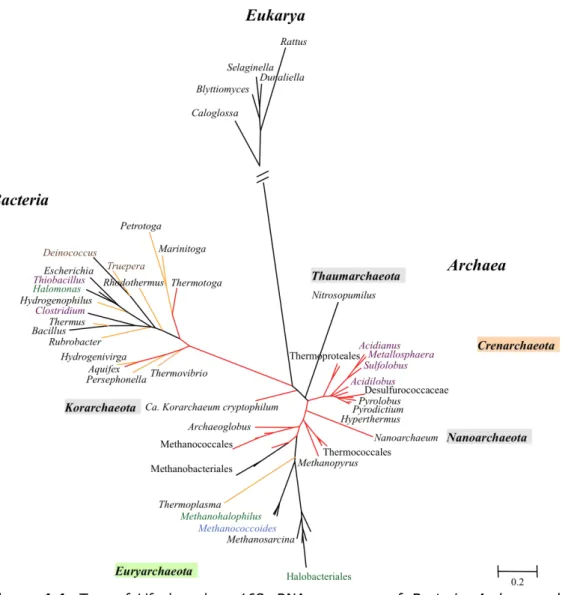
Figure 1.1. Tree of Life based on 16S rRNA sequences of Bacteria, Archaea and
Eukarya. The tree is drawn to scale, with branch lengths measured in the number of substitutions per site. The 59 nucleotide 16S rRNA sequences were retrieved from Silva database (http://www.arb-silva.de/) and the analysis involved one species of the genus represented in the Tree. Sequences were aligned using MUSCLE and the maximum likelihood tree was computed by PHYML. Hyperthermophilic and thermophilic branches are represented in red and orange, respectively. Thermoproteales include the genera Caldivirga, and Pyrobaculum; Desulfurococcaceae include the genera Ignisphaera, Aeropyrum, Ignicoccus, and Staphylothermus; Thermococcales include the genera Palaeococcus, Thermococcus, and Pyrococcus; Methanococcales include the genera Methanocaldococcus, Methanotorris, and
Methanobacterium, and Methanothermus; Halobacteriales include the genera
Halobacterium, Natronobacterium, Haloferax, Natronococcus, and Halococcus.
Within the domain Bacteria, hyperthermophiles are restricted to members of the orders Aquificales and Thermotogales, more specifically to the genera Aquifex and Thermocrinis for the Aquificales, and the genus
Thermotoga for the Thermotogales. Thermotoga maritima and Aquifex pyrophilus withstand the highest growth temperatures, 90ºC and 95ºC, respectively (with optimal growth temperatures of 80ºC for Thermotoga maritima and 85ºC for Aquifex pyrophilus). The position of these bacteria near the root of the Tree of Life makes them good candidates for evolutionary studies (Vieille and Zeikus 2001, Stetter 2006). One interesting aspect that arouse from the sequencing of the Thermotoga maritima genome was the abundance of foreign genes originating from archaeal species or other bacterial species, which supported the occurrence of lateral gene transfer between (hyper)thermophilic bacteria and archaea (Nelson et al. 1999, Mongodin et al. 2005).
Hyperthermophiles are much better represented among the domain
Archaea than in the domain Bacteria. Species of the genera Thermococcus,
Pyrolobus, Hyperthermus, and Sulfolobus are examples of hyperthermophiles that belong to the phylum Crenarchaeota; Thermococcus, Pyrococcus, and
Archaeoglobus are examples of genera belonging to the phylum
Euryarchaeota, and the phylum Nanoarchaeota is represented by
of a new archaeal microbe, strain 121, which supposedly was able to grow up to 121ºC (Kashefi and Lovley 2003), however, this result could not be validated by the scientific community since the strain was not made available. The domain Eukarya is represented by the peculiar “worm” Alvinella pompejana, isolated from hydrothermal vents, which withstands temperatures above 80ºC and live symbiotically with hyperthermophilic bacteria that grow on its tail (Cary et al. 1998, Burgess et al. 2007).
Hyperthermophiles can thrive in a diverse array of biotopes and within each type several factors such as pH, pressure, or NaCl concentration, introduce additional variety. Natural habitats include terrestrial hot springs, geysers, sulfataras, marine hydrothermal vents or even subsurface environments as petroleum reservoirs or geothermally heated lakes. Artificial water-containing hot environments include hot outflows from geothermal power plants, coal refuse piles or even household compost piles (Burgess et al. 2007). The bottleneck for the discovery of new species does not reside in sampling, because nowadays it is possible to access hyperthermophilic biotopes almost routinely. The major problem in studying hyperthermophilic physiology has been, and still is, isolation and cultivation of new species. A good example is the bacterium Thermocrinis rubber, described by Brock in the sixties, which was successfully cultivated by Huber and co-workers only in 1998 (Brock 1967, Huber et al. 1998).
The energy sources and lifestyle of hyperthermophiles are very simple. These organisms are mainly chemolithoautotrophs, using inorganic compounds as energy source and CO2 as the single carbon source to build up
organic cell material. Molecular hydrogen (H2) is an important electron donor,
but sulfide, sulfur, and ferrous iron can be used for that matter as well. Energy yielding reactions are mainly anaerobic and include nitrate (NO3-),
electron acceptors. However, microaerophilic respiration can be found for example in Aquifex pyrophilus, which may use oxygen as electron acceptor. Some hyperthermophiles are facultative heterotrophs, using organic material (from dying cells, for example) instead of inorganic nutrients as electron donors, and conserve energy either by aerobic respiration, different types of anaerobic respiration, or even by fermentation (Stetter 2006).
Hyperthermophiles are adapted to distinct environmental factors such as pH, redox potential, salinity, minerals, gas composition and temperature. These extraordinary organisms are unable to grow below 80ºC but amazingly they can survive at ambient temperature for decades (Stetter 2006). The primitive and yet extraordinary physiology and biochemistry of these organisms has drawn the attention of scientists to several lines of investigation, like the evolutionary aspects of Life on Earth, adaptation strategies to extreme environments, and to the biotechnological potential of hyperthermophiles (Huber et al. 2000).
Extreme biocatalysts
sources of proteases, lipases or amylases for utilization in detergent, food or cosmetics industry (Antranikian et al. 2005). The degradation process of polymers such as starch or cellulose is improved if performed at high temperature, for the sake of solubility improvement and consequent better substrate accessibility. Hence enzymes that are resistant and active at high temperatures are preferred for such application. Furthermore, a few studies were also conducted for the utilization of live cultures of extremophiles in industrial processes, namely desulfurization of ground rubber with the hyperthermophilic Pyrococcus furiosus, bioremediation of waters following water spills by psychrophilic Antarctic bacteria, or production of β-carotenes and glycerol by the halophilic Dunaliella salina (Hubber and Stetter 1998, Bredberg et al. 2001, Rothschild and Mancinelli 2001).
and the molecular mechanisms adopted by these organisms to support life under extreme conditions is particularly interesting and poses a major challenge to the scientific community.
Adaptation to extreme environments
Intrinsic factors for thermoadaptation
that are related with phylogeny, namely due to the close relationship between hyperthermophiles and the domain Archaea.
i) Lipids
The maintenance of membrane fluidity, ion gradients or transport systems across the cellular membrane is vital for cell growth. However, high temperature affects chemical and physical properties of this barrier, causing increased membrane fluidity and permeability. Deficient ion gradients are sufficient to abolish cell growth. In fact, membrane permeability may as well be one of the most important factors in determining the upper temperature limit for life, given the vulnerability of ion gradients at elevated temperature (van de Vossenberg et al. 1998). Hyperthermophiles have evolved mechanisms for the maintenance of membrane integrity at high temperatures, assuring an appropriate matrix for proteins and thus allowing the generation of concentration gradients across the membrane. Such mechanisms include alterations in the length and branching of the acyl chains, saturation, cyclization, or the arrangement of phospholipids across the membrane (Daniel and Cowan 2000, Ulrih et al. 2009).
hydrocarbon chains are highly methyl-branched while its bacterial counterparts are mainly composed by straight chain fatty acids. Most of the archaeal isoprenoid lipid acyl chains are fully saturated, and so they are more resistant to hydrolysis and oxidation. Membrane spanning (monolayer) tetraether lipids (C40 isoprenoid acyl chains) are usually found in extreme
thermophiles and in acidophiles. In contrast, diether lipids (C20 isoprenoid acyl
chains) are present in moderate archaea and halobacteria, forming bilayers similar to those found in the bacterial ester-linked lipids (Koga and Morii 2007). The addition of cyclic structures to the transmenbrane portions results in enhanced stability and reduced membrane fluidity (Daniel and Cowan 2000).
Interestingly, lipids in hyperthermophilic bacteria appear to mimic archaeal lipids. Although some exceptions can be found within the domain
Bacteria, the presence of membrane-spanning lipids and mixed ether/ester lipids in hyperthermophilic bacteria of the genera Thermotoga and Aquifex, corroborates the idea that these features may be associated with life at high temperature (Damsté et al. 2007).
ii) Nucleic acids
pair is more stable and thus, more heat resistant than the A-T pair. However, a correlation between high G+C content in DNA and hyperthermophily is not observed. Some mesophiles have genomes with higher G+C content than hyperthermophiles and even amongst hyperthermophiles there is a huge variation of genomic G+C contents. For example, Pyrolobus fumarii (Topt
106ºC), Pyrococcus furiosus (Topt 100ºC) and Aquifex aeolicus (Topt 85ºC)
have genomic G+C contents of 53, 41, and 44%, respectively. However, the same is not true for RNA. In fact, studies performed in the nineties revealed that the secondary structure of tRNA and rRNA are stabilized by an increased number of G+C pairs near the stem areas and that the increase in G+C content correlates positively with the optimal growth temperature of the organism (Galtier and Lorby 1997). These studies have been confirmed by recent comparative genomic analysis (Dutta and Chaudhuri 2010). Additionally, RNA molecules in hyperthermophiles are stabilized by post-translational covalent modification of nucleosides. These modifications involve increased methylation of the ribose, thiolation and acetylation or replacement of N by C atoms in adenine rings (Kowalak et al 1994, Dutta and Chaudhuri 2010). Salts protect DNA and RNA from thermal degradation: in vitro studies have proved that Na+, K+ and Mg2+ protect DNA from chemical degradation by shielding the negatively charged phosphate backbone of the nucleic acids molecules, hence protecting the phosphodiester bond and aiding the correct folding of these molecules (Hensel and Konig 1988, Marguet and Forterre 2001, Serebov et al 2001). The same effect could be justified by the presence of high intracellular concentrations of salts (Molar range) or the presence of intracellular polycationic polyamines in some hyperthermophiles (Terui et al. 2005, Grosjean and Oshima 2007).
proteins, that compact DNA and have in vitro stabilizing effects, have been identified in hyperhermophiles (Daniel and Cowan 2000). Reverse gyrase is a unique DNA topoisomerase responsible for generating positive supercoils in dsDNA. Reverse gyrase is present in all hyperthermophiles and may be an important hallmark for thermoadaptation (Lopes-Garcia and Forterre 2000, Grosjean and Oshima 2007). However, the in vivo role of reverse gyrase is not completely understood. A Thermococcus kodakarensis mutant with deletion on the reverse gyrase gene revealed that growth was only compromised at extreme temperatures. These results and other recent data suggest that positive supercoiling is not essential for hyperthermophily and that the role of this enzyme may be associated with protection of the DNA molecule against damage such as depurination or DNA breakage (Atomi et al. 2004, Brochier-Armanet and Forterre 2007, Heine and Chandra 2009).
iii) Proteins
studies of meso-, thermo-, and hyperthermophilic proteins show that with increasing temperatures there is a general tendency for an increase in the amount of charged amino acids (Glu, Asp, Arg and Lys), and a decreased frequency of uncharged polar amino acids (Ser, Gln, Tyr and Asn). Furthermore, recent work revealed a positive correlation between the number of intra-subunit ionic interactions and the growth temperature. It is clear that charged residues can promote pair formation, but the exact role that ion-pairs have in the thermal stability of proteins is still unclear (Elcock 1998, Das and Gerstein 2000). In general, stable proteins have more compact structures with fewer internal spaces, a consequence of smaller loops, but also form oligomeric assemblies and inter-subunit interactions (Arnott et al. 2000). Extrinsic factors such as molecular chaperones, intracellular salt concentration, compatible solutes, metabolites, or cofactors may also contribute to protein stability (Ladenstein and Antranikian 1998).
The key for success at high temperatures is a correct balance between flexibility and stability. Proteins are dynamic structures with conformational flexibility, and it is important that at high temperatures correct protein flexibility is achieved to allow for enzyme activity. This concept is of extreme importance, for example in industrial applications, since it is crucial the existence of a balance between protein stability and functionality for optimal enzyme activity.
iv) Metabolites
(Stetter 1999, Cowan 2004). Again, Nature appears to have found alternative ways to bypass the problem of thermal instability of metabolites and coenzymes. Such mechanisms include micro-environmental compartmentalization or metabolic channeling, rapid turnover or increased catalytic efficiency, utilization of alternative more stable metabolites, or alternative biochemical pathways (Daniel and Cowan 2000, Massant 2007). Interestingly, the clustering of functionally related genes into operons, a feature exclusive to bacteria and archaea, facilitates the formation of multienzyme complexes and mutual stabilization of proteins, as well as the channeling of thermolabile metabolites (Glasdorff 1999).
The replacement of the thermolabile NAD+/P for more heat stable non-haem iron proteins in redox reactions has been observed in several members of the domain Archaea. The same observation was made in hyperthermophilic bacteria of the genus Thermotoga, suggesting that although it may be a characteristic of primitive organisms there is also a correlation with hyperthermophily (Daniel and Cowan 2000). Furthermore, hyperthermophilic archaeal kinases show some preference for the utilization of more stable metabolites like pyrophosphate or ADP instead of ATP, as is the case of
Pyrococcus furiosus hexokinase and phosphofructokinase, which are
exclusively ADP-dependent (Daniel and Cowan 2000).
Functional genomics studies, arising from the whole-genome sequencing era, have provided global methods (microarrays, proteomics, metabolomics or mutational analysis) to investigate the molecular basis of heat adaptation. High-throughput studies are presently being carried out for extremophiles such as Thermotoga maritima, Sulfolobus solfataricus,
The heat-shock response
Cellular stress is caused by a sudden change in the surrounding environment of the cell. The type of stress may be physical, like temperature variation, or chemical, such as changes in pH, salinity or oxygen concentration. In all cases, the stress response includes protein denaturation, down-regulation of housekeeping genes and activation of heat-shock genes. The stress proteins encoded by the heat-shock genes are multifunctional, ubiquitous, and serve functions as molecular chaperones, proteases, rRNA processing and signal transduction (Macario et al. 1999).
The heat-shock response has been first described in Drosophila melanogaster (Ritossa 1962) and then in Escherichia coli (Yamamori and Yura 1980), but this is a ubiquitous protective strategy (Schumann 2007). The beginning of the whole process involves thermosensors that will recognize the temperature shift, and trigger a stress response. This response is usually transitory and involves the expression of specific genes that will counteract the external physical effects allowing the cell to reach a new steady state.
and Forterre 2000, Klinkert and Narberhaus 2009). At low temperature the Shine-Dalgarno sequence and the start codon are trapped in a hairpin structure within the mRNA molecules. This prevents binding of the ribosome and thus translation initiation. Upon an increase in the temperature the secondary structure is destabilized and the ribosome binding site becomes accessible (Schumann 2007, Klinkert and Narberhaus 2009).
Protein thermosensors include transcription regulators, chemosensory proteins, chaperones or proteases. Regulators of transcription include the repressor proteins TlpA, RheA, and Phr found in Salmonella enterica,
Streptomyces albus, and Pyrococcus furiosus, respectively (Servant et al. 2000, Hurme et al. 1997, Vierke et al. 2003). Phr was the first transcription factor of Archaea to be discovered and it acts as a repressor of heat-shock genes at physiological temperature, but upon heat-shock (107ºC) it is released from the promoters (Liu et al. 2007, Klinkert and Narberhaus 2009). Molecular chaperones and proteases are part of more complex thermosensor systems and their transcription is induced under heat-shock conditions. These proteins participate in protein folding, assembly, transport and repair under stress conditions (Schumann 2007, Klinkert and Narberhaus 2009). Not all heat-shock proteins are molecular chaperones and likewise, not all molecular chaperones are heat-shock proteins. In fact, molecular chaperones are present and functional in the cell under normal conditions (Lopez-Garcia and Forterre 2000).
The heat-shock response has been extensively studied in Escherichia coli (Guisbert et al. 2008). The master regulon σ32, encoded by the rpoH
increase in the temperature, occurs due to an increase in the levels of the σ32
regulon, which directs RNA polymerase to transcribe from the heat-shock promoters. At optimal physiological conditions (e.g., at 30ºC), σ32 is relatively unstable and the intracellular concentration is kept very low, but upon temperature up-shift (e.g., from 30ºC to 42ºC) the intracellular concentration of σ32 transiently increases (nearly 20-fold in 5 min). During this time the transcription of some heat-shock genes is activated. The increase in σ32 levels is transient and rapidly starts to decline reaching a steady-state level approximately 2 or 3-fold higher than its original value. After a temperature downshift (e.g. from 42ºC to 30ºC) the transcription of heat-shock genes decreases as a result of the decrease in the activity of σ32, mediated by the heat-shock proteins. Hence, transcription of heat-shock genes can be regulated by controlling the synthesis, degradation or activity of σ32 (Yura et al. 2000, Guisbert et al. 2008). While σ32 offers protection against cytoplasmatic stress, another σ factor, σE, was found to be involved in the extracytoplasmatic stress response of Escherichia coli. This second regulon provides cytoplasmatic and periplasmatic heat-shock proteins that are essential for cell survival at very high temperatures (e.g., from 45ºC to 50ºC) (Yura et al. 2000).
Little is known about the heat-shock response of hyperthermophilic organisms. So far, the heat-shock response was investigated in the archaea
Pyrococcus furiosus, Archaeoglobus fulgidus, Sulfolobus solfataricus,
Generally, the transcriptional heat-shock response of hyperyhermophilic archaea has some common trends: in Pyrococcus furiosus,
Methanococcus janaschii, and Archaeoglobus fulgidus the thermosome, small heat-shock proteins, and the proteosome are up-regulated. However, small differences can be found and other genes are differentially regulated: while in
Methanococcus janaschii genes encoding an α-subunit of the prefoldin is highly up-regulated, in Archaeoglobus fulgidus all the prefoldin related genes are down-regulated upon heat-shock (Shockley et al 2003, Rohlin et al. 2005, Boonyaratanakornkit et al. 2005). The heat-shock response of Sulfolobus solfataricus showed that genes encoding small heat-shock proteins were up-regulated, but genes encoding the thermosome and proteases were not affected or were down-regulated by the temperature increase. Interestingly, several DNA-binding proteins were highly up-regulated as well as many insertion sequence elements (Tachdjian and Kelly 2006). The heat-shock response of the hyperthermophilic bacteria Thermotoga maritima was also investigated: it shares some common elements with mesophilic bacteria like the up-regulation of chaperones such as DnaK, but also significant differences were noticed, specially the down-regulation of ATP-dependent proteases (Pysz et al. 2004).
Compatible solutes
The content of salt or sugar in the environment influences the amount of water available to microbes and, the availability of water affects the survival and growth of microorganisms. Most organisms were isolated from aqueous environments where salts and sugars are relatively diluted, but some can be found in extremely saturated brines or concentrated sugar solutions. In either case, microorganisms are able to adjust to osmotic variations that occur in the surroundings, by a process called osmoadaptation. Alterations in the external osmolarity of the medium, usually due to changes in the external Na+
concentration, rapidly triggers a cascade of responses, including water movement across the cellular membrane, that could result in cell swelling and bursting in hypotonic environments, or cell dehydratation in hypertonic environments.
Empadinhas and da Costa 2008). However, the majority of prokaryotes accumulate preferentially small organic osmolytes, i.e., compatible solutes, to cope with hypertonic environments.
The notion of “compatible solute” was put forward in the early seventies by Brown (Brown and Simpson 1972, Brown 1976) and referred to organic or inorganic compounds that could accumulate to high concentrations and did not interfere with normal cell function. The accumulation of compatible solutes does not require modifications of the genetic or enzymatic machinery and is an efficient way to adjust to osmotic fluctuations. Perhaps due to the specific requirements implied in the definition of compatible solutes, only a limited number of compounds meet these criteria and apparently they were widely adopted as osmoprotectants. In fact, this strategy is widespread in Nature and is present in the three domains of Life (da Costa et al. 1998, Roeβler and Müller 2001).
Diversity and distribution of compatible solutes