Amnion-Epithelial-Cell-Derived Exosomes
Demonstrate Physiologic State of Cell under
Oxidative Stress
Samantha Sheller1,2, John Papaconstantinou2, Rheanna Urrabaz-Garza1, Lauren Richardson1, George Saade1, Carlos Salomon3, Ramkumar Menon1*
1Division of Maternal-Fetal Medicine & Perinatal Research, Department of Obstetrics & Gynecology, The University of Texas Medical Branch at Galveston, Galveston, Texas, United States of America,
2Department of Biochemistry and Molecular Biology, The University of Texas Medical Branch at Galveston, Galveston, Texas, United States of America,3Exosome Biology Laboratory, Centre for Clinical Diagnostics, UQ Centre for Clinical Research, Faculty of Health Sciences, University of Queensland, Herston,
Queensland, Australia
Abstract
At term, the signals of fetal maturity and feto-placental tissue aging prompt uterine readi-ness for delivery by transitioning quiescent myometrium to an active stage. It is still unclear how the signals reach the distant myometrium. Exosomes are a specific type of extracellular vesicle (EVs) that transport molecular signals between cells, and are released from a wide range of cells, including the maternal and fetal cells. In this study, we hypothesize thati)
exosomes act as carriers of signals in utero-placental compartments andii) exosomes
reflect the physiologic status of the origin cells. The primary aims of this study were to deter-mine exosomal contents in exosomes derived from primary amnion epithelial cells (AEC). We also determined the effect of oxidative stress on AEC derived exosomal cargo contents. AEC were isolated from amniotic membrane obtained from normal, term, not in labor pla-centae at delivery, and culture under standard conditions. Oxidative stress was induced using cigarette smoke extract for 48 hours. AEC-conditioned media were collected and exo-somes isolated by differential centrifugations. Both growth conditions (normal and oxidative stress induced) produced cup shaped exosomes of around 50 nm, expressed exosomes enriched markers, such as CD9, CD63, CD81 and HSC70, embryonic stem cell marker Nanog, and contained similar amounts of cell free AEC DNA. Using confocal microscopy, the colocalization of histone (H) 3, heat shock protein (HSP) 70 and activated form of pro-senescence and term parturition associated marker p38 mitogen activated protein kinase (MAPK) (P-p38 MAPK) co-localized with exosome enrich marker CD9. HSP70 and P-p38 MAPK were significantly higher in exosomes from AEC grown under oxidative stress condi-tions than standard condicondi-tions (p<0.05). Finally, mass spectrometry and bioinformatics analysis identified 221 different proteins involved in immunomodulatory response and cell-to-cell communication. This study determined AEC exosome characteristics and their cargo reflected the physiologic status of the cell of origin and suggests that AEC-derived exoso-mal p38 MAPK plays a major role in determining the fate of pregnancy. Understanding the
a11111
OPEN ACCESS
Citation:Sheller S, Papaconstantinou J, Urrabaz-Garza R, Richardson L, Saade G, Salomon C, et al. (2016) Amnion-Epithelial-Cell-Derived Exosomes Demonstrate Physiologic State of Cell under Oxidative Stress. PLoS ONE 11(6): e0157614. doi:10.1371/journal.pone.0157614
Editor:Kang Sun, Shanghai Jiaotong University School of Medicine, CHINA
Received:April 14, 2016
Accepted:June 1, 2016
Published:June 22, 2016
Copyright:© 2016 Sheller et al. This is an open access article distributed under the terms of the Creative Commons Attribution License, which permits unrestricted use, distribution, and reproduction in any medium, provided the original author and source are credited.
Data Availability Statement:All relevant data are within the paper and its Supporting Information files.
Funding:This study is supported by Innovative catalyst grant funding from March of Dimes Center of Ohio, Cincinnati, OH to RM and partially by RM’s Faculty development funds from University of Texas Medical Branch at Galveston, TX. Authors declare
allthe funding or sources of support received
propagation of fetal signals and their mechanisms in normal term pregnancies can provide insights into pathologic activation of such signals associated with spontaneous preterm parturitions.
Introduction
Normal term human parturition is initiated between 37 and 40 weeks of gestation when in utero fetal growth and development are completed [1–3]. The signals from mature fetal organs prompt uterine readiness for delivery by transitioning quiescent myometrium to an active stage (contractile phenotype) [2,4,5]. Various endocrine, immune and mechanical signals from feto-maternal compartments enhance overall uterine inflammatory load leading to functional progesterone withdrawal causing myometrial contractility [6–8]. Therefore, term labor and delivery results from well-orchestrated activities of various endocrine and paracrine factors. Nonetheless, the signature of these signals and their precise mechanisms in initiating parturi-tion are still unclear and under investigaparturi-tion by many laboratories. Understanding the mecha-nisms of these signals in normal term pregnancies can provide insights into pathologic activation that can cause spontaneous preterm parturitions.
Recently, our laboratory has reported fetal membrane (amniochorionic membrane) senes-cence as a mechanism associated with normal term parturition [9,10]. Placental membranes undergo an oxidative stress associated telomere dependent cellular senescence at term [11–13]. In addition, placental membrane senescence is also associated with sterile inflammation in the amniotic fluid [9,14]The unique inflammation seen in senescent cells is defined senescence associated secretory phenotype (SASP) [15]. SASP is characterized by proinflammatory cyto-kines and chemocyto-kines that are reported to be associated with term labor and delivery [9]. The findings in clinical specimens have been reproduced in vitro in primary amnion epithelial cells in culture exposed to oxidative stress induced by cigarette smoke extract (CSE) [12]. Genera-tion of senescence and SASP were reduced by inhibiting p38 mitogen activated kinase (MAPK), a stress associated pro-senescence protein, suggesting that sterile inflammation can be generated by senescent cells [12,14,16].
It is still unclear how proinflammatory SASP signals from senescent fetal membranes can reach distant myometrium or whether they are confined to enforcing local (fetal) tissue damage and inflammation until parturition. Senescent signals may be transported to distant tissues indicating a dysfunctional fetal membrane status that prompts delivery of the fetus, as well as placenta and membrane. Although localized activities of SASP can be achieved through direct cell-cell contact or through ligand-receptor interactions, distant feto-maternal communication is likely facilitated through specific carriers that can transport and deliver signals from senes-cent cells. Thus, prior to projecting senessenes-cent fetal cells signaling parturition at a distant myo-metrium, the mode of delivery of such signals must be established. Several recent reports and reviews propose a role for exosomes as such carriers of parturition signals to utero-placental compartments [17–20].
Exosomes are 30–100 nm endosome-derived vesicles with specific characteristics that sepa-rate them from other larger particles such as microvesicles and apoptotic bodies [21–24]. Exo-some biogenesis is a process that begins with the endocytosis of transmembrane proteins [25,26]. Once sorted to late endosomes, the endosomal sorting complex required for transport (ESCRT) complex, recruits proteins and other cargo, while also mediating the inward budding of the late endosome, creating the intraluminal vesicles inside the multivesicular body (MVB) [27–30]. The MVB can either follow a degradation pathway fusing with lysosomes or proceed
to release the intraluminal vesicles into the extracellular space through exocytosis as exosomes [27,28]. Placental derived exosomes have been well characterized during normal and abnormal pregnancies and their functional roles have also been documented [4–13,31–39]. Their size facilitates easy transportation between cells and tissues, while their contents reflect the state of the source cell and regulate the phenotype of the target cell [23,34,36,40–42]. Exosomes inter-act with the target cell by direct fusion with the cell membrane, thus releasing the contents directly into the cytosol; through active uptake via endocytosis or by binding to the target cell via receptor-ligand interactions thereby inducing a signaling cascade which changes the pheno-type of the target cell.
No reports exist on fetal membrane- derived exosomes or their contents. Therefore, the objectives of this study are: 1) determine the generation of exosomes from primary amnion epi-thelial cells and characterize their contents, 2) determine the changes in specific exosome con-tents in primary amnion cells in response to oxidative stress. Since p38 MAPK was identified as a crucial stress response-signaling pathway that activates oxidative stress induced senescence at term, we specifically examined exosomal p38 MAPK cargo. Using an in vitro primary amnion epithelial cell (AEC) model of oxidative stress induced by cigarette smoke extract, a well characterized model of in vitro oxidative stress, and using Liquid Chromatography (LC)/ Mass Spectrometry (MS) we characterize the contents of AEC -derived exosomes and their potential role in parturition.
Materials and Methods
Placental samples were obtained for this study from John Sealy Hospital at The University of Texas Medical Branch (UTMB) at Galveston, TX, USA. No subjects were recruited or con-sented for this study as we used discarded placenta from normal term, not in labor cesarean sections. The study protocol was submitted and approved by the institutional review board at UTMB, whereby placental samples could be collected without consenting subjects. Placentae from women (18–40 years old) undergoing elective repeat cesarean delivery (between 37 and 41 weeks of gestation) prior to the onset of labor were included in the study. Women with prior history of preterm labor and delivery, preterm premature rupture of the membranes, pre-eclampsia, placental abruption, intrauterine growth restriction, and gestational diabetes were excluded. Additionally, group Bstreptococcuscarriers, treated for urinary tract infection, sexu-ally transmitted diseases, chronic infections like HIV, hepatitis, and women who smoked ciga-rettes or reported drug and alcohol abuse, were also excluded.
Isolation and Culture of human Amnion Epithelial Cells (AECs)
All reagents and media were warmed to 37°C prior to use. The amniotic membrane was pro-cessed as described previously [6,12,14] Briefly, the amnion membrane was manually peeled from normal, term, not in labor caesarean section placentas, rinsed in saline and transferred to a petri dish containing Hanks Balanced Salt Solution (HBSS; Mediatech Inc., Manassas, VA). After cutting the amnion into 2 cm x 2 cm pieces, they were digested twice in 0.25% trypsin and 0.125% Collagenase A (Sigma–Aldrich, St. Louis, MO) in HBSS for 35 minutes at 37°C. After each digestion, the tissue was filtered through a 70μm cell strainer (Thermo Fisher
Scien-tific, Waltham, MA) and trypsin was inactivated using complete Dulbecco's Modified Eagle Medium: Nutrient Mixture F-12 media (DMEM/F12; Mediatech Inc.) supplemented with 15% fetal bovine serum (FBS; Sigma-Aldrich), 10% Penicillin/Streptomycin (Mediatech Inc.) and 100μg/mL epidermal growth factor (EGF; Sigma-Aldrich). The collected filtrate was
flasks containing complete DMEM/F12 media at 37°C, 5% CO2, and 95% air humidity to 70–
80% confluence.
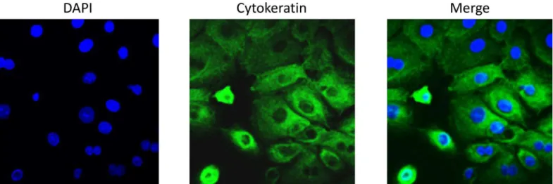
To ensure the purity of our primary AEC cultures, immunofluorescent staining was per-formed. Cells were seeded on glass coverslips at a density of 30,000 cells per slip and incubated overnight. Cells were fixed with 4% paraformaldehyde (PFA), permeablized with 0.5% Triton X and blocked with 3% BSA in PBS prior to incubation with Cytokeratin 18 (Abcam, Cam-bridge, United Kingdom) primary antibody diluted 1:300 in 3% BSA overnight at 4°C. After washing with PBS, slides were incubated Alexa Fluor conjugated secondary antibodies (Life Technologies, Carlsbad, CA) diluted 1:400 in PBS for 1 hour in the dark. Slides were washed with PBS then treated with NucBlue1Live ReadyProbes1Reagent (Life Technologies) then mounted using Mowiol 4–88 mounting medium (Sigma-Aldrich). Images were captured using LSM 510 Meta UV confocal microscope (63x) (Zeiss, Germany).
Stimulation of AEC with cigarette smoke extract (CSE)
To induce oxidative stress in AECs, CSE was used as detailed in our prior studies, [12,43,44] with modifications. Smoke from a single lit commercial cigarette (unfiltered CamelTM, R.J. Reynolds Tobacco Co, Winston Salem, NC) was infused into 25 mL of exosome-free media, consisting of DMEM/F12 supplemented with 10% exosome-free FBS (System Biosciences, Mountain View, CA). The stock CSE was sterilized using 0.25 mm Steriflip1filter unit (Milli-pore, Billerica, MA). CSE concentrate was diluted 1:10 in exosome-free media prior to use. Once cells reached 70–80% confluence, each flask was rinsed with sterile 1x PBS followed by treatment with exosome-free media (control) or CSE containing media and incubated at 37°C, 5% CO2, and 95% air humidity for 48 hours.
Cell cycle analysis of AECs using flow cytometry
CSE treated and control AECs were harvested after media collection using trypsin EDTA (Corning, Corning, NY) and centrifuged for 10 minutes at 3000 RPM. The supernatant was removed and cells were resuspended in 50μL PBS. Cell cycle analysis was performed using the
Coulter DNA Prep Reagents Kit (Beckman Coulter, Indianapolis, IN). Briefly, 50μL of DNA
Prep LPR was added to each sample and vortexed. Then 1.0 mL DNA Prep Stain was added to the tubes, vortexed and run immediately on the Cytoflex flow cytometer (Beckman Coulter). After selecting for single cells, gating was set for the control cells and applied to histograms for the CSE treated AECs using Cytexpert (Beckman Coulter).
Activation of p38 MAPK in AECs using flow cytometry
Activation of p38 MAPK was also performed. After harvesting cells using trypsin EDTA and centrifugation for 10 minutes at 3000 RPM, the pellet was resuspended in 500μL 4%
parafor-maldehyde and vortexed. After incubation for 10 minutes at room temperature, cells were placed on ice for 1 minute then centrifuged for 5 minutes at 2000 RPM at 4°C. The supernatant was removed and the pellet was resuspended in 500μL 90% ice cold methanol, gently vortexing
while adding methanol slowly. Once the pellet was completely resuspended, the cells were incu-bated on ice for 10 minutes then stored at -20°C until use.
centrifuged and resuspended in 400μL PBS and run immediately on the Cytoflex flow
cytome-ter (Beckman Coulcytome-ter). Afcytome-ter gating for single cells, data analysis based was performed using Cytexpert (Beckman Coulter). Flow Cytometry analysis for exosome markers and DNA
Exosome isolation
The culture media were collected and stored at -80°C until exosome isolation. Media was thawed overnight then isolated using differential ultracentrifugation as described previously, with modifications [21,45,46]. Briefly, the culture media was centrifuged sequentially at 4°C for 10 minutes at 300g and 20 minutes at 2,000g using Sorvall Legend X1R and TX-400 swinging bucket rotor (Thermo Fisher Scientific), 30 minutes at 10,000g and 2 hours at 100,000g using a Beckman Optima LX-80 ultracentrifuge with 50.1Ti and 70.1Ti rotors (Beckman Coulter). The pellet was resuspended in 1x PBS then centrifuged again at 100,000g for 1 hour. Pellet was resuspended in RIPA (Western Blot) or 1x PBS (electron microscopy, Flow Cytometry, DNA quant and sizing).
Transmission Electron Microscopy (TEM) of whole mounted exosomes
For TEM studies, 5μL suspended exosomes in PBS were dropped onto a formvar-carbon
coated 300-mesh copper grid and left to dry at room temperature for 10 min. Grids were treated with 10 seconds of Hydrogen-Oxygen plasma in a Gatan Solarus 950 plasma cleaning system (Gatan, Inc., Pleasanton, CA) prior to use. After three washes in purified water, the exo-some samples were negatively stained using PhosphoTungstic Acid (PTA). The grids were dried at room temperature then viewed in a 120 keV JEM 1400 electron microscope (Jeol, Pea-body, MA). A minimum of 10 frames were viewed per sample.
Exosome particle sizing analysis
Dynamic light scattering analysis (DLS) was used to determine the mean size distribution and purity of our exosome preps. 50μL of concentrated exosomes was brought to a volume of 1.0
mL in 1x PBS and sized using Malvern High Performance Particle Sizer (HPPS) (Malvern Instruments, Worcestershire, UK).
Western blot analysis
Exosomal pellets were resuspended in RIPA lysis buffer (50 mM Tris pH 8.0, 150 mM NaCl, 1% Triton X-100, and 1.0 mM EDTA pH 8.0, 0.1% SDS) supplemented with protease and phosphatase inhibitor cocktail and PMSF. After centrifugation at 10,000 RPM for 20 minutes, the supernatant was collected and protein concentrations were determined using BCA (Pierce, Rockford, IL). The protein samples were separated using SDS-PAGE on a gradient (4–15%) Mini-PROTEAN1TGX™Precast Gels (Bio-Rad, Hercules, CA) and transferred to the mem-brane using iBlot1Gel Transfer Device (Thermo Fisher Scientific). Membranes were blocked in 5% nonfat milk in 1x Tris buffered saline-Tween 20 (TBS-T) buffer for a minimum of 1 h at room temperature then probed (or re-probed) with primary antibody overnight at 4°C. The membrane was incubated with suitable secondary antibody conjugated with horseradish per-oxidase and immunoreactive proteins were visualized using Luminata Forte Western HRP sub-strate (Millipore, Billerica, MA). The stripping protocol followed the instructions of Restore Western Blot Stripping Buffer (Thermo Fisher). No blots were used more than three times.
Embryonic stem cell markers Nanog andOCT-4, (Cell Signaling, Beverly, MA) were diluted 1:500. Heat shock protein (HSP) 70 (Abcam), a damage associated molecular pattern, was diluted 1:500. Total p38 MAPK and phospho p38 MAPK (Cell Signaling) were diluted 1:400.
Flow Cytometry analysis for exosome markers and DNA
For flow cytometry analysis of exosome tetraspanin markers CD9, CD63 and CD81, a total of five samples per condition were prepared using the ExoFlow kit (System Biosciences) protocol with modifications. Briefly, exosomes isolated from treated and untreated amnion cell cultures were resuspended in 150μL 1x PBS and all kit reagent volumes were halved. Streptavidin
coated 9.1μm beads were washed then incubated with biotinylated anti-CD9, CD63 or CD81
for 2 hours on ice, flicking intermittently to mix. Beads were washed and resuspended in 200μL bead wash buffer prior to incubation overnight at 4°C with 50μL exosomes (total
vol-ume 250μL). The following day, samples were washed and stained using the Exo-FITC
exo-some stain according to manufacturer then run on the Cytoflex flow cytometer (Beckman Coulter). Negative controls with isotype-matched antibodies were used for gating, applied according to manufacturer instructions. Data analysis based on fluorescein isothiocyanate (FITC) signal shift was performed using Cytexpert (Beckman Coulter).
After initial analysis for tetraspanin markers, the samples were tested for DNA content using the Coulter DNA Prep Reagents Kit (Beckman Coulter), which uses propidium iodide to tag DNA. Since DNA fragments are expected to stick to exosomal membranes and can confound with true exosomal DNA cargo, AEC exosomes were pretreated with DNase to remove all exter-nal DNA. To address the potential contamination, the samples were split in half, one set of sam-ples were pretreated with DNase while the other half remained untreated. Pretreated samsam-ples were washed with exosome wash buffer provided in the ExoFlow kit then resuspended in DNase 1 set (Zymo Research, Irvine, CA) solution according to the manufacturer’s protocol, substituting exosome wash buffer for ethanol. Samples were incubated at room temperature for 10 minutes. To inactivate the DNase, samples were heated to 65°C for 15 minutes. After washing with exo-some wash buffer three times, the pretreated and untreated exoexo-somes were resuspended in 50μL
PBS then stained using the Coulter DNA Prep kit (Beckman Coulter). Samples were run on the Beckman Coulter CytoFlex Flow Cytometer. Negative controls, consisting of beads without exo-somes attached, were used for gating, applied according to ExoFlow (SBI) instructions.
Quantitation of exosomal DNA
To quantify the amount of DNA, exosomes were resuspended in 200μL 1x PBS. DNA was
extracted following DNeasy Blood and Tissue kit protocol (Qiagen, Hilden, Germany). Quanti-fication was performed on the Synergy H4 Hybrid Microplate Reader (Biotek, Winooski, VT) using the Take 3 microvolume plate and Gen5 software (Biotek). We corrected for the blank and DNA concentration was calculated based on the absorbance ratio of 280/260.
Immunofluorescence staining and microscopy
(Sigma-Aldrich). Images were captured using LSM 510 Meta UV confocal microscope (63x) (Zeiss, Germany). Exosomes were tagged using anti-CD9 diluted 1:300, while P-p38 MAPK, Histone 3 (H3), and HSP70 were diluted 1:250. Colocalization of proteins of interest with exo-somes was determined using LSM software (Zeiss) and Image J (open source). A total of five images per condition and 5 regions of interest per image were used to determine red and green fluorescence intensity values per distance (μm). Intensity was graphed versus distance for both
colors using GraphPad Prism (GraphPad Software, La Jolla, CA). Areas of overlap indicate colocalization. The images were also analyzed for Pearson's Correlation Coefficient using Coloc 2 from Fiji (open source), selecting 5 regions of interest for each image. Mean Pearson’s values were graphed using Excel. Any image modifications (brightness, contrast, and smooth-ing) were applied to the entire image using Image J (open source).
Proteomic analysis of AEC-derived exosomes by mass spectrometry
Protein profile of exosomes isolated from AEC culture under normal or oxidative stress condi-tions were established by Liquid Chromatography (LC)/ Mass Spectrometry (MS) as previously described with modifications [41,42]. Briefly, exosome pellets were lysed in 500μL modified
RIPA buffer (2.0% SDS, 150mM NaCl, 50mM Tris, pH 8.5, 1X Complete Protease inhibitor (Roche)) at 100°C for 15 minutes. The lysate was clarified by centrifugation and the protein concentration determined by Qubit fluorometry (Invitrogen). 10μg of extracted protein was
processed by SDS-PAGE using 10% Bis Tris NuPage mini-gel (Invitrogen) in the MES buffer system. The migration window (2cm lane) was excised and in-gel digestion performed using a ProGest robot (DigiLab) using ammonium bicarbonate (25mM), dithiothreitol (reduction step, 10mM at 60°C) and iodoacetamide (alkylation step, 50mM). Samples were digested with sequencing grade trypsin (Promega) at 37°C for 4h and quenched with formic acid. The super-natant was analyzed directly without further processing. Digested samples were analyzed by nano LC-MS/MS with a Waters NanoAcquity HPLC system interfaced to a ThermoFisher Q Exactive. Peptides were loaded on a trapping column and eluted over a 75μm analytical column
at 350nL/min using a 2hr reverse phase gradient; both columns were packed with Jupiter Pro-teo resin (Phenomenex). The mass spectrometer was operated in data-dependent mode, with the Orbitrap operating at 60,000 FWHM and 17,500 FWHM for MS and MS/MS respectively. The fifteen most abundant ions were selected for MS/MS. False discovery rate (FDR) was esti-mated using a reversed sequence database.
Functional analysis of exosome proteome
Proteins identified by MS/MS were analyzed by PANTHER (Protein Analysis THrough Evolu-tionary Relationships;http://www.pantherdb.org) as previously described [41,42]. Differen-tially identified proteins were analyzed further by bioinformatic pathway analysis (Ingenuity Pathway Analysis [IPA]; Ingenuity Systems, Mountain View, CA;www.ingenuity.com).
Statistical analysis
SPSS software (IBM, Armonk, NY) was used for statistical evaluation. Samples were analyzed using pairedt–test and aPvalue less than 0.05 was considered statistically significant.
Results
Primary AEC cultures are positive for cytokeratin
Cell cycle analysis and p38 MAPK activation of control and CSE treated
AECs
To determine the pattern of the cell cycle in control and CSE AECs, cell cycle analysis was per-formed.Table 1summarizes the results of the cell cycle analysis. While majority of the control and CSE treated AECs were in G1 phase, noticeable differences between the two groups can be seen (Table 1). As can be seen inTable 1, the percentage of SubG0G1 phase cells was much higher in CSE treated AECs than controls. The SubG0G1 phase includes cellular debris, as well as late apoptotic and necrotic cells and suggestive of cell cycle arrest and senescence associated changes after CSE treatment. The percentage of control cells in S phase and G2 (indicating active proliferation) were almost double in control compared to CSE treated AECs showing normal progression of cell cycles in these cells. These data represent the physiologic state of the cell.
To further confirm this physiologic state, we also examined p38MAPK activation, an oxida-tive stress response indicator activated by various stressors, using flow cytometry. AECs treated with CSE showed higher p38 MAPK activation than control cells, indicating CSE AECs were undergoing oxidative stress and cellular senescence.
Characteristics of exosomes released by primary amnion epithelial cells
under control and CSE conditions
Exosomes isolated from AECs under standard (control) and oxidative (CSE) conditions were characterized using four different methods–electron microscopy (for shape and morphology), particle sizing (size), western blot and flow cytometry (specific markers and cargo contents) and Nano drop analysis (DNA quantitation).
Fig 1. Cytokeratin staining of primary AECs.Primary AECs were fluorescently labeled for cytokeratin 18 to show cultures were predominantly epithelial cells. AEC-amnion epithelial cell.
doi:10.1371/journal.pone.0157614.g001
Table 1. Cell cycle analysis and p38 MAPK activation of control and CSE treated amnion cells.
SubG0G1 G1 S G2 P-p38 MAPK
Control (untreated) AEC
6.9% 73.4% 15.6% 4.1% 1.8%
CSE Treated AEC 36.4% 53.6% 7.9% 2.1% 35.9%
Exosome size and morphology from Control and CSE cells
TEM analysis (Fig 2A) revealed vesicles with classic exosomal morphology. Both untreated AECs and CSE treated AECs produced cup shaped vesicles with a size distribution between 30–50 nm, which is consistent with published reports for exosomes in general, as well for exosomes from reproductive tissues [31,34,47,48]. Size distribution by intensity graphs for control and CSE treated AEC exosomes indicate the absence of larger material in each sample (Fig 2B), such as microvesicles and apoptotic bodies validating our isolation procedures and purity of exosome preparations. DLS analysis also confirmed the size distribution of 3 control and 3 CSE exosome isolations (mean ±STD) (control 39.1±2.97; CSE 45.5±2.34). CSE treated AECs produced slightly larger exosomes than exosomes from untreated AECs (p<0.05). The number of exosomes as determined by total protein concentrations per 1 million cells were similar under both condi-tions. However, we acknowledge that this is not the best approach to quantitate exosomes.
Exosome and AEC specific markers from Control and CSE cells
Western blot analysis was performed to determine exosome enriched markers and cargo con-tents. AEC exosomes showed the presence of markers CD81 and HSC70 (Fig 2C) regardless of
Fig 2. Characterization of exosomes released from amnion cells grown under standard (control) and oxidative (CSE) conditions.A: Electron microscopy showing cup-shaped vesicles that have a size distribution of 30–50 nm (arrow indicates exosomes, scale bar represents 50 nm). B: Size distribution analysis of exosomes from amnion cells untreated control (39.16±2.97) and CSE (45.53±2.34) exosomes. P<0.05 using paired sample t test. C: Western blot analysis showed the presence of exosome markers CD81 and HSC70, as well as embryonic stem cell marker, Nanog, indicating amnion epithelial cell origin. D: Western blot analysis shows lack of expression of Alix in AEC derived exosomes. E: Flow cytometric characterization of exosome tetraspanin markers exosome markers CD9 and CD63. X-axis is FITC intensity, y-axis is count, or number of beads positive for exosomes. Green represents negative control (neg). F: Western blot analysis showing differences in specific markers. Presence of stress responsive and pro-senescence marker p38 mitogen activated protein kinase (MAPK) and one of the DAMP) markers, HSP70, in exosomes from both control and CSE treated AECs. Expression of P-p38MAPK was higher in exosomes from AECs treated with CSE compared to control. AEC-amnion epithelial cell, CSE-cigarette smoke extract, DAMP-damage associated molecular pattern.
treatment. We used two embryonic and amnion stem cell specific markers, Nanog (a transcrip-tion factor) and octamer-binding transcriptranscrip-tion factor 4 (Oct-4), to confirm origin of exosomes [49–54]. Although these two proteins are not exclusive to AECs, they are well documented in both amniotic epithelial and mesenchymal cells and are well reported to characterize AECs. AECs from both untreated and treated AECs demonstrated Nanog but Oct-4 was not localized in any of our exosome preparations.
We also used western blot analysis to detect the expression of Alix in AEC derived exo-somes. As shown inFig 2D, western blot analysis did not demonstrate the presence of Alix in either untreated or CSE treated AEC exosomes.
Flow cytometry was performed to further characterize the exosomes using tetraspanin markers CD9 and CD63 (Fig 2E), common markers used for exosome identification [55]. The shift in FITC intensity on the representative histograms inFig 2Eindicates beads positive for either CD9 or CD63 exosomes (pink peaks) relative to the negative control (no exosomes) (green peaks). By graphing forward scatter versus FITC intensity, we calculated the percentage of beads positive for exosome markers. After subtracting the negative control to account for background, results are expressed as percentage of beads positive for CD9 or CD63 expressing exosomes. Exosomes from control AECs showed an average of 70% for CD9 compared to 38% in CSE treated AEC exosomes. Similar decrease was also seen for CD63 (64% and 19% in con-trols vs CSE respectively). Difference in exosome marker expression was found to be statisti-cally significant (p<0.05).
Characterization of exosomal cargo from Control and CSE treated AECs
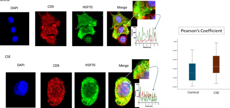
Western blot analysis showed the presence of active forms of pro-senescence and parturition associated marker p38 MAPK (P-p38 MAPK) and one of the damage associated molecular pat-tern (DAMP) molecule HSP70 in exosomes from both control and CSE treated AECs. As shown inFig 1FHSP70, P-p38 MAPK and total p38 MAPK were seen in both untreated and treated exosomes. However, the intensity of bands for P-p38 MAPK was higher in exosomes from AECs treated with CSE.
Cell free fetal DNA are associated with human parturition through inflammatory activation [56]. Telomere fragments from fetal cells can also cause sterile inflammation in fetal tissues associated with parturition [13]. Exosomes are known to carry double and single stranded DNA fragments and cause immune activation in recipient cells [57]. Therefore, we examined untreated and CSE treated AEC derived exosomes for DNA fragments. Flow cytometry analy-sis for DNA content of exosomes from control and CSE treated cells (Fig 3) showed DNA within exosome preparations. There was no significant difference in DNA content between DNase treated and untreated nor between control and CSE treated AEC exosomes suggesting that external contaminant DNA are unlikely in our preparations and DNA fragmentation expected after CSE treatment did not result in increased exosomal content. We further con-firmed the presence of DNA in both control and CSE exosomes by quantifying the amount of DNA using A260/280 and confirmed data obtained from flow cytometric analysis. Average DNA concentration from control exosomes was 4.7 ng/μL ± 0.794 while CSE exosomes
aver-aged 5.1 ng/μL ± 1.86.
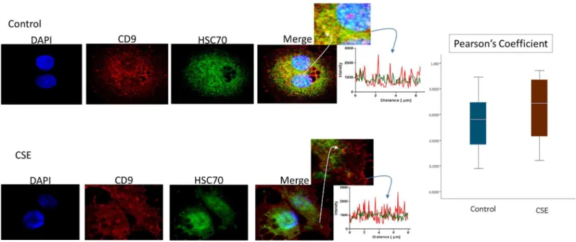
Immunofluorescence staining was used to colocalize markers within exosomes. As shown in
Fig 4, HSC70 was colocalized with CD9 markers (Fig 4). The colocalizations were similar in
P-p38 MAPK (Fig 5), Histone 3 (H3,Fig 6), and HSP70 (Fig 7) were colocalized with exo-some marker CD9 using immunofluorescent microscopy to determine whether exoexo-somes reflect the physiologic state of AECs. All studied markers are associated with oxidative stress Fig 3. Documentation of DNA by flow cytometry in exosomes using propidium iodide (PI).There was no significant difference in DNA content in exosomes untreated with DNase (top) and those pretreated with DNase (bottom). There was also no significant difference in DNA content between CD9, CD63 and CD81 positive exosomes. The average DNA content was similar for both control (4.7 ng/μL) and CSE (5.1 ng/μL) derived exosomes. AEC-amnion epithelial cell, CSE-cigarette smoke extract.
doi:10.1371/journal.pone.0157614.g003
Fig 4. Colocalization of exosome marker HSC70 and CD9 in AECs.Control (untreated AECs) (top panel) have a similar amount of HSC70 (green) colocalization with CD9 exosome marker (red) compared to AECs exposed to CSE (bottom panel). The line graphs represent overlap between CD9 and HSC70 signal at the region of interest. The bar graph represents no significant differences between the two groups (Pearson’s Coefficient p = ns). AEC-amnion epithelial cell, CSE-cigarette smoke extract.
Fig 5. Colocalization of exosome marker H3 and CD9 in AECs.Immunofluorescence imaging of control (top panel) and CSE treated amnion cells (bottom panel) show colocalization differences of H3 (green) and CD9 (red). Significantly higher colocalization (line graph and bar graph) of H3 was seen after CSE treatment compared to control (Pearson’s Coefficient P<0.0001) AEC-amnion epithelial cell, CSE-cigarette smoke extract.
doi:10.1371/journal.pone.0157614.g005
response and activation of p38 MAPK in amnion cells, which is the signaler in senescence response to CSE. H3 and HSP70 are p38 MAPK responder genes in stress associated cellular signaling [58,59]. Colocalization was quantified using Pearson’s correlation coefficient and graphed comparing control and CSE mean values. Colocalization of all three markers were sig-nificantly higher in CSE treated AEC exosomes compared to control AEC exosomes (mean Pearson’s correlation coefficients for each were statistically significant (p<0.05)) confirming CSE causes increased cargo of these markers by exosomes. CSE induced oxidative stress dam-age leads to senescence of AEC through p38 MAPK signaling (12] and current data confirm that P-p38 MAPK and its responder proteins H3 and HSP70 can also get packaged inside exo-somes at a higher level reflecting the physiologic state of AECs.
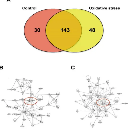
Proteomic analysis of CTs-derived exosomes
Mass spectrometry analysis identified over 200 exosomal proteins (S1 Table). We also identi-fied unique proteins for each condition (Fig 8A; Tables2and3). The number of proteins iden-tified in exosomes isolated from AEC exposed to CSE was higher (193) compared to control (173). We investigated the molecular network that can be activated by the proteins identified in exosomes isolated from AEC cultured under normal (8B) and oxidative stress conditions (8C). Interestingly, NF-κβcomplex seems to be a central regulator in the molecular network that can be activated by exosomes from AEC cultured under normal conditions, where TGF-β
might regulate the molecular network from exosomes from AEC treated with CSE. We have already reported that CSE treatment produce minimal activation of NF-κB compared to con-trol and the exosomal proteomic analysis further supports our earlier findings [60]. We have also seen evidence of epithelial-mesenchymal transition (EMT) of amnion epithelial cells under oxidative stress conditions. TGF-βis a major mediator of EMT [61–65]. Molecular Fig 7. Colocalization of exosome marker P-p38MAPK and CD9 in AECs.Colocalization of pro-senescence marker P-p38MAPK and CD9: Immunofluorescence imaging of control (top panel) and CSE treated amnion cells (bottom panel) show colocalization differences of HSP70 (green) and CD9 (red). Significantly higher colocalization (line graph and bar graph) of H3 was seen after CSE treatment compared to control (Pearson’s Coefficient P<0.0001) AEC-amnion epithelial cell, CSE-cigarette smoke extract.
networks in CSE treated exosomes confirms that TGF-βmediated EMT may be functional in AECs under oxidative stress. Using Ingenuity Pathway Analysis (IPA), a bioinformatics approach, we examined the biological pathways represented by differentially expressed pro-teins from our proteomic analysis. The canonical pathways determined by exosomal cargo con-tents showed ERK/MAPK (Fig 9A), PI3K/AKT (Fig 9B) and epithelial adherent junctions (Fig 9C) was significantly higher in exosomes from AEC under oxidative stress conditions com-pared to the control. On the other hand, canonical pathways as LPS/IL-1 mediated inhibition of the nuclear receptor retinoid X receptor (RXR) function and IL-6 signaling were significantly lower and unchanged in exosomes from AEC cultured under oxidative stress conditions com-pared to control, respectively. Finally, analysis of diseases and functions showed that exosomes isolated from AEC under oxidative stress conditions might significantly increase the eosino-philic inflammation compared to control. Interestingly, higher amount of eosinophil cells in the amniotic fluids has been associated with preterm labor [66].
Fig 8. Proteomic analysis of AEC-derived exosome proteins.(A) The Venn diagram represents the distribution of common and unique proteins identified by nanospray LC-MS/MS in exosomes released from AEC cultured under normal or stress conditions. List contain 221 unique proteins. (B and C) Proteins identified in exosomes isolated from AEC under normal (B) or oxidative stress (C) conditions were submitted to IPA network analysis. Red circle: central molecules involved in the signaling pathways.
Discussion
Ongoing studies suggest the development of fetal membrane senescence as a mechanism asso-ciated with term parturition [9,12,14]. Oxidative stress, antioxidant depletion, oxidative stress
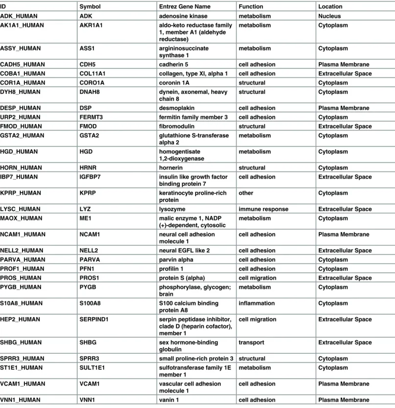
Table 2. Proteomics analysis of exosomal cargo identified 30 unique markers in exosomes derived from amnion epithelial cells grown in normal culture conditions.
ID Symbol Entrez Gene Name Function Location
ADK_HUMAN ADK adenosine kinase metabolism Nucleus
AK1A1_HUMAN AKR1A1 aldo-keto reductase family
1, member A1 (aldehyde reductase)
metabolism Cytoplasm
ASSY_HUMAN ASS1 argininosuccinate
synthase 1
metabolism Cytoplasm
CADH5_HUMAN CDH5 cadherin 5 cell adhesion Plasma Membrane
COBA1_HUMAN COL11A1 collagen, type XI, alpha 1 cell adhesion Extracellular Space
COR1A_HUMAN CORO1A coronin 1A structural Cytoplasm
DYH8_HUMAN DNAH8 dynein, axonemal, heavy
chain 8
structural Cytoplasm
DESP_HUMAN DSP desmoplakin cell adhesion Plasma Membrane
URP2_HUMAN FERMT3 fermitin family member 3 cell adhesion Cytoplasm
FMOD_HUMAN FMOD fibromodulin structural Extracellular Space
GSTA2_HUMAN GSTA2 glutathione S-transferase
alpha 2
metabolism Cytoplasm
HGD_HUMAN HGD homogentisate
1,2-dioxygenase
metabolism Cytoplasm
HORN_HUMAN HRNR hornerin structural Cytoplasm
IBP7_HUMAN IGFBP7 insulin like growth factor
binding protein 7
cell adhesion Extracellular Space
KPRP_HUMAN KPRP keratinocyte proline-rich
protein
other Cytoplasm
LYSC_HUMAN LYZ lysozyme immune response Extracellular Space
MAOX_HUMAN ME1 malic enzyme 1, NADP
(+)-dependent, cytosolic
metabolism Cytoplasm
NCAM1_HUMAN NCAM1 neural cell adhesion
molecule 1
cell adhesion Plasma Membrane
NELL2_HUMAN NELL2 neural EGFL like 2 cell adhesion Extracellular Space
PARVA_HUMAN PARVA parvin alpha cell adhesion Cytoplasm
PROF1_HUMAN PFN1 profilin 1 cell adhesion Cytoplasm
PROS_HUMAN PROS1 protein S (alpha) cell migration Extracellular Space
PYGB_HUMAN PYGB phosphorylase, glycogen;
brain
metabolism Cytoplasm
S10A8_HUMAN S100A8 S100 calcium binding
protein A8
inflammation Cytoplasm
HEP2_HUMAN SERPIND1 serpin peptidase inhibitor,
clade D (heparin cofactor), member 1
cell migration Extracellular Space
SHBG_HUMAN SHBG sex hormone-binding
globulin
transport Extracellular Space
SPRR3_HUMAN SPRR3 small proline-rich protein 3 structural Cytoplasm
ST1E1_HUMAN SULT1E1 sulfotransferase family 1E
member 1
metabolism Cytoplasm
VCAM1_HUMAN VCAM1 vascular cell adhesion
molecule 1
cell adhesion Plasma Membrane
VNN1_HUMAN VNN1 vanin 1 cell adhesion Plasma Membrane
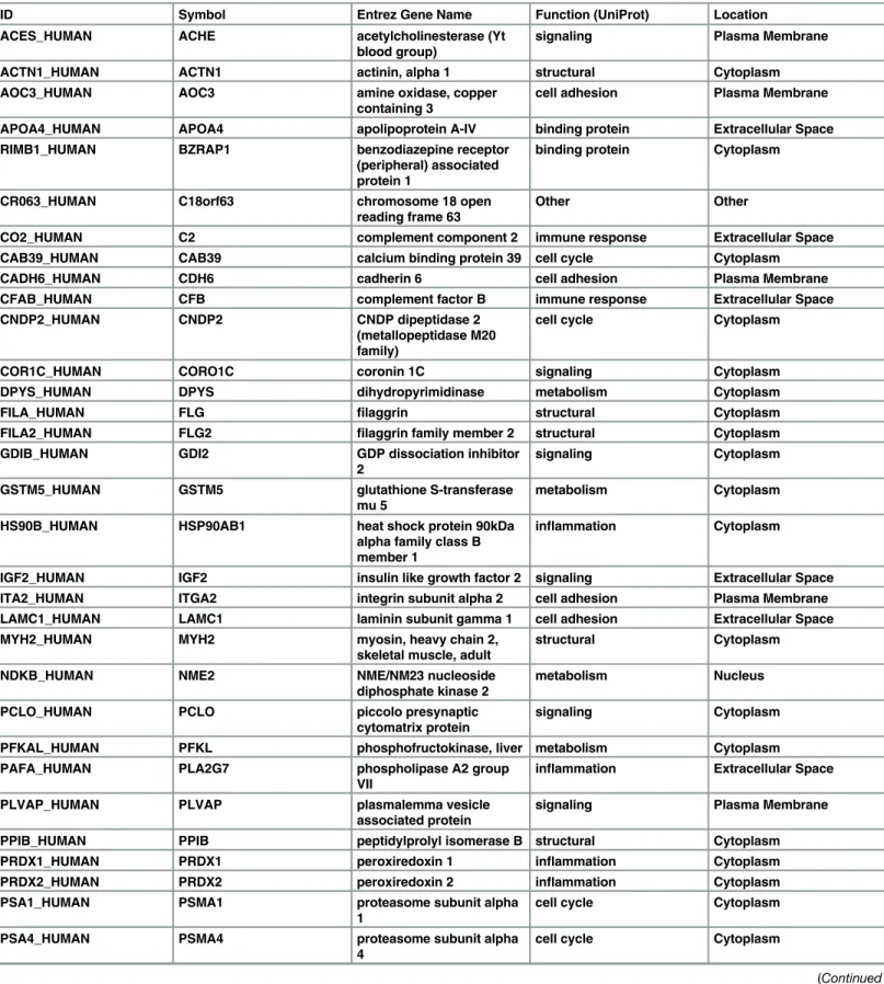
Table 3. Proteomics analysis of exosomal cargo identified 48 unique markers in exosomes derived from amnion epithelial cells grown under oxi-dative stress conditions.
ID Symbol Entrez Gene Name Function (UniProt) Location
ACES_HUMAN ACHE acetylcholinesterase (Yt
blood group)
signaling Plasma Membrane
ACTN1_HUMAN ACTN1 actinin, alpha 1 structural Cytoplasm
AOC3_HUMAN AOC3 amine oxidase, copper
containing 3
cell adhesion Plasma Membrane
APOA4_HUMAN APOA4 apolipoprotein A-IV binding protein Extracellular Space
RIMB1_HUMAN BZRAP1 benzodiazepine receptor
(peripheral) associated protein 1
binding protein Cytoplasm
CR063_HUMAN C18orf63 chromosome 18 open
reading frame 63
Other Other
CO2_HUMAN C2 complement component 2 immune response Extracellular Space
CAB39_HUMAN CAB39 calcium binding protein 39 cell cycle Cytoplasm
CADH6_HUMAN CDH6 cadherin 6 cell adhesion Plasma Membrane
CFAB_HUMAN CFB complement factor B immune response Extracellular Space
CNDP2_HUMAN CNDP2 CNDP dipeptidase 2
(metallopeptidase M20 family)
cell cycle Cytoplasm
COR1C_HUMAN CORO1C coronin 1C signaling Cytoplasm
DPYS_HUMAN DPYS dihydropyrimidinase metabolism Cytoplasm
FILA_HUMAN FLG filaggrin structural Cytoplasm
FILA2_HUMAN FLG2 filaggrin family member 2 structural Cytoplasm
GDIB_HUMAN GDI2 GDP dissociation inhibitor
2
signaling Cytoplasm
GSTM5_HUMAN GSTM5 glutathione S-transferase
mu 5
metabolism Cytoplasm
HS90B_HUMAN HSP90AB1 heat shock protein 90kDa
alpha family class B member 1
inflammation Cytoplasm
IGF2_HUMAN IGF2 insulin like growth factor 2 signaling Extracellular Space
ITA2_HUMAN ITGA2 integrin subunit alpha 2 cell adhesion Plasma Membrane
LAMC1_HUMAN LAMC1 laminin subunit gamma 1 cell adhesion Extracellular Space
MYH2_HUMAN MYH2 myosin, heavy chain 2,
skeletal muscle, adult
structural Cytoplasm
NDKB_HUMAN NME2 NME/NM23 nucleoside
diphosphate kinase 2
metabolism Nucleus
PCLO_HUMAN PCLO piccolo presynaptic
cytomatrix protein
signaling Cytoplasm
PFKAL_HUMAN PFKL phosphofructokinase, liver metabolism Cytoplasm
PAFA_HUMAN PLA2G7 phospholipase A2 group
VII
inflammation Extracellular Space
PLVAP_HUMAN PLVAP plasmalemma vesicle
associated protein
signaling Plasma Membrane
PPIB_HUMAN PPIB peptidylprolyl isomerase B structural Cytoplasm
PRDX1_HUMAN PRDX1 peroxiredoxin 1 inflammation Cytoplasm
PRDX2_HUMAN PRDX2 peroxiredoxin 2 inflammation Cytoplasm
PSA1_HUMAN PSMA1 proteasome subunit alpha
1
cell cycle Cytoplasm
PSA4_HUMAN PSMA4 proteasome subunit alpha
4
cell cycle Cytoplasm
induced senescence, stress associated p38 MAPK activation, and sterile inflammation, are asso-ciated with normal term human parturition [9,12,67,68]. These findings inin vivoclinical sam-ples were recapitulated in vitro using normal term not in labor fetal membrane explant cultures or AECs where oxidative stress induced transition of fetal membranes to a senescence phenotype mimicked term labor status [12–14]. This suggests that exogenous oxidative stress at term promotes senescence and senescent fetal membrane cells to signal parturition by enhancing the overall inflammatory load in the uterine cavity. In this study, we demonstrated that AEC-derived exosomes may serve as carriers of signals of communication between various tissue layers by senescent fetal cells. Exosomes, generated as a consequence of multivesicular endosome (MVE) fusion with the plasma membranes [69–71], and their contents (protein, DNA, and all forms of RNAs), represent the character and physiologic state of the cell of origin that makes them good vectors of paracrine signaling.
The primary aim of this study was to isolate, characterize and demonstrate that AEC derived exosomes reflect the physiological status of the cells of origin. Our key findings are as follows: 1) AEC derived exosomes demonstrated classic shape, size and markers (CD9, 63, 81, HSC 70) along with amnion cell-stem cell specific transcription factor Nanog, regardless of treatment. 2) AEC derived exosomes do not show the presence of ESCRT-associated protein Alix and amnion cell stem cell marker Oct-4 in their cargo. 3) CSE treatment caused increased Table 3. (Continued)
ID Symbol Entrez Gene Name Function (UniProt) Location
PSA6_HUMAN PSMA6 proteasome subunit alpha
6
cell cycle Cytoplasm
PSA7_HUMAN PSMA7 proteasome subunit alpha
7
cell cycle Cytoplasm
PSMD5_HUMAN PSMD5 proteasome 26S subunit,
non-ATPase 5
cell cycle Other
PYGL_HUMAN PYGL phosphorylase, glycogen,
liver
metabolism Cytoplasm
RAN_HUMAN RAN RAN, member RAS
oncogene family
cell cycle Nucleus
RAP1B_HUMAN RAP1B RAP1B, member of RAS
oncogene family
transport Cytoplasm
S10A7_HUMAN S100A7 S100 calcium binding
protein A7
inflammation Cytoplasm
S10A9_HUMAN S100A9 S100 calcium binding
protein A9
inflammation Cytoplasm
TAGL2_HUMAN TAGLN2 transgelin 2 binding Cytoplasm
TFR1_HUMAN TFRC transferrin receptor transport Plasma Membrane
TENX_HUMAN TNXB tenascin XB cell adhesion Extracellular Space
TPM1_HUMAN TPM1 tropomyosin 1 (alpha) structural Cytoplasm
TBA4A_HUMAN TUBA4A tubulin alpha 4a structural Cytoplasm
RL40_HUMAN UBA52 ubiquitin A-52 residue
ribosomal protein fusion product 1
inflammation Cytoplasm
WDR1_HUMAN WDR1 WD repeat domain 1 transport Extracellular Space
1433T_HUMAN YWHAQ tyrosine
3-monooxygenase/ tryptophan 5-monooxygenase activation protein, theta
signaling Cytoplasm
colocalization of H3, HSP70 and active p38 (P-p38) MAPK in AEC exosomes. Increased locali-zation of these proteins demonstrated a pathophysiological phenotype of AECs in response to CSE induced oxidative stress. Although the functional relevance is unclear, this is the first report to demonstrate P-p38 MAPK as an exosomal cargo. We propose that AEC-derived exo-somes demonstrate the characteristics of a pathophysiological state of the cell of origin and that the presence of P-p38 MAPK, a marker of inflammation, is a key mediator of senescence induction and sterile inflammation. Our studies suggest the importance of AEC derived exo-some cargo in causing potential functional changes in feto-maternal compartments. Although we do not report any functional role of AEC exosomes during pregnancy, ongoing research is focused on determining such a role.
Fig 9. Ingenuity pathway analysis of AEC-derived-exosomes proteins.Exosomal protein identified under normal (control) or oxidative stress conditions were analyzed using the IPA software. Comparison of canonical pathways: (A) ERK/MAPK, (B) PI3K/AKT, (C) epithelial adherens junctions, (D) LPS/IL-1 mediated inhibition of RXR function and (E) IL-6 signaling. Diseases and functions analysis: (F) eosinophilic
inflammation. Values are mean±SD. In A and F,**p<0.001. In B, C and D,*p<0.005.
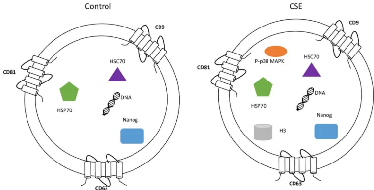
The impact of p38 MAPK activation is well documented in fetal tissues but no data exist on its functional contributions on myometrial side whose contractility determines pregnancy out-come. Functional progesterone withdrawal and subsequent myometrial activation (i.e. increased contractility and excitability) are key events in the initiation of labor [72–75]. Func-tional progesterone withdrawal, which is mediated, in part, by switching of the PR-A:PR-B ratio in myometrial cells from PR-B-dominance (mediates anti-inflammatory and relaxing actions of progesterone) to PR-A-dominance (inhibits the anti-inflammatory actions of pro-gesterone and increase contractility) [72–75]. A key finding in breast cancer cell lines is that the PR-A: PR-B ratio is determined by the stability of the PR-A and PR-B proteins, which is caused by post-translational modifications, especially phosphorylation by MAPKs at specific serine residues in the N-terminal domain [76–79]. Khan et al found that PR-A stability is increased by MEKK1-induced p38 MAPK activation, which increased the PR-A: PR-B ratio; whereas PR-B stability was increased by activation of ERK1/2, leading to a decrease in the PR-A: PR-B ratio. Our unpublished data (performed in collaboration with Dr. Sam Mesiano) in myometrial cells demonstrate that CSE can cause functional progesterone withdrawal in myocytes through p38 MAPK mediated mechanism, a process reversed by p38 MAPK inhibi-tor SB203580. This determined the impact of p38 MAPK in human parturition. Therefore exo-somal transport of fetal cell derived P-p38 MAPK to the maternal side may be influential in determining the status of pregnancy. This is also dependent on the number of exosomes and load of p38 MAPK that can reach the maternal side. Exosomes may carry other inflammatory molecules (SASP) from senescent fetal cells and they may also enhance the inflammatory load on the maternal side. We postulate that based on the physiologic state of cell, exosome cargo and signaling may determine the outcome of pregnancy. Placental derived exosomes have been well documented in maternal liquid biopsies and their changes (quantity and contents) have been implicated in various pregnancy associated pathologies [31–37,40,42,46–48,80–94]. Fig 10. Characterization of AEC exosomes.The exosomes isolated from untreated (control) and CSE treated primary AEC carry cargo representative of the state of the origin cell. AEC exosomes contain tetraspnins (CD9, CD63 and CD81), Nanog, HSC70, HSP70 and DNA regardless of treatment, while CSE exosomes contain significantly increased amounts of P-p38MAPK and H3 compared with control exosomes.
Although AEC exosomes demonstrated the consistent presence of classical exosomal mark-ers and the amnion stem cell marker Nanog, we did not show one of the ESCRT class of pro-teins, Alix. ESCRT complex dependent and independent mechanisms of exosomal assembly and secretion have been described as a tissue/cell specific physiological phenomenon [27]. Based on our experimental approaches, AEC exosomes are likely derived without the participa-tion of the ESCRT protein; however, we do not rule out mediaparticipa-tion by other classes of ESCRT proteins. We have documented three of the tetraspanin protein markers (CD9, CD63 and CD81) that can participate in exosomal assembly and release of cargo.
In summary, we have demonstrated AECs produce exosomes and that their cargo reflects the status of the cell (Fig 10). Furthermore, we identified active p38 MAPK as one of the cargo proteins in exosomes derived from oxidative stress-treated AECs. Our studies highlight the sig-nificance of AEC derived exosomes as donors of p38 MAPK which plays a major role in deter-mining the fate of pregnancy. This study demonstrated a limited number of markers and further characterization of AEC exosome cargo using proteomic approaches are warranted to elucidate the functional role of exosomes in human parturition and feto-maternal
communication.
Supporting Information
S1 Table. Proteomics analysis of exosomal cargo identified over 200 proteins in exosomes derived from amnion epithelial cells grown under control and oxidative stress conditions.
(DOCX)
Acknowledgments
Authors acknowledge support by all the staff from the Maternal-Fetal Medicine and Perinatal Research Laboratory, University of Texas Medical Branch at Galveston (UTMB), TX, USA. We specifically acknowledge the contributions of Talar Kechichian, MS, (laboratory manager) for her expertise in protein chemistry and western blot analysis of samples, Jayshil Trivedi, MS for providing cell and molecular biological expertise.
TEM was performed at the electron microscopy core laboratory at UTMB with support and guidance from Dr. Michael Woodson, PhD.
This study is supported by Innovative catalyst grant funding from March of Dimes Center of Ohio, Cincinnati, OH to R Menon and partially by R. Menon’s Faculty development funds from University of Texas Medical Branch at Galveston, TX. Authors declare all the funding or sources of support received during this specific study. The funders had no role in study design, data collection and analysis, decision to publish, or preparation of the manuscript.
Author Contributions
Conceived and designed the experiments: RM. Performed the experiments: SS LR RU. Ana-lyzed the data: RM SS CS. Contributed reagents/materials/analysis tools: RM JP CS GRS. Wrote the paper: RM SS JP CS.
References
1. Casey ML, Macdonald PC. The endocrinology of human parturition. Ann N Y Acad Sci. 1997; 828: 273–84. PMID:9329848
2. Challis JRG, Bloomfield FH, Booking AD, Casciani V, Chisaka H, Connor K, et al. Fetal signals and par-turition. J Obstet Gynaecol Res. 2005; 31: 492–9. PMID:16343248
4. Smith R, Mesiano S, McGrath S. Hormone trajectories leading to human birth. Regul Pept. 2002; 108: 159–64. PMID:12220740
5. Phaneuf S. AGLBARJE-FG. Parturition: activation of stimulatory pathways or loss of uterine quies-cence? Adv Exp Med Biol. 1995; 395: 435–51. PMID:8713997
6. Blackburn S. Maternal, Fetal, & Neonatal Physiology. Elsevier Health Sciences; 2014. 768 p.
7. Shynlova O, Tsui P, Jaffer S, Lye SJ. Integration of endocrine and mechanical signals in the regulation of myometrial functions during pregnancy and labour. Eur J Obstet Gynecol Reprod Biol. 2009; 144: S2–10. doi:10.1016/j.ejogrb.2009.02.044PMID:19299064
8. Shynlova O, Lee Y-H, Srikhajon K, Lye SJ. Physiologic uterine inflammation and labor onset: integra-tion of endocrine and mechanical signals. Reprod Sci. 2013; 20: 154–67. doi:10.1177/
1933719112446084PMID:22614625
9. Behnia F, Taylor BD, Woodson M, Kacerovský M, Hawkins H, Fortunato SJ, et al. Chorioamniotic mem-brane senescence: a signal for parturition? Am J Obstet Gynecol. 2015; 213: 1–16.
10. Fox H, Faulk WP. The placenta as an experimental model. Clin Endocrinol Metab. 1981; 10: 57–72. PMID:6261997
11. Menon R, Yu J, Basanta-Henry P, Brou L, Berga SL, Fortunato SJ, et al. Short fetal leukocyte telomere length and preterm prelabor rupture of the membranes. PLoS One. 2012; 7: 1–6.
12. Menon R, Boldogh I, Urrabaz-Garza R, Polettini J, Syed TA, Saade GR, et al. Senescence of primary amniotic cells via oxidative DNA damage. PLoS One. 2013; 8: e83416. doi:10.1371/journal.pone. 0083416PMID:24386195
13. Polettini J, Behnia F, Taylor BD, Saade GR, Taylor RN, Menon R. Telomere Fragment Induced Amnion Cell Senescence: A Contributor to Parturition? Sun K, editor. PLoS One. 2015; 10: e0137188. doi:10. 1371/journal.pone.0137188PMID:26397719
14. Menon R, Boldogh I, Hawkins HK, Woodson M, Polettini J, Syed TA, et al. Histological Evidence of Oxi-dative Stress and Premature Senescence in Preterm Premature Rupture of the Human Fetal Mem-branes Recapitulated in Vitro. Am J Pathol. 2014; 184: 1740–51. doi:10.1016/j.ajpath.2014.02.011 PMID:24832021
15. Coppé J-P, Desprez P-Y, Krtolica A, Campisi J. The Senescence-Associated Secretory Phenotype: The Dark Side of Tumor Suppression. Annu Rev Pathol Mech Dis. 2010; 5: 99–118.
16. Bredeson S, Papaconstantinou J, Deford JH, Kechichian T, Syed TA, Saade GR, et al. HMGB1 pro-motes a p38MAPK associated non-infectious inflammatory response pathway in human fetal mem-branes. PLoS One. 2014; 9: e113799. doi:10.1371/journal.pone.0113799PMID:25469638
17. De Toro J, Herschlik L, Waldner C, Mongini C. Emerging Roles of Exosomes in Normal and Pathologi-cal Conditions: New Insights for Diagnosis and Therapeutic Applications. Front Immunol. 2015; 6: 1– 12.
18. Luo S-S, Ishibashi O, Ishikawa G, Ishikawa T, Katayama A, Mishima T, et al. Human Villous Tropho-blasts Express and Secrete Placenta-Specific MicroRNAs into Maternal Circulation via Exosomes. Biol Reprod. 2009; 81: 717–29. doi:10.1095/biolreprod.108.075481PMID:19494253
19. Zhang B, Yin Y, Lai RC, Lim SK. Immunotherapeutic Potential of Extracellular Vesicles. Front Immunol. 2014; 5: 1–11.
20. Buzas EI, György B, Nagy G, Falus A, Gay S. Emerging role of extracellular vesicles in inflammatory diseases. Nat Rev Rheumatol. 2014; 10: 356–64. doi:10.1038/nrrheum.2014.19PMID:24535546
21. Liga A, Vliegenthart ADB, Oosthuyzen W, Dear JW, Kersaudy-Kerhoas M. Exosome isolation: a micro-fluidic road-map. Lab Chip. 2015; 15: 2388–94. doi:10.1039/c5lc00240kPMID:25940789
22. Lin J, Li J, Huang B, Liu J, Chen X, Chen X-M, et al. Exosomes: novel biomarkers for clinical diagnosis. ScientificWorldJournal. 2015; 2015: 657086. doi:10.1155/2015/657086PMID:25695100
23. Frydrychowicz M, Kolecka-Bednarczyk a, Madejczyk M, Yasar S, Dworacki G. Exosomes—Structure, Biogenesis and Biological Role in Non-Small-Cell Lung Cancer. Scand J Immunol. 2015; 81: 2–10. doi: 10.1111/sji.12247PMID:25359529
24. Cai X, Janku F, Zhan Q, Fan J-B. Accessing Genetic Information with Liquid Biopsies. Trends Genet. 2015; 31: 564–75. doi:10.1016/j.tig.2015.06.001PMID:26450339
25. Rana S, Zöller M. Exosome target cell selection and the importance of exosomal tetraspanins: a hypothesis. Biochem Soc Trans. 2011; 39: 559–62. doi:10.1042/BST0390559PMID:21428939
26. Raposo G, Stoorvogel W. Extracellular vesicles: Exosomes, microvesicles, and friends. J Cell Biol. 2013; 200: 373–83. doi:10.1083/jcb.201211138PMID:23420871
28. Urbanelli L, Magini A, Buratta S, Brozzi A, Sagini K, Polchi A, et al. Signaling pathways in exosomes biogenesis, secretion and fate. Genes. 2013; 4: 152–70. doi:10.3390/genes4020152PMID:24705158
29. Corrado C, Raimondo S, Chiesi A, Ciccia F, De Leo G, Alessandro R. Exosomes as intercellular signal-ing organelles involved in health and disease: Basic science and clinical applications. Int J Mol Sci. 2013; 14: 5338–66. doi:10.3390/ijms14035338PMID:23466882
30. Bobrie A, Colombo M, Raposo G, Théry C. Exosome Secretion: Molecular Mechanisms and Roles in Immune Responses. Traffic. 2011; 12: 1659–68. doi:10.1111/j.1600-0854.2011.01225.xPMID: 21645191
31. Mitchell MD, Peiris HN, Kobayashi M, Koh YQ, Duncombe G, Illanes SE, et al. Placental exosomes in normal and complicated pregnancy. Am J Obstet Gynecol. 2015; 213: S173–81. doi:10.1016/j.ajog. 2015.07.001PMID:26428497
32. Sabapatha A, Gercel-taylor C, Taylor DD. Specific isolation of placenta-derived exosomes from the cir-culation of pregnant women and their immunoregulatory consequences. Am J Reprod Immunol. 2006; 56: 345–55. PMID:17076679
33. Salomon C, Torres MJ, Kobayashi M, Scholz-Romero K, Sobrevia L, Dobierzewska A, et al. A gesta-tional profile of placental exosomes in maternal plasma and their effects on endothelial cell migration. PLoS One. 2014; 9: e98667. doi:10.1371/journal.pone.0098667PMID:24905832
34. Salomon C, Yee S, Scholz-Romero K, Kobayashi M, Vaswani K, Kvaskoff D, et al. Extravillous tropho-blast cells-derived exosomes promote vascular smooth muscle cell migration. Front Pharmacol. 2014; 5: 175. doi:10.3389/fphar.2014.00175PMID:25157233
35. Sarker S, Scholz-Romero K, Perez A, Illanes SE, Mitchell MD, Rice GE, et al. Placenta-derived exo-somes continuously increase in maternal circulation over the first trimester of pregnancy. J Transl Med. 2014; 12: 204. doi:10.1186/1479-5876-12-204PMID:25104112
36. Ouyang Y, Mouillet JF, Coyne CB, Sadovsky Y. Review: Placenta-specific microRNAs in exosomes— Good things come in nano-packages. Placenta. 2014; 35: S69–73. doi:10.1016/j.placenta.2013.11. 002PMID:24280233
37. Donker RB, Mouillet JF, Chu T, Hubel CA, Stolz DB, Morelli AE, et al. The expression profile of C19MC microRNAs in primary human trophoblast cells and exosomes. Mol Hum Reprod. 2012; 18;: 417–24. doi:10.1093/molehr/gas013PMID:22383544
38. Sadovsky Y, Mouillet J, Ouyang Y, Bayer A. The Function of TrophomiRs and Other MicroRNAs in the Human Placenta. Cold Spring Harb Perspect Med. 2015; 5: a023036. doi:10.1101/cshperspect. a023036PMID:25877393
39. Salomon C, Scholz-Romero K, Sarker S, Sweeney E, Kobayashi M, Correa P, et al. Gestational Diabe-tes Mellitus Is Associated With Changes in the Concentration and Bioactivity of Placenta-Derived Exo-somes in Maternal Circulation Across Gestation. Diabetes. 2016; 65: 598–609. doi: 10.2337/db15-0966PMID:26718504
40. Rice GE, Scholz-Romero K, Sweeney E, Peiris H, Kobayashi M, Duncombe G, et al. The Effect of Glu-cose on the Release and Bioactivity of Exosomes From First Trimester Trophoblast Cells. J Clin Endo-crinol Metab. 2015; 100: E1280–8. doi:10.1210/jc.2015-2270PMID:26241326
41. Salomon C, Kobayashi M, Ashman K, Sobrevia L, Mitchell MD, Rice GE. Hypoxia-induced changes in the bioactivity of cytotrophoblast-derived exosomes. PLoS One. 2013; 8: e79636. doi:10.1371/journal. pone.0079636PMID:24244532
42. Salomon C, Ryan J, Sobrevia L, Kobayashi M, Ashman K, Mitchell M, et al. Exosomal Signaling during Hypoxia Mediates Microvascular Endothelial Cell Migration and Vasculogenesis. PLoS One. 2013; 8: 1–24.
43. Polettini J, Silva MG, Kacerovsky M, Syed TA, Saade G, Menon R. Expression profiles of fetal mem-brane nicotinamide adenine dinucleotide phosphate oxidases (NOX) 2 and 3 differentiates spontane-ous preterm birth and pPROM pathophysiologies. Placenta. 2014; 35: 188–94. doi:10.1016/j.placenta. 2013.12.012PMID:24439294
44. Polettini J, Silva M, Syed T, Saade G, Menon R. 828: Screening of lysyl oxidase (LOX) and lysyl oxi-dase-like (LOXL) enzyme expression and activity in human fetal membranes. Am J Obstet Gynecol. 2014; 210: S402–3.
45. Lässer C, Eldh M, Lötvall J. Isolation and characterization of RNA-containing exosomes. J Vis Expnet]. 2012: e3037.
46. Bullerdiek J, Flor I. Exosome-delivered microRNAs of“chromosome 19 microRNA cluster”as immuno-modulators in pregnancy and tumorigenesis. Mol Cytogenet. 2012; 5: 27. doi:10.1186/1755-8166-5-27 PMID:22559272
48. Kshirsagar SK, Alam SM, Jasti S, Hodes H, Nauser T, Gilliam M, et al. Immunomodulatory molecules are released from the first trimester and term placenta via exosomes. Placenta. 2012; 33: 982–90. doi: 10.1016/j.placenta.2012.10.005PMID:23107341
49. Izumi M, Pazin BJ, Minervini CF, Gerlach J, Ross M a, Stolz DB, et al. Quantitative comparison of stem cell marker-positive cells in fetal and term human amnion. J Reprod Immunol. 2009; 81: 39–43. doi:10. 1016/j.jri.2009.02.007PMID:19501410
50. Zhao H, Li Y, Jin H, Xie L, Liu C, Jiang F, et al. Rapid and efficient reprogramming of human amnion-derived cells into pluripotency by three factors OCT4/SOX2/NANOG. Differentiation. 2010; 80: 123–9. doi:10.1016/j.diff.2010.03.002PMID:20510497
51. Koike C, Zhou K, Takeda Y, Fathy M, Okabe M, Yoshida T, et al. Characterization of Amniotic Stem Cells. Cell Reprogram. 2014; 16: 298–305. doi:10.1089/cell.2013.0090PMID:25068631
52. Teng Z, Yoshida T, Okabe M, Toda A, Higuchi O, Nogami M. Establishment of Immortalized Human Amniotic Mesenchymal Stem Cells. Cell Transplant. 2013; 22: 267–78. doi:10.3727/
096368912X655055PMID:23006979
53. Gao Y, Pu Y, Wang D, Hou L, Guan W, Ma Y. Isolation and biological characterization of chicken amnion epithelial cells. Eur J Histochem. 2012; 56: e33. doi:10.4081/ejh.2012.e33PMID:23027349
54. Saito S, Lin Y-C, Murayama Y, Hashimoto K, Yokoyama KK. Human amnion-derived cells as a reliable source of stem cells. Curr Mol Med. 2012; 12: 1340–9. PMID:23016591
55. Andreu Z, Yáñez-Mó M. Tetraspanins in Extracellular Vesicle Formation and Function. Front Immunol.
2014; 5: 1–12.
56. Phimister EG, Phillippe M. Cell-free Fetal DNA—A Trigger for Parturition. N Engl J Med. 2014; 370: 2534–6. doi:10.1056/NEJMcibr1404324PMID:24963574
57. Olivieri F, Albertini MC, Orciani M, Ceka A, Cricca M, Procopio AD, et al. DNA damage response (DDR) and senescence: shuttled inflamma-miRNAs on the stage of inflamm-aging. Oncotarget. 2015; 6: 35509–21. doi:10.18632/oncotarget.5899PMID:26431329
58. Karrasch T, Steinbrecher KA, Allard B, Baldwin AS, Jobin C. Wound-induced p38MAPK-dependent histone H3 phosphorylation correlates with increased COX-2 expression in enterocytes. J Cell Physiol. 2006; 207: 809–15. PMID:16508963
59. Valbonesi P, Ricci L, Franzellitti S, Biondi C, Fabbri E. Effects of cadmium on MAPK signalling path-ways and HSP70 expression in a human trophoblast cell line. Placenta. 2008; 29: 725–33. doi:10. 1016/j.placenta.2008.05.004PMID:18571719
60. Behnia F, Sheller S, Menon R. Mechanistic Differences Leading to Infectious and Sterile Inflammation. Am J Reprod Immunol. 2016; 75: 505–18. doi:10.1111/aji.12496PMID:26840942
61. Derynck R, Muthusamy BP, Saeteurn KY. Signaling pathway cooperation in TGF-β-induced epithelial-mesenchymal transition. Curr Opin Cell Biol. 2014; 31: 56–66. doi:10.1016/j.ceb.2014.09.001PMID: 25240174
62. Papageorgis P. TGFβSignaling in Tumor Initiation, Epithelial-to-Mesenchymal Transition, and Metas-tasis. J Oncol. 2015; 2015: 587193. doi:10.1155/2015/587193PMID:25883652
63. Fabregat I, Malfettone A, Soukupova J. New Insights into the Crossroads between EMT and Stemness in the Context of Cancer. J Clin Med. 2016;5.
64. Da C, Liu Y, Zhan Y, Liu K, Wang R. Nobiletin inhibits epithelial-mesenchymal transition of human non-small cell lung cancer cells by antagonizing the TGF-β1/Smad3 signaling pathway. Oncol Rep. 2016; 35: 2767–74. doi:10.3892/or.2016.4661PMID:26986176
65. Yeh Y-H, Wang S-W, Yeh Y-C, Hsiao H-F, Li T-K. Rhapontigenin inhibits TGF-β-mediated epithe-lial‑mesenchymal transition via the PI3K/AKT/mTOR pathway and is not associated with HIF-1α degra-dation. Oncol Rep. 20169; 35: 2887–95. doi:10.3892/or.2016.4664PMID:26986649
66. Romero R, Kusanovic JP, Gomez R, Lamont R, Bytautiene E, Garfield RE, et al. The clinical signifi-cance of eosinophils in the amniotic fluid in preterm labor. J Matern Fetal Neonatal Med. 2010; 23: 320– 9. doi:10.3109/14767050903168465PMID:19900034
67. Menon R, Fortunato SJ, Milne GL, Brou L, Carnevale C, Sanchez SC, et al. Amniotic fluid eicosanoids in preterm and term births: effects of risk factors for spontaneous preterm labor. Obstet Gynecol. 2011; 118: 121–34. doi:10.1097/AOG.0b013e3182204eaaPMID:21691170
68. Menon R. Oxidative stress damage as a detrimental factor in preterm birth pathology. Front Immunol. 2014; 5: 567. doi:10.3389/fimmu.2014.00567PMID:25429290
70. György B, Szabó TG, Pásztói M, Pál Z, Misják P, Aradi B, et al. Membrane vesicles, current state-of-the-art: Emerging role of extracellular vesicles. Cell Mol Life Sci. 2011; 68: 2667–88. doi:10.1007/ s00018-011-0689-3PMID:21560073
71. Hornung D, Lebovic DI, Shifren JL, Vigne JL, Taylor RN. Vectorial secretion of vascular endothelial growth factor by polarized human endometrial epithelial cells. Fertil Steril. 1998; 69: 909–15. PMID: 9591502
72. Mesiano S, Chan EC, Fitter JT, Kwek K, Yeo G, Smith R. Progesterone withdrawal and estrogen activa-tion in human parturiactiva-tion are coordinated by progesterone receptor A expression in the myometrium. J Clin Endocrinol Metab. 2002; 87: 2924–30. PMID:12050275
73. Merlino A a., Welsh TN, Tan H, Li JY, Cannon V, Mercer BM, et al. Nuclear progesterone receptors in the human pregnancy myometrium: Evidence that parturition involves functional progesterone with-drawal mediated by increased expression of progesterone receptor-A. J Clin Endocrinol Metab. 2007; 92(5): 1927–33. PMID:17341556
74. Mesiano S. Myometrial progesterone responsiveness. Semin Reprod Med. 2007; 25: 5–13. PMID: 17205419
75. Mesiano S, Wang Y, Norwitz ER. Progesterone receptors in the human pregnancy uterus: do they hold the key to birth timing? Reprod Sci. 2011; 18: 6–19. doi:10.1177/1933719110382922PMID:20889955
76. Dressing GE, Knutson TP, Schiewer MJ, Daniel AR, Hagan CR, Diep CH, et al. Progesterone receptor-cyclin D1 complexes induce cell cycle-dependent transcriptional programs in breast cancer cells. Mol Endocrinol. 2014; 28: 442–57. doi:10.1210/me.2013-1196PMID:24606123
77. Beck C a., Zhang Y, Altmann M, Weigel NL, Edwards DP. Stoichiometry and Site-specific Phosphoryla-tion of Human Progesterone Receptor in Native Target Cells and in the Baculovirus Expression Sys-tem. J Biol Chem. 1996; 271(32): 19546–55. PMID:8702648
78. Beck CA, Zhang Y, Weigel NL, Edwards DP. Two Types of Anti-progestins Have Distinct Effects on Site-specific Phosphorylation of Human Progesterone Receptor. J Biol Chem. 1996; 271: 1209–17. PMID:8557652
79. Zhang Y, Beck CA, Poletti A, Edwards DP, Weigel NL. Identification of a group of Ser-Pro motif hor-mone-inducible phosphorylation sites in the human progesterone receptor. Mol Endocrinol. 1995; 9: 1029–40. PMID:7476977
80. Delorme-Axford E, Bayer A, Sadovsky Y, Coyne CB. Autophagy as a mechanism of antiviral defense at the maternal-fetal interface. Autophagy. 2013; 9: 2173–4. doi:10.4161/auto.26558PMID:24231730
81. Li JYZ, Yong TY, Michael MZ, Gleadle JM. MicroRNAs: are they the missing link between hypoxia and pre-eclampsia? Hypertens pregnancy. 2014; 33: 102–14. doi:10.3109/10641955.2013.832772PMID: 24354525
82. Vargas A, Zhou S, Ethier-Chiasson M, Flipo D, Lafond J, Gilbert C, et al. Syncytin proteins incorporated in placenta exosomes are important for cell uptake and show variation in abundance in serum exo-somes from patients with preeclampsia. FASEB J. 2014; 28: 3703–19. doi:10.1096/fj.13-239053 PMID:24812088
83. Kambe S, Yoshitake H, Yuge K, Ishida Y, Ali MM, Takizawa T, et al. Human exosomal placenta-associ-ated miR-517a-3p modulates the expression of PRKG1 mRNA in Jurkat cells. Biol Reprod. 2014; 91: 129. doi:10.1095/biolreprod.114.121616PMID:25273530
84. Mincheva-Nilsson L, Baranov V. The Role of Placental Exosomes in Reproduction. Am J Reprod Immu-nol. 2010; 63: 520–33. doi:10.1111/j.1600-0897.2010.00822.xPMID:20331583
85. Mincheva-Nilsson L, Baranov V. Placenta-Derived Exosomes and Syncytiotrophoblast Microparticles and their Role in Human Reproduction: Immune Modulation for Pregnancy Success. Am J Reprod Immunol. 2014; 72: 440–57. doi:10.1111/aji.12311PMID:25164206
86. Record M. Intercellular communication by exosomes in placenta: A possible role in cell fusion? Pla-centa. 2014; 35: 297–302. doi:10.1016/j.placenta.2014.02.009PMID:24661568
87. Stenqvist A-C, Nagaeva O, Baranov V, Mincheva-Nilsson L. Exosomes secreted by human placenta carry functional Fas ligand and TRAIL molecules and convey apoptosis in activated immune cells, sug-gesting exosome-mediated immune privilege of the fetus. J Immunol. 2013; 191: 5515–23. doi:10. 4049/jimmunol.1301885PMID:24184557
88. Southcombe J, Tannetta D, Redman C, Sargent I. The immunomodulatory role of syncytiotrophoblast microvesicles. PLoS One. 2011; 6: e20245. doi:10.1371/journal.pone.0020245PMID:21633494
90. Atay S, Gercel-Taylor C, Suttles J, Mor G, Taylor DD. Trophoblast-derived exosomes mediate mono-cyte recruitment and differentiation. Am J Reprod Immunol. 2011; 65: 65–77. doi:10.1111/j.1600-0897. 2010.00880.xPMID:20560914
91. Atay S, Gercel-Taylor C, Taylor DD. Human trophoblast-derived exosomal fibronectin induces pro-inflammatory IL-1βproduction by macrophages. Am J Reprod Immunol. 2011; 66: 259–69. doi:10. 1111/j.1600-0897.2011.00995.xPMID:21410811
92. Redman CWG, Tannetta DS, Dragovic R a, Gardiner C, Southcombe JH, Collett GP, et al. Review: Does size matter? Placental debris and the pathophysiology of pre-eclampsia. Placenta. 2012; 33: S48–54. doi:10.1016/j.placenta.2011.12.006PMID:22217911
93. Salomon C, Yee SW, Mitchell MD, Rice GE. The Possible Role of Extravillous Trophoblast-Derived Exosomes on the Uterine Spiral Arterial Remodeling under Both Normal and Pathological Conditions. Biomed Res Int. 2014; 2014: 1–10.