STUDY OF VE VIRUS AND ISOLATION OF SLE, EE, GROUP C,
AND GUAMA GROUP ARBOVIRUSFS IN THE
AMAZON REGION OF PERU, 1975l
William F. Scherer, * Jo& Madalergoitia, 3 Oswald0 Menesis,3 and Ma&e1 Acosta”
The source of the Venezuelan encephalitis (VE) virus that has caused many of the human and equine disease outbreaks in Peru has not yet been determined. This article reports findings from a study in northern Peru consistent with the theory that
the virus is silently enzootic in the Peruvian Amazon Basin, and that movements of virus out of this region are the cause of
the periodic human and equine VE disease outbreaks along Peru’s dry Pacific Coast.
Introduction
Venezuelan encephalitis (VE) virus has intermittently produced equine epizootics and human epidemics along the dry Pacific coastal plain of Peru for at least the last 35 years (I, 2). Equine epizootic disease, like ,that caused by VE virus, was recorded on the northern coastal plain of Peru in 1925, and VE outbreaks of proven etiology oc- curred from the early 1940s to the early 1950s and again in 1969 and 1973 (1-5).
The source of VE virus for these epi- demics and equine epizootics has not been determined except in 1969, when the dis- ease was limited to northwestern coastal
1Also amwaring in Spanish in the Roletin de la Ofi- ._ -
cina Sanitaria Panamericana, 1980. The research re- ported here, performed in collaboration with the Pan American Hialth Organization and the Government of Peru, was sponsored by the United States Army Me- dical Research and Development Command (Wash- ington, D.C. 20314) under Contract No. DADA-17.7% C-2140 and by the University of the Peruvian Ama- zon and the Institute of Public Health of Peru.
ZDepartment of Microbiology, Cornell University Medical College, New York, New York 10021, U.S.A.
3Institute of Public Health of Peru. 4Universi ty of the Peruvian Amazon.
Peru adjacent to Ecuador. On that occasion the disease resulted from southward exten- sion of a large Ecuadorian epizootic and epidemic (3, 6). But other Peruvian epidemics, the most recent recorded in 1973, have not bordered Ecuador.
The 1973 VE outbreak in northern Peru was described in Spanish by Madalengoitia and associates (4) and by Terry (5). The VE virus involved was isolated from both hu- mans and equine animals, and its hemag- glutination-inhibition (HI) subtype was established as I-B by the U.S. Center for Disease Control in Atlanta, Georgia, and by other studies (4, 7). Cases of disease in 93 human subjects 1 to 68 years of age were investigated. (Thirty per cent of those involved were under 10 years of age; 59 were males and 23 females, the gender of the remaining 11 not being recorded.) Cen- tral nervous system symptoms were present in about 25 per cent of the cases, although most patients were ambulatory.
A diagnosis of Venezuelan encephalitis was confirmed in 41 cases by isolation of the virus from blood and/or by VE anti- body development in serum. A presump- tive diagnosis was made in 28 cases, based on VE IgM antibodies or unusually high anti-
Schemer
et al. *
STUDY OF VE VIRUS IN PERU273
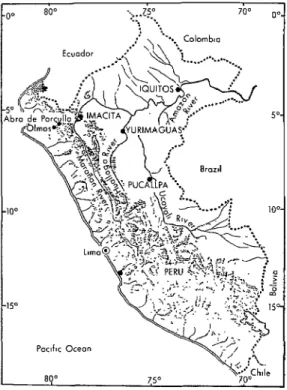
body titers in single sera. Specimens from the other 24 cases were inadequate for diag- nostic purposes. All of the 41 confirmed cases came from three departments along the Pacific Coast of northern Peru (4-g” south latitude), 1 coming from Lambaye- que, 17 from La Libertad, and 23 from Piura (see Figure I). The number of equine cases was estimated at over 2,500, equine mortality being estimated at about 40 per cent. The outbreak occurred during Jan- uary-June 1973, but some equine cases (as indicated by clinical diagnoses) had been recorded in the region the previous year (in May 1972) following heavy rains. Inacti- vated VE vaccine was used to immunize equine animals in the early months of 1973, and use of attenuated live-virus vaccine from Mexico (about 50,000 doses) began in March 1973.
This epidemic drew renewed attention to an important question; namely, What is the source or sources of VE virus for such an epidemic when the disease first appears in an irrigated region on the dry Pacific Coast of northern Peru that is neither contiguous with Ecuador nor associated with concur- rent disease in Ecuador? Two possibilities are either (a) that the virus comes from tro- pical rain forests in Peru’s Amazon region or (b) that persistent small foci of virus, cy- cling between mosquitoes and vertebrates, exist in irrigated river valleys along the otherwise desert Pacific Coast of Peru. Studies of these possible sources of Peruvian VE epidemics and equine epizootics were carried out during 1970-1971 and have been reported (2, 8). However, because of the 1973 outbreak, further investigations were done in 1975. This article describes the results of these latter investigations.
As in 1970 and 1971, when arboviruses other than VE (including eastern encepha- litis, group C, and Guama group viruses) were encountered in Peru’s Amazon region (9), additional arboviruses were recovered from tissues of mosquitoes and sentinel hamsters in 1975. Information about those
isolations-of St. Louis encephalitis (SLE), eastern encephalitis (EE), group C (Maritu- ba), and Guama group arboviruses-are included in this article.
Materials and Methods
Study Sites
andField Techniques
Two types of study sites were employed. These were: (1) in Loreto Department, tro- pical wet forests near Yurimaguas on the Huallaga River and Iquitos on the Amazon River, sites located at respective altitudes of 180 and less than 200 m; and (2) in Ama- zonas Department, tropical dry forest near Imacita on the Maraiion River, a site situ- ated at an altitude of 240 m and at 5O5’ south, 7S022’ west (see Figure 1). Only patches of forest remained in the environs of Yurimaguas, since most of the land there had been cleared for agricultural purposes. In contrast, the regions around Iquitos and Imacita were mostly forested in 1975.
Mosquitoes were collected with Center for Disease Control (CDC) light traps at two forest habitats within 6 km of Yurima- guas (Muniches Airport Road and San Ra- m&r), and sentinel Syrian hamsters (Meso-
cricetus auratus)
were exposed in theseareas (see Photographs l-3). The site near Iquitos was the Quistococha forest previous- ly studied in 1970-1971 (2, 9). The third site investigated was a small patch of forest within 1 km of Imacita (Photo 4) on the pro- perty of Benign0 Chiozo (Photo 5).
274
PAHO BULLETIN
l vol.I3, no. 3, 1979
(4)
(5)
(1) The forest at the airport mad site near Yurimaguas
is seen in the distance to the left. The Yuri-
maguas airport is within a kilometer to the right of this road. (2) The forest at the San Ram&
site, also
near Yurimaguas,
is seen in the distance acnxs cleared fields. (3) A close-in view of forest at the San
Ram&
site. (4) The village of Imacita,
located along the Mara?&
River in Amazonas Department.
Scherer et al.
l STUDY OF VE VIRUS IN PERU 27500 80’= . . . . ..!S” . ...* -.. 700 0’
Figure 1. A map of Peru showing the 1975 arbovirus study sites.
shipped by airplane to Peru. The hamsters exposed at Iquitos and Yurimaguas had been previously immunized in New York with group C and Patois arboviruses, since it was known from experience in Central America that these viruses could kill sentinel hamsters if transmitted to them by mosquitoes, and thus might interfere with VE virus isolations. This immunization was accomplished by inoculating the ham- ster with live Patois virus (Mexican strain 63A49) on day 0; then with killed (forma- linized) Nepuyo virus (Mexican strain 63U 11) with complete Freund’s adjuvant on days 14, 20, and 27; with live Nepuyo virus on day 41; and with live Oriboca virus on day 47. Of the 156 hamsters 7 to 8 weeks old that were inoculated, 31 died after receiving Patois virus, 1 died after receiving inactivated Nepuyo virus, 1 died after receiving live Nepuyo virus, and 62 died after receiving live Oriboca virus.
This left 61 immune hamsters 14-15 weeks of age when their exposure began in Peru from late March to early May 1975. The
normal unimmunized hamsters were 10
weeks of age when exposure began.
Vz’rus
Isolationand Identzyication
All tissue suspensions from mosquitoes and from sick or dead sentinel hamsters were tested by combined intracranial (ic) and subcutaneous (SC) inoculation of suck- ling Swiss albino mice l-4 days of age that were obtained from Taconic Farms in Germantown, New York. Mosquito suspen- sions and sentinel hamsters yielding virus were tested in New York during the period May-July 1975. Complement fixation (CF) tests were performed as previously described (10) with saline-extracted antigens from in- fected suckling mouse brains.
Neutralization (N) tests were based on plaque-reductions-in primary chicken em- bryonic cell cultures for SLE and EE vi- ruses, and in Vero African green monkey kidney cell cultures for group C and Guama group viruses (II). Some N tests for Guama group viruses were done with suckling mice inoculated ic (II).
276 PAHO BULLETIN l vol. 13, no. 3, 1979
701-567; Guama group: G202-701-567; Ilheus: V509-701-562; Rio Bravo: V536-701- 562; Bussuquara: V561-701-562; Modoc: V538-701-562; Cowbone Ridge: V533-701- 562; Montana Myotis Leucoencephalitis (MML): V541-701-562; EE strain Massachu- setts: V515-701-562; and Mayaro: V507-701- 562 (12). Oriboca, Caraparu, and Marituba antisera were kindly supplied by Dr. C. J . Gibbs in 1965 after preparation in rabbits by Dr. W. Pond; and specific Guama, Moju, and Bimiti mouse ascitic fluids were generously provided by Dr. R. Shope.
Antibody Tests
N and HI tests were done as previously described (II). The antigenic subtype de- signations used for VE viruses were based on the classification of Young and Johnson (13). For N tests, the strains used were VE strains 52/73, 71D1249, and 71D1252 from Peru (2, 4); SLE strain 75D90; and EE
strain 75U40. The N tests were performed by means of plaque-reduction in cultures of primary chicken embryonic cells prepared as described elsewhere-in plastic plate wells with an area of either 2 or 8 square centimeters (II). HI tests used hemaggluti- nins from Mexican VE strain 63U2, Guate- malan EE strain 68U230, Mexican SLE strain 65V310, and United States western encephalitis (WE) strain 1985-60.
Results
VE Virus
As Table 1 indicates, a pool of 100 female mosquitoes collected on the night of 7-8 April 1975 at the Quistococha forest site near Iquitos yielded a strain of VE virus. All the inoculated mice in two litters died on days 3 or 6 following inoculation. The strain (75D143) was identified as VE virus by means of the plaque-reduction neutraliza- tion test; the logI neutralization index
Table 1. A listing of the VE, SLE, EE, group C, and Guama group arboviruses isolated from mosquitoes and adult sentinel hamsters in Peru’s Amazon region
fnxn late March to early May 197.5.
Cuama Gr C-Pat. Un- ovrr tmal No SLE group lmmunc ,mmun1,rd cxposrd’ EE
hamstcrsb
Gmup c: hamstrrs
lquitos site
(Quistococha) 25.436 245 1 0 2 702 2/18 0 0 Yurimaguas
site (Muniches 4,774 44 0 1 0 270 o/10
Airport Road) 122 l/5 0 0
Yurimaguas site 8,980 56 0 0 0 297 o/11
(San RamBn) 87 2/5 1 1
lmacita site
Scherer et al.
l STUDY OF VE VIRUS IN PERU277
(LNI) with VE antiserum was greater than 2.5 and with EE antiserum was 0.5. In a one-hour, virus-dilution hemagglutination- inhibition test using early rooster antibo- dies, 7513143 hemagglutinin was inhibited 250-fold by antibody to a Colombian sub- type I-D VE strain (V209A), 90- or 120-fold by antibodies to two 1971 Peruvian Amazon strains (71D1316 and 71D1249), 8-fold by antibodies to subtypes I-A (strain Kubes), I-B (strain 52/73), and III (strain BeAn8), and less than 8-fold by antibodies to strains of subtypes I-C, I-E, II, IV, and a possible new subtype V. Strain 75D143 (at the pas- sage level of suckling mouse 2 and chicken embryonic cell culture 1) killed both of two adult English short-hair guinea pigs inocu- lated SC with 16,000 chicken embryonic cell culture (CEC) plaque forming units (pfu).
Sentinel hamsters exposed at Iquitos, Yurimaguas, and Imacita (see Table 1) yielded no VE virus. Plasmas obtained from 28 hamsters on 5 May 1975, at the end of their exposure at Yurimaguas, and from 10 other hamsters on 7 May, following
their exposure at Imacita, contained no detectable HI antibody to VE, EE, or SLE viruses at 1:lO dilutions. Plasmas from hamsters exposed at the Muniches Airport Road site near Yurimaguas yielded HI titers of 1:lO and 2 1:20 with western ence- phalitis virus. No CF antibodies to group Capim or Patois arboviruses were detected in 1:4 dilutions of plasma from 6 nonim- mune hamsters exposed at Yurimaguas or from 10 exposed at Imacita.
N antibodies to VE were found in 21 per cent of the sera from 29 horses bled at Yurimaguas in April 1975. As Table 2 in- dicates, sera from animals as young as 2-4 years yielded positive results. Higher per- centages of these sera were positive to strains 71D1249 and 7101252 isolated dur- ing 1971 in the Peruvian Amazon region than were positive to epizootic strain 52/73 obtained from the outbreak on the north- ern Pacific Coast of Peru in 1973.
Sera obtained from people at Yurimaguas in April 1975 also had detectable VE N
antibodies. In contrast to the results with
Table 2. Prevalences of VE, EE, and SLE viral neutralizing antibodies in sera from horses bled in Loreto Depaament, Peru, during 1970 and 1975.
Approximate
age of horses
Proportion (No. positive= over No. tested) of horses with sera showing detectable N antibody to particular virus strains
Yurimaguas, April 1975 Sept. 1970 Iquitos, Sept. 1970 Pucallpa. (in years)
VE HI subtypes and strains
I-B, strain I-D, strain ?V. strain EE strain SLE strain SLE strain SLE strain 52173 ‘71D1249 71Dl252 75u 40 75D90 75D90 75D90 > 10
54 2-4c
<2 Total (percent)
O/6 2/6 O/6 4/6 l/6 2/2 -
l/16 3/16 S/16 12/16 3/15 2/3 5/12
2/7 l/7 2/7 5/7 l/7 3/6 3/10
- - - l/2 o/7
3/29 6/29 5/29 21/29 5/28 8/13 8/29
(10%) (21%) (17%) (72%) (18%) (62%) (28%)
aPositive = LNI > 1.6 with 1:4dilution of serum heated at 6O’C for 20 minutes and tested by plaque-reduction against about 100 pfu (plaque forming units) in CEC (chicken embryonic cell culture).
bone serum gave a positive response to all three VE strains, while four others only responded positively to one strain.
278
PAHO BULLETIN lvol. 13, no. 3, 1979
horse sera, however, the highest percentage of positive results (38 per cent of 14 human sera) was obtained with epizootic strain 52/73; only 7 and 14 per cent of the human sera yielded positive results when tested with the Amazonian enzootic strains (see Table 3). Each of the three sera yielding a positive result with the Amazonian strains also yielded positive results with the epi- zootic strain, and all the people found to have antibodies were over 29 years of age.
VE N antibodies found in these horse and human sera did not correlate with EE N antibodies. Two horse and four human sera tested positively with VE and negative- ly with EE, while 13 horse and two human sera were negative with VE and positive with EE.
As Table 3 shows, none of the sera from 13 people bled at Imacita during April 1975 showed detectable levels of VE N antibody.
SLE Virus
A suspension made from 60 female mosquitoes collected on 14-15 April 1975 at the Muniches Airport Road site near Yuri- maguas contained SLE virus (see Table 1). All of eight suckling mice inoculated with the mosquito suspension died on day 5 after inoculation. Virus was reisolated from frozen mosquito suspension 6 months later when five more suckling mice were inocu- lated and died 9 days later. This strain of SLE virus (75D90) was identified by plaque-reduction neutralization tests. An immune mouse ascitic fluid made with SLE virus (Mexican strain 65V310) produced
LNI of >2.4 and 3.1 in two tests. LNI ob- tained with immune mouse ascitic fluids made with Ilheus, Rio Bravo, Bussuquara, Modoc, Cowbone Ridge, and MML flavi-
Table 3. Prevalences of VE, EE, and SLE viral neutralizing antibodies in sera from 27 human subjects bled in April 1975 at two locations in the Amazon Basin of central and
northern Peru, just east of the Andes Mountains.
Proportion (No. positivea over No. tested) of people with sera showing detectable N antibody to particular virus strains
Location
Age of subjects (in years)
VE H 1 subtypes and strains I-B, strain I-D, strain ?V, strain
52/73 71D1249 71D1252
EE strain SLE strain 75u40 75D90
Yurimaguas 40.52 3o-3gb 20-29 Total (percent) Imacita 50-57 30-39 20-29 10-19 Total (percent) 2/4 3/8 o/2 5/14 (36%) o/3 O/l o/5 o/4 o/13 o/4 l/8 o/2 l/14 (7%) o/3 O/l o/5 o/4 o/13 o/4 2/8 o/2 2/14 (14%) o/3 O/l o/5 o/4 o/13 o/4 2/8 l/2 3/14 (21%) o/3 O/l 2/5 o/4 2/13 (15%) o/4 O/8 o/2 o/14 o/3 O/l o/5 o/4 o/13 apositive = LNI > 1.6 with a 1:4 dilution of serum heated at 60°C for 20 minutes and tested by plaque-reduc- tion against about 100 pfu (plaque forming units) in CEC (chicken embryonic cell cultures).
Scherer et al.
l STUDY OF VE VIRUS IN PERU279
viruses were in the range of 1.1-1.3. LNI obtained with polyvalent group A, Bun- yamwera group, and group C mouse ascitic fluids-and with specific VE and EE rabbit antisera-ranged from 0 to 0.7.
As indicated in Table 2, N antibodies to SLE virus, strain 75D90, were detectable in sera from horses bled at Yurimaguas in 1975 and at Iquitos and Pucallpa (see Figure 1) in 1970. Small series of human sera from Yurimaguas and Imacita were negative for SLE N antibody, but sera from 2 of 13 people 15-53 years of age who were bled at Pucallpa in September 1970 showed detectable levels of SLE N anti- body. The two subjects were 26 and 28 years old; the LNI obtained were >2.4 and 1.7 at fourfold serum dilutions.
EE Virus
A sentinel hamster died on 26 April 1975 after 18 days of exposure at the San Ramon site near Yurimaguas; brain tissue from this hamster yielded a strain of EE virus (75U40) .upon inoculation into nine suck- ling mice, all of which died 2 days after inoculation (see Table 1). This strain was neutralized by polyvalent group A mouse ascitic fluid (LNI 3.5) and by each of three antisera to different strains of EE virus (Massachusetts, 448, and Guatemalan 68U230); in the three latter cases, each LNI was >4.8. LNI obtained with VE rabbit antiserum (Guatemalan strain 69Zl),
WE rabbit antiserum (North Dakota
strain), and Mayaro mouse ascitic fluid (TRVL 15537 strain) were respectively 0.3, 0, and 0.
The 75U40 strain of EE virus thus ob- tained was classified by Dr. C. Calisher of the U.S. CDC facility at Ft. Collins, Colo- rado, as belonging to the South American subtype. Dr. Calisher also found two of the EE strains obtained from Pucallpa in 1970 (7OU1104 and 7OU1114) to be of the South American subtype.
Sera from 21 (72 per cent) of 29 horses bled at Yurimaguas during April 1975 had detectable levels of N antibody to the 75U40 strain of EE virus (see Table 2). In addition, sera from 3 (21 per cent) of 14 people bled at Yurimaguas and 2 (15 per cent) of 13 people bled at Imacita neutralized EE virus (see Table 3).
One strain of group C arbovirus (75U41), tentatively identified as Marituba virus, was recovered from the brain of a nonim- munized sentinel hamster that died at the San Ramon site near Yurimaguas on 5 May 1975, following 27 days of exposure (see Table 1). All of the 11 suckling mice in two inoculated litters died 2 or 3 days after in- oculation. Polyvalent group C mouse ascitic fluid produced an LNI > 2.3 in a plaque- reduction neutralization test, and the LNI with Marituba antiserum was >2.1. LNI obtained with antisera to other group C viruses (Oriboca and Caraparu) were 0.1 and 0.6 respectively, while those obtained with polyvalent group A, group B, and Guama group mouse ascitic fluids were
< 0.4.
Guama Group Virus
280
PAHO BULLETIN lvol. 13, no. 3, 1979
mouse ascitic fluids and specific Guama and Moju mouse ascitic fluids neutralized 71D125 virus in suckling mouse tests, the respective LNI obtained being 1.8, 2.4, and 2.2. But the virus was not neutralized by antibodies to Bimiti, Bertioga, Catu, or Mahogany Hammock viruses of the Guama group, respective LNI obtained in these cases being 0.7, ~0.2, 0.3, and ~1.7.
Another Guama group virus (75D235) was obtained from a pool of 100 female mos- quitoes captured on 16-17 April 1975 in another light trap. This mosquito suspen- sion killed two of eight suckling mice 7 days after inoculation; the virus involved was identified as Bimiti by means of plaque- reduction neutralization tests in Vero cell cultures. These tests yielded LNI > 2.3 with Guama group immune mouse ascitic fluid and >2.1 with Bimiti mouse ascitic fluid.
Discussion
Possible sources of VE epidemics and equine epizootics on the dry Pacific Coast of Peru were studied during 1970-1971 (2). The Amazon region was examined at two locations by testing mosquitoes for virus, testing sera for antibodies, and searching for VE virus activity with sentinel hamsters. Irrigated river valleys on the Pacific Coast, one in northern and one in southern Peru, were studied for evidence of VE virus activi- ty by means of equine antibody tests and ex- posure of sentinel hamsters. Sentinel ham- sters detected no VE virus activity in parts of north-coastal Peru bordering Ecuador dur- ing the dry season of 1970. However, VE virus was isolated from mosquitoes and sen- tinel hamsters across the Andes Mountains at the eastern extreme of Peru’s Amazon region, near the city of Iquitos.
These findings were compatible with the possibility that the VE epidemics and equine epizootics on the Pacific Coast of Peru were caused by movements of virus carried by in- fected vertebrates traversing Andean passes
or carried in infected vertebrates or mos- quitoes transported out of the Amazon region by airplanes. However, because the isolations made during 1971 were from a location about 750 km east of the Pacific coastal plain, it seemed advisable to examine regions of the Peruvian Amazon nearer the Andes Mountains for evidence of VE virus activity. Though simple in concept, this examination was difficult to perform. The two sites studied during 1970 and 1971 in the Peruvian Amazon (Iquitos and Pucallpa in Loreto Department) were chosen because they were among the few locations that could be reached by airplane; movement by roads or rivers was impractical because of the time required, the need for special vehicles, and the expense.
Another factor that had to be considered was how VE virus, if it were present just east of the Andes Mountains, might reach the Pacific Coast. As just noted, besides possible transportation in infected vertebrates and mosquitoes by airplane, there existed the possibility that infected vertebrates were traversing Andean passes. The lowest pass between the Amazonian and Pacific water- sheds in the entire Andean system from southern Chile to eastern Colombia occurs between Olmos and Tambo in Peru’s Piura Department. This place, known as the Abra de Porculla, is located at about 5”5’ south, 79”30’ west (see Figure 1). There are also other passes almost as low in nearby regions.
Scherer et al. . STUDY OF VE VIRUS IN PERU 281
Abra de Porculla; the second, situated in tropical dry forest at Imacita, was accessible by road through the Abra de Porculla, being about 200 km away from the Pacific side of the pass at Olmos by line-of-sight and 340 km away by road (see Figure 1). Iquitos, at the eastern extreme of Peru’s Amazon region, was studied again in 1975 as a con- trol site for evaluating the mosquito and sentinel hamster methods employed to iso- late VE virus. Since VE virus was isolated there in 1971, it seemed likely that it could be found again in 1975.
Indeed, VE virus was recovered from mosquitoes collected near Iquitos. Closer to the Andes Mountains, at Yurimaguas, a location with only patches of tropical wet forest, N antibodies to VE virus were found in sera from both horses and people. The studies at Imacita, near the Andes Moun- tains but farther north than Yurimaguas, were made in tropical dry forest and were limited in time and small in scope; not un- expectedly, they revealed no evidence of VE virus activity.
These observations of VE virus in Peru during 1975 thus revealed virus activity in the eastern portion of the Amazon region near the Andes Mountains and also con- firmed VE virus activity farther west near Iquitos. The VE antibodies found in horse sera from Yurimaguas were particularly informative, since most of these horses were born and raised in the region. Moreover, the existence of VE antibody in horses only 2-4 years of age indicated recent cycling of the virus. The VE antibodies found in hu- man sera obtained during 1975, like those found in people on the eastern side of the Andes Mountains during 1965 (14), are harder to interpret as indicating local virus activity, since people travel periodically.
All knowledge to date of VE virus activi- ty in Peru is compatible with the theory that the virus travels from tropical Amazo- nian rain forests to the irrigated valleys of the Pacific Coast to produce epidemics and equine epizootics. Before air travel between
these regions became extensive, the virus might have been carried by viremic people and equine animals moving through An- dean passes. Nowadays, airplanes could easily move viremic people or infected mos- quitoes.
This theory of origin, however, is cur- rently difficult to prove. Comparisons made between VE strains from the Amazon region and those from coastal outbreaks show that they (a) exhibit similarities in their virulence for English short-hair guinea pigs inoculated SC with about 1,000 CEC pfu of virus and (b) exhibit both simi- larities and differences in virus-dilution HI tests. The VE strain obtained from Iquitos in 1975 (75D143) killed these guin- ea pigs, as did two strains obtained from Iquitos in 1971 (71D1249 and 7OU1129) and one strain (52/73) from the 1973 coastal outbreak (15 and unpublished observations). The strains from both re- gions have the same HI subtype (subtype I) but have been reported to represent differ- ent varieties within that subtype. Iquitos strains 71D1249, 7OU1129, and 75D143 have been related to variety D of subtype I, whereas coastal outbreak strains isolated in the 1940s-Piura andHoja Redonda(Ica)- and in 1973 (52/73) are in variety B of sub- type I (4, 7, 8). However, further HI tests using chicken antibodies have shown cross- reactions between Amazonian strains and variety B, especially Peruvian coastal strain
282
PAHO BULLETIN lvol. 13, no. 3, 1979
disease and thus be recognized. Such change could result from mutation and/or selection processes, but whether it actually does so re- mains to be determined.
The isolation of SLE virus from the Peru- vian Amazon region is a new observation. This virus is widespread throughout the U.S.A. and has been recovered in Argen- tina, Brazil, Canada, Ecuador, Guatema- la, Haiti, Jamaica, Mexico, and Trini- dad (16, 17). The Brazilian isolations of SLE virus have occurred in SHo Paulo State, and near Belem on the eastern coast. In 1965 Madalengoitia, Flores, and Casals (14) tested sera from people residing in eastern Peru for arbovirus antibodies. Eleven sera-from both Indians and mestizos-had HI antibody patterns considered specific for SLE virus infection; these sera came from both the eastern foothills of the Andes and the lowland regions further east. Because of HI test cross-reactions among flavivirus antibodies, these data did not constitute un- equivocal proof of the existence of SLE virus in eastern Peru. However, when these data are considered together with the isola- tion of SLE virus in 1975 and the finding of SLE N antibodies in sera collected during 1970 and 1975, they seem to establish the existence of SLE virus in the Amazon region
of eastern Peru. Whether the virus is actually causing human encephalitis has now to be determined by careful diagnostic studies of patients. Unfortunately, in such a remote region many patients may not re- ceive medical help. Nevertheless, there are hospitals and clinics in Peru’s Amazon re- gion, and the physicians there should be alerted to send appropriate diagnostic spe- cimens (especially acute and convalescent sera) to a diagnostic laboratory equipped to test for SLE antibodies.
With regard to other matters, the 1975 isolation of EE virus from a sentinel ham- ster just east of the Andes Mountains at Yurimaguas extends the distribution of this virus westward in the Peruvian Amazon from the locations (Pucallpa and Iquitos) where its activity was demonstrated in 1970-1971 (9). Likewise, the recovery of a group C arbovirus from a sentinel hamster at Yurimaguas and of Guama group viruses from mosquitoes at Iquitos fits in with pre- vious isolations of these viruses near Iquitos
in 1970 and 1971 (9). The extent to which these viruses cause disease in Peru now needs to be determined by close collabora- tion between clinicians of the Amazon region and a qualified diagnostic labora- tory.
ACKNOWLEDGMENTS
The able assistance of J ayne Chin, Ameri- of Dr. Jorge Sibina, Chief of the Eastern co Leyva, Eugenio Sangama, and Alonso Health Region, of Dr. Luis Saavedra at
Ramirez was vital to this research. We also Yurimaguas, and of Major Germ%n Galvez greatly appreciate the generous cooperation Lanchashire at Imacita.
SUMMARY
The studies reported here show that Venezue- was found further west at Yurimaguas (Loreto Ian encephalitis virus (VE) was present in the Department) in lowlands near the Andes Moun-
Schemer et al. l STUDY OF VE VIRUS IN PERU 283
outbreaks of human and equine disease that have occurred in irrigated, inhabited locations on the dry Pacific Coast.
Historically, and most recently in 1973, the majority of these coastal epidemics and equine epizootics have taken place along Peru’s north- ern coast-but in areas that (except in 1969) were not contiguous with Ecuador, where VE virus also causes disease. This fact fits in with the theory that VE virus may have traditionally moved through low mountain passes, which are
limited in Peru, to the northern portion of the Andes Mountains. Nowadays, of course, air- planes could also move viremic people or in- fected mosquitoes across the mountains.
Other mosquito-borne viruses, including St. Louis encephalitis (a flavivirus), eastern ence- phalitis (an alphavirus), and a group C bunya- virus were also isolated during 1975 near Yuri- maguas; and two Guama group bunyaviruses were isolated near Iquitos in Peru’s Amazon lowlands.
REFERENCES
(I) Scherer, W. F. History and importance of VE virus (Discussion). In: Venezuelan Ence- phalitis-Proceedings
of
the Workshop-Sympo- sium on Venezuelan Encephalitis Virus. PAHO Scientific Publication 243. Pan American Health Organization, Washington, D.C., 1972, p. 26.(2) Scherer, W. F., J. Madalengoitia, W. Flores, and M. Acosta. Ecologic studies of Vene- zuelan encephalitis virus in Peru during 1970-
1971. Am J Epidemiol 101:347-355, 1975. (3) Madalengoitia, J., 0. Palacios, J. Cornejo Ubiliuz, and S. Alva. Natural history of VE in- fection: epidemic behavior (Discussion). In: Venezuelan Encephalitis-Proceedings
of
the Workshop-Symposium on Venezuelan Encepha- litis Virus. PAHO Scientific Publication 243. Pan American Health Organization, Washing- ton, D.C., 1972, pp. 198-201.(4) Madalengoitia, J., F. Bullon, P. SBenz, M. Arbulu, S. Urbina, and S. Sanchez. Infec- cibn por virus de encefalitis equina venezolana en humanos y diagnostico virolbgico. In: Anales de I Symposium International sobre Enferme- dades de Equines. Jockey Club de1 Peru, Insti- tuto de Zoonosis e Investigation Pecuaria de1 Ministerio de Salud, y DirecciBn General de ProducciBn Agraria de1 Ministerio de Agri- cultura, 1974.
(5) Terry, T. La encefalomielitis equina ve- nezolana en el Peru y su control. In: Anales de I Symposium International sobre Enfermedades de Equinos. Jockey Club de1 Peru, Instituto de Zoonosis e InvestigaciBn Pecuaria de1 Ministe- rio de Salud, y Direction General de Produc- cidn Agraria de1 Ministerio de Agricultura, 1974.
(6) Gutierrez, V., E., T. P. Monath, A. Alava A., D. Uriguen B., M. Arzube R., and R. W. Chamberlain. Epidemiologic investiga- tions of the 1969 epidemic of Venezuelan ence- phalitis in Ecuador. Am J Epidemiol 102:400- 413, 1975.
(7) Scherer, W.F., and B. A. Pancake. Com- parisons of Venezuelan encephalitis virus strains by hemagglutination-inhibition tests with chick- en antibodies: J Clin Microbial 6:578-585, 1977.
(8) Scherer, W. F., and K. Anderson. Anti- genie and biologic characteristics of Venezuelan encephalitis virus strains including a possible new subtype, isolated from the Amazon region of Peru in 1971. Am J Epidemiol 101:356-361,
1975.
(9) Scherer, W. F., J. Madalengoitia, W. Flores, and M. Acosta. The first isolations of eastern encephalitis, group C, and Guama group arboviruses from the Peruvian Amazon region of western South America. Bull Pan Am Health Organ 9:19-26, 1975 (and in Spanish in Bol Of Sanit Panam 78:485-493, 1975).
(10) Scherer, W. F., and N. D. Lewis. Immu- nologic studies of Japanese encephalitis virus in Japan: VI. An evaluation of the direct com- plement-fixation test for detecting infection of swine. Am J Vet Res 23:1157-1163, 1962.
(II) Scherer, W. F., C. Campillo-Sainz, J. de Mucha-Macias, R. W. Dickerman, C. Wong Chia, and M. L. Zarate. Ecologic studies of Venezuelan encephalitis virus in southeastern Mexico: VII. Infection of man. Am J Trap Med Hyg 21:79-85, 1972.