DEVELOPMENT OF AUTOMATIC METHODS
BASED ON FLOW TECHNIQUES FOR
EVALUATION OF ANTIOXIDANT CAPACITY
DEVELOPMENT OF AUTOMATIC METHODS
BASED ON FLOW TECHNIQUES FOR
EVALUATION OF ANTIOXIDANT CAPACITY
IN PHARMACEUTICAL AND FOOD PRODUCTS
Abstract
In the present work, different automatic analytical flow-based methodologies for the determination of antioxidant/radical scavenging capacity were developed. The automation was performed by the computer-controlled multisyringe flow injection analysis (MSFIA) using spectrophotometric and chemiluminometric measurements as detection systems. These methods were applied to pharmaceutical compounds and to several types of food products including red and white wines, blond and dark beers, juices, herbal and tea infusions, and isotonic and soft drinks.
A multisyringe flow injection system was developed for the determination of 2,2-diphenyl-1-picrylhydrazyl radical (DPPH•) scavenging capacity in different reaction media. For this study, a stopped-flow approach was implemented in order to monitor spectrophotometrically the reduction of DPPH• radicals by several recognised antioxidant compounds in different solvents including methanol and ethanolic solution 50% v/v (unbufered, apparent pH 4.1 and apparent pH 7.6). Moreover, the number of DPPH• molecules reduced by one molecule of antioxidant was determined whenever a stable absorbance value was attained within the period of measurement.
An automatic method based on MSFIA system using the spectrophotometric DPPH• scavenging reaction was developed for the determination of antioxidant capacity of several food products. The determination was based on the colour disappearance due to the reduction of DPPH• by antioxidant compounds present in the sample. The influence of initial DPPH• concentration and sample dilution in the performance of the analytical methodology was also studied. The results were expressed as vitamin C equivalent antioxidant capacity, calculated as the equivalent amount of ascorbic acid (mg) present in 100 mL of sample.
strategies for mixing of sample and reagent were tested, through software control without manifold reconfiguration. The proposed method was applied to compounds with known antioxidant/reducing capacity (both phenolic and nonphenolic) and to several types of food products using gallic acid as the standard.
Exploiting the flexibility of flow management associated to computer-control features of the MSFIA systems, the sequential spectrophotometric determination of FC reducing capacity and 2,2′-azinobis(3-ethylbenzothiazoline-6-sulfonic acid) radical cation (ABTS•+)
scavenging capacity were carried out in the same manifold using gallic acid and trolox as standard compounds. The proposed flow system configuration allowed the performance of each method separately or in tandem by changing the parameters in the controlling software. The determination of FC assay relies on the absorbance increase due to the reduction of FC reagent, whilst the ABTS•+ scavenging capacity was assessed by the absorbance decrease due to reduction of colored ABTS•+ radical cation by antioxidant compounds. This method was applied to a large number of beverages (n = 72) with recognised antioxidant properties and the results were compared within and between methods.
A chemiluminometric automatic flow methodology for in vitro determination of hypochlorous acid (HOCl) scavenging capacity, under pH and concentration conditions similar to those found in vivo, was developed. The determination was based on the inhibition of chemiluminescence reaction of HOCl and luminol after prior scavenging reaction of antioxidant compounds towards the oxidant species (HOCl). The proposed method was applied to nonsteroidal anti-inflammatory drugs of different chemical families, and to positive controls (cysteine, gallic acid, lipoic acid). The HOCl scavenging capacity at different pH values (7.4 and 10.0, for comparison purposes) was evaluated.
Resumo
Neste trabalho, foram desenvolvidas várias metodologias automáticas de fluxo para a determinação da capacidade antioxidante. Para tal, recorreu-se à análise por injecção em fluxo baseada em multi-seringa com detecção baseada em espectrofotometria na zona do visível ou em quimioluminescência. As metodologias desenvolvidas foram aplicadas a compostos farmacêuticos e a diferentes produtos alimentares, tais como vinhos (tintos e brancos), cervejas (louras e pretas), sumos, infusões e bebidas isotónicas.
Visando a determinação da capacidade antioxidante baseada na captura do radical 2,2-difenil-1-picrilhidrazilo (DPPH•) em diferentes meios reaccionais, foi desenvolvido um sistema automático de fluxo baseado em multi-seringa que incluiu a paragem da mistura reaccional no detector para monitorização espectrofotométrica da redução do radical DPPH• provocada por compostos antioxidantes. Foram testados diferentes solventes, nomeadamente metanol e soluções etanólicas a 50% v/v (sem ajuste de pH, pH 4,1 e pH 7,6). A metodologia desenvolvida foi aplicada a vários compostos reconhecidos como antioxidantes e permitiu determinar o número de moléculas de DPPH• reduzidas por uma molécula de antioxidante quando a absorvância atingiu um valor estável dentro do intervalo de medição.
Com base nos resultados anteriores, foi desenvolvido um sistema automático de fluxo baseado em multi-seringa recorrendo à mesma reacção para a determinação da capacidade antioxidante de vários produtos alimentares. A determinação foi baseada na diminuição da absorvância devido à redução do radical DPPH• por reacção com os compostos antioxidantes presentes na amostra. Foram estudadas a influência da concentração inicial do radical DPPH• e da diluição da amostra na performance da metodologia analítica. Os resultados foram expressos como a quantidade de ácido ascórbico equivalente (mg) presente em 100 mL de amostra.
redutores presentes na amostra para os complexos oriundos dos ácidos fosfotungstico e fosfomolíbdico (reagente FC). Foram estudadas diferentes estratégias de mistura entre amostra e o reagente FC recorrendo ao controlo do sistema por computador, sem necessidade de qualquer reconfiguração física. O ácido gálico foi usado como composto padrão e a metodologia proposta foi aplicada a compostos com reconhecida capacidade antioxidante (fenólicos e não fenólicos) e a vários tipos de produtos alimentares.
A determinação sequencial da capacidade redutora da amostra usando o reagente FC e da capacidade de captura do radical catiónico 2,2′-azinobis(3-etilbenzotiazolina-6-sulfonato)
Résumé
Ce travail décrit le développement de plusieurs méthodes automatiques de flux pour la détermination de la capacité antioxydant. Ainsi, nous avons eu recours à l’analyse avec injection en flux avec multi seringue et avec détection par spectrophotométrie dans le visible et par chimiluminescence. Les méthodes développées ont été appliquées à quelques composés pharmaceutiques et à différents produits alimentaires, tels que vins (rouges et blancs), bières (blondes et brunes), jus, infusions et boissons isotoniques.
Un système automatique de flux avec multi seringue incluant un arrêt du mélange réactionnel dans le détecteur pour contrôler spectophotométriquement la réduction du radical 2,2-diphényl-1-picrylhidrazyl (DPPH•), provoquée par les composés antioxydants, dans différents milieux réactionnels a été développé. Différents solvants ont été testés, à savoir: méthanol et solutions éthanoliques à 50% (v/v) sans contrôle du pH, à pH 4,1 et 7,6. La méthode développée a été appliquée à plusieurs composés reconnus comme étant antioxydants et a permis de déterminer le nombre de molécules de DPPH• réduites par une molécule de antioxydant quand l’absorbance avait atteint une valeur stable.
Basé sur les résultats précédents, un système automatique de flux avec multi seringue utilisant la même réaction pour la détermination de la capacité antioxydant de différents produits alimentaires a été développé. La détermination était basée sur la diminution de l’absorbance due à la réduction du radical DPPH• par réaction avec les composés antioxydants présents dans l’échantillon. L’influence de la concentration initiale du radical DPPH• et l’influence de la dilution de l’échantillon sur la performance da la méthodologie analytique ont été étudiées. Les résultats ont été exprimés en quantité d’acide ascorbique équivalent (mg) présent dans 100 mL d’échantillon.
différentes stratégies de mélange de l’échantillon et du réactif FC ont été étudiées recourant au contrôle du système par ordinateur, sans la nécessité de quelconque reconfiguration physique. L’acide gallique a été utilisé comme composé étalon et la méthodologie proposée a été appliquée à des composés dont l’activité antioxydant est reconnue (phénoliques et non phénoliques) et à plusieurs types de produits alimentaires. La détermination séquentielle de la capacité réductrice de l’échantillon utilisant le réactif FC et la capacité de capture du radical cationique ABTS•+ (acide 2,2'-azinobis(3-éthylbenzothiazoline-6-sulfonique)) a été réalisée recourant au même système de flux utilisant l’acide gallique et le trolox comme composés étalons. La grande flexibilité de la gestion des fluides associée au contrôle par ordinateur des systèmes avec multi seringue a permis de réaliser les méthodes de manière isolée ou séquentielle simplement en modifiant les paramètres du programme de contrôle du système. La détermination des propriétés antioxydants de la méthode Folin-Ciocalteu est basée sur l’augmentation de l’absorbance due à la réduction du réactif FC. La capacité de capture du radical ABTS•+, quant à elle, a été déterminée par la diminution de l’absorbance due à la réduction du radical coloré par les composés antioxydants. L’application à un nombre élevé de boissons (n = 72) a permis la réalisation d’une étude comparative entre les deux méthodes.
Acknowledgements
Though the following dissertation is an individual research work, I could never have reached the heights or explored the depths without the help, support, guidance and efforts of a lot of people. Therefore, here is a small tribute to all those people.
To Faculdade de Farmácia da Universidade do Porto (FFUP) for receiving me as a PhD student.
To Fundação para a Ciência e Tecnologia (FCT), in the scope of the III Quadro Comunitário de Apoio, for the PhD grant SFRH/BD/12539/2003.
To my supervisor Dr. Marcela Segundo, for her excellent guidance and for inspiring me the qualities of being a “good scientist”. Special thanks also for her friendship, enthusiasm, availability and effort made for the realization of this work.
To Prof. Dr. Salette Reis, my special thanks for accompanying the progress of my work and for being always available when I needed.
To Prof. Dr. José Luís Costa Lima for receiving me in his department during all these years and for providing me all the necessary conditions to do my work. I am also grateful for the careful proofread of my papers as well as for the valuable suggestions.
To Prof. Dr. António Rangel for receiving me in his department in Escola Superior de Biotecnologia, Universidade Católica Portuguesa, during practically one year of my PhD program. The time spent there had an enormous contribution in my knowledge about how to do research. Thanks also for his friendship and for the precious discussions taken at the end of the days.
To Dr. Ildikó Tóth (Ildi) for her friendship and all the help given. Special thanks also to all the people that received me in Escola Superior de Biotecnologia for the excellent hospitality and companionship offered.
To Prof. Dr. Víctor Cerdà for receiving me in his department in Universitat des Illes Ballears during three months. I am also grateful for his attention and special enthusiasm devoted to the chemilumescence detector. Special thanks also to Prof. Dr. José Manuel Estela for all the helpful discussions and advices. To the colleagues and friends of this lab my sincere gratitude for the good times which I spent there.
To all my friends who were always present and available.
To my parents and my sister, all my thanks. They were always supporting and encouraging me along these years to pursue my interests.
Framework and objectives
During the past decade, the formation of reactive of oxygen species (ROS) and reactive nitrogen species (RNS) have been implicated in the oxidative deterioration of food products as well as in the pathogenesis of several human diseases such as atherosclerosis, diabetes mellitus, chronic inflammation, neurodegenerative disorders and certain types of cancer. On the other hand, antioxidant compounds present in food and in biological systems play an important role in the protection of biomolecules from these free radical redox-reactions. In this regard, in a broad spectrum of areas, including biomedical, nutrition and agrochemical, the assessment of antioxidant/radical scavenging capacity has become a topic of increasing attention. This situation demands the availability of simple, convenient, rapid and reliable in vitro analytical methodologies.
In this context, analytical chemistry researchers have been devoted efforts to develop and/or to improve the antioxidant capacity assays. Thus, in the last few years, several in vitro analytical methods for the assessment of scavenging capacity against specific ROS/RNS using different target/probe species and reaction conditions were developed. The scavenging capacity assays against stable and non-biological chromogen radicals as well as the evaluation of total reduction capacity have also been implemented. At present moment, a validated in vitro assay that can reliably measure the total antioxidant capacity is not yet available. Among the present methods, undoubtedly the most widely used especially for screening/routine purposes are (i) the 2,2-diphenyl-1-pycrylhydrazyl radical (DPPH•) assay; (ii) the 2,2′-azinobis-(3-ethylbenzothiazoline-6-sulphonate) radical cation
assays are also susceptible to operational errors, such as inadequate sample/reagent mixing or poor reproducibility of time events.
These limiting aspects can be overcome by automation. In this way, flow injection analysis and its predecessor computer-controlled flow procedures represent a useful analytical tool to improve antioxidant methodologies mainly due to the strict control of reaction conditions, the reproducible contact between oxidant and antioxidant/scavenger molecule and the high determination rate. The later flow procedures present greater potentialities, as it allows more versatile flow management including sample manipulation, improved precision on timing events and capacity to accommodate a wide variety of assays in the same manifold by changing the parameters in the controlling software.
Organisation of the dissertation
This dissertation is organised in eight chapters.
Chapter 1 is a general overview about the analytical methods used for the determination of antioxidant capacity. For this, a brief description regarding the oxidants and antioxidants species found in biological systems is given. The chemistry principles, some of its variants, recent applications and the merits and the disadvantages of the most common in vitro analytical methods used for the assessment of this property are discussed in detail. Particular emphasis is given to flow-based methods developed for determination of antioxidant capacity. Furthermore, the current state-of-the-art of these flow-based methodologies as well as a survey and critical discussion about their features and limitations to assess the antioxidant capacity in food and biological samples are also discussed in this chapter.
In Chapter 2, the materials and methods that were used throughout the experimental work are presented. The devices used to assemble the flow injection systems are also fully described. In addition, the assessment of antioxidant capacity and expression of this parameter are also described in detail in this chapter.
parameters, the analytical features of the developed method, the application to food products or to pharmaceutical compounds and the comparison of the results obtained by the proposed methodology with that attained by batch procedures as well as with results reported in the literature; and iv) conclusions, where the key findings of the research are included and the main characteristics and advantages of the developed automatic antioxidant capacity assays are highlighted.
List of acronyms and respective names
Acronym Name
AAPH 2,2′-Azobis(2-amidinopropane) dihydrochloride
ABTS 2,2'-Azino-bis(3-ethylbenzthiazoline-6-sulphonic acid)
ABTS•+ 2,2′-Azinobis-(3-ethylbenzothiazoline-6-sulphonate) radical cation
AH Antioxidant compound(s)
AMVN 2,2′-Azobis(2,4-dimethylvaleronitrile)
α1-AP α1-Antiproteinase
AUC Area under curve
BODIPY 581/591 4,4-difluoro-5-(4-phenyl-1,3-butadienyl)-4-bora-3a,4a-diaza-s -indacene-3-undecanoic acid
BPEA 9,10-Bis-(phenylethynyl)anthracene
BSA Bovine serum albumin
Carboxy-PTIO 2-(4-Carboxyphenyl)-4,4,5,5-tetramethylimidazoline-1-oxyl-3-oxide
CCPO 2-Carbopentyloxy-3,5,6-trichlorophenyl oxalate
CL Chemiluminescence
CLA 2-Methyl-6-phenyl-3,7-dihydroimidazo[1-2-a]pyrazin-3-one CO3•– Carbonate radical anion
CV Cyclic voltammetry
DAF-2 4,5-Diaminofluorescein
DAF-2T Triazolofluorescein
DCF Dichlorofluorescein
DCFH-DA Dichlorofluorescein-diacetate
DHBA Dihydroxybenzoic acid
Acronym Name
DMPO 5,5-Dimethyl-1-pyrroline-N-oxide DPPH• 2,2-Diphenyl-1-picrylhydrazyl radical
DPPH 2,2-Diphenyl-1-picrylhydrazyne
DTNB 5,5-Dithiobis(2-nitrobenzoic acid)
EC50 Efficient concentration
EDTA Ethylenediamine tetraacetic acid
ESR Electron spin resonance
ET Electron transfer
FIA Flow injection analysis
FC Folin-Ciocalteu
FRAP Ferric reducing antioxidant power
GC-FID Gas chromatography with flame ionization detector
HAT Hydrogen atom transfer
HPLC High performance liquid chromatography
HPLC-ED High performance liquid chromatography with electrochemical detection
HO• Hydroxyl radical
H2O2 Hydrogen peroxide
HOBr Hypobromous acid
HOCl Hypochlorous acid
HRP Horseradish peroxidase
HVA Homovanillic acid
Ia Intensity of the anodic current
IC50 Concentration of the antioxidant compound that correspond to 50% of the blank analytical signal
KMBA α-Keto-γ-methiolbutyric acid
Acronym Name
LPO Lactoperoxidase
MCLA 2-Methyl-6-(4-methoxyphenyl)-3,7-dihydroimidazo[1,2-a]pyrazin-3-one
MPFS Multipumping flow systems
MSFIA Multisyringe flow injection analysis NADH Nicotinamide adenine dinucleotide
NADPH Nicotinamide adenine dinucleotide phosphate
NBT Nitroblue tetrazolium
NDPO2 Disodium 3,3'-(1,4-naphthalene)bispropionate
NO• Nitric oxide radical
NO2• Nitrogen dioxide radical N2O3 Dinitrogen trioxide
NOC-7 3-(2-Hydroxy-1-methyl-ethyl-2-nitrosohydrazino)-N -methyl-1-propanamine
NOSs Nitric oxide synthases
NSAIDs Non-steroidal anti-inflammatory drugs 1O
2 Singlet oxygen
O2•– Superoxide anion radical
O3 Ozone
OCl– Hypochlorite anion
ONOO– Peroxynitrite anion
ONOOH Peroxynitrous acid
ONOOCO2– Nitrosoperoxycarbonate anion ORAC Oxygen radical absorbance capacity
PABA p-Aminobenzoic acid
β-PE Phycoerythrin
Acronym Name
PMS Phenazine methosulphate
POCL Peroxyoxalate chemiluminescence
R• Carbon-centered radicals
R–N=N–R Thermolabile azo-compounds
RNS Reactive nitrogen species
RO• Alkoxyl radicals
ROO• Peroxyl radicals
ROOH Hydroperoxides
ROS Reactive oxygen species
RSD Relative standard deviation
RSE Radical scavenging efficiency
S Area under the anodic wave
SIA Sequential injection analysis
SIN-1 3-Morpholinosydnonimine N-ethylcarbamide
–SH Thiol group
TBARS Thiobarbituric acid reactive substances
TCHQ Tetrachlorohydroquinone
TEC50 Time needed to reach steady state with EC50 concentration
SOD Superoxide dismutase
TEAC Trolox equivalent antioxidant capacity
TNB 5-Thio-2-nitrobenzoic acid
TOSC Total oxyradical scavenging capacity TPTZ 2,4,6-Tripyridyl-s-triazine
TRAP Total radical-trapping antioxidant parameter VCEAC Vitamin C equivalent antioxidant capacity
Table of contents
Abstract i
Resumo iii
Résumé v
Acknowledgements vii
Framework and objectives ix
Organisation of the dissertation xi
List of acronyms and respective names xiii
CHAPTER 1.
General introduction 1
1.1. Oxidants and antioxidants in biological systems 2
1.2. Scavenging capacity assays against specific ROS/RNS 10 1.2.1. Peroxyl radical (ROO•) scavenging capacity assays 11 1.2.1.1. Oxygen radical absorbance capacity (ORAC assay) 13 1.2.1.2. Total radical-trapping antioxidant parameter (TRAP assay) 15
1.2.1.3. Crocin bleaching assay 17
1.2.1.4. Inhibition of low-density lipoproteins (LDL) oxidation 18 1.2.1.5. Total oxyradical scavenging capacity (TOSC assay) 19
1.2.1.6. Chemiluminescence-based assays 20
1.2.8. Peroxynitrite (ONOO–) scavenging capacity assays 35 1.3. Scavenging capacity assays against stable, non-biological radicals and
evaluation of total reduction capacity 43
1.3.1. Scavenging of 2,2′-azinobis-(3-ethylbenzothiazoline-6-sulphonate) radical
cation (ABTS•+) or Trolox equivalent antioxidant capacity (TEAC) assay 44 1.3.2. Scavenging of 2,2-diphenyl-1-picrylhydrazyl radical (DPPH• assay) 46 1.3.3. Ferric reducing antioxidant power (FRAP assay) 49
1.3.4. Folin-Ciocalteu reducing capacity (FC assay) 51
1.3.5. Total reducing capacity estimated by electrochemical methods 52 1.4. Flow-based methods for determination of antioxidant capacity 56 1.4.1. Present situation of automatic antioxidant flow-based methods 60 1.4.2. Flow-based methods for determination of scavenging capacity of specific
ROS/RNS 64
1.4.3. Flow-based methods for determination of scavenging capacity of stable,
non-biological radicals (DPPH• and ABTS•+) 74
1.4.4. Flow-based methods for determination of total reducing/antioxidant
capacity 83
1.5. References 89
CHAPTER 2.
General materials and methods 116
2.1. Introduction 117
2.2. Reagents, solutions and samples 117
2.3. Multisyringe flow injection systems 118
2.3.1. Multisyringe burette 118
2.3.2. Flow network 121
2.3.3. Detectors 122
2.4. Development of the automatic antioxidant capacity assays 125
2.5. Determination of antioxidant capacity 126
2.6. Validation of the results 128
2.7. References 130
CHAPTER 3.
Multi-syringe flow injection system for the determination of the scavenging
capacity of the diphenylpicrylhydrazyl radical in methanol and ethanolic media 132
CHAPTER 4.
Automatic method for determination of total antioxidant capacity using
2,2-diphenyl-1-picrylhydrazyl assay 139
CHAPTER 5.
Automatic method for the determination of Folin-Ciocalteu reducing capacity
in food products 149
CHAPTER 6.
Automatic flow system for sequential determination of ABTS•+ scavenging capacity
and Folin-Ciocalteu index: a comparative study in food products 156
CHAPTER 7.
Automatic in vitro determination of hypochlorous acid scavenging capacity
CHAPTER 8.
General conclusions 177
8.1. Analytical features of the developed flow-based antioxidant capacity assays 178 8.2. Contributions for the improvement of antioxidant capacity assays 184
8.3. Perspectives and future trends 186
CHAPTER 1
1.1. Oxidants and antioxidants in biological systems
Chemically, any compound that can accept electrons is an oxidant or oxidizing agent, and the chemical reaction is defined as a reduction. In contrast, a substance that donates electrons is a reductant or reducing agent, and the chemical reaction is defined as an oxidation (Petrucci et al., 2006). An oxidation is impossible without a reduction elsewhere, and when such reactions characterize a chemical mechanism, it is called redox reaction. Such reactions are the heart of numerous biochemical pathways and cellular chemistry, biosynthesis, and regulation (Kohen and Nyska, 2002). They are also important for understanding the oxidative stress phenomena and radical/antioxidant effects. While oxidant and reductant are chemical terms, in biological environments they are usually termed as pro-oxidant and antioxidant, respectively (Cao and Prior, 1998). Pro-oxidant is a substance that can induces oxidative damage to various biological targets such as nucleic acids (e.g. base modification, single and double-strand breaks), lipids (e.g. peroxidation, fatty acid loss), and proteins (e.g. oxidation of specific amino-acid residues, formation of carbonyls). An antioxidant is a substance that can efficiently reduce a pro-oxidant with concomitant formation of products having no or low toxicity. Indeed, a broader definition of antioxidant was suggested by Halliwell et al. (1995) as “any substance that when present at low concentrations, compared to those of an oxidizable substrate significantly delays or prevents oxidation of that substrate”. The term “oxidizable substrate” includes almost everything found in biological systems including nuclei acids, lipids, and proteins. Therefore, according to this definition, not all reductants involved in a chemical reaction are antioxidants; only those compounds which are capable of protecting the biological target meet these criteria.
hydroxyl radical (HO•) is considered the most powerful oxidizing radical formed in biological systems. Due to its high reactivity, it can react at the site of its production with most organic and inorganic molecules (Halliwell and Gutteridge, 1999). The group of non-radical compounds contains a large variety of substances, some of which are extremely reactive. For instance, the protonated form of peroxynitrite (ONOOH) is a powerful oxidizing agent that may cause depletion of thiol groups (-SH) and oxidation of many biomolecules causing damage similar to that observed when HO• is involved.
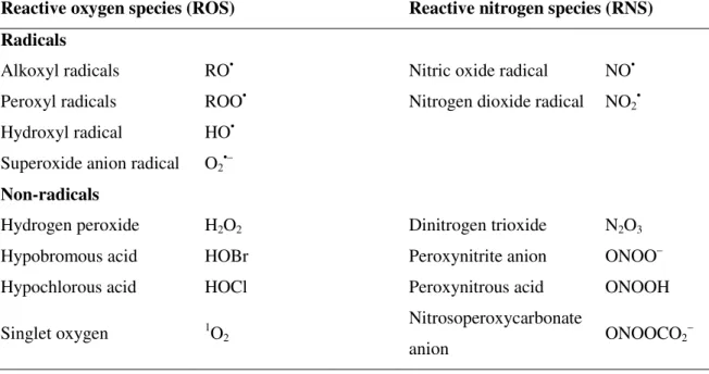
Table 1.1. Pro-oxidants: most common reactive oxygen species (ROS) and reactive
nitrogen species (RNS).
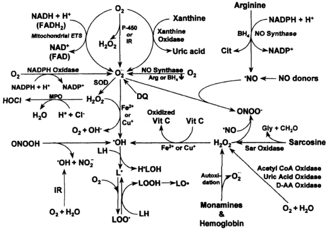
The major pathways for the production of ROS and RNS in vivo are illustrated in Fig. 1.1.
Reactive oxygen species (ROS) Reactive nitrogen species (RNS)
Radicals
Alkoxyl radicals RO• Nitric oxide radical NO•
Peroxyl radicals ROO• Nitrogen dioxide radical NO
2•
Hydroxyl radical HO•
Superoxide anion radical O2•–
Non-radicals
Hydrogen peroxide H2O2 Dinitrogen trioxide N2O3
Hypobromous acid HOBr Peroxynitrite anion ONOO–
Hypochlorous acid HOCl Peroxynitrous acid ONOOH
Singlet oxygen 1O
2
Nitrosoperoxycarbonate
anion ONOOCO2
Figure 1.1. Production of reactive oxygen and nitrogen species in mammalian cells (Fang
et al., 2002). AA, amino acid; Arg, L-arginine; BH4, (6R)-5,6,7,8,-tetrahydro-L-biopterin; CH2O,
formaldehyde; Cit, L-citrulline; DQ, diquat; ETS, electron transport system; FAD, flavin adenine dinucleotide (oxidized); FADH2, flavin adenine dinucleotide (reduced); Gly, glycine; H2O2, hydrogen
peroxide; HOCl, hypochlorous acid; H•LOH, hydroxy lipid radical; IR, ionizing radiation; L•, lipid radical;
LH, lipid (unsaturated fatty acid); LO•, lipid alkoxyl radical; LOO•, lipid peroxyl radical; LOOH, lipid
hydroperoxide; MPO, myeloperoxidase; NAD+, nicotinamide adenine dinucleotide (oxidized); NADH, nicotinamide adenine dinucleotide (reduced); NADP+, nicotinamide adenine dinucleotide phosphate (oxidized); NADPH, nicotinamide adenine dinucleotide phosphate (reduced); NO•, nitric oxide; O2•–,
superoxide anion radical; •OH, hydroxyl radical; ONOO–, peroxynitrite anion; ONOOH, peroxynitrous acid;
P-450, cytochrome P-450; Sar, Sarcosine; SOD, superoxide dismutase; Vit C, vitamin C.
•– •–
These reactive species, radicals and non-radicals, can be easily formed from exogenous and endogenous sources (Kohen and Nyska, 2002). The exogenous sources include exposure of biological systems to γ- or UV-irradiation that results in the production of a
wide range of non-radical and radical species such as hydrogen peroxide (H2O2), hydroxyl radical (HO•), and superoxide anion radical (O2•–). Ozone (O3), which presence in the upper atmosphere is essential in scavenging deleterious UV-irradiation, is also used as disinfection agent by the food industry to destroy food-borne pathogens. Nevertheless, it can oxidize biomolecules yielding the formation of various reactive species. Additionally, a large variety of xenobiotics (e.g. drugs, pollutants, toxins, pesticides, and herbicides) produce ROS/RNS as a by-product of their in vivo metabolism. Although the exposure to exogenous sources is high, the exposure to endogenous sources is much more important and extensive, because it is a continuous process. The main endogenous sources are related with the mitochondrial electron-transport chain, and also with the activity of some enzymes, such as nitric oxide synthases (NOSs) and xanthine oxidase (XOD) that catalyzes the production of nitric oxide radical (NO•) and O2•–, respectively. Moreover, activated phagocytes produce a variety of reactive oxygen, halogen and nitrogen species that play an important role in the mechanism of defense against infectious agents (Halliwell, 2006). To counteract the assault of these ROS/RNS species, living cells have developed a complex defense system composed of enzymatic and non-enzymatic antioxidants that convert them to harmless species. Enzymatic antioxidant defenses include superoxide dismutase (SOD) that detoxifies the O2•– to water and H2O2, catalase which converts H2O2 to oxygen and water, and glutathione peroxidase whose function is to detoxify cellular peroxides to alcohols and water. Non-enzymatic antioxidants are represented by dietary antioxidants such as ascorbic acid (vitamin C), α-tocopherol (vitamin E), carotenoids, and
polyphenolic compounds. Uric acid, glutathione, bilirubin, albumin, and other proteins (transferrin, ceruloplasmin, myoglobin and ferritin) are considered as endogenous antioxidants for protecting essential biological targets against ROS/RNS action.
state is called ‘oxidative stress’, and it can be triggered by several factors such as diseases, diet, lifestyle, and environmental conditions (Kohen and Nyska, 2002). As mentioned before, the excess of ROS/RNS can oxidize cellular lipids, proteins, or nucleic acids, inhibiting their normal function. Because of this, oxidative stress has been implicated in the pathogenesis of several human diseases, including atherosclerosis, cancer, cardiovascular diseases, diabetes mellitus, inflammatory diseases, ischemia/reperfusion injury, and neurodegenerative disorders (Alzheimer’s and Parkinson’s diseases) as well as in the ageing process (Valko et al., 2007). Oxidation can also affect foods, where it is one of the major causes of chemical spoilage, resulting in rancidity and/or deterioration of the nutritional quality, colour, flavour, texture and safety of foods (Frankel, 1996).
Regarding the protective effects of antioxidants against these deleterious oxidative-induced reactions, interest in antioxidant research has become a topic of increasing attention in the last few years, especially within biological, medical, nutritional, and agrochemical fields. In fact, a literature search performed on the ISI Web of Knowledge search engine for articles containing the expression “antioxidant or antioxidants”, revealed that the number of publications has increased about 345% in the past decade (16080 until 1996, while between 1997 and 2006 around 55493 papers were published). This situation demands the existence of simple, convenient, and reliable in vitro analytical methodologies for the fast determination of antioxidant capacity of pure compounds or in complex matrices, such as foods and biological samples (Sánchez-Moreno, 2002; Huang et al., 2005; Prior et al., 2005; Roginsky and Lissi, 2005; Wood et al., 2006).
methods that allow the evaluation of antioxidant capacity in the whole sample, taking into consideration the interactions between all compounds present in the matrix, may offer an additional advantage when compared to those based on separation techniques that are time-consuming, expensive, and often not suitable for screening/routine determinations.
results from different studies difficult and stresses the necessity to standardize antioxidant methods to bring some order to the present chaos in this field.
In this context, Prior et al. (2005) have proposed some guidelines for consideration in method selection for standardization. Thus, a standardized method for determination of antioxidant capacity in routine analysis should meet the following requirements/criteria: (i) measurement of the chemistry actually occurring in potential applications; (ii) utilization of biological relevant molecules; (iii) technically simple; (iv) with a defined endpoint and chemical mechanism; (v) readily available instrumentation; (vi) good repeatability and reproducibility; (vii) adaptable for assay of both hydrophilic and lipophilic antioxidants; (viii) and adaptable to high-throughput analysis.
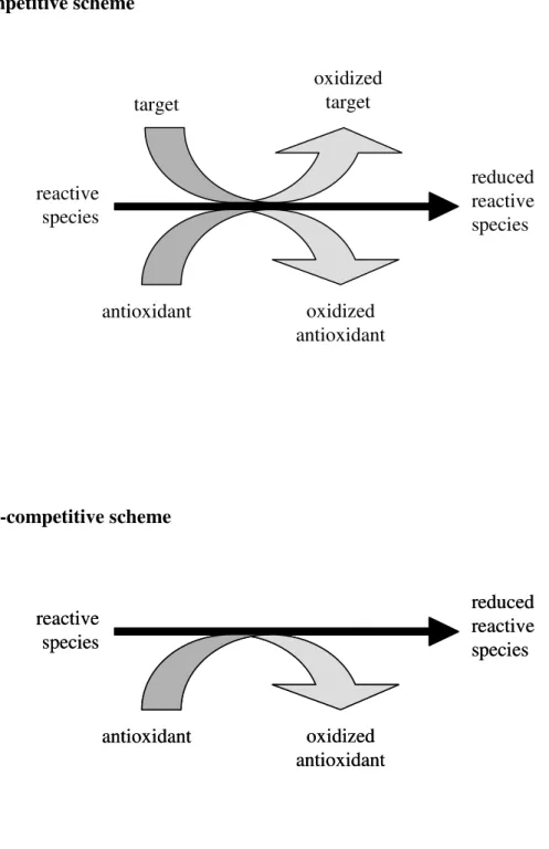
In this regard, knowledge of the chemistry principles of the available methodologies is of utmost importance to select the adequate technique(s). Generally, the in vitro analytical methods for determination of antioxidant capacity rely on two different approaches, named here as competitive or non-competitive scheme (Fig. 1.2).
Competitive scheme
Non-competitive scheme
Figure 1.2. Principle of competitive and non-competitive schemes for the antioxidant
capacity assays.
target
oxidized target
reactive species
reduced reactive species
antioxidant oxidized antioxidant
reactive species
reduced reactive species
antioxidant oxidized antioxidant reactive
species
reduced reactive species
In the non-competitive assays, putative antioxidant compounds react with reactive species without the presence of any other competing target molecule. In this way, these assays involve two components in the initial reaction mixture: the antioxidant compound(s) and the reactive species, which may also be the probe for reaction monitoring. Otherwise, the remaining reactive species may be measured after addition of some derivative reagent. In this context, some of the most commonly used methods for in vitro determination of antioxidant capacity are reviewed in the following sections, where the chemical principles, some of its variants, recent applications as well as the advantages and shortcomings are outlined. These assays were roughly divided into two categories according to the type of the oxidant species: i) scavenging capacity assays against specific ROS/RNS; ii) scavenging capacity assays against stable, non-biological radicals and evaluation of total reducing capacity. The determination of the activity of antioxidative enzymes (superoxide dismutase, catalase, and glutathione peroxidase), or of biomarkers of oxidative stress is out of the scope of this review.
1.2. Scavenging capacity assays against specific ROS/RNS
Reactive oxygen species (ROS) and reactive nitrogen species (RNS) are regularly produced in food and biological systems and may damage biomolecules (Valko et al.,
1.2.1. Peroxyl radical (ROO•) scavenging capacity assays
Peroxyl radicals (ROO•) are commonly found in food and biological substrates and they are formed during lipid oxidation chain reactions, such as the oxidation of polyunsaturated fats (Halliwell and Gutteridge, 1999). They have harmful effects on health and they are also associated to quality deterioration of foods. Their impact on these areas foster the existence of several methods for determining the peroxyl radical scavenging capacity, which were subject of review recently (Laguerre et al., 2007; Roginsky and Lissi, 2005).
In general, methods for examination of ROO• scavenging capacity measure the ability of an antioxidant to scavenge peroxyl radicals by hydrogen atom transfer (HAT) reactions. In these assays a competitive scheme is applied, where antioxidants or target molecules react with ROO•. Hence, the assay system is composed of three components: i) thermolabile azo-compound (R–N=N–R), which yields carbon-centered radicals (R•) that react fast with O2 to give a steady flux of ROO• radicals; ii) oxidizable target (PH); and iii) antioxidant compound(s) (AH), as represented schematically in the following equations (1.1–1.5):
R–N=N–R → 2 R• + N2 (1.1)
R• + O2 → ROO• (1.2)
ROO• + PH → ROOH + P• (1.3)
ROO• + AH → ROOH + A• (1.4)
ROO• + A• → ROOA (1.5)
-azobis(2,4-dimethylvaleronitrile) (AMVN); their molecular structure are represented in Fig. 1.3. The rate of their spontaneous decomposition and production of ROO• radicals is primarily determined by temperature of the reaction medium (Niki, 1990).
AAPH AMVN
2,2′-azobis(2-amidinopropane) dihydrochloride 2,2′-azobis(2,4-dimethylvaleronitrile)
Figure 1.3. Molecular structure of thermolabile azo-compounds used for generation of
peroxyl radicals.
In these competitive assays, the presence of antioxidant compounds inhibits or retards the rate of target/probe oxidation induced by peroxyl radicals (eq. 1.3). Therefore, in the
beginning of the assay, insignificant spectroscopic changes of the target/probe would be observed (induction period or lag phase) because the antioxidants protect the target/probe from ROO•-oxidation. As the reaction proceeds, the antioxidants are consumed by the constant flux of ROO• and the oxidation of the target/probe would progress at a slower rate when compared with the control (absence of antioxidant compounds/samples). Finally, when the antioxidants are depleted, the reaction rate of oxidation of the target/probe is similar to that obtained for the control. As mentioned before, the beginning and duration of these three phases is dependent on the rate of reaction between antioxidant and ROO•, the rate of reaction between the target/probe and ROO•, and the concentration ratio between antioxidant and target/probe.
N N CH 3 CH3 N H2 NH C H3 C H3 N H NH2 HCl HCl C
H3 N N
N C
H3 HC 3
CH3
Although the competitive scheme applied resembles in vivo conditions, the concentration of the target species is usually smaller than the concentration of antioxidants. This is in contradiction with the “definition of antioxidant” (Halliwell et al., 1995) and with what is
found in real situations, where the antioxidant concentration is much smaller than that of the oxidizable substrate (lipids or proteins, for instance). Moreover, these assays apply a ROO• reaction without taking into account the essential propagation step in lipid autoxidation, such as the breakdown of hydroperoxides (ROOH) yielding peroxyl and alkoxyl radicals (RO•) (Huang et al., 2005). Finally, most of these assays are rather
time-consuming and their application requires a significant expertise and experience in chemical kinetics. As a consequence, HAT-assays are commonly not suitable for routine determinations.
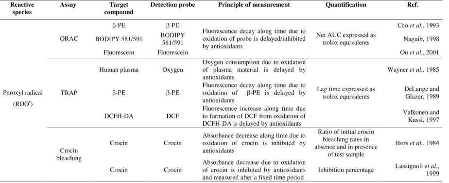
The analytical methods comprising these features include the oxygen radical absorbance capacity (ORAC) assay, the total radical-trapping antioxidant parameter (TRAP) assay, and the crocin bleaching assay. The inhibition of low-density lipoproteins (LDL) oxidation, the total oxyradical scavenging capacity (TOSC), and the chemiluminescence-based assays employing peroxyl radicals may also be included. The major difference among these assays lies mostly in the approach used for quantification of antioxidant capacity: the ORAC assay applies the area under curve (AUC) that represents the oxidation of the target along time, the TRAP assay relies on the “lag time”, that corresponds to the time period between the beginning of the assay and the beginning of the oxidation of the target, whilst the crocin-bleaching assay utilises the initial oxidation rate of target species.
1.2.1.1. Oxygen radical absorbance capacity (ORAC assay)
fluorescent target/probe, which reacted with ROO• to form a nonfluorescent product (Cao
et al., 1993). Nevertheless, some shortcomings were observed such as large lot-to-lot
variability, photobleaching of the β-PE after exposure to excitation light, and interaction with polyphenols by non-specific protein binding. To overcome these limitations, the synthetic, non-protein fluorescein has been used as the fluorescent target/probe, instead of the original β-PE (Ou et al., 2001). The application to both lipophilic and hydrophilic
chain-breaking antioxidants was carried out using a mixture of acetone/water containing 7% of randomly methylated β-cyclodextrin as a water solubility enhancer (Huang et al.,
2002a). Lipophilic compounds were also quantified by ORAC assay using either organic media or liposomes, AMVN as a lipophilic peroxyl radical generator, and 4,4-difluoro-5-(4-phenyl-1,3-butadienyl)-4-bora-3a,4a-diaza-s-indacene-3-undecanoic acid (BODIPY
581/591 C11) as a fluorescent target/probe (Naguib, 1998). To improve the throughput, Huang et al. (2002b) developed a high-throughput assay using a multichannel liquid
handling system coupled with a microplate fluorescence reader in 96-well format.
In the ORAC assay, the reaction is monitored for extended periods (≥ 30 min) and the quantification is based in the area under curve (AUC) that represents the oxidation of the probe along time. The protective effect of antioxidants is evaluated from the net integrated area under the fluorescence decay curves (AUCsample – AUCblank) and results are expressed as µM of trolox equivalents. The advantage of the AUC approach is that it can be applied for antioxidants that exhibit distinct lag phases and to those samples that have no lag phases. Moreover, it takes into account the initial reaction rate and the total extent of inhibition, which includes the action of slow-reacting or secondary antioxidant products formed. The principles of the ORAC assay can be adapted to determine the action against other reactive oxygen species (Ou et al., 2002a). The main limitation of the ORAC assay is
This methodology has provided substantial information regarding the antioxidant capacity of lipophilic and hydrophilic antioxidant compounds (Cho et al., 2007), food products (Wu et al., 2004), animal tissues (Cao et al., 1996), and in bioavailability studies
(Mertens-Talcott et al., 2006; Prior et al., 2007).
1.2.1.2. Total radical-trapping antioxidant parameter (TRAP assay)
The total radical-trapping antioxidant parameter (TRAP) assay was introduced by Wayner
et al. (1985), for the determination of the antioxidant status of human plasma. This method
was based on the measurement of the time period in which oxygen uptake was inhibited by plasma during a controlled ROO• peroxidation reaction induced by the thermal decomposition of an azo-compound. In this assay, the target was the human plasma while the oxygen consumed in the oxidation of plasma material was the probe used to follow the action of antioxidants. The measurement is based on the “lag time” that corresponds to the time period between the beginning of the assay and the beginning of the oxidation of the target molecules. Later, the same research group evaluated the relative contributions of the main antioxidants of human plasma to the TRAP value (Wayner et al., 1987).
One of the major problems with the original TRAP assay lies in the utilization of the oxygen electrode as detector, since it may not maintain its stability over the period of time required (Rice-Evans and Miller, 1994). To overcome this limitation, this assay was later improved using β-phycoerythrin (β-PE) as the fluorescent target/probe, and the ability of the plasma to protect β-PE from peroxyl radical oxidation was fluorimetrically monitored (DeLange and Glazer, 1989). Ghiselli et al. (1995), proposed some modifications in order
competitively inhibits the increase of fluorescence signal. For the analysis of lipophilic samples as olive and seed oils, peroxyl radicals were generated by the lipophilic AMVN azo compound (Cabrini et al., 2001). Due to the lower solubility of fluorescent
target/probes in the reaction medium, the TRAP value was determined by measuring the time period during which oxygen uptake was inhibited.
Disregarding the different variations discussed above, the quantification is based on the lag phase duration, in which oxidation is inhibited by the antioxidants, compared to the lag phase of trolox. The antioxidant capacity expressed as trolox equivalents (XAO) is calculated by the following equation:
XAO = (CTrolox/TTrolox) * TAO (1.6)
where CTrolox is the trolox concentration, whilst TTrolox and TAO are the lag time of the kinetic curve of target oxidation in the presence of trolox or in the presence of antioxidant/sample, respectively; XAO is then multiplied by 2.0, the stoichiometric factor of trolox, and by the dilution factor of the sample to give the TRAP value (µM).
The main shortcoming of this assay is the use of the lag phase for quantifying antioxidant capacity, since not every antioxidant possesses an obvious lag phase and also the antioxidant capacity profile after the lag phase is totally ignored. Moreover, the application of different criteria for establishing the endpoint makes difficult the comparison between laboratories. Another important limitation of this assay, and also of the ORAC assay, is that the oxidative deterioration and antioxidant protection of fluorescent target/probe does not necessarily mimic a critical biological substrate.
This assay has been applied to in vitro evaluation of antioxidant capacity of foods (Pellegrini et al., 2003a), and to assess the plasma status after ingestion of food-rich
1.2.1.3. Crocin bleaching assay
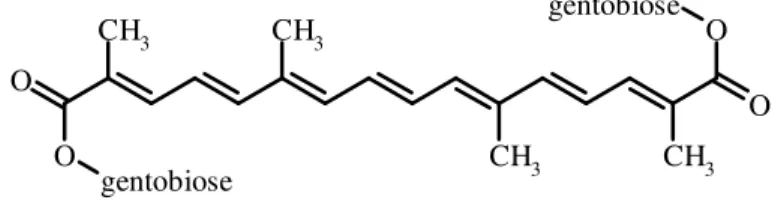
This assay measures the ability of antioxidant compounds to protect the naturally occurring carotenoid derivative crocin (Fig. 1.4) from oxidation by ROO• radicals formed through thermolysis of AAPH (Bors et al., 1984). The reaction is initiated by the addition of AAPH
and the bleaching rate (absorbance decrease/time) of crocin is monitored at 443 nm during 10 min. Antioxidants compete with crocin for ROO•, and the degree of inhibition of crocin oxidation depends on the antioxidant capacity of tested samples. The quantification of antioxidant capacity is based on the ratio of initial crocin bleaching rates in absence (V0) and in presence (V) of antioxidants, and is given by the Stern-Volmer-like relation:
V0/V = 1+ (kAO/kC)*([AO]/[C]) (1.7)
where [AO] and [C] are the concentrations of antioxidant and crocin, respectively, kAO and kC are the rate constants for the reaction of the peroxyl radicals with antioxidant and crocin, respectively. After measuring V0/V value at known ratio of [AO] to [C], kAO/kC is given by the slope value obtained from eq. (1.7) and indicates the relative peroxyl radical
scavenging capacity. A microplate-adapted crocin bleaching assay based on the inhibition percentage at a fixed time instead of kinetic analysis have also been reported (Lussignoli et al., 1999).
Figure 1.4. Chemical structure of crocin, a natural carotenoid compound formed from
diester of the disaccharide gentobiose and the dicarboxylic acid crocetin; when dissolved in water, it forms an orange solution (λmax = 443 nm).
CH3 CH3
CH3 CH3 O O
O O
gentobiose
This assay was used to determine the structure-antioxidant activity relationships of flavanones present in Citrus fruit (Di Majo et al., 2005). Nevertheless, it has limited
applications in food samples since many food pigments, such as carotenoids, absorb at the same wavelength of the determination. Besides this drawback, crocin is a natural food pigment extracted from saffron, which may confer a low reproducibility between assays. In fact, Chatterjee et al. (2005) described a more affordable crocin assay using the Indian
spice saffron instead of the commercial chemical product and found that the antioxidant capacity values of pure natural compounds, plant extracts, and human plasma from healthy individuals using either approach were similar.
Recently, Ordoudi and Tsimidou (2006a) have examined the crocin bleaching assay performance and validation procedures. The studies were focused on target/probe and test compound characteristics, conditions for peroxyl radical generation, reaction monitoring, and expression of results. They observed that any authentic commercial saffron can be used for target/probe preparation given that: interferences, such as tocopherols, are removed; the concentration of working solution is adequately adjusted; and the changes of the stock target/probe solution during storage are not neglected. Results are expressed as “percent inhibition of crocin bleaching value” instead of the ratio of initial crocin bleaching rates. This assay has been applied to structure-activity relationship studies of selected phenolic compounds (Ordoudi and Tsimidou, 2006b).
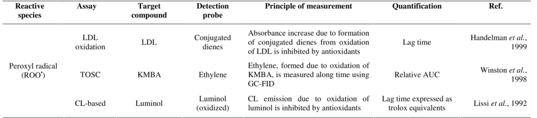
1.2.1.4. Inhibition of low-density lipoproteins (LDL) oxidation
In this assay the low-density lipoproteins (LDL), isolated from blood samples, are the oxidizable target. The peroxidation of LDL is initiated by thermal decomposition of AAPH or by Cu(II) and assessed through the formation of conjugated dienes, determined spectrophotometrically at 234 nm after HPLC separation (Handelman et al., 1999).
Sánchez-Moreno et al. (2000) studied the oxidation of LDL induced by Cu(II) and
compared to the control (CLT50) was determined graphically upon the representation of the ratio lag time antioxidant/lag time control as a function of antioxidant concentration. In this assay the use of peroxyl radicals and LDL as oxidant and target species, respectively, allows a strong resemblance to oxidative reactions that might occur in biological systems. This assay has been applied to evaluate the capacity to inhibit in vitro LDL oxidation of pure compounds, plant foods and beverages (Saura-Calixto and Goni, 2006). Recently, Gomes-Ruiz et al. (2007) used this method to determine the antioxidant capacity of
different compounds present in coffee or which are produced as a result of the metabolism of this beverage; 1-methyluric acid was particularly effective at inhibiting oxidation of LDL. However, the major limitation is that the LDL has to be isolated on a regular basis from different individuals, and therefore a high inter-batch variation is verified. Moreover, problems with this assay arise because it is difficult to measure the small lag times that occur, and many substances also absorb at the wavelength of the determination.
1.2.1.5. Total oxyradical scavenging capacity (TOSC assay)
In the original total oxyradical scavenging capacity (TOSC) assay, peroxyl radicals generated by thermal homolysis of AAPH cause the oxidation of α-keto-γ-methiolbutyric acid (KMBA) to ethylene, which is monitored by gas chromatographic analysis of head space from the reaction vessel (Winston et al., 1998). The antioxidant capacity of the
to scavenge different oxidants. In the TOSC assay, the approach for quantification of antioxidant capacity is similar to that applied for the ORAC assay. In this case it is based in the area under the curve that represents the inhibition of ethylene formation as a function of time. Nevertheless, there is not a linear relationship between AUC and antioxidant concentration or dilution factor of the sample. Therefore, the concentrations or dilution factors that correspond to TOSC values of 20, 50, and 80% are calculated, and DT50 is also determined, which is the first derivative of the curve at a TOSC of 50% (Lichtenthäler and Marx, 2005a). The long reaction time (>100 min), the short shelf-life of the assay solutions, and the necessity of multiple manual injections from the same sample into a gas chromatograph to monitor the formation of ethylene, make this assay difficult to implement to routine analysis. To overcome this last drawback, Lichtenthäler and Marx (2003) monitored the time course of ethylene formation resorting to automated headspace gas chromatography. Later, the same research group applied this automated version for a comparative and detailed survey of the antioxidant capacities of 14 common European fruit and vegetable juices (Lichtenthäler and Marx, 2005a). The antioxidant capacity of methanol and ethanol seed extracts (Rodrigues et al., 2006) and fruit pulp (Lichtenthäler et al., 2005b) from Euterpe oleracea Mart. (açai) were also studied with the TOSC assay in a
modified and automated version.
1.2.1.6. Chemiluminescence-based assays
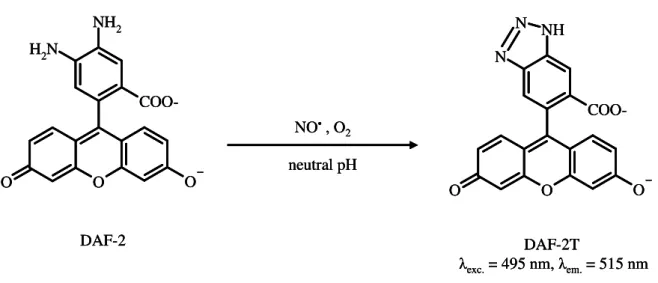
The general principle of chemiluminescence (CL) assays is based on the reaction of oxidants (ROS/RNS) with chemiluminogenic probes (luminol or lucigenin) to produce excited state species that emit light. A recent outlook on chemiluminescent methods for ROS is given by Lu et al. (2006). These assays are based on competitive or on
results in inhibition of the light production and the intensity of CL-quenching is directly proportional to the antioxidant capacity of the sample. Nevertheless, it should be stressed that the antioxidant/sample may reduce or scavenge the reactive species as well as the radicals involved in the target/probe CL-oxidation (Fig. 1.5). The interference on CL reaction will depend both on the rate of reaction of antioxidants with ROS/RNS and with radicals formed during CL-probe oxidation. In this regard, these interferences may be minimized and reliable results are obtained with a non-competitive approach, since the antioxidants react with oxidants before the addition of CL-probe. This strategy is easily attained using the flow-based methods, which are discussed further in this chapter.
Peroxyl radical scavenging capacity assays have also been implemented using CL as detection system. The principles of ROO• induced luminol-CL assays were described in detail by Alho and Leinonen (1999). Lissi et al. (1992 and 1995) evaluated the effect of
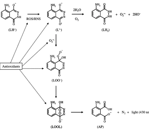
Figure 1.5. Mechanism of luminol oxidation induced by reactive species (ROS/RNS) and
antioxidant inhibition pathways (Giokas et al., 2007). At alkaline pH, monodissociate
luminol (LH–) reacts with ROS/RNS to yield the diazasemiquinone radical (L•–). The later reduces O2 to superoxide anion (O2•–) and is oxidised to 5-aminohyphenthalazine-1,4-dione (LH2). Diazasemiquinone radical and O2•– yield the carbon-centered hydroperoxide anion (LOO–) that rearranges to a transient endoperoxide (LOOL), which decomposes to give light emission and products (N2 and aminophthalate (AP)). Antioxidants may react with ROS/RNS as well as with the radicals formed during luminol oxidation pathway.
O2•–
NH2 O N N OH O O
(LOO–)
NH2 O O OH O (AP) Antioxidants O2 2H2O
ROS/RNS NH2 NH N O O
(LH–) (LH
2) NH2 O
O NH NH
+ O2•– + 2HO–
(L•–) NH2 N N O O• (LOOL) NH2 N N OH O O
O + N2 + light (430 nm)
(LOOL) NH2 N N OH O O O O2•–
O2•–
NH2 O N N OH O O
(LOO–) NH2 O N N OH O O
(LOO–)
NH2 O O OH O (AP) NH2 O O OH O (AP) Antioxidants O2 2H2O
O2 2H2O
ROS/RNS NH2 NH N O O
(LH–) NH2
NH N O
O
(LH–) (LH
2) NH2 O
O NH NH
+ O2•– + 2HO–
(LH2)
NH2 O
O NH NH
+ O2•– + 2HO– NH2 O
O NH NH
+ O2•– + 2HO–
(L•–) NH2
N N O
O•
(L•–) NH2 N N O O• NH2 N N O O• (LOOL) NH2 N N OH O O O (LOOL) NH2 N N OH O O O NH2 N N OH O O
O + N2 + light (430 nm)
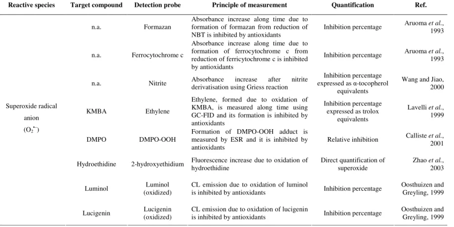
1.2.2. Superoxide radical anion (O2•–) scavenging capacity assays
Superoxide radical anion (O2•–) is produced as a result of the donation of one electron to oxygen. This radical arises either from several metabolic processes or following oxygen activation by irradiation (Fig. 1.1). Nicotinamide adenine dinucleotide phosphate (NADPH) oxidase from activated neutrophils generates O2•– in the respiratory burst necessary for bacteria destruction (Halliwell, 2006). Moreover, under oxidative stress conditions, xanthine oxidase (XOD) transfers electrons to oxygen and produces O2•– and hydrogen peroxide. Most of the O2•– generated in vivo undergoes a dismutation reaction catalysed by the antioxidative enzyme superoxide dismutase to give hydrogen peroxide. Despite the low reactivity of O2•–, it is considered the “primary” ROS that interacts with other molecules to generate “secondary” ROS, either directly or prevalently through enzyme- or metal-catalysed processes. In the last case, it originates the production of the highly reactive hydroxyl radical.
The analytical methods for determination of O2•– scavenging capacity make use of the system XOD/hypoxanthine or xanthine at pH 7.4 to generate superoxide radical anion. To a minor extent, O2•– is also generated using a non-enzymatic reaction of phenazine methosulphate (PMS) in the presence of nicotinamide adenine dinucleotide (NADH). In both generation systems, O2•– may reduce nitroblue tetrazolium (NBT) into formazan, which is spectrophotometrically monitored at 560 nm (Aruoma, et al. 1993; Fernandes et al., 2003; Floriano-Sanchez et al., 2006). Antioxidant compounds compete with NBT for
O2•– and decrease the rate of reaction. Another widely used probe for O2•– is cytochrome c. The kinetic analysis of reduction of ferricytochrome c to ferrocytochrome c was monitored at 550 nm (Aruoma, et al. 1993; Quick et al., 2000). In fact, Aruoma et al. (1993) observed
spectrophotometrically at 530 nm after addition of sulfanilic acid and α-naphthylamine (Griess reaction) (Elstner and Heupel, 1976). This assay was applied for determining the O2•– scavenging capacity of fruit juices and the results were expressed as µmol of α -tocopherol equivalent per 10 g of fresh weight. The scavenging capacity towards O2•–, using XOD/hypoxanthine generating system, has also been measured by reaction with KMBA to produce ethylene, which is measured by gas chromatography (Lavelli et al.,
1999). The scavenging capacity against this radical can also be measured by using electron spin resonance (ESR) spectrometry (Calliste et al., 2001). Here, the O2•– is trapped by 5,5-dimethyl-1-pyrroline-N-oxide (DMPO), and the resultant DMPO-OOH adduct is detected
by ESR.
The CL-based determination of O2•– scavenging capacity has also been described. Luminol or lucigenin are frequently applied as target compounds (Lu et al., 2006), but neither of
them is selective towards O2•– (Oosthuizen and Greyling, 1999; Li et al., 1998). CLA (2-methyl-6-phenyl-3,7-dihydroimidazo[1-2-a]pyrazin-3-one) and MCLA [2-methyl-6-(4-methoxyphenyl)-3,7-dihydroimidazo[1,2-a]pyrazin-3-one], analogs of coelenterazine, have also been described as more specific targets of O2•– but their application is more focused to in vivo monitoring of superoxide formation (Lu et al., 2006). Ogawa et al. (1999)
developed an HPLC system with indirect luminol-based CL for screening individual antioxidants present in extracts of green tea leaves (essentially catechins and flavones). The determination of antioxidants was based on the decrease of CL intensity derived from luminol and O2•– (generated from the hypoxanthine/XOD system). Epicatechin was detected as the main antioxidant among the various intrinsic substances present in green tea extracts. The O2•– scavenging capacity of some therapeutic compounds (NSAIDs, β -blockers) and medicinal plant extracts have also been measured by monitoring the O2•– induced lucigenin chemiluminescence (Costa et al., 2006a; Gomes et al., 2006; Abreu et al., 2006; Chen and Yen, 2007).
reduced antioxidant formed by attack of O2•– could also reduce NBT or ferricytochrome c (Halliwell et al., 1995). The nonfluorescent hydroethidine has also been used as the
target/probe for measuring O2•– scavenging capacity (Zhao et al., 2003). The target is oxidized by O2•– (generated from XOD/xanthine system) to form a species that exhibits a strong fluorescence signal (2-hydroxyethidium). This approach can circumvent the problem of direct reduction of the target/probe by antioxidants, but possible inhibition of xanthine oxidase by antioxidants/sample remains an issue.
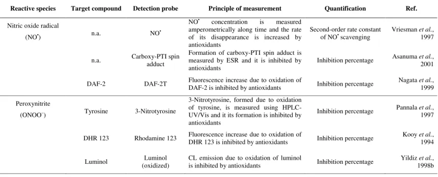
1.2.3. Hydrogen peroxide (H2O2) scavenging capacity assays
Hydrogen peroxide (H2O2) is generated in vivo, under physiological conditions by peroxisomes, by several oxidative enzymes including glucose oxidase and D-amino acid oxidase, and by dismutation of superoxide radical, catalysed by superoxide dismutase. Additionally, H2O2 produced in the respiratory burst of activated phagocytes is known to play an important role in killing of several bacterial and fungal strains (Halliwell, 2006). Hydrogen peroxide is very diffusible within and between cells but it is rather inert at low concentrations and reacts quite slowly with most biological compounds. Nevertheless, its oxidant power is associated to the presence of transition metals ions, especially Fe(II) or Cu(I), generating the potent oxidant hydroxyl radical (Fig. 1.1).
formed from enzyme and H2O2 (it is possible that the superoxide radical is produced during enzyme activity). Moreover, antioxidants such as ascorbic acid, quercetin dihydrate, and thiols can be a substrate for the peroxidase enzyme, introducing errors in the determination of scavenging capacity. Therefore, if the compound does interfere with peroxidase-based system, other assays for H2O2 should be used. For instance, the direct reaction of H2O2 and titanium(IV) was applied as it originates a complex Ti-H2O2 that is dissolved in acidic medium and further measured spectrophotometrically measured at 410 nm. This approach was applied to evaluate H2O2 scavenging capacity of fruit juices and results were expressed as µmol of ascorbate equivalent per 10 g of fresh weight (Wang and Jiao, 2000). A valid alternative to these methods was proposed by Arnous et al. (2002).
The enzyme-free methodology implemented relied on the peroxyoxalate chemiluminescence (POCL) using 9,10-diphenylanthracene and imidazole as a fluorophore (probe) and catalyst, respectively. Briefly, POCL involves hydrogen peroxide imidazole-catalysed oxidation of an aryl oxalate ester yielding a high-energy intermediate (dioxetanedione) that transfers its energy to the fluorophore. The transition of the excited state of the fluorophore to its ground state causes the emission of light. Therefore, any compound with capacity to scavenge H2O2 would lead in CL inhibition. In this way, several antioxidants including β-carotene, α-tocopherol, butylated hydroxytoluene, quercetin, and L-ascorbic acid were investigated and the results were compared to other in vitro tests. Later, this assay was applied to investigate a wide range of natural antioxidants (cinnamic and benzoic acids) and to examine possible structure-H2O2 scavenging capacity relationships (Mansouri et al., 2005). Due to the non-polar environment employed (ethyl
1.2.4. Hydroxyl radical (HO•) scavenging capacity assays
The hydroxyl radical (HO•) is extremely reactive in vivo (rate constants > 109 M-1 s-1) and it can hydroxylate any molecule found in the food matrix or in living cells (including proteins, polyunsaturated fatty acids, sugars, and nucleic acids). Biologically, the HO• radical can be generated by several mechanisms: i) homolytic fission of oxygen-hydrogen bonds in water driven by continuous exposure to background ionizing radiation; ii) reaction between Fe2+ released under stress conditions and H2O2 (eq. 1.8, Fenton reaction); iii) Haber-Weiss reaction involving superoxide radical (eq. 1.9); and iv) reaction of HOCl
with O2•– (Halliwell, 2006).
Fe2+ + H2O2→ Fe3+ + HO• + HO– (1.8)
O2•– + H2O2→ O2 + HO• + HO– (1.9)
Due to the high reactivity of hydroxyl radicals, almost anything in biological systems can be regarded as an HO• scavenger. Hence, this task is not performed by any specific molecule or enzyme. Thus, the evaluation of direct scavenging of HO• may be irrelevant for evaluation of antioxidant action of a compound or matrix, simply because very high concentrations of scavenger are required to compete with adjacent molecules in vivo or in the food matrix for any HO• generated. For these reason, it is more relevant and useful to quantify the capacity of putative antioxidants to scavenge or block the formation of its precursors (O2•–, H2O2, HOCl) and/or to sequester free metal ions related to HO• formation. Scavenger compounds that act in this way would behave as preventive antioxidants.
Despite this remark, several in vitro methodologies for determination of HO• scavenging capacity are available, mostly based on Fe3+ + EDTA + H2O2 + ascorbic acid system to generate a constant flux of HO• radicals. Those radicals attack the sugar 2-deoxy-D-ribose (used as target), degrading it into a series of fragments, some or all of which react upon heating with thiobarbituric acid at low pH to give a pink chromogen (Halliwell et al.,