https://doi.org/10.1007/s11694-020-00661-4
ORIGINAL PAPER
Antioxidant and cytotoxic activities of sulfated polysaccharides
from five different edible seaweeds
K. Arunkumar1 · Rathinam Raja2 · V. B. Sameer Kumar3 · Ashna Joseph1 · T. Shilpa1 · Isabel S. Carvalho2
Received: 19 June 2020 / Accepted: 11 September 2020
© Springer Science+Business Media, LLC, part of Springer Nature 2020 Abstract
In recent times, there has been a growing interest in the exploration of antioxidants and global trend toward the usage of seaweeds in the food industries. The low molecular weight up to 14 kDa sulfated polysaccharides of seaweeds (Portieria
hornemannii, Spyridia hypnoides, Asparagopsis taxiformis, Centroceras clavulatum and Padina pavonica) were evaluated for
in vitro antioxidant activities and cytotoxic assay using HeLa cell line and also characterized by FTIR. The high yield (7.74% alga dry wt.) of sulfated polysaccharide was observed in P. hornemannii followed by S. hypnoides (0.69%), C. clavulaum (0.55%) and A. taxiformis (0.17%). In the brown seaweed P. pavonica, the sulfated polysaccharide yield was 2.07%. High amount of sulfate was recorded in the polysaccharide of A. taxiformis followed by C. clavulaum, P. pavonica, S. hypnoides and P. hornemannii as indicative for bioactivity. The FTIR spectroscopic analysis supports the sulfated polysaccharides of S.
hypnoides, C. clavulatum and A. taxiformis are similar to agar polymer whereas the spectral characteristics of P. horneman-nii have similarities to carrageenan. The higher DPPH activity and reducing power were recorded in the polysaccharide of
brown seaweed P. pavonica than the red seaweeds as follows: DPPH activities: S. hypnoides > A. taxiformis > C.
clavula-tum > P. hornimanii; Reducing power: A. taxiformis > P. hornimanii > S. hypnoides > C. clavulaclavula-tum. The polysaccharide
fractions contain up to 14 kDa from red seaweeds P. hornemannii and S. hypnoides followed by brown seaweed P. pavonica exhibit cytotoxic activity in HeLa cancer cell line (and are similar to structural properties of carrageenan extracted from P.
hornemannii). The low molecular weight agar like polymer of S. hypnoides and alginate like brown seaweed P. pavonica
showing better in vitro antioxidant activities that are capable of exhibiting cytotoxicity against HeLa cell line can be taken up further in-depth investigation for nutraceutical study.
Keywords Seaweeds · Sulfated polysaccharides · Antioxidants · Cytotoxic assay · HeLa cell lines
Introduction
Marine macroalgae (seaweeds) are the only resource for industrially important polymers such as agar and carra-geenan from red seaweeds; alginate, fucoidan and lami-narin from brown seaweeds. These polymers are extracted
only from few seaweed species that meet certain industrial applications. This seaweed sulfated polysaccharides possess a variety of biological activities and immunomodulatory activities to mitigate associated negative effects including inflammation [1]. These polysaccharides have been used in food industries as gelling agents, thickening, and stable excipients for control release of drugs [2]. Recently, in a test of antiviral effectiveness against the virus which causes COVID-19, an extract from edible seaweeds substantially outperformed remdesivir, the current standard antivi-ral agent used to combat the disease. Further Heparin, a common blood thinner, and a heparin variant stripped of its anticoagulant properties, performed on par with rem-desivir in inhibiting SARS-CoV-2 infection in mammalian cells [3]. Beside, this seaweed hydrocolloids have great economic importance because of their various bioactive (anticoagulant, antiviral, anticancer, antioxidant, antitumor, * Rathinam Raja
1 Department of Plant Science, Central University of Kerala,
Periye, Kerala 671 320, India
2 M ED- Med ite rra nean Institute for Agriculture, Environment
and Development, Food Science Laboratory, FCT, University of Algarve, Building 8, Gambelas, 8005-139 Faro, Portugal
3 Department of Biochemistry and Molecular Biology, Central
immunomodulating, antihyperlipidemic and antihepatotoxic activities) and unique rheological properties, hence they are being widely used in the pharmaceutical and biomedi-cal sectors [4–6]. The structural features like the composi-tion of monomers, sulfate level and its posicomposi-tion in the sugar moiety, chain length, molecular weight etc. of the polymer make them ideal materials for various biomedical applica-tions [7, 8]. Cholesterol and lipid-controlling properties of carrageenan have been demonstrated in a clinical trial and found significant reduction in the serum cholesterol and tri-glyceride levels [9]. Carrageenan is well known in control-ling the inflammation and a complete review on carrageenan biological activities was reported [10, 11]. Recently, Zhong et al. [8] reviewed the cytotoxic and antioxidant activity in various seaweeds polysaccharides.
Among five seaweeds, the red Portieria hornemannii,
Spyridia hypnoides, Asparagopsis taxiformis and Cen-troceras clavulatum; and brown seaweed Padina pavon-ica were taken up, a monoterpene possessing cytotoxicity
against a diverse panel of human tumor cell line was isolated from the red alga Portieria hornemannii [12]. The antioxi-dant and in vitro cytotoxic activity of extracts prepared using methanol, chloroform, petroleum ether and ethyl acetate from red alga Asparagopsis taxiformis were demonstrated [13]. In another study, ethanol extract significantly exhib-iting antibacterial activity against fish pathogenic bacteria was found [14]. Ethanolic extract of brown alga, Padina
pavonica having antimicrobial, antioxidant, and anticancer
activities was proved [15]. Padina pavonica extract contain phenolic, flavonoid and tannin which exhibits antioxidant activity [16] whereas there is no report on Spyridia
hyp-noides and Centroceras clavulatum. Further, in vitro
anti-oxidant activity and cytotoxic assay using cancer cell-line from these five algae sulfated polysaccharides are signifi-cant. Because taping the potential of these seaweeds which are not exploited for polysaccharides of industrial quality and by considering its food value, they can be utilized for the source of compounds of biomedical and nutraceutical val-ues. Hence, in the present study, results of sulfated polysac-charides extracted from these five seaweeds were evaluated also for antioxidant and cytotoxic properties.
Materials and methods
Collection of seaweedsThe fresh and healthy specimens of red seaweeds about 3 kg of Portieria hornemannii (Lyngbye) P.C. Silva, Spyridia
hyp-noides (Bory) Papenfuss, Asparagopsis taxiformis (Delile)
Trevisan and Centroceras clavulatum (C. Agardh) Montagne; and brown seaweed Padina pavonica (Linnaeus) Thivy occur-ring along the Coast of Pamban (9.2798° N, 79.2291° E), Gulf of Mannar, Tamil Nadu, India were collected during the month of September 2018.
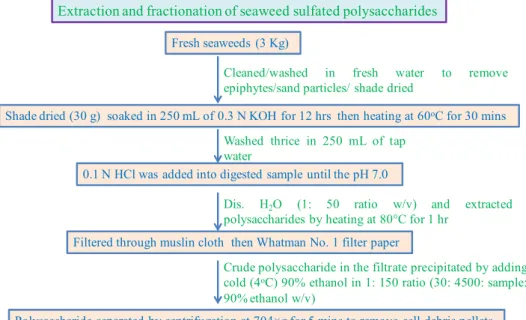
Extraction and fractionation of polysaccharides
The extraction procedure of Distantina et al. [17] was followed with minor modifications. The collected five seaweeds were cleaned with fresh water to remove epiphytes and sand parti-cles. The samples were shade dried under air in dark for 5 days. Thirty gram of dried seaweeds was soaked in 250 mL of 0.3 N KOH solutions for 12 h and then heated at 60 °C for 30 min. The alkaline digested sample was washed thrice in 250 mL of tap water and then 0.1 N HCl was added into digested sample until the pH was neutral (7.0). Then distilled water was added to sample to reach 1:50 ratio (30:1500 w/v) and extracted by heating at 80 °C for 1 h. The hot water extract was filtrated through muslin cloth followed by Whatman No. 1 filter paper and cooled. The crude polysaccharide in the filtered extract was precipitated by adding cold (4 °C) 90% ethanol in 1:150 ratio (30:4500 sample: 90% ethanol w/v). The precipitate was collected and stored at 4 °C over night and thawed further, the crude polysaccharide was separated by centrifugation at 704×g for 5 min to remove cell debris. Then the supernatant was col-lected and again centrifuged at 7826×g for 10 min to obtain pellets of crude polysaccharides which were stored at 4 °C. These pellets were dialyzed using dialysis tubing of 14 kDa (Sigma Aldrich D9652) molecular weight cut off against two volumes of distilled water for 24 h at room temperature then freeze-dried and weighed (Fig. 1. Flow chart).
Estimation of sulfate in fractionated polysaccharides
The total sulfate [18] content of dialyzed polysaccharides were estimated.
Characterization of polysaccharides by FTIR spectra
The extracted polysaccharides were characterized by FTIR spectroscopy (Perkin-Elmer Version 10.5.1 spectrometer, Bos-ton, USA). The polysaccharide (5 mg) of each seaweeds were ground with spectroscopic grade potassium bromide (KBr) powder and pressed into 1 mm pellet for FTIR measurement in the wavelength that ranges from 400 to 4000 cm−1 [19]. The
FTIR spectrum was finalized on the basis of spectral consist-ency recorded from triplicate KBr powder samples from each seaweed.
In vitro antioxidant activities of the polysaccharide
DPPH radical scavenging activity
The scavenging effects of polysaccharide were deter-mined using ascorbic acid as a positive control [20]. For the assay, 2 mL of 0.16 mM DPPH in methanol was added into the test tube containing 2 mL aliquot of test sample at various concentrations (0.1, 0.5 and 1 mL from stock of 1 mg mL−1). The mixture was vortexed for 1 min and kept
at room temperature for 30 min in the dark. The absorbance was measured at 517 nm in a UV–visible spectrophotometer (Shimadzu UV-2600). The capacity to scavenge the DPPH radical was calculated by the following equation:
where, A control is the absorbance of the control (DPPH solu-tion without sample); A sample is the absorbance of the test
sample (DPPH solution plus test sample); A sample blank is the
absorbance of the sample (sample without DPPH solution). Reducing power
The reducing power of fractioned polysaccharide was determined by following the method described by Yen and Chen [20]. One mL of polysaccharide solution in various Scavenging effect(%)
= [1 − (Asample− Asample blank) ∕Acontrol] × 100
concentrations (0.1–1 mg mL−1) was mixed with 2.5 mL
of phosphate buffer (0.2 M, pH 6.6) and potassium ferri-cyanide (2.5 mL, 1%). The mixture was incubated at 50 °C
for 20 min and rapidly cooled. A 2.5 mL of trichloroacetic acid (10%) was added into the mixture and centrifuged at 704×g for 10 min. The upper layer of the solution (2.5 mL) was mixed with distilled water (2.5 mL) and 0.5 mL FeC13 (0.1%) was measured at 700 nm absorbance. An increased absorbance of the reaction mixture indicated the increased reducing power activity. The ascorbic acid (1 mg mL−1) was
used as a control. MTT assay
The cytotoxic activities of the five algal polysaccharides were examined using colorimetric 3-(4,5-dimethylthiazolyl-2-yl)-2,5-diphenyltetrazolium bromide, a tetrazole (MTT) assay [21]. Formation of purple formazan by reduction takes place only when mitochondrial reductase enzymes are active, and therefore conversion can be directly related to the number of viable (living) cells. The effectiveness of this agent in causing cell death can be deduced, through the production of dose–response curve. The resulting purple solution is spectrophotometrically measured.
HeLa cells cultured in a complete DMEM (10% FBS, 1% antibiotic–antimycotic solution) were seeded 5000 cells per well in 96-well plate and incubated in 5% CO2 for 24 h. After attachment of cells, 100 µL of polysaccharide of increasing Fig. 1 Flow chart for the
extrac-tion and fracextrac-tionaextrac-tion of sea-weed sulfated polysaccharides
dilutions (starting from 1 mg mL−1 to 0.0019 mg mL−1)
were added to the wells and incubated for 24 h. Blank wells containing DMEM alone and control wells with cells and no added drug were maintained for each plate. MTT reagent at 0.5 mg mL−1 was added followed by incubation for 3 h.
Formazon crystals were dissolved in DMSO and the absorb-ance was read at 570 nm with a reference wavelength of 630.
Statistical analysis
The differences in the biological activities among the poly-saccharides were tested for significance (p < 0.05) by one-way analysis of variance (ANOVA) by the Tukey post hoc comparison test using SPSS14.0.
Results and discussion
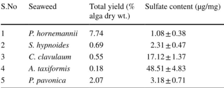
The seaweed polysaccharides were extracted from
Portie-ria hornemannii, Spyridia hypnoides, Asparagopsis taxi-formis, Centroceras clavulatum and Padina pavonica and
their sulfate content was analyzed (Table 1). Agar and car-rageenan are present in red seaweeds and glugan is found in green seaweeds whereas brown seaweeds contain alginate and fucoidan [22]. The red seaweed species such as
Gelid-ium, Gracilaria, Gelidiella, Ahnfeltia, Pterocladia, Acon-thopeltis, and Annfeltia are the major source for agar
collec-tively called agarophytes [22] and the agar content is varied depending on the species for example, 53% in Gracilaria sp. whereas in Gelidium 44% [23]. These seaweed polysaccha-rides are commercially utilized worldwide in the processed foods, cosmetics, pharmaceutical products and medicine as gelling and stabilizing agents [24].
Carrageenans, an another major cell wall polysaccharides extracted from certain species of red seaweeds
(Rhodophy-ceae) belong to the family members of Solieriaceae, Rhab-doniaceae, Hypneaceae, Phyllophoraceae, Gigartinaceae, Furcellariaceae and Rhodophyllidaceae [22]. This group of algae constitutes carrageenan up to 75% (dry wt.) in
Kap-paphycus with 15 to 40% of ester-sulfate content [25] and these species polymers are being used in various food indus-tries as texturing and gelling agents [26, 27]. It also exhibits
various biological and biomedical properties [4, 6]. The brown seaweeds (Phaeophyceae) contain alginate up to 40% in some species, example Ascophylum nodosum 22–30%,
Laminaria digitata 25–44%, Sargassum sp. 17–45% [28,
29]. Agar extracted in red seaweeds in the molecular weight range of 100–30,000 kDa [30]. Among the different fraction of polysaccharides (3.2, 10.5, 29.0, and 48.8 kDa) isolated from red seaweed Pyropia yezoensis, 3.2 kDa found as effec-tive against stress protection [31]. The yield of carrageenan varies depending on species and up to 70% (dry basis) was recorded from some species such as Betaphycus gelatinum and Kappaphycus alvarezii. Other species like Eucheuma
denticulatum and Chondrus crispus had 30% yields. Sulfate
content of carrageenans varied depends on the types (20% in κappa, 33% in ioda and 41% in λ) [32]. The molecular weight of commercially values carrageen in the range of 100 to 800 kDa was recorded [25] and low molecule weights possess more bioactivity properties [33]. Sulfated galactan of 16 kDa showing anticoagulant and antioxidant activities was purified from red Spyridia hypnoides [34]. Sulfated polysaccharide of 60 to 500 kDa isolated from Red seaweed
Asparagopsis taxiformis exhibit anticoagulant activity [35]. In this study, polysaccharide from five seaweeds (P.
hornemannii, S. hypnoides, A. taxiformis, C. clavulatum and P. pavonica) up to 14 kDa was isolated by considering the
significance of high bioactivity recorded in low molecular weight polysaccharides [36]. Whereas the alginate polymer of brown seaweeds in the range of 300–1000 kDa is com-mercial valuable [37, 38] but low molecule weight alginate polymers have potential bioactive properties [39]. Among the four red seaweeds, highest yield of 7.74% dry wt. of sulfated polysaccharide was extracted from P. hornemannii followed by S. hypnoides (0.69%), C. clavulaum (0.55%) and A. taxiformis (0.176%). Whereas the yield of sulfated polysaccharide extracted from the brown seaweed, Padina
pavonica was 2.07% dry wt. (Table 1). Comparing the poly-saccharides of terrestrial plant origin, seaweed polysaccha-rides constitute unique monomers like galactose, mannose, fucose, xylose etc. with varying degree of sulfation in their sugar residues [40] that influence the structure and biologi-cal properties of polymer isolated from each seaweed species [41]. This sulfate group of sugar moieties is found respon-sible for exhibiting anionic charge to the polymers [27, 39,
40]. Antibacterial and antiviral activity of anionic seaweed sulfated polysaccharides exhibit by binding the positively charged glycoprotein virus envelope and bacteria cell sur-face thereby integrating with virus or bacteria thus it pre-vent the pathogens entry into host cells [42–44]. Positive charge glycoprotein receptor of bacteria cell surface bind sulfate polysaccharides thereby inhibiting the bacteria [44]. So, high sulfate content in seaweed polysaccharides propor-tionally display more bioactivity [45, 46]. This study results showing high amount of sulfate in the polysaccharides of A. Table 1 Yield and sulfate content of seaweed polysaccharides
S.No Seaweed Total yield (%
alga dry wt.) Sulfate content (µg/mg) 1 P. hornemannii 7.74 1.08 ± 0.38
2 S. hypnoides 0.69 2.31 ± 0.47
3 C. clavulaum 0.55 17.12 ± 1.37 4 A. taxiformis 0.18 48.51 ± 4.83
taxiformis (48.51 ± 4.83 µ mg−1) followed by C. clavulaum, P. pavonica, S. hypnoides and P. hornemannii (Table 1) indicate the potential of these polysaccharides for biologi-cal activity.
FTIR spectroscopy characterization of polysaccharides
Structure of agar, carageenan and alginate
Agar and carrageenan extracted from Rhodophyceae (red seaweeds), the latter is highly sulfated ones. Thus, it consti-tutes repeating disaccharide units of 3-linked β-d-galactose
(G-units) and 4-linked α-galactose (D-units) or 3,6-anhydro-α-galactose (AnGal-units) whereas agar is less sulfated poly-mer composed of agarose, agaropectin, fibre, protein and ash. The agaropectin of agar is a charged sulfated non-gelling polymer constitutes d-glucuronic acid, and small amounts
of pyruvic acid. The gelling part of the agar, agarose is a neutral polymer with molecular weight of 120 kDa free of sulfate constitute repeating unit of agarobiose. It is a disac-charide made by β-1,3-linked- d-galactose and α-1,4-linked
3,6-anhydro-l-galactose [47] that is constituted by repeating
units of d-galactose and l-galactose [48, 49]. The specific
band at 890 cm−1 for agar attributed to anomeric C–H of
β-galactose residues generally absent in carrageenan [50]. At least fifteen different types of carrageenan categorized on the basis of structural characteristics, they are: sulfation patterns and presence or absence of AnGal on D-units are reported from various red seaweed species and most preva-lent are ioda, lamda and kappa [51] with molecular weight in the range of 100 to 800 kDa was recorded [25]. The cell wall of brown seaweeds mainly contain alginate polymer which constitute the blocks of β-l,4-linked d-mannuronic
acid and blocks of α-l,4-1inked l-guluronic acid or blocks
of d-mannuronic acid alternatively by l-guluronic acid [52].
Red seaweed polysaccharides
The FTIR spectroscopy remains a best tool to characterize the seaweed polysaccharides by identifying their functional groups [53]. The broad band between 3500 and 3200 cm−1
due to stretching vibrations of OH group is recorded in the FTIR spectra of all seaweed polysaccharides and standard spectra of agar, carrageenan and alginate (Figs. 2 and 3). The FTIR spectral characters of polysaccharides of red sea-weeds Portierea hornemanii, Spiridia hypnoides, A.
taxi-formis and C. clavulatum were compared with both agar and
carrageenan standards (Fig. 2). A peak at 930 cm−1 shows
the presence of C–O–C of glycosidic bond for
3,6-anhydro-l-galactose of agar and 3,6-anhydro-d-galactose of
carra-geenan is found in the spectra of both standards (agar and carrageenan), Spyridia hypnoides, A. taxiformis and C.
clavulatum [53, 54]. A band at 1650 cm−1 for carbonyl group
of a carboxylic acid; a peak at 1375 cm−1 and a broad band
in between 1210 and 1280 cm−1 indicate the presence of
sul-fate esters and a peak at 1140 cm−1 for C–O glycosidic bond
are recorded in all red seaweed polysaccharide samples as well as both agar and carrageenan standards [Fig. 2; 53, 54].
A peak at 840 cm−1 show the presence of galactose
sul-fated at C4 is found in the spectra of carrageeenan standard and polysaccharides of Spyridia hypnoides and A. taxiformis (Fig. 2b). Agar differs from carrageenan by having L-config-uration for the 4-linked galactose residue; nevertheless, they have some structural similarities with carrageenan. Peaks at 770 cm−1 and 740 cm−1 for pyranose ring are recorded in
all the spectra (Figs. 2ab, 4). Agar specific peak at 890 cm−1
due to anomeric CH in the β-galactose residues and a band at 930 cm−1 correspond to 3,6-anhydro-l-galactose recorded
in the spectra of agar standard, Spyridia hypnoides and
Centroceras clavulatum belong to the order Ceramiales
and Asparagopsis taxiformis of the Order Florideophyceae confirmed the presence of polysaccharides in agar forms [34, 50] and the spectral characteristics of P. hornemannii of order Gigartinales shows more spectral similarities with the carrageenan standard. The FTIR spectral characteristics found the presence of agar form of low molecular weight up to 14 kDa polysaccharides in S. hypnoides, C. clavulatum and A. taxiformis and carrageenan form in P. hornemannii, among the four red seaweeds.
Brown seaweed polysaccharides
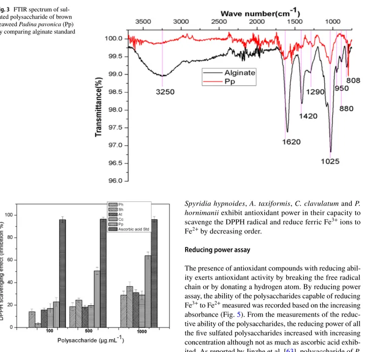
The FTIR spectral characteristics of polysaccharide of brown seaweed, Padina pavonica show spectral similarities with alginate standard (Fig. 3). Peaks at 3250 cm−1 assigned
for OH groups; 1620 cm−1 for S=O; 1420 cm−1 for C–O and
1025 C–O–C groups are recorded from the spectra of both alginate standard and sample [55]. Peaks at 1290 cm−1 for
M/G block (Mannose/Galactose) and at 808 cm−1 assigned
to M block are recorded from the spectra of both alginate standard and sample [7, 56]. The anomeric region of finger-print (950–750 cm−1) showed three characteristic absorption
bands in alginate standard and sample. A band at 950 cm−1
is assigned to the C–O stretching vibration of uronic acid residues, the one at 880 cm−1 is assigned to the C1–H
defor-mation vibration of β-mannuronic acid residues and the band at 808 cm−1 is characteristic of mannuronic acid residues
[57]. From this spectral data the extracted polysaccharide of Padina pavonica shows similar characteristics of alginate polymer.
Antioxidant activities
In vitro antioxidant examinations are used to measure the degree of protection against free radicals [58]. Seaweed pol-ysaccharides having antioxidant properties are the promising source for manufacture of food products and pharmaceuti-cals [58]. The molecular weight, type of monomer residues, degree of sulfation and sulfate position significantly influ-ence their antioxidant activity of seaweed polysaccharides [8]. In this study, among the in vitro antioxidant activities, DPPH hydroxyl radical scavenging and reducing power of the sulfated polysaccharides were measured.
DPPH assay
The free radical scavenging activities of different sulfated polysaccharides assessed by DPPH assay based on the
hydrogen ion donating ability of the polysaccharide are presented (Fig. 4). DPPH is a stable free radical appears in purple color in methanol/ethanol turns colorless by reduction in the presence of hydrogen donating antioxi-dants [59]. The DPPH, a stable free radical shows max-imum absorbance at 517 nm in methanol. When DPPH encounters a proton donating substance like antioxidant, radical would be scavenged and the absorbance reduced. For every sulfated polysaccharide tested, there was a con-centration dependent increase in scavenging activity where 1 mg mL−1 polysaccharide of P. pavonica exhibited an
increase in DPPH free radical scavenging activity up to 63%. Khaled et al. [60] explained that the ethyl acetate fraction of the algae Padina pavonica showed the highest antioxidant activity (42.5%); those activities may be due to phenolic compounds present in significant amounts in this fraction (8.98 GAE/g). In another study, Nouf et al. Fig. 2 FTIR spectra of
sul-phated polysaccharide of red seaweeds (Portieria
horneman-nii-Ph, Spyridia hypnoides-Sh, Asparagopsis taxiformis-At, Centroceras clavulatum-Cc and Padina pavonica-Pp) by
com-paring agar and carrageenan standards
[61] described the extracts of Padina pavonica showed a concentration-dependent manner with maximum scaveng-ing activity of 77.6%, IC50 = 5.59 µg/mL. Among the red
seaweeds, high DPPH activity was recorded by polysac-charide (1 mg mL−1) of Spyridia hypnoides followed by A. taxiformis (31%) and C. clavulatum (28%) compared
to ascorbic acid as standard (96%). Results from previous studies [62] are in accordance with the present study that polysaccharides of P. pavonica followed by red seaweeds
Spyridia hypnoides, A. taxiformis, C. clavulatum and P. hornimanii exhibit antioxidant power in their capacity to
scavenge the DPPH radical and reduce ferric Fe3+ ions to
Fe2+ by decreasing order.
Reducing power assay
The presence of antioxidant compounds with reducing abil-ity exerts antioxidant activabil-ity by breaking the free radical chain or by donating a hydrogen atom. By reducing power assay, the ability of the polysaccharides capable of reducing Fe3+ to Fe2+ measured was recorded based on the increasing
absorbance (Fig. 5). From the measurements of the reduc-tive ability of the polysaccharides, the reducing power of all the five sulfated polysaccharides increased with increasing concentration although not as much as ascorbic acid exhib-ited. As reported by Jinzhe et al. [63], polysaccharide of P.
pavonica had the greatest reducing power closely followed
by red seaweeds A. taxiformis then P. hornimanii, Spyridia
hypnoides and C. clavulatum implicated on the basis of
sul-fate content and monosugar constituents [64]. The Ferric reducing antioxidant power (FRAP) of P.pavonica reveals a higher antioxidant activity with IC50 = 0.4 mg mL−1 [65].
El-Shazoly and Fawzy [66] described that the ethyl alco-hol extract of Padina pavonica showed significant activity (339.92 mg g−1 dry wt.).
In vitro cytotoxic assay using HeLa cell‑line
The five sulfated polysaccharides were assayed for the cyto-toxic effect in HeLa cell line at different concentrations from 0.0019 mg mL−1 to 1 mg mL−1. The effect has measured Fig. 3 FTIR spectrum of
sul-fated polysaccharide of brown seaweed Padina pavonica (Pp) by comparing alginate standard
Fig. 4 DPPH free radical scavenging activity of sulfated polysaccha-rides (Portieria hornemannii-Ph, Spyridia hypnoides-Sh,
Asparagop-sis taxiformis-At, Centroceras clavulatum-Cc and Padina
based on the cell survivability percent (Fig. 6) and IC50
values. Figure 6 evidently shows the concentration of poly-saccharide increases the cell survivability decreases by the red seaweeds P. hornemannii and S. hypnoides. Ktari and Guyot [67] evaluated the cytotoxic activity of dichlorometh-ane extract of Padina pavonica against KB cells and the results showed significant activity (IC50 10 μg mL−1). The
antitumor activity of Padina pavonica against breast cancer cells (MCF-7) and several strains of prostate cancer (DU-145, LNCaP and PC3) by in vitro cytotoxicity assay with methanolic extract found that P. pavonica has more toxicity
against cell strains tested according to Taskin et al. [68]. The cell survivability was decreased by low concentrations of P. pavonica polysaccharide but at high concentrations it promotes the cell survivability (Fig. 6) since the IC50 value was, 1.059.
Indeed polysaccharides of P. hornemannii and S.
hyp-noides showed cytotoxic effects at higher concentrations
but at low concentrations promote cell growth hence, IC50 values recorded were 0.428 and 0.755, respectively (Fig. 7). Instead IC50 values for polysaccharides of C. clavulatum and A. taxiformis were 1.27 and 3.27, respectively indicate these polysaccharides have nutracetical values. This inves-tigation results suggest that crude polysaccharides at high dose of red seaweeds P. hornemannii and S. hypnoides and at low dose of brown seaweed P. pavonica exhibit cytotoxic activity in HeLa cancer cell line whereas polysaccharides of
C. clavulatum and A. taxiformis have nutraceutical values.
Conclusion
The sulfate recorded in the polysaccharide of A. taxiformis was higher followed by C. clavulaum, P. pavonica, S.
hyp-noides and P. hornemannii. The FTIR spectroscopy
char-acteristics support that this low molecular weight sulfated polysaccharide fractions up to 14 kDa extracted from S.
hyp-noides, C. clavulatum and A. taxiformi were similar to agar
polymer whereas P. hornemannii similar to carrageenan and polysaccharide of P. pavonica shows similarity to alginate possessing varying biological activities. Higher DPPH activ-ity and reducing power were recorded in the polysaccharide Fig. 5 Reducing power assay of sulfated polysaccharides (Portieria
hornemannii-Ph, Spyridia hypnoides-Sh, Asparagopsis
taxiformis-At, Centroceras clavulatum-Cc and Padina pavonica-Pp). Error bars indicate replicates standard deviation of each analysis
Fig. 6 Cell survivability of sulfated polysaccharides (Portieria
hornemannii-Ph, Spyridia hypnoides-Sh, Asparagopsis taxiformis-At, Centroceras clavulatum-Cc and Padina pavonica-Pp) tested by MTT
Assay using HeLa cell line
Fig. 7 IC50 value of cell survivability of different concentrations of polysaccharides (Portieria hornemannii-Ph, Spyridia hypnoides-Sh,
Asparagopsis taxiformis-At, Centroceras clavulatum-Cc and Padina pavonica-Pp) tested by MTT Assay using HeLa cell line
of brown P. pavonica than red seaweeds. Thus, the study clearly suggest that low molecular weight carrageenan like polysaccharide extracted from P. hornemannii, agar like from S. hypnoides and alginate polymer from P. pavonica shows in vitro antioxidant activities which are capable of inhibiting HeLa cell line could be used for biomedical appli-cations whereas polysaccharides of C. clavulatum and A.
taxiformis for nutraceutical products.
Acknowledgements The corresponding author Dr.R.Raja, sin-cerely acknowledges the University of Algarve, Faro for funding the researcher under the rule DL 57/2016, Portugal.
References
1. J. Guangling, Y. Guangli, Z. Junzeng, H.E. Stephen, Mar. Drugs. 9(2), 196–223 (2011)
2. P. Seema, Biotechnology 2(3), 171–185 (2012)
3. P.S. Kwon, S.J. Kwon, J. Weihua, Z. Fuming, F. Keith, Cell Dis-cov. (Online) (2020)
4. N.-H. Ngo, K. Se-Kwon, Int. J. Biol. Macromol. 62, 70–75 (2013) 5. R. Raja, S. Hemaiswarya, K. Arunkumar, I.-S. Carvalho, Braz. J.
Bot. 39(1), 9–17 (2016)
6. K.K. Sanjeewa, L. Asanka, K. Nalae, A. Ginnae, J. Youngheun, K. Young-Tae, L. You-Jin, Food Hydrocoll. 81, 200–208 (2018) 7. L. Pereira, S.F. Gheda, J. Paulo, A. Ribeiro-Claro, Int. J.
Carbo-hydr. Chem. 537202, 7 (2013)
8. Q. Zhong, B. Wei, S. Wang, S. Ke, J. Chen, H. Zhang, H. Wang, Mar. Drugs 17(12), 674 (2019)
9. L.N. Panlasigui, O.Q. Baello, J.M. Dimatangal, B.D. Dumelod, Asia Pac. J. Clin. Nutr. 12, 209–214 (2003)
10. V.D. Prajapati, P.M. Maheriya, G.K. Jani et al., Carbohydr. Polym. 105, 97–112 (2014)
11. R. Pangestuti, S.-K. Kim, Adv. Food Nutr. Res. 72, 113–124 (2014)
12. R.W. Fuller, J.H. Cardellina, Y. Kato, L.S. Brinen, J. Clardy, K.M. Snader, M.R. Boyd, J. Med. Chem. 35, 3007–3011 (1992) 13. P.V. Neethu, K. Suthindhiran, M.A. Jayasri, Pharmacogn. Res.
9(3), 238–246 (2017)
14. M. Fabio, D.C. Gianfranco, C. Gugliandolo, S. Antonio, F. Cate-rina, G. Giuseppa, M. MaCate-rina, R. Annamaria, B. Davide, F. Franc-esco, S. Andrea, Front. Physiol. 7, 459 (2016)
15. N.M. Al-Enazi, A.S. Awaad, E.Z. Mohamed, S.I. Alqasoumi, Saudi Pharm. J. 26(1), 44–52 (2018)
16. G. Bernardini, M. Mariagiulia, P. Giuseppe, B. Manuele, S. Annalisa, Mar. Drugs 16, 504 (2018)
17. S. Distantina, A. Wiratni, M. Fahrurrozi, L. Rochmadi, World Acad. Sci. Eng. Technol. 78, 738–742 (2011)
18. A.G. Lloyd, K.S. Dodgson, R.G. Price, F.A.I. Rose, Biochem. Biophys. Acta 1, 108–115 (1961)
19. B.W. Souza, M.A. Cerqueira, A.I. Bourbon, A.C. Pinheiro, J.T. Martins, J.A. Teixeira, A.A. Vicente, Food Hydrocoll. 27(2), 287–292 (2012)
20. G.C. Yen, H.Y. Chen, J. Agric. Food Chem. 43, 27–32 (1995) 21. T. Mosmann, J. Immunol. Methods 65, 55–63 (1983)
22. S. Istini, M. Ohno, H. Kusunose, Bull. Mar. Sci. Fish. Kochi Univ. 14, 49–55 (1994)
23. D.J. Mc Hugh, Hydrobiology 221, 19–29 (1991)
24. E. Marinho-Soriano, E. Bourret, Biores. Technol. 96(3), 379–382 (2005)
25. L. Pereira, A.T. Critchley, A.M. Amado, P.J.A. Ribeiro-Claro, J. Appl. Phycol. 21, 599–605 (2009)
26. M.L. Weiner, D. Nuber, W.R. Blakemore, J.F. Harriman, S.M. Cohen, Food Chem. Toxicol. 45(1), 98–106 (2007)
27. B. Tanna, A. Mishra, Comp. Rev Food Sci. Food Saf. 18(3), 817–831 (2019)
28. L.S. Costa, G.P. Fidelis, S.L. Cordeiro, R.M.D.A. Oliveira, R. Sabry, B.G. Câmara, L.B. Nobre, M.P. Costa, J. Almeida-Lima, E.C. Farias, E.L. Leite, H.O. Rocha, Biomed. Pharmacol. 64(1), 21–28 (2010)
29. S. Yu-Fong, Y. Hui-Chun, L. Yen, Int. J. App. Sci. Eng. 7(1), 25–41 (2009)
30. A. Al-Alawi, P. Chitra, A. Al-Mamun, I. Al-Marhubi, M.S. Rah-man, Int. J. Food Eng. 14, 20170353 (2018)
31. P. Zou, X. Lu, C. Jing, Y. Yuan, Y. Lu, C. Zhang, L. Meng, H. Zhao, Y. Li, Front Plant Sci. 9, 427 (2018)
32. M. Ghanbarzadeh, A. Golmoradizadeh, A. Homaei, Phytochem. Rev. 17, 535–571 (2018)
33. A.A. Kalitnik, A.O.B. Barabanova, V.P. Nagorskaya, A.V. Reunov, V.P. Glazunov, T.F. Solov’eva, I.M. Yermak, J Appl. Phycol. 25, 65–72 (2013)
34. S. Sudharsan, S. Giji, P. Seedevi, S. Vairamani, A. Shanmugam, Int. J. Biol. Macromol. 109, 589–597 (2018)
35. A. Manilal, S. Sujith, J. Selvin, M.V. Nataraja Panikkar, S. George, Thalassas Int. J. Mar. Sci. 28(2), 9–15 (2012)
36. D. Guo, Y. Kai, S. Xin-Yuan, O. Jian-Ming, Biological effi-cacy of medicinal plant extracts in preventing oxidative dam-age. Oxid. Med. Cell. Longev. 15, 1 (2018). https ://doi. org/10.1155/2018/79043 49
37. E. Fourest, B. Volesky, Appl. Biochem. Biotechnol. 67, 215–226 (1997)
38. C.K. Larsen, O. Gåserød, O. Smidsrød, Carbohydr. Polym. 51, 125–134 (2003)
39. M. Sen, E.N. Erboz, Food Res. Int. 43, 1361–1364 (2010) 40. E. Fourest, B. Volesky, Environ. Sci. Technol. 30(1), 277–282
(1996)
41. M. Xing, Q. Cao, Y. Wang et al., Mar. Drugs. 18(3), 144 (2020) 42. L.N. Callahan, M. Phelan, M. Mallinson, M.A. Norcross, J. Virol.
65, 1543–1550 (1991)
43. Z. Mellouk, I. Benammar, D. Krouf, M. Goudjil, M. Okbi, W. Malaisse, Exp. Ther. Med. 13(6), 3281–3290 (2017)
44. H. Jinzhe, Y. Xu, H. Chen, P. Sun, Int. J. Mol. Sci. 17(12), E1988 (2016)
45. J. Trinchero, M. Nora, M.A. Ponce, O.L. Córdoba, M. Lujan Flo-res, Phytother. Res. 23(5), 707–712 (2009)
46. L.B. Talarico, E.B. Damonte, Virology 363, 473–485 (2007) 47. S.T. Moe, K.I. Draget, G. Skjak-Brack, O. Smidsrod, Alginates,
ed. (New York, USA, 1995), pp. 245–286
48. J. Craigie, (Cambridge University Press, Cambridge, UK, 1990), pp. 221–257
49. V. Jagatheesan, B.K. Pramanik, J. Chen et al., Biores. Technol. 204, 202–212 (2016)
50. D. Christiaen, M. Bodard, Bot. Mar. 26, 425–427 (1983) 51. M. Lahaye, Cah. Biol. Mar. 42, 137–157 (2001)
52. J. Wilma, A. Limewood, R. North Hamptonshire NN9 6NG (UK, 1990), pp. 53–60
53. I.P.S. Fernando, A. Sanjeewa, K.W. Samarakoon, W.W. Lee, H.-S. Kim, E.-A. Kim, D.K.S. Gunasekara, D.T.U. Abeytunga, C. Nanayakkara, E.D. Silva, H.-S. Lee, Y.-J. Jeon, Algae 32(1), 75–86 (2017)
54. T. Chopin, B.F. Kerin, R. Mazerolle, Phycol. Res. 47, 167–188 (1999)
55. D.M. Abid, L. Sirine, H.A. Hiba, C. Dora, E. Nejeh, M. Hatem, B. Abderrahman, Trends. Appl. Sci. Res. 4(2), 62–67 (2019) 56. R.M. Amir, F.M. Anjum, M.I. Khan, M.R. Khan, I. Pasha, M.
57. N.P. Chandía, B. Matsuhiro, E. Mejías, A. Moenne, J. Appl. Phy-col. 16, 127–133 (2004)
58. S. Castro, J. de Paula Lima, L. Costa, L. Eduardo Castanheira, P.M.P. Francisco, T. dos Santos, P.H. de Menezes, F.L. Mistrello, D.R. Moschini, S.G. dos Ramalho Cardoso, C.N. Sérgio Medei-ros, F.A. Lúcia Ponte, Polímeros 28(2), 178–186 (2018) 59. C.M.P.G. Dore, C. Faustino Alves, M.G.E.P. Will et al.,
Carbo-hydr. Polym. 91, 467–475 (2013)
60. N. Khaled, M. Hiba, C. Asma, Adv. Environ. Biol. 6, 42–48 (2012)
61. M.A.-E. Nouf, S.A. Amani, E.Z. Mohamed, I.A. Saleh, Saudi Pharm. J. 26(1), 44–52 (2018)
62. T. Ghosh, K. Chattopadhyay, M. Marschall, P. Karmakar, P. Man-dal, B. Ray, Glycobiology 19, 2–15 (2009)
63. A. Rodrigo, N. Santos, R.J.A. Gurgel, H. Márjory Lima, Q. Ana Luíza Gomes, P. Regina Célia Monteiro, M. Vânia Maria Maciel, B.N. Maria Barros, Braz. Arch. Biol. Tech. 55(2), 171–181 (2012)
64. P. Seedevi, M. Moovendhan, S. Viramani, A. Shanmugam, Car-bohydr. Polym. 155, 516–524 (2017)
65. M.D. Alshaikheid, A. Abdelhamid, A. Bouraoui, J. Adv. Res. Bio-tech. 4(1), 1–6 (2019)
66. R.M. El-Shazoly, M.A. Fawzy, Egypt Eur. J. Biol. Res. 8(4), 232–242 (2018)
67. L. Ktari, M. Guyot, J. App. Phycol. 11, 511–513 (1999) 68. E. Taskin, Z. Caki, M. Ozturk, E. Taskin, African. J. Biotechnol.
27, 4272–4277 (2010)
Publisher’s Note Springer Nature remains neutral with regard to jurisdictional claims in published maps and institutional affiliations.