Filippo Maria Rotatori
B.Sc. Natural Sciences
Orientador: Octávio Mateus, Prof. Associado FCT-UNL
Co-orientador: Miguel Moreno-Azanza, Pós-Doc, FCT-UNL
Ornithopod dinosaurs from the Late Jurassic of
Portugal
Dissertação para obtenção do Grau de Mestre em Paleontologia
September 2018
September 2018
Filippo Maria Rotatori
B.Sc. Natural Sciences
Ornithopod dinosaurs from the Late Jurassic of Portugal
Dissertação para obtenção do Grau de Mestre em Paleontologia
Orientador: Octávio Mateus, Prof. Associado FCT-UNL
ii
Acknowledgements
This is the end of a journey, as a student, as a scientist and most importantly as a person. As any other achievement, this thesis is not just the fruit of myself, but there are so many to thank. Since no one is a student if there is no one who teaches, I need to thank my main advisor, Prof. Octávio Mateus and my co-advisor Dr. Miguel Moreno-Azanza. Thanks for their patience and enthusiasm in teaching and leading this thesis project. Also thanks to the unknown arguent, whose work will surely improve this small piece of research. Thanks to Dr. Hübner for sharing with me his work and for being so enthusiastic about basal iguanodontians (which I was not at the time) and to Dr. Madzia for giving me advises about neornithischians and phylogenetics. Thanks to Prof. Will Harcourt-Smith for helping me out with Principal Component Analysis, morphometrics and multivariate statistics. Thanks also to Dr. Federico Fanti, my former advisor, for always giving me suggestions and sharing cool ideas. For the same reasons as wel, thanksl to Dr. Emanuel Tschopp, for sharing his experience in the field and as a research.
Thanks to Dr. Bruno Camilo Silva of Sociedade de Historia Natural in Torres Vedras for giving me access to the holotye of Eousdryosaurus nanohallucis, and to Joana Ferreira for her assistance.
The data taken there, were greatly useful for the realization of this thesis.
Thanks also to my family, who gave me the possibility to live and study abroad, and kept sustaining me during these years: Mom, Dad, my two sisters: Anna and Rebecca. Also thanks to uncle Andrea, Tommaso, Renato and all the cousins. Thanks to my Grandma and all the aunties who actually fed me during this year of research and writing.
Here in Lourinhã I need to thank all the members of Lourinhã Paleoteam: Alex, Darìo, Andrè, for they close supports, laughs and reciprocal help. Thanks also to Francisco, Catia, an indefinite number of Joãos, Edu, Xanna, Alex, Carla, Marco, Hugo. A special thank goes to Carla-Alexandra for her patience in helping with (and teaching me about) preparation. Also thanks to her for the affection she showed me in all this time. Another one goes to Micael, for sharing his work with me, since most of the material here described was introduced to me by him! Thanks also to Vincent for his kindess and joy shared with us this year. Thanks to Miggi for the fun, the honest talks about life, paleontology and many other things.
iii
iv
Abstract
Ornithopods are the most successful group of herbivorous dinosaurs, ranging from the Middle Jurassic to the Cretaceous, becoming extinct at the Cretaceous Paleogene boundary. However, most of the attention has been given to derived forms (Hadrosaurids). Herein, ornithopod material coming from Upper Jurassic of Lourinhã Fm. and housed at Museu da Lourinhã (ML) is described and discussed. Specimens studied include cranial and dental material, including cranial remains of a dryosaurid, axial skeleton elements, pectoral girdle elements and limb bones. Comparison with reference literature and phylogenetic analyses has allowed the attribution of the material either to Dryosauridae or and to Ankylopollexia. Since the lack of autapomorphic characters, it was not possible to assign the material to the two valid ornithopod Portuguese taxa, Draconyx loureiroi and
Eousdryosaurus nanohallucis, although phylogenetic analyses hint a close relationship between the
Lourinha dryosaurid material and E. nanohallucis. Principal Component Analysis (PCA) plotting limb
bones proportions indicates a not fully mature ontogenetic stage for the Portuguese specimens. Comparing the Portuguese ornithopod fauna with the one in Morrison Formation and Kimmeridge Clay Formation, it is remarked the key-role of Portugal to understand biogeographic patterns in the distribution of iguanodontians.
v
Resumo
Os dinossauros ornitópodes são o grupo mais bem sucedido de dinossauros herbívoros, tendo aparecido no Jurássico Médio extinguindo-se no limite Cretácico/Paleogénico. É aqui descrito e discutido material das coleções do Museu da Lourinhã (ML) de ornitópode do Jurássico Superior da Formação da Lourinhã. Os espécimes estudados incluem material craniano e dentário, incluindo restos cranianos de um dinossauro driossaurídeo, elementos do esqueleto axial, elementos da cintura escapular e ossos dos membros. A comparação e análise filogenética permitiu a atribuição do material tanto a Dryosauridae como a Ankylopollexia. Não é possível atribuir o material aos dois táxons portugueses válidos de ornitópodes, Draconyx loureiroi e Eousdryosaurus nanohallucis,
embora análises filogenéticas indiquem uma estreita relação entre o material de driossaurídeo da Lourinhã e E. nanohallucis. Análise de Componentes Principais (PCA) representando as proporções
dos ossos dos membros indica um estágio ontogenético não maduro para os espécimes portugueses. É debatida a importância dos espécimes portugueses na distribuição de Iguanodontia na Formação Morrison e na Formação Kimmeridge Clay.
vi
vii
Table of contents
Acknowledgements ... ii
Abstract ... iv
Resumo ... v
Graphical Abstract ... vi
1.Introduction ... 5
1.1 Systematics of ornithopod dinosaurs ... 6
1.2 Ornithopod anatomy: state of the art and main synapomorphies ... 12
Basal ornithopods (non-iguanodontians) ... 12
Iguanodontians ... 17
Hadrosaurids ... 24
1.3 Ornithopods in Portugal: history of discoveries ... 30
1.4 Geological framework: Late Jurassic of Portugal ... 31
1.5 Objectives ... 36
2. Material and methods ... 36
3.Results ... 38
3.1 Systematic Palaeontology ... 38
Dryosauridae ... 38
Cranial skeleton ... 38
Axial skeleton ... 44
Appendicular skeleton ... 46
Ankylopollexia ... 50
Cranial skeleton ... 50
Appendicular skeleton ... 55
3.2 Phylogenetic analysis ... 56
3.3 Principal Component Analysis ... 59
4. Discussion ... 63
Paleobiogeographical and paleoecological Implications for the Lourinhã ornithopod fauna ... 68
5. Conclusions ... 70
Bibliography ... 72
Annex I – cranial and post-cranial material measurements ... 80
Annex II – Dental material measurements... 81
Annex III – Data matrix... 82
Annex IV – Linear Measurements used for PCA (Femur) ... 91
viii
Annex VI – Principal Components (Femur) ... 94
Annex VII – Principal Components (Tibia) ... 95
Annex VIII – Loading scores and Linear Bivariate model of GM ... 97
Annex IX – Morphometrics variables from Hübner 2018 ... 99
Table of figures
Fig. 1.1.1: Sereno Ornithischia cladogram………..………. 6Fig. 1.1.2: Butler Ornithischia cladogram……….8
Fig. 1.1.3: Boyd Ornithischia cladogram……….……… .9
Fig. 1.2.1: Hypsilophodon skull………13
Fig. 1.2.2: Hypsilophodonaxial skeleton………..15
Fig. 1.2.3: Hypsilophodon appendicular skeleton………..17
Fig. 1.2.4: Iguanodontians skull………18
Fig. 1.2.5: Iguanodontian teeth………20
Fig.1.2..6: Iguanodontian axial skeleton……….22
Fig. 1.2.7: Iguanodontian pelvic skeleton……….………..24
Fig.1.2.8: Hadrosaurid cranial skeleton……….……….26
Fig. 1.2.9: Hadrosaurid dental battery………27
Fig. 1.2.10: Hadrosaurid axial skeleton………..28
Fig. 1.2.11: Hadrosaurid pelvic girdle……….29
Fig. 1.4.1: Geological framework………..………..32
Fig. 1.4.2: Lourinhã Fm. outcrops………35 Fig. 3.1.1: ML 1851 Parietal………..………39
Fig. 3.1.2: ML 768 Dentary………..……… 41
Fig. 3.1.3: ML 2304, ML 2305 Dentary teeth………..………..………..…43
ix
Fig. 3.1.5: ML 2055, ML 563 Femora……….47
Fig. 3.1.6: ML 505, ML2055 Tibiae……..……….……….49
Fig. 3.1.7: ML 818 Dentary………..……….51
Fig. 3.1.8: ML Dentary teeth wearing process………..………..53
Fig. 3.1.9: ML Maxillary teeth wearing process………..………54
Fig. 3.1.10: ML 2206 Coracoid……….………55
Fig. 3.2.1: Strict consensus trees of separate analyses………..……….57
Fig. 3.2.2: Strict consensus trees of composite analyses………...………..58
Fig. 3.3.1: Principal Component Analysis (Femora) scatter plot……….……….61
Fig. 3.3.2: Principal Component Analysis (Tibiae) scatter plot……….62
Fig.4.1: Comparison table……….65
Table index
Table 1.1.1: Systematic definitions………..………105
1.Introduction
Ornithopod dinosaurs were among the most successful taxa of dinosaurian groups during the Mesozoic. They span from the Middle Jurassic (Callovosaurus leedsi, Callovian) to
the latest Cretaceous, becoming extinct at the end- Maastrichtian mass extinction. To Ornithopoda (sensu Butler et al., 2008) belong some iconic species and some of the first
dinosaur taxa ever described. Iguanodon is one of the three species which Owen (1842) used
to define the group Dinosauria, and the second dinosaur species to appear in a scientific publication. It was made popular to the general public firstly thanks to the work of Waterhouse Hawkins, who made the first physical reconstructions of this animal (Romano et al., 2016). A
few years later, another iconic taxon was described in North America: Hadrosaurus. During
the infamous ‘Bone Wars’ in the American West, other famous species were discovered, such
as Dryosaurus and Camptosaurus (Forster, 2007). In 1878, a huge concentration of Iguanodon
specimens was discovered in the Bernissart coal mine of Belgium (Godefroit et al., 2012). At
the beginning of the 1900s, a large variety of diverse forms were unearthed by Barnun Brown in the Late Cretaceous Dinosaur Park Formation, Canada: Kritosaurus, Prosaurolophus,
Corythosaurus (Weishampel, 2014). Later in the 20th century, many other taxa were unearthed
around the globe and the pace of ornithopod discoveries has since not slowed down in the present days (Weishampel, 2014).
Although ornithopods have a long history of research, some aspects of their systematic relationships remain somewhat poorly understood. This is particularly true for the most basal forms, which have undergone some dramatic revisions in their taxonomy (Butler et al. 2008;
Boyd 2015). The more derived taxa (i.e. iguanodontians) are generally better understood whith respect to Cretaceous species; nevertheless, the Jurassic forms still present some enigmatic issues regarding their systematics and paleobiology.
6
In this section, some general aspects of ornithopod anatomy and systematics are presented. It is then proceeded to revise the history of discoveries in Portugal and the geological framework of the Late Jurassic ornithopod-bearing formations.
1.1 Systematics of ornithopod dinosaurs
Systematic studies with cladistic approaches which comprised Ornithopoda, began in the 1980s (Norman, 1984; Sereno, 1984, 1986) and for long time the relationships within the clade remained stable. The traditional concept of Ornithopoda, included at the base Heterodontosauria (=Heterodontosauridae) and Hypsilophodontia, and basal iguanodontians in a more derived position (Dryosauridae and Tenontosaurus). Ankylopollexians
(Camptosaurus + Styracosterna) were regarded as the most derived taxa within Ornithopoda
(Sereno, 1986).
7
The analysis by Sereno (1986) dominated Ornithopoda (and, more in general, Ornithischia) taxonomy and systematics without many challengers (fig.1.1.1), although the dataset was never published (Butler, et al. 2008). In recent years, the discovery of many new
taxa which apparently showed basal ornithopods affinities (i.e.: Jeholosaurus shangyuanensis,
Changchunsaurus parvus and Haya griva) has led to many systematics revisions, with some
contradictory results (Butler et al., 2011; Makovicky et al., 2011).
Butler et al. (2008), in their comprehensive analysis of Ornithischia, recovered
Heterodontosauridae, once thought to be the basal-most group of ornithopods, in a basal position outside Cerapoda (fig.1.1.2). The same occurs for the taxa Othnielia (=
Othnieliosaurus rex), Agilisaurus and Hexinlusaurus, although weak support has been found
for these positions. They also recovered a paraphyletic ‘Hypsilophodontidae’, ephasizing that the ‘hypsilophodontid plexus’ represents just a grade of many basal forms, as many other authors previously suggested (Scheetz, 1998, 1999; Winkler et al., 1998).
To date, the only analysis which recovers a monophyletic hypsilophodontid clade is Norman (2015), grouping together Hypsilophodon foxii, Tenontosaurus, and Rhabdodontidae.
Norman (2015), completely changes phylogenetic definition and diagnosis of Hypsilophodontidae in comparison to previous works; further studies may validate a monophyletic hypsilophodontid clade. In any event, the study of Butler et al., (2008) pointed
out the need of deep revision within Ornithopoda as originally conceived. The phylogenetic analyses of Butler et al. (2011) and Makovicky et al. (2011) which recovered a clade of basal
asian ornithopods composed of Jeholosaurus, Haya and Changchunsaurus, did little to change
the topology obtained from Butler et al. (2008). McDonald (2012) focused his attention on the
relationships of basal Iguanodontians, supporting the splitting of traditional ‘Camptosaurus’
and ‘Iguanodon’. McDonald (2012) points out that Iguanodontia presents a patchy fossil
records, which can lead to the instability of many basal taxa. Dryomorpha, with the inclusion of the genus Callovosaurus, is acknowledged as the most ancient clade of basal
8
Fig. 1.1.2: Cladogram showing the ornithischian interrelationships recovered from Butler et al., 2008.
Regarding Ornithopoda, the major changes is the position of Heterodontosauria. From Butler et al.,
9
The most heterodox hypothesis regarding ornithopod inter-relationships was proposed by Boyd (2015), in his extensive revision of ornithischian systematics and paleobiogeography of (fig.1.1.3). It was recovered the most inclusive Ornithopoda to date, consisting of just
Hypsilophodon + Iguanodontia. Many of the basal taxa that once constituted the
‘hypsilophodontid plexus’ (i.e. Thescelosaurus, Talenkauen, Zephyrosaurus, Orodromeus…)
and the taxa included in the ‘basal asian ornithopod clade’ from Butler et al. (2011) and
Makovicky et al. (2011), are recovered as part of a monophyletic clade called Parksosauridae,
sister taxon to Cerapoda. Strickson et al. (2016) in their study in which calculated evolutionary
10
rates among ornithopods and obtained a super-tree in which recovered a topology more consistent with Butler et al. (2008).
Madzia et al. (2017) included the new basal species Burianosaurus augustai in the data
matrix of McDonald (2012) and Boyd (2015), and furtherly complicated the phylogenetic scenario of basal ornithopods relationships, recovering another different topology. The results of Madzia et al., (2017) strongly suggest that density of sampling has a great influence on
Ornithopoda topology. This is particularly evident in the case of two enigmatic taxa:
Kulindadromeus zabaikalicus and Gideonmantellia amonsanjuanae. Kulindadromeus is the
first ornithischian which preserves trace of feather-like structures, and it is originally recovered by Godefroit et al. (2014) within Neornithischia sister taxon to Cerapoda. This topology was
recovered inserting Kulindadromeus in the dataset of Butler et al. (2011). Gideonmantellia,
was included in the same dataset by Ruiz-Omeñaca et al. (2012), resulting in a politomy with
Talenkauen, the node composed by Gasparinisaura, Parksosaurus and Iguanodontia. The
dataset of Madzia et al. (2017), which includes the highest number of basal neornithischian
taxa so far, recovered Kulindadromeus nested at the base of Neornithischia basal to the node
composed by (Jeholosauridae (Othnielosaurus (Clypeodonta, Thescelosauridae).
Gideonmantellia in the same dataset is recovered as the basal-most ornithopod, while in the
dataset of Han et al. (2017) which does not comprise a wide sampling of taxa in Neornithischia (due to the fact it was designed to test the affinities of Yilong within Ceratopsia) is recovered
as basal-most iguanodontian. These results suggest that the base of Ornithopoda, and more in general Neornithischia, will remain unstable in the near future. Table 1.1.1 summarizes the phylogenetic definitions used in this work, and their relative diagnoses.
Taxon Definition Diagnosis Reference
Ornithopoda All genasaurians more closely related to Parasaurolophus walkeri, than to Triceratops horridus.
Presence of fossa-like depression on the premaxilla – maxilla boundary; possession of narrow and elongate (more than twice as long as wide) frontals that contact the nasals anterior to the orbits; possession of a foramen positioned on the dorsal part of the surangular-dentary joint.
Butler et al., 2008.
11 Iguanodontia All ornithopods more closely
related to Parasaurolophus walkeri, than to Hypsilophodon foxii or Thescelosaurus neglectus.
Premaxilla with a transversely expanded and edentulous margin; a predentary with a smoothly convex rostral margin when viewed in dorsal aspect; a deep dentary ramus; with parallel dorsal and ventral borders; no evidence of sternal rib ossifications; the loss of one phalanx in the digit III of the manus; a prepubic process that is compressed and blade-shaped
Norman, 2004
Dryosauridae Dryosaurus altus and all the taxa more closely related to it than to Parasaurolophus walkeri.
Lacrimal inserts into notch in the maxilla; very wide brevis shelf of the ilium; large, deep pit on the femoral shaft, at the base of 4th trochanter; digit I of the pes lost or vestigial.
Sereno, 1998.
Ankylopollexia Camptosaurus dispar, Parasaurolophus walkeri, their common ancestor and all its descendants.
Specially modified manus and fore limb to accommodate spine-like thumb; more elongate preorbital and generally lower skull-profile; the frontal participates less in the orbital margin than in dryomorphs.
Norman, 2004.
Styracosterna All ankylopollexians more closely related to Parasaurolophus than to Camptosaurus.
Hatchet-shape sternal bones; flattened manus unguals.
Norman, 2004
Hadrosauridae Parasaurolophus, Telmatosaurus their common ancestor and all its descendants.
Three or more replacements of teeth for tooth family; distal extension of dentary tooth row terminating caudal to coronoid process; caudal-most termination of the dentary well behind the coronoid process; absence of surrangular foramen; contact of the ectoperygoid and jugal with supraorbital; elevation of cervical zygapophyseal peduncles well above the neural arch; long and dorsally arched post-zygapophyses; coracoid with a long cranioventral process that extends well below the glenoid; dorsoventrally narrow scapula with acromion process which projects horizontally; reduced area for coracoid articulation; deep intercondylar extensor groove on the femur.
12
1.2 Ornithopod anatomy: state of the art and main
synapomorphies
Basal ornithopods (non-iguanodontians)
In light of recent phylogenetic analyses, there is little agreement about what a basal ornithopod is (Butler et al., 2008; Boyd 2015). Traditionally, Heterodontosauridae was considered the most primitive clade belonging to Ornithopoda, but it is now recovered as a distinct group within Cerapoda (Butler et al., 2008, 2011; Boyd 2015). A plethora of possible
basal forms have been discovered in recent years, although the position of these taxa is unstable as we saw in the previous chapter. In this work Hypsilophodon foxii is acknowledged
as the best model to illustrate basal ornithopod bauplan. This is because Hypsilophodon is the
basal-most taxon with a stable position at the base of Ornithopoda (Butler et al., 2008, 2011;
Boyd 2015, Dieudonné et al., 2016); and this species is represented by various specimens at
different ontogenetic stages (Galton, 1974a). The following description is based on the original osteological analysis by Galton (1974a), integrated with the general revision of basal ornithopods by Norman et al. (2004).
Cranial skeleton (fig.1.2.1) – Hypsilophodon presents a comparatively short and high
skull. The antorbital, orbital and postorbital openings are large compared to the size of the skull. The general skull proportions present in the juveniles do not change much in adult specimens. The orbit is circular, the antorbital fossa forms a pocket-like recess, bounded by a sub-triangular antorbital fenestra (Galton, 1974a). The rostral region of the premaxilla is edentulous, with a rugose outer surface, supporting probably a keratinous rhamphoteca in life.
Hypsilophodon has five premaxillary teeth. As a primitive condition in ornithopods, the ventral
margin of the premaxilla is offset to a level below that of the maxilla. Several foramina are well distinguishable around the narial opening. The rostrodorsal margin of the maxilla contacts the lateral process of the premaxilla along a shallow, convex articular surface (Galton, 1974a; Norman et al., 2004). In the maxilla, the tooth row is inset from the side of the skull and creates
an overhang (a similar inset is present in the dentary). The longitudinal facial recess, formed by the inset of maxillary dentition suggests the presence of fleshy cheeks (Norman et al.,
2004). In Hypsilophodon, the maxillary lower lateral portion presents vascular canals. The
13
frontal bones. The two frontals have an approximate compressed pentangle-shape in dorsal view, indented laterally where it forms the rugose part of the orbit. Caudally it forms interdigitated transverse suture with the parietal (Galton 1974a, Norman et al., 2004).
The predentary is scoop shaped and cap the rostral end of each dentary. It has a short median ventral process that underlies the dentary symphysis. The oral margin is sharp and unserrated. The dentary becomes progressively thicker, deeper and divergent caudally. Various neurovascular foramina are present in the body of the dentary, and a discrete coronoid process is present. he teeth row extends along the medial edge of the dentary, terminating in correspondence with the coronoid process. The surangular forms a buttress for the coronoid process, and the contact with the dentary is marked by a small foramen. The coronoid bone
overlies the coronoid process of the dentary (Galton 1974a, Norman et al., 2004). In medial
view, the mandible is constituted for the majority of its length by a large splenial (it covers the mandibular canal). The prearticular, which forms the medioventral wall of the abductor fossa,
Fig. 1.2.1: Hypsilophodon foxii. Skull in (A) lateral and (B) dorsal view. Mandible in (C-1) lateral and
14
contacts the dentary, articular and surangular. The articular forms the medial portion of the jaw joints and overlaps laterally the surrangular.
Dental anatomy (fig.1.2.1)–Hypsilophodon possesses ten premaxillary teeth. They do
not differ from one another, presenting a pointy, slightly curved crown mesio-distally and an elongated tapering root. The predentary is toothless. The maxillary teeth range from ten to twelve in number, while the dentary teeth from 13 to 14. The root and the crown are divided by small cingulum. The crown of maxillary teeth is slightly recurved lingually, presenting on the enamelled labial surface, a series of longitudinal ridges. The dentary teeth are oriented on the opposite direction with respect to the maxillary teeth, being recuverved labially, and having the ridges on the lingual surface. The ridges of dentary teeth are slightly different from the maxillary ones, with some being on more developed than the others (main or primary ridge) and forming an apex on the tooth crown. Both the maxillary and dentary crown present numerous dentilcles on the crown edge.
Axial skeleton (fig.1.2.2)– Hypsilophodon, presents nine cervical vertebrae, which is
the primitive condition for Ornithopoda (Galton, 1971, 1974a; Norman et al., 2004). The
proatlas is a recurved element connecting the atlas to the occiput. The unfused atlas comprises an intercentrum, well developed pleurocentrum sutured to the axis and separated neural arches. The prezygapophises of the axis face laterally, to receive the atlas. The axis is cylindrical, with a prominent neural spine. It is possible to recognize a trend in the cervical series: the centra are parallelogram-like cranially, rectangular in middle, and trapezoidal caudally. The centra are longer in the cranial part of the series (Norman et al., 2004). This
gives a sigmoidal outline to the neck. The centra from the third to the seventh cervical are slightly opisthocoelus, while the 8th and 9th are slightly amphicoelus. A lateral concavity is present on the centra, which becomes deeper caudally. The parapophyses are situated on or just below the neurocentral suture. The diapophysis, in the cranial portion of the series, is near the parapophysis but migrates caudodorsally. The neural spines form a ridge at the contact with postzygapophyses for most of the cervical series, with a slight increase in elevation toward dorsal vertebral series (Galton, 1971, 1974a; Norman et al., 2004). Cranially, cervical
zygapophyses are nearly horizontal, becoming slightly recurved in the middle portion of the series, and returning sub-horizontal caudally. The cervical ribs are double headed: the capitulum and tuberculum are closely situated cranially, while caudally they become more widely separated, and the tubercular process shorter. Dorsal vertebrae are 15 in
Hypsilophodon, and are generally slightly amphicoelous. The centra are spool-shaped, and
15
cranial end of the series. The cranial dorsal zygapophyses are horizontal and become inclined of 45° caudally. The dorsal ribs, as the cervical one, are double-headed; the tuberculum and capitulum are close together, with the first becoming progressively reduced caudally. In
Hypsilophodon is reported intra-specific dimorphism regarding the structure of the sacrum.
Some specimens present five sacral vertebrae, while other six (Galton, 1974a; Norman et al.,
2004). The specimens which show 5 sacral vertebrae have the last dorsal vertebra expanded caudally, forming an extensive sutural contact with the first sacral (Galton, 1974a; Norman et al., 2004). By contrast, the specimens which possess six sacral vertebrae, show the ribs of the
first one sutured to the centrum and the neural arch and contact the pubic peduncles of the ilia. Hypsilophodon has 45-50 caudal vertebrae, all of them are amphicoelous and become progressively lower and more-slender towards the terminal end of the tail (Galton, 1971, 1974a; Norman et al., 2004). The proximal part of the caudal series, presents more concave
centra, because of the facets of articulation for the chevrons; the distal part of the caudal series the articulation is reduced and the ventral border is flatter (Galton, 1971, 1974a; Norman et al.,
2004). Transverse processes are horizontal and directed laterodistally, located ventrally to the neurocentral suture, and are reduced progressively towards the distal part of the tail, until they completely disappear in the distal-most part of the caudal series. Zygapophyses in the caudal series are inclined of 45° proximally and well developed, while distally become more vertical. Chevrons are found throughout all the caudal series. As for the other vertebral processes, proximally are more robust while become more reduced distally (Galton, 1974a; Norman et al.,
2004).
Appendicular skeleton (fig. 1.2.3) – the scapula in Hypsilophodon is the same length
as the humerus (Galton, 1971, 1974a; Norman et al., 2004). It is slightly bowed, to
accommodate around the rib cage. Distally is more flattened than proximally, in mid-section. The proximal end of the base of the scapula, bears a triangular facet with a rounded articular
Fig. 1.2.2: Reconstruction of Hypsilophodon foxii. Scale bar = 50cm.
16
surface for the insertion of the clavicle. The dorsal end of the scapula probably carried a cartilagineous suprascapula (Galton, 1971, 1974a; Norman et al., 2004). The coracoid is thin,
concave dorso-ventrally and convex antero-posteriorly. The coracoid foramen is located in the posterior part of a raised area present dorsally in the inner surface. The glenoid fossa faces caudolaterally. The sternal plates are reniform in ventral view. Both the sagittal rim and the caudolateral margin are thickened, where they contact other bone elements. The humerus is robust and slightly twisted, with the head of the humerus broader than the distal end (Galton, 1974a; Norman et al., 2004). The deltapectoral crest is restricted to the proximal end of the
humerus, and it is angular and directed craniolaterally. Radius and ulna are about 70 – 80 % of the length of humerus. The wrist is composed by ulnare and intermedium, the manus retains 5 digits.
The ilium has a low lateral profile, a primitive condition among ornithopods. In
Hypsilophodon, the ilium dorsal margin is broadly convex. It is present a bulbous lateral
expansion above ischial peducle. The pubic peduncle is slender, and is positioned at 20° with the horizontal (Galton, 1971, 1974a; Norman et al., 2004). The pubis has a well-developed
prepubic process, which is flat and striated on external surface, and ventrally grooved. The pubic shaft is a long straight rod, the obturator foramen is enclosed by the pubis. The ischium presents an expanded proximal end, which is separated by the blade-like distal one by a short, restricted shaft. The long bones appear proportionally gracile. The pelvic-limbs are more developed than the fore-limbs, respect to the ones present in the more derived iguanodontians (Galton, 1971, 1974a; Norman et al., 2004). The shaft of the femur is straight in cranial view,
and slightly bowed laterally. The head of the femur is positioned ad 90° respect to the shaft. The 4th trochanter is positioned in proximal half the femoral shaft. The tibia appears to be a straight shaft, transversely expanded distally. Hypsilophodon retains two tarsals and five
metatarsals. The ungueals are narrow and claw-like (Galton, 1971, 1974a; Norman et al.,
17
Iguanodontians
Iguanodontian ornithopods show a great variety in shape and dimensions, ranging from the cursorial, two m long Dryosaurus altus, to the facultative biped, seven m long
Ouranosaurus nigeriensis. They are the most successful group of ornithopods, spanning from
the Middle Jurassic to the latest Cretaceous. Their success is linked to increased efficiency in the processing of food, which reached its apex with Hadrosauridae (Horner et al., 2004;
Norman, 2004). Iguanodontians are distinguished from basal ornithopods by a premaxilla with a transversely expanded and edentulous margin; a predentary with a smoothly convex rostral margin when in dorsal view; a deep dentary ramus; with parallel dorsal and ventral borders; no evidence of sternal rib ossifications the loss of one phalanx in digit III of the manus; a prepubic process that is compressed and blade-shaped. The following introduction is based on the general review of Norman (2004), integrated with Norman (1980,1986).
Fig. 1.2.3: Manus (A) and pelvis (B) of Hypsilophodon foxii. Scale bar: 2,5 cm and 5 cm
18
Cranial skeleton (fig.1.2.4) – generally, iguanodontians present a long, laterally
compressed skull. On the dorsal part of the skull-plate, two well-proportioned oval temporal fenestrae are present. Dryosaurus and Zalmoxes represent an exception, showing a more
robust and compressed skull (Galton, 1983; Norman, 2004). The palpebral bone is thin and, in the more derived forms (i.e. Iguanodon) is smoothly arched, along the rostro-dorsal part of
the orbit. The two nasals are long and narrow. The frontals are a big part of the skull table, being flat instead of slightly arched as in basal ornithopods. The antorbital fossa associated to the antorbital fenestra is reduced with respect to basal ornithopods, and in the most derived forms (i.e Probactrosaurus), the fossa is completely obliterated by the surrounding bones
(Norman, 1980, 1986, 2004). The premaxilla is arched to form a beak which is most- developed in more derived forms. The predentary is deep, more developed respect to basal ornithopods, being completely smooth in Dryosaurus and Zalmoxes, and exhibiting denticles in more
derived forms (Galton, 1983, 2006; Norman, 2004). The most derived species show denticles that resemble the ones present in hadrosaurids. The dentary bone is generally thick and robust in all iguanodontians; caudally the coronoid process articulates with a small and oblique coronoid ramus in Dryosaurus and Camptosaurus (Galton & Powell, 1980; Galton, 1983;
Norman. 2004) and in the more derived forms, the coronoid ramus is larger and nearly perpendicular. In the most derived non-hadrosauroid iguanodontians, the coronoid ramus is slightly offset laterally respect with to the dental series (Norman, 2004; Bertozzo et al., 2017).
Fig. 1.2.4: Skull comparisons of some basal iguanodontians: Mantellisaurus atherfieldensis (A), Ouranosaurus nigeriensis (B), Dysalotosaurus lettoworbecki (C), Camptosaurus dispar (D). Modified
19
Dental anatomy (fig. 1.2.5) – basal iguanodontians possess mesiodistally expanded
20
Fig. 1.2.5: I g u a n o d o n t i a n t e e t h , A-B: labial and lingual view of a maxillary and a dentary tooth of Dysalotosaurus lettowvorbecki; C-D: lingual and labial view of dentary and maxillary
21
Axial skeleton (fig.1.2.6) – like other parts of the skeleton, the axial skeleton show
variability in non-hadrosauroid iguanodontians. Tenontosaurus has twelve cervical vertebrae,
16 dorsal vertebrae, five sacral and 60-65 caudal vertebrae (this feature is thought to be an autapomorphic character) (Norman, 2004). Camptosaurus and Dryosaurus show similar
numbers in vertebral series: nine cervical vertebrae, 15 dorsal vertebrae, six sacral vertebrae, up to 30 caudal vertebrae are present in Dryosaurus, and nine cervical vertebrae, 15 to 16
dorsal vertebrae, five sacral vertebrae, 45 caudal vertebrae are present in Camptosaurus. The
axial skeleton of Dryosaurus is not significantly different from Hypsilophodon (Galton, 1971,
1974a, 1981); Camptosaurus cervical vertebrae become progressively more opisthocoelous
toward caudal end of the series (Norman, 2004). They are cubic in shape, presenting a ventral keel. Dorsal vertebrae are similar in shape, but towards the middle of the series they become amphiplatyan. The two first sacral vertebrae fuse before the other sacral vertebrae present in the same series. The neural spines become progressively thicker along the vertebral column (Norman, 2004).
More derived iguanodontians show an increase number of vertebrae: Mantellisaurus
atherfieldensis presents ten to eleven cervical vertebrae, 17 dorsal vertebrae, six sacral
vertebrae and up to 45 caudal vertebrae; Iguanodon bernissartensis exhibit eleven cervical
vertebrae, 17 dorsal vertebrae, seven sacral vertebrae, 50 caudal vertebrae (Norman, 1980,1986, 2004); Ouranosaurus nigeriensis eleven cervical vertebrae, 17 dorsal vertebrae,
sixsacral vertebrae, more than 40 caudal vertebrae (Bertozzo et al., 2017). Among the more
derived forms peculiar evolutionary trends are recognizable. In the cervical series, the neural spines become more prominent with respect to the more basal forms, the zygapophyses recede to the large dorsal platform which also supports the neural spine, parapophyses become larger and move to the neurocentral suture. The dorsal vertebrae, cranially retain some cervical features, further in the series they become narrower and taller. The neural arch is supported by a broad neural platform, proportionally larger than the more basal forms. The parapophyses migrate to the side of the neural arch. In Ouranosaurus the neural spines are
22
23
Appendicular skeleton (fig.1.2.7)– in Tenontosaurus the scapula is longer than the
humerus. Generally, in all iguanodontians these proportions are maintained. Some taxa exhibit scapula and humerus of the same length (Norman, 2004). The scapula flares distally,
Dryosaurus and derived iguanodontians present a well-developed acromion (Galton, 1981;
Norman, 2004). The coracoid is a large and tabular structure. The sternal bones are highly variable: in Camptosaurus, Dryosaurus and Tenontosaurus are large, flat and reniform in
shape, while in more derived iguanodontians are hatchet-like (Galton, 1981; Norman, 2004).
Camptosaurus has well-ossified inter-carpals, while more derived iguanodontians have more
heavily-built carpals. The manus is laterally compressed, in ankylopollexians the first ungueal has the typical spike shape, and the other digits show hyperphalangic condition (Norman, 1980,1986,2004). The elements which compose the hips show great variability: the ilium in
Dryosaurus has a low, elongated profile, in Camptosaurus is higher in lateral profile, and more
derived iguanodontians have stout and thick ilia (Galton, 1981; Norman, 2004). The preacetabular process becomes more pronounced in derived iguanodontians. The pubis, in basal-most iguanodontians is similar to basal ornithopods, while more derived iguanodontians show an extremely more developed prepubic process, and their pubic-shaft terminates towards the midshaft of the ischium. The ischium does not show as much variation as the other hip-bones, beside the peculiar shape of Zalmoxes and Tenontosaurus (Dodson, 1980;
Weishampel, 2003). In more derived taxa, the ischium presents an obturator process and it is slightly curved (Norman, 2004).
In Dryosaurus and Valdosaurus The femur is slender and arched, with the 4th
trochanter proximal and pointy (Galton, 1981,2009; Barrett et al., 2011). Camptosaurus and
more derived iguanodontians have more straight femora (Gilmore, 1909; Norman, 1980, 1986, 2004). The 4th trochanter is positioned in the mid-shaft of the femur and is blade-like in shape. The extensor groove is deeper. The tibia is longer than the femur in dryosaurids and
Tenontosaurus, while in Camptosaurus and derived iguanodontians the tibia is shorter than
the femur (Gilmore, 1909; Dodson, 1980; Galton 1981, 1983; Norman, 1986, 2004). The pes shows interesting evolutionary trends: the number of lateral digits are progressively reduced.
Tenontosaurus has the V metatarsal extremely reduced to a splint. Dryosaurus shows the loss
of the functional digit I, as in more derived forms, reduced to a vestigial structure as autapomorphic, the metatarsal V is absent (Galton, 1981). Camptosaurus has two phalanges
in digit I, and the ungueals are pointed as the ones present in Dryosaurus (Gilmore, 1909;
Galton, 1981). More derived iguanodontians (Mantellisaurus, Iguanodon, Ouranosaurus)
24
Hadrosaurids
Hadrosaurids are the most derived group of iguanodontian dinosaurs, they were extremely successful in the latest Cretaceous, showing a great variety of forms. They are the best-known group of ornithopods, thanks to the hundreds of findings in North America, Asia and Europe. Not only does the information come from dozens of complete adult skeletons, but also from nests, juveniles, and trackways. The key innovations which led these animals to be one of the most abundant herbivorous groups of dinosaurs during the Late Cretaceous, are the better developed dental batteries and coronoid process, which implemented the masticatory function of basal iguanodontians (Horner et al., 2004; Williams et al., 2009). They
are distinguished from other iguanodontians by: three or more replacements of teeth for tooth family; distal extension of dentary tooth row terminating caudal to coronoid process;
caudal-Fig. 1.2.7: A-E: Pelvis in lateral view of various basal iguanodontians: A, Tenontosaurus tilletti; B Dryosaurus altus; C, Camptosaurus dispar; D Mantellisaurus atherfieldensis; E, Ouranosaurus nigeriensis.
Scale bar: 10 cm.
F-H: Femora of various basal iguanodontians: F 1-3, Dysalotosaurus lettowvorbecki,1, posterior view; 2,
25
most termination of the dentary well behind the coronoid process; absence of surrangular foramen; contact of the ectoperygoid and jugal with supraorbital; elevation of cervical zygapophyseal peduncles well above the neural arch; long and dorsally arched post-zygapophyses; coracoid with a long cranioventral process that extends well below the glenoid; dorsoventrally narrow scapula with acromion process which projects horizontally; reduced area for coracoid articulation; deep intercondylar extensor groove on the femur (Horner et al., 2004).
The following presentation of hadrosaurid general anatomy is based on the revision of Horner
et al., 2004.
Cranial skeleton (fig. 1.2.8) – the skull is a well-known part of hadrosaurid, it’s most
peculiar characters are the dental batteries, the alternation of nasal cavities and the expanded rostrum. The dental batteries are the result of close packing of small teeth, with 3-5 teeth replacement for tooth units. The teeth are leaf-shaped with a median carina. Facial skeleton is often arranged to accommodate a facial crest which is typical of hadrosaurines, or reorganized as high supracranial crest distinctive of lambeosaurines. The high circumnarial depression in hadrosaurines was most likely the anchor for a fleshy narial diverticulum, while the supracranial crest hosts a hollow nasal cavity, composed mainly by nasals and premaxillae, and constituted by the frontals and prefrontals laterally (Horner et al., 2004;
Prieto-Màrquez, 2010)). The premaxillae are edentulous and present denticles, which shape and size are species-related (Horner et al., 2004; Williams et al., 2009; LeBlanc et al., 2016). The
26
Dental anatomy (fig. 1.2.9) – hadrosaurids have one of the most complex dentitions
among vertebrates. Both maxillary and dentary teeth are packed in dental batteries, with up to 60 tooth families, consisting from three to five teeth per family. The teeth are diamond-shaped, possessing enamel just on one side of the crown (labial or lingual, as more basal iguanodontians). Only a medial ridge is present on the enamelled surface, and no denticles are present on the crown margin (Williams et al., 2009). Recently, LeBlanc et al. (2016)
proposed a strikingly different model to describe replacement patterns in hadrosaurid dentition: the teeth, instead of being shed after root resorption, as in other reptiles and basal ornithopods, are eroded completely during mastication. In their model, the pulp cavity is filled with dentine very early in tooth development stages, so the tooth itself, has a very short life as ‘living tissue’ and it acquires its functional characteristics through wearing.
Fig.1.2.8: Comparison of the skulls in lateral vew of the hadrosaurine Edmontosaurus saskatchewanensis (A) and the lambeosaruine Lambeosaurus lambei (B). Scale bars: 20 cm.
27
Axial skeleton (fig.1.2.10) – the number presacral vertebrae varies from 29 to 32 in
primitive hadrosaurids, while ranges from 30 to 34 in lambeosaurines. In the former group twelve to 15 cervical vertebrae are present, while in lambeosarines have from 13 to 15 (Horner
et al., 2004; Prieto-Màrquez, 2010). All the vertebrae are held horizontal to the ground, being
the cervical vertebrae dorsiflexed and the dorsal ventriflexed, giving the typical hadrosaurid lateral profile. The cervical vertebrae are dorsoventrally compressed in cranial part of the series, while become heart shaped caudally. There is not much difference between last cervical vertebrae and the first dorsal vertebrae, they retain the heart-shape centra of the last cervical vertebrae and become bigger caudally. Dorsal vertebrae range from 16 to 20 (Horner
et al., 2004). The neural arches and spines become progressively higher along the
cervical-dorsal series, being in lambeosaurines much higher than in other hadrosaurids. Sacral series comprises nine to twelve vertebrae, the fusion degree of the sacrum depends from the species. Generally, the parapophyses of the sacral vertebrae contacts the sacral plates formed from the sacral ribs, which contact the inner part of the ilium. The caudal series ranges from 50 to
28
70 vertebrae, and as many part of the post-cranial skeleton, the number is species-related (Horner et al., 2004). The general outline of the centra is hexagonal, compressed axially; the
high of neural arch and spine decrease progressively along the series. Typically, the tallest neural spine is the one belonging to the second caudal vertebra.
29
Appendicular skeleton (fig.1.2.11) – pectoral girdle and fore-limb are distinctive among
hadrosaurids. The scapula flares distally exhibiting a well-developed acromion process. The coracoid is proportionally small, compared to other ornithischians. The sternal bones have various shape, they are held together by cartilage (Horner et al., 2004; Prieto-Marquez, 2010).
The fore – limb presents a massive humerus, with a well-developed deltapectoral crest. The rest of the forelimb (radius, ulna) is a straight slender structure, articulating with the manus which retains four digits supported by two carpals. The pelvic elements are not fused together, but held by connective tissue. The ilium presents as most distinctive feature a long, ventrally deflected and laterally compressed, cranial process. The pubis has a long prepubic process, expanded distally; and a short, curved pubic shaft. The obturator process is completely open. The ischium shaft is long, lighter built in hadrosaurines (Horner et al., 2004; Prieto-Marquez,
2010). The leg is generally massive, as the fore-limb, presenting a robust straight femur having a distinct head articulating with the pelvis, articulating with a slender tibia, which expands both proximally and distally. The pes, as in more basal iguanodontians, retains three functional toes, with three digits. The metatarsal presents are: II, III, and IV (Horner et al., 2004;
Prieto-Marquez, 2010).
30
1.3 Ornithopods in Portugal: history of discoveries
Ornithopod remains have been known in Portugal since the late 19th century. The very first reports are ascribed to Sauvage (1898), who described fragmentary dental material and part of an appendicular skeleton referred to Iguanodon mantelli (= Mantellodon carpenteri) and
I. prestwichii (= ‘Cumnoria’ prestwichii), coming from Boca do Chapim and Porto de Mos
localities. The specimens proceed from the Lower Cretaceous Papo-Seco Formation, a succession constituted by lagoonal and transitional deposits (Figuereido et al., 2016).
Recently, new iguanodontian material from Papo-Seco Fm. was reported (Figuereido et al.,
2015) consisting of both cranial and post-cranial material. In the lower part of the same succession, a large footprint is ascribed to Ornithopoda (Figuereido et al., 2017).
Guimarota Mine lignites yielded a rich fossiliferous assemblage, providing significant insights on the structure of Late Jurassic ecosystems (Martin & Krebs, 2000). Among the dinosaurian fauna, in 1973 Thulborn erected the species Phyllodon henkeli, a small
ornithischian which appears to be closely related to Drinker nisti and Othnielosaurus rex from
Morrison Formation according to Martin & Krebs (2000). Rauhut (2001) revised a large sample of herbivorous dinosaur material from Guimarota. He reported over 100 teeth and a fragmentary dentary ascribed to Phyllodon, proposing a diagnosis of the species based on the
arrangement and proportion of tooth ridges. Along the Phyllodon material, Rauhut (2001)
reported three dentary teeth belonging to indeterminate iguanodontians. Close to Guimarota, from Pedrògao locality, Thulborn (1973) instituted the species Alocodon kuehnei based on
isolated teeth.
Most of the vertebrate material dated to Late Jurassic in Portugal comes from the outcrops of Lourinhã Formation (Mateus & Antunes, 2001). In 1973 Thulborn established
Trimucrodon cuneatus and reported the presence of cf. Hypsilophodon from Porto Dinheiro
locality. Hypsilophodon was later reported also by Galton (1980) from Praia da Areia Branca.
Mateus & Antunes (2001) describe the first iguanodontian species from Portugal: Draconyx
loureiroi, from Vale de Frades locality, Lourinhã municipality. The holotype (ML 357) is
constituted by two maxillary teeth, two mid-anterior caudal centra, one chevron, one manual phalanx, three ungual manual phalanges, distal epiphysis of the femur, epiphyses of tibia and fibula, astragalus and calcaneum, three tarsals, four metatarsals and pedal phalanges. ML 434, an isolated femur from Praia do Caniçal, was subsequently attributed to this species.
Draconyx loureiroi was ascribed to Camptosauridae by Mateus & Antunes (2001) based on
31
a strongly bowed femur displaying prominent lesser trochanter. It is distinguished from
Camptosaurus dispar in having the medial condyle of the femur without a lateral projection on
the flexor groove; lateral condyle of the femur wider mediolaterally than long anteroposteriorly; a vestigial first pedal digit and absent pedal digit V. Given the fragmentation of the holotype,
Draconyx loureiroi was excluded by recent revisions of basal iguanodontian taxonomy
(McDonald, 2011; 2012, Carpenter & Lamanna, 2015) being regarded as wildcard.
Other putative ankylopollexian ornithopods were reported by Escaso (2014) in his doctoral dissertation, in which he refers some isolated remains (SHN 015) to a taxon close to ‘Uteodon’
(= Camptosaurus) aphanoecetes.
Escaso et al., (2014) erected the first dryosaurid species from Portugal,
Eousdryosaurus nanohallucis, recovered from Porto das Barcas locality. The holotype (SHN
170) is constituted by a partial skeleton which includes nine proximal caudal vertebrae, the left ilium, right femur, proximal epiphysis of left femur, left tibia, left fibula, left astragalus, left calcaneum, left distal tarsals, and a complete left pes. The identification of Eousdryosaurus
nanohallucis as dryosaurid is supported by the presence of a proximally placed 4th trochanter
and an insertion scar of the M. caudifemoralis longus restricted to medial surface of femoral shaft and widely separated from the 4th trochanter. Escaso et al., 2014 in their phylogenetic analysis found a close relationship between Eousdryosarus, Dryosaurus, Callovosaurus and
Kangnasaurus, while Dysalotosaurus, Elhrazosaurus and Valdosaurus are resolved in a
different clade. The most notable characteristic of Eousdryosaurus nanohallucis is the anatomy
of its pes, which displays a phalangeal formula of 1-3-4-5-0, showing a reduction of the number of phalanges in the first digit, being incongruent in the general trend present in Ornithopoda.
1.4 Geological framework: Late Jurassic of Portugal
Portugal yields one of the richest and most diverse Late Jurassic vertebrate assemblages of all Europe. Generally, the Portuguese faunas (including ornithopods) show affinities with the ones recovered from the Morrison Formation, USA and Tendaguru Beds, Tanzania (Mateus, 2006). The main concentration of fossiliferous beds is located in the Lusitanian Basin, a meso-cenozoic sedimentary basin which occupies an area of 20.000 km² in the central part of the oriental Iberian border (fig. 1.4.1) (Kullberg et al., 2014). The Lusitanian
Basin ranges from the Aveiro Fault southward to the Arrabida Fault and it is subdivided in three main sectors: northern, central and southern (Kullberg et al., 2014). The central sector is
delimited north by the Nazarè Fault and south by Lower Tagus Fault (Kullberg et al., 2014). In
32
Mateus et al., 2017). Alcobaça Formation consists of a succession of marls, sandstones and
limestones; representing shallow marine and transitional environments (Mateus et al., 2017).
Fig. 1.4.1: A, General view of the Iberian Peninsula and study area, Google Satellite Pictures; B, general geological map around Lourinhã area, Mateus, 2006; C, structural map of the Lusitanian Basin, Henriques & Canales, 2013; D, Geographical and stratigraphical settings of the material here reported, Mateus et al., 2017. Stars represent the localities of provenence of the holotypes of Draconyx loureiroi
33
Most of the vertebrate material comes from the central sector of the Lusitanian Basin, in particular from the Lourinhã Formation (Antunes & Mateus, 2003), which ranges in an area of approximately 5001 km² around Lourinhã municipality (Mateus, 2006). This complex lithostratigraphic unit is composed by a succession of mudstones and sandstones, representing braided fluvial systems, alluvial fans and upper deltas, with the occasional presence of shallow marine sediments (Mateus et al., 2014). The stratigraphical subdivision of
Lourinhã Fm. has been revised several times since its definition (Hill, 1988, 1989; Manuppella, 1996, 1998; Manuppella et al., 1999; Taylor et al., 2013). The material presented in this work,
was collected during the last two decades from the following localities: Porto Dinheiro, Porto das Barcas, Peralta, Zimbral and Vale Frades. The exact stratigraphical procedence of each specimen was not recorded in the museum collection catalog, only including notes on the formation and member where the fossils come from. Volunteers that collected the specimens have been contacted in an attempt to recover this information and improve the museum records, but this work is still ongoing. Thus, all the stratigraphic procedence is here discussed at the member level. The members cropping out in the aforementioned localities are two: Praia Azul member (Hill, 1988, 1989; Porto das Barcas member sensu Taylor et al., 2013) and Porto
Novo/Praia da Amoreira members (São Bernardino sensi Taylor et al., 2013). In this work, it is
followed Mateus et al., (2017) in considering Porto Novo/Praia da Amoreira as two
sub-divisions of the same unit.
Porto Novo/Praia da Amoreiramembers - they are the two oldest units in Lourinhã Fm.
The base is marked by the transition of the brackish water facies of the Alcobaça Fm., and the top is marked by the first bioclastic carbonated layer of Praia Azul Member (Taylor et al., 2013).
Along the Lourinhã coastline, these units crop out from north of Santa Cruz until Areia Branca locality, and in São Bernardino locality. This stratigraphical arrangement, is due to the presence of a syncline with the axis located at Areia Branca (Taylor et al., 2013). These two
members are characterized by a percentage of sand which ranges from 34 to 44% (Taylor et al., 2013), being mud more abundant in Praia da Amoreira member (lower part of São
Bernardino member) reaching up to 60% in percentage (Mateus et al., 2017). These two
members are dominated by deposits of meandering fluvial channels (Hill, 1988,1989; Taylor
et al., 2013; Mateus et al., 2017), red-brown mature paleosols (Taylor et al., 2013). As a whole,
these units are constituted by fluvial channels sands, calcrete-bearing floodplain muds with occasional massive and fine sand lenses (Taylor et al., 2013; Mateus et al., 2017). The channel
infillings show a dominant granitic source (Taylor et al., 2013; Mateus et al., 2017), especially
34
are present (Taylor et al., 2013; Mateus et al., 2017). Crevasse splay deposits are common
(Mateus et al., 2017), and often bear vertebrate remains. The most abundant ichnofossil are
Taenidium barrettii and Planolites montanus, with the former more abundant in sand bodies,
while the latter occurs equally in sand and laminated muds (Taylor et al., 2013). The
environment represented by these two members be is interpreted to be either the distal mudflat of alluvial fan, or braided river system for Praia da Amoreira mb., and distal fluvial to upper deltaic for Porto Novo mb. (Mateus et al., 2017). From these units, a diverse ornithopod faunal
assemblage is resported, incuding cf. Hypsilophodon, Trimucrodon (Mateus et al., 2017),
Ankylopollexia and Dryosauridae. (Escaso, 2014) and the holotype of Eousdryosaurus
nanohallucis (Escaso et al., 2014).
Praia Azul member - crops out south to Areia Branca syncline (Taylor et al., 2013) near
Santa Cruz locality, and from Porto Dinheiro to Paimogo localities. It lies directly on top of Porto Novo/Praia da Amoreira members. Northward in respect to Areia Branca syncline, Praia Azul member crops out in the locality of Vale Frades and Vale Pombas (Taylor et al., 2013). The
main characteristic of this unit are three laterally extensive carbonated bioclastc layers, which extend for the entirety of the member (Hill, 1988; Taylor et al., 2013; Mateus et al., 2017). The
type section was recognized by Fürsich (1981) in Praia Azul locality, in which he noted a sequence of grey mudstones cut by lenticular, cross bedded channel sands, and reddish mudstones bearing calcareous nodules (paleosols). Ichnofossils, ascribed to the genus
Thalassinoides, indicate the presence of crustaceans (Hill, 1988). The percentage of sands is
lower respect to the underlying Porto Novo/Praia da Amoreira member, being estimated around 12% to 25% (Taylor et al., 2013). The percentage of sands rises from south to north
(Taylor et al., 2013). The base of this unit is considered the first carbonated bioclastic unit,
while the upper boundary is defined by the third and last carbonated bioclastic unit (Taylor et al., 2013; Mateus et al., 2017). From this member, Draconyx loureiroi holotype was recovered
and other indeterminate ornithopods were reported (Mateus et al., 2017).
Assenta member/Santa Rita member – this lithostratigraphic unit consists mainly of
35
Beside Lourinhã Formation, one of the most productive sites for Jurassic vertebrates is the Guimarota mine, located in the Leiria district. Being dated from Oxfordian to Kimmeridgian (Mohr 1989; Krebs, 1991; Helmdach, 1971), this coal deposit yielded a diverse faunal assemblage, comprising dinosaurs, mammals, reptiles and amphibians. The fossiliferous beds are assigned to Alcobaça Formation, which is in part equivalent to Lourinhã Fm., although Alcobaça has a more marine participation. The environment is interpreted to be a swamp (van Erve & Mohr, 1988). Among ornithischian dinosaurs, Thulborn (1973) reported some teeth material on which he instituted Phyllodon henkeli. Other indeterminate iguanodontians are
reported from Rauhut (2001).
Most of the material discussed here (ML 768, ML 2304, ML 2305, ML 2055, ML 505, ML 818, ML 814, ML 962, ML 2303, ML 957, ML 2321, ML 2294) comes from Praia Azul mb. ML 2206 was recovered from Santa Rita mb., while for ML 563 no precise indication was reported (fig.1.4.1).
36
1.5 Objectives
The present work aims to review and describe the material presented here. The obectives include:
To provide an updated anatomical descriptions of ML material.
Assess its inter-relationships, via direct comparison and phylogenetic analysis. Elucidate, when possible, aspects of the paleobiology of the specimens here
discussed, with enmphasis on ontogeny.
Discuss the material in a biogeographical context, assessing the relevance of Portuguese fauna for events of dispersal and vicariance of ornithopods during the Late Jurassic.
2. Material and methods
Here we report and describe unpublished material currently housed at Museu da Lourinhã (ML), Lourinhã municipality (Tab.2.1). The holotype of Eousdryosaurus nanohallucis
(SHN 177), housed at Sociedade de Historia Natural in Torres Vedras municipality was studied. Nomenclature follows Galton (1981,1983), Weishampel (1984) and Norman (1980, 1986). Further photographic comparison was carried out using photographic material provided by Octávio Mateus, Dryosaurus: CM 3392; CM 11340; CM 21786; NHM 723, NHM 724, NHM
725; NMNH 812. Institutional abbreviations: CM, Canregie Museum; NHM, National History Museum. For descriptive measurements, see annexes I-II.
Inventory number Material Provenance
ML 768 Right dentary Zimbral, Lourinhã
ML 1851 Parietal Peralta, Lourinhã
ML 2304 Dentary tooth Peralta, Lourinhã
ML 2305 Dentary tooth Zimbral, Lourinhã
ML 2055 Femur and Tibia Zimbral, Lourinhã
ML 505 Tibia Vale Pombas, Lourinhã
ML 563 Femur Lourinhã
ML 2321 Neural arches Porto Dinheiro, Lourinhã
ML 818 Right Dentary Vale Frades, Lourinhã
ML 2206 Coracoid Lourinhã
ML 814 Dentary/Maxillary teeth Porto das Barcas, Lourinhã
ML 962 Dentary tooth Porto da Barcas, Lourinhã
ML 2303 Dentary/Maxillary teeth Valmitão, Lourinhã
37
ML 957 Maxillary tooth Zimbral, Lourinhã
ML 2294 Dentary tooth Porto Dinheiro, Lourinhã
In order to assess their phylogenetic relationships, ML 768, ML 2055, ML 563, ML 505 and ML 818 were included in matrix of Dieudonné et al. (2016). A heuristic search of 1000
replicates holding 10 trees per replicate was performed for each of the multiple analysis carried (see section 3.2 for further details). The analyses were carried out in TnT, version 1.5. (Goloboff et al., 2008).
Dryosaurus altus was scored as follows, based on literature (Galton, 1981, 1983) and
photographs: Ch. 251:1, 252:1, 253:1, 255:0, 257:1, 258:0, 259:1, 260:1, 261:0.
Dysalotosaurus lettowvorbecki was scored as follows, based on literature (Janensch, 1955;
Galton, 1981, 1983; Hübner, 2018) and photographs: Ch. 250:1, 252:1, 253: 2, 258:0, 259:1). The complete data matrix used in the phylogenetic analysis is in annex III.
In order to estimate the ontogenetic stage of ML 2055, ML 563 and ML 505 a Principal Component Analysis (PCA), following the methodology described by Hammer & Harper (2006). The ML specimens were incorporated in a sub-set of the dataset of Hübner (2018), estimating post-cranial ontogenetic allometric variation into a population of Dysalotosaurus
lettowvorbecki. Dysalosaurus is a good term for comparison due to (1) phylogenetic proximity
38
3.Results
3.1 Systematic Palaeontology
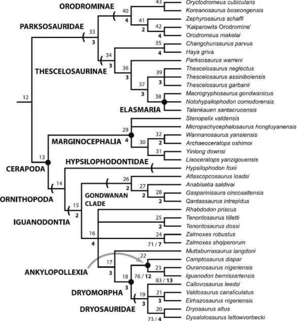
Dinosauria Owen, 1842 Ornithischia Seeley, 1887 Ornithopoda Marsh, 1881 Iguanodontia Sereno, 1986
Dryosauridae Milner and Norman, 1984
Aff. Eousdryosaurus nanohallucis Escaso et al., 2014
Figs.3.1.1-3.1.6
Description
Cranial skeleton
Parietal (fig. 3.1.1)
ML 1851 is 18 mm in length and 22 mm in width and sub-triangular in shape, wider than long, with the right lateral half fractured. The contact with the frontals, is W-shaped, presenting an interdigitated suture as seen in Dysalotosaurus (Janensch, 1955). Laterally, a
ventrally arched parietal process is preserved, showing a smooth surface, as seen in
Dysalotosaurus (Janensch, 1955; Galton, 1983: Hübner & Rauhut, 2010). Posteriorly at the
39
40
Dentary (fig.3.1.2)
ML 768 is a fragment of a right dentary bone (length: 30 mm, height: 7 mm, latero-medial thickness: 8 mm), fractured on both caudal and rostral ends, first reported by Mateus (2007) as aff. Dryosaurus sp. It preserves fifteen close-packed alveoli (grouped in seven pairs
and one isolated, two erupting teeth and six roots. In dorsal view, it is sinuous in shape and the medio-lateral section is slightly concave-convex. The lateral surface is smooth, bearing seven visible foramina on two different levels. On the medial surface, a deep Meckelian sulcus runs for the entire length of the tooth-row. The margins of the sulcus are neat and straight. Caudally, on the medial surface, and dorsally with respect to the Meckelian sulcus, the splenial contact is preserved exhibit a highly dense capillary vascular system. The preserved teeth are diamond-shaped, with a rounded apex. Numerous apicobasally extended ridges are distributed on the lingual surface of the tooth-crown. The margin of the crown possesses various well-developed tongue-shape denticles, which are more numerous than the crown ridges. The primary ridge is positioned towards the midline of the crown, but slightly distally offset, resembling the condition seen in some teeth of Dryosaurus altus and Dysalotosaurus
41
42
Isolated dentary teeth (fig. 3.1.3)
43