Cláudia Marisa Monteiro Saraiva
Mestre em Bioquímica
MicroRNA-124-loaded nanoparticles as a new
promising therapeutic tool for neural stem cell-based
brain repair strategies
Dissertação para obtenção do Grau de Doutor
em Bioengenharia (MIT)
Orientador:
Professora Doutora Liliana Bernardino, Universidade da
Beira Interior
Co-orientadores: Professor Doutor Lino Ferreira, Universidade de Coimbra;
Professor Doutor Manuel Nunes da Ponte, Universidade
Nova de Lisboa.
Júri:
Presidente: Professor Doutor Fernando Santana, Universidade Nova de Lisboa. Arguente(s): Professor Doutor Luís de Almeida, Universidade de Coimbra;
Doutor Ricardo Pires das Neves, Investigador CNC, Universidade de Coimbra. Vogal(ais): Professora Doutora Liliana Bernardino, Universidade da Beira Interior;
Doutora Teresa Casimiro, investigadora principal – LAQV/REQUIMTE, Universidade Nova de Lisboa;
Doutor Karsten Ruscher, Invesigador BMC, Universidade de Lund, Suécia; Professora Doutora Paula Alves, Universidade Nova de Lisboa.
MicroRNA-124-loaded nanoparticles as a new promising therapeutic tool for neural stem
cell-based brain repair strategies
Copyright © Cláudia Marisa Monteiro Saraiva, Faculdade de Ciências e Tecnologia, Universidade Nova de Lisboa.
A Faculdade de Ciências e Tecnologia e a Universidade Nova de Lisboa têm o direito, perpétuo e sem limites geográficos, de arquivar e publicar esta dissertação através de exemplares impressos reproduzidos em papel ou de forma digital, ou por qualquer outro meio conhecido ou que venha a ser inventado, e de a divulgar através de repositórios científicos e de admitir a sua cópia e distribuição com objectivos educacionais ou de investigação, não comerciais, desde que seja dado crédito ao autor e editor.
Agradecimentos •
Acknowledgements
Após uma longa (por vezes atribulada) caminhada dou como concluída mais uma etapa da minha vida profissional e, não poderia deixar de agradecer a todos os que de uma forma directa ou indirecta contribuíram para o sucesso deste projecto.
Começo por agradecer ao MIT-Portugal por me dar a oportunidade de integrar o Programa Doutoral em Bioengenharia de Sistemas, por todos os ensinamentos que me transmitiu sempre de olhos postos no futuro e na inovação sustentável, mas acima de tudo pela rede de contactos que me permitiu estabelecer. Obrigada a todos e, em especial ao Doutor Manuel Nunes da Ponte pela sua co-orientação, disponibilidade e, auxílio sempre que necessário.
À Profª. Doutora Liliana Bernardino, que um pouco “às cegas” me aceitou como sua orientanda, o meu mais sincero obrigada. Obrigada por todos os ensinamentos que me transmitiste, por todo o tempo que me dispensaste, pela confiança que me depositaste no laboratório e, pelos incentivos ao meu crescimento profissional e pessoal. Se hoje me posso chamar investigadora a muito o devo a ti! Tenho muito orgulho de te ter como orientadora e és para mim um exemplo de sucesso. Obrigada ainda por todos os momentos não laborais e aos muitos risos que tornaram esta minha jornada mais agradável!
Ao Doutor Lino Ferreira agradeço a sua co-orientação, a sua pronta disponibilidade, todo o seu apoio, amabilidade e, sentido crítico que em muito contribuíram para o sucesso deste trabalho. Obrigada!
Doctor Karsten Ruscher thank you for the supervision, for making me more critical about research, for the support, for all the time you dedicated helping me and above all to open my scientific horizons. I have learned a great deal of teachings from you!
I am also grateful to Doctor Tadeusz Wieloch for accepting me in his Laboratory as well as all the members of the Laboratory for Experimental Brain Research Group, who were always very friendly and supportive. Thank you for making my passage in Lund very enjoyable, it was a pleasure to meet you all. And of course, I must thank the ones you made my staying in Lund more entertaining and not only worked related.
Agradeço à Doutora Graça Baltazar e a todo o seu grupo que sempre demonstraram disponibilidade para me ajudar nas alturas em que precisei.
À Doutora Francisca Eiriz agradeço a disponibilidade e toda a ajuda aquando da determinação do método analítico usado para os estudos in vivo.
O meu muito obrigada à Profª. Doutora Raquel Ferreira e ao Doutor Tiago Santos que acompanharam quase toda a minha caminhada durante a aventura que foi o Doutoramento. Obrigada por tudo o que me ensinaram, ambos contribuíram em muito para o meu crescimento no mundo da investigação. Obrigada também por toda a ajuda laboratorial, pelo apoio, pelas críticas construtivas e, por todos os conselhos/dicas. Muita energia matinal (por vezes tanta que cansava só de olhar), extrema organização, um pouco de agilidade e perfecionismo, uma pitada de conversa (ou queixas), risos até as lágrimas nos caírem dos olhos, azeitonas qb, ingredientes perfeitos de um dia bem passado no CICS. Obrigada!
Agradeço também à Sandra Rocha, à Marta Esteves, à Patrícia Lindeza, à Doutora Ana Clara Cristóvão e, a todos os elementos do Brain Repair Group, quer os membros actuais quer os que por ele passaram nos últimos 5 anos. Todos vocês me ajudaram a percorrer este caminho!
Aos meus amigos começo por agradecer a sua presença constante na minha vida, apesar de ser uma desnaturada. Muito obrigada por ouvirem os meus queixumes e desabafos; por me animarem e
encorajarem nas “derrotas” e, celebrarem comigo as vitórias. Sou muito grata por todos os momentos que passo convosco. Momentos sempre repletos de boas aventuras, muitas gargalhadas e que me enchem o coração! Muito obrigada! Agora, deixemo-nos de lamechices e vamos mas é beber um copo, pago eu ;)
This work was conducted in the Brain Repair Group at Health Sciences Research Centre (CICS), University of Beira Interior, under the scientific supervision of Professor Doctor Liliana Bernardino and co-supervision of Doctor Lino Ferreira. Part of the work was also performed in the Laboratory for Experimental Brain Research, Wallenberg Neuroscience Center, Biomedical Centre (BMC), University of Lund, under supervision of Doctor Karsten Ruscher.
Cláudia Saraiva was a student of the Bioengineering Systems Doctoral program from MIT Portugal at the Faculty of Science and Technology, Universidade Nova de Lisboa, and a recipient of the fellowship SFRH/BD/90365/2012 from the Portuguese Foundation for Science and Technology (FCT). This work was financial supported by FCT (Project EXPL/BIM-MED/0822/2013 and Project UID/Multi /00709/2013), the European Community through the European Research Council (ERC; Project Nanotrigger, 307384) and ERC chair (Project ERA@UC, 669088). This work was also supported by FEDER funds through the POCI - COMPETE 2020 - Operational Programme Competitiveness and Internationalisation in Axis I - Strengthening research, technological development and innovation (Project POCI-01-0145-FEDER-007491; Project 3386 - “StrokeTherapy”), the Swedish Brain Fund, the Crafoord Foundation, the Swedish Research Council, Sveriges Stroke Riksförbundet, the Hans-Christian and Alice Wachtmeister Foundation, the Stiftelsen Sven-Olof Jansons livsverk, SFO Multipark and Region Skåne (ALF).
The work developed during the PhD and herein described resulted in the publication or submission of two original research articles and one commentary article in international peer-review journals:
C. Saraiva, J. Paiva, T. Santos, L. Ferreira, L. Bernardino, MicroRNA-124 loaded nanoparticles enhance
brain repair in Parkinson’s disease., J. Control. Release. 235 (2016) 291–305. doi:10.1016/j.jconrel.2016.06.005. – Chapter 3
Comment in:
C. Saraiva, L. Ferreira, L. Bernardino, Traceable microRNA-124 loaded nanoparticles as a new
promising therapeutic tool for Parkinson’s disease, Neurogenesis. 3 (2016) e1256855. doi:10.1080/23262133.2016.1256855. – Chapter 3
C. Saraiva, D. Talhada, A. Rai, R. Ferreira, L. Ferreira, L. Bernardino, K. Ruscher, MicroRNA-124-loaded nanoparticles increase survival and neuronal differentiation of neural stem cells in vitro but do not contribute to stroke outcome in vivo., Experimental Neurology (submitted). – Chapter 4
Additionally, three review articles and one book chapter were published or submitted:
T. Santos, C. Boto, C.M. Saraiva, L. Bernardino, L. Ferreira, Nanomedicine Approaches to Modulate Neural Stem Cells in Brain Repair, Trends Biotechnol. 34 (2016) 437–439. doi:10.1016/j.tibtech.2016.02.003.
C. Saraiva, C. Praça, R. Ferreira, T. Santos, L. Ferreira, L. Bernardino, Nanoparticle-mediated brain drug delivery: Overcoming blood–brain barrier to treat neurodegenerative diseases, J. Control. Release. 235 (2016) 34–47. doi:10.1016/j.jconrel.2016.05.044.
C. Saraiva, M. Esteves, L. Bernardino, MicroRNA: basic concepts and implications for regeneration and repair of neurodegenerative diseases, Biochemical Pharmacology (2017). 10.1016/j.bcp.2017.07.008.
The subventricular zone (SVZ) represents the major neurogenic niche of the rodent brain and its modulation upon brain injury may enhance brain repair. Herein, we used biocompatible and traceable polymeric nanoparticles (NPs) to deliver microRNA-124 (miR-124) into SVZ cells. miR-124 is a key neuronal fate determinant and was recently described as anti-inflammatory and neuroprotective. Therefore, the efficiency of NPs to deliver miR-124 and prompt SVZ neurogenesis and brain repair in
Parkinson’s disease (PD) and stroke models was evaluated.
Firstly, we showed that miR-124 NPs were efficiently internalized by neural stem/progenitors cells and neuroblasts and promoted their differentiation into matured neurons by targeting Sox9 and Jagged1 (two stemness-related proteins) in vitro. Likewise, intracerebral administration of miR-124 NPs increased the number of migrating neuroblasts reaching the olfactory bulb, both in control mice and in mice subjected to a 6-hydroxydopamine (6-OHDA), a model for PD. Moreover, miR-124 NPs increased the levels of new neurons in the lesioned striatum of 6-OHDA-challenged mice, correlating with functional improvement.
Interestingly, miR-124 NPs also demonstrated the ability to decrease cell death and enhance neuronal differentiation of SVZ cultures after oxygen and glucose deprivation, suggesting a potential beneficial role in stroke. Therefore, miR-124 NPs were intravenously injected in mice subjected to photothrombotic (PT) stroke. In contrast to intracerebral administration, miR-124 NPs were unable to promote recovery of lost neurological function in PT mice. miR-124 NPs did not affect SVZ neurogenesis nor post-stroke inflammation.
We conclude that miR-124 NPs treatment modulates PD outcome in vivo, while in stroke it improves survival and neuronal differentiation of SVZ cells. However, the promising in vitro data from stroke could not be verified in vivo. Thus, we provide evidence supporting that miR-124 NPs are well tolerated and positive modulators of endogenous SVZ neurogenesis as well as their potential application in brain repair strategies.
Keywords: Neural Stem Cells; MicroRNA-124-loaded nanoparticles; Neurogenesis; Parkinson’s
A região subventricular (SVZ) representa o maior nicho neurogénico do cérebro de roedores e, a sua modulação após lesão pode promover a reparação cerebral. Neste trabalho, usámos nanopartículas (NPs) poliméricas biocompatíveis e rastreáveis como veículos de entrega de microRNA-124 (miR-microRNA-124), um promotor essencial da neurogénese, recentemente descrito como anti-inflamatório e neuroprotector. A eficiência das NPs na entrega de miR-124 e, o seu consequente efeito neurogénico e reparador foram avaliados em modelos animais da doença de Parkinson (PD) e acidente vascular cerebral (AVC).
Inicialmente provámos que as miR-124 NPs foram internalizadas por células estaminais/progenitoras neurais e neuroblastos, promovendo a sua diferenciação e maturação neuronal através da diminuição da expressão das proteínas Sox9 e Jagged1. Posteriormente, mostrámos que a administração intracerebroventricular de miR-124 NPs aumenta o número de novos neurónios no bolbo olfativo de murganhos saudáveis e com PD. As miR-124 NPs também aumentaram o número de novos neurónios no estriado de murganhos com PD, culminando na diminuição dos seus défices motores.
Curiosamente, as miR-124 NPs demonstraram ser capazes de reduzir a morte celular e aumentar a diferenciação neuronal de células SVZ privadas de oxigénio e glucose, sugerindo um possível envolvimento no AVC. Como tal, administrámos as miR-124 NPs intravenosamente em murganhos previamente sujeitos a fototrombose (modelo de AVC). Inesperadamente, as miR-124 NPs demonstraram ser incapazes de melhorar os défices neurológicos dos murganhos, aumentar a neurogénese na SVZ e, modular a resposta inflamatória.
Assim, o uso de miR-124 NPs permitiu diminuir os danos causados pela PD e, aumentar a sobrevivência e diferenciação neuronal de células SVZ num modelo in vitro de AVC. Contudo, in vivo, no modelo de AVC usado, estes efeitos benéficos não foram verificados. Concluindo, este trabalho evidencia o papel das miR-124 NPs no aumento da neurogénese da SVZ e a sua potencial utilização em estratégias de reparação cerebral.
Table of contents
Direitos de cópia • Copyright ... iii
Agradecimentos • Acknowledgements ...v
Support and financial acknowledgements ... vii
Publications ... ix
Abstract... xi
Resumo ... xiii
Table of contents ... xv
List of Figures ... xix
List of Tables ... xxi
Abbreviations and Acronyms ... xxiii
Chapter 1 – INTRODUCTION ... 1
1.1 Neural Stem Cells ... 3
1.1.1 Subventricular zone ... 4
1.1.2 Subgranular zone ... 7
1.2 Neurogenesis in pathology ... 8
1.3 MicroRNAs ... 12
1.4 MicroRNA-124 ... 15
1.5 Nanoparticles for drug/genetic material delivery ... 18
1.5.1 Brain delivery routes ... 21
1.6 Objectives ... 23
Chapter 2 – MATERIALS AND METHODS ... 25
2.1 Synthesis of PLGA nanoparticles ... 27
2.2 Complexation of nanoparticles and microRNAs ... 27
2.3 Characterization of nanoparticles ... 27
2.4 Subventricular zone cell culture ... 27
2.5 Cell transfection ... 28
2.6 Oxygen and glucose deprivation and experimental treatments ... 28
2.7 Internalization studies of miRNA NPs ... 28
2.8 Immunocytochemistry ... 29
2.9 Cell Survival studies ... 30
2.9.1 Propidium iodide incorporation ... 30
2.10 BrdU incorporation ... 30
2.11 Quantitative PCR analysis ... 31
2.12 In vivo studies ... 31
2.13 Animal models of disease and treatments ... 31
2.13.1 6-OHDA model of Parkinson’s disease and miRNA-loaded NPs treatments (intraventricular administration) – Chapter 3 ... 31
2.13.2 Photothrombotic model of stroke and miRNA-loaded NPs treatments (intravenous administration) – Chapter 4 ... 32
2.13.3 Proliferating cell labelling – BrdU incorporation ... 33
2.14 Behavior analysis ... 33
2.14.1 Apomorphine-induced rotation test ... 33
2.14.2 Rotating pole test ... 33
2.14.3 Grid test ... 33
2.15 Tissue collection ... 34
2.16 Lesion evaluation ... 34
2.16.1 Dopaminergic neurons evaluation ... 34
2.16.2 Infarct volume measurements ... 34
2.17 Immunohistochemistry... 35
2.18 Preparation of protein extracts ... 36
2.19 Cytokine analysis from brain extracts and serum samples ... 36
2.20 Statistical analysis ... 37
Chapter 3 – MicroRNA-124 LOADED NANOPARTICLES ENHANCE BRAIN REPAIR IN PARKINSON’S DISEASE ... 39
3.1 Introduction ... 41
3.2 Results ... 42
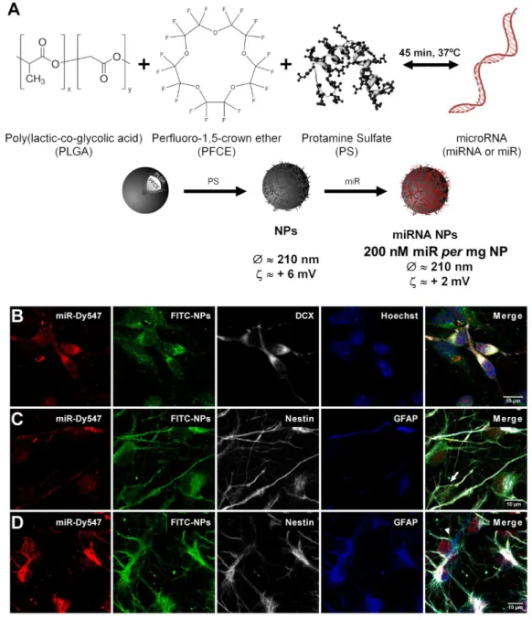
3.2.1 miRNA-loaded NPs are efficiently internalized by SVZ cells ... 42
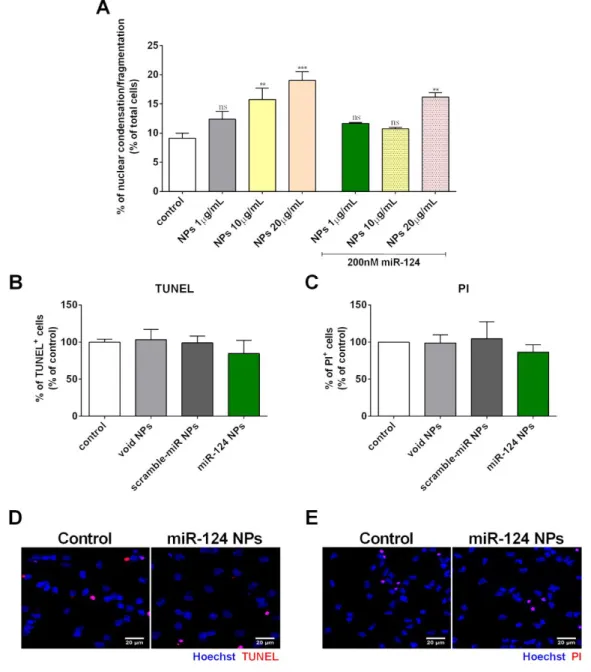
3.2.2 Cellular toxicity of NPs ... 43
3.2.3 miR-124 NPs prompt neuroblasts proliferation ... 45
3.2.4 miR-124 NPs induce neuronal differentiation by repressing key non-neuronal genes ... 46
3.2.5 miR-124 NPs promote axonogenesis ... 49
3.2.6 miR-124 NPs increase the number of migrating neuroblasts that reach the OB and the lesioned striatum leading to motor amelioration of the PD symptoms ... 50
3.3 Discussion ... 54
3.4 Conclusions ... 57
4.2. Results ... 62
4.2.1 miR-124 NPs protect SVZ cells and stimulate their differentiation after OGD ... 62
4.2.2 Treatment with miR-124 NPs does not affect lesion volume and functional outcome after photothrombosis ... 64
4.2.3 SVZ Neurogenesis after miR-124 NPs treatment in PT mice ... 67
4.2.4 Effects of miR-124 NPs on the post-ischemic inflammatory response ... 68
4.3 Discussion ... 69
4.4. Conclusions ... 74
Chapter 5 – GENERAL CONCLUSIONS ... 75
List of Figures
Figure 1.11 Localization and composition of neural stem cell (NSC) niches in the adult rodent brain. .. 5
Figure 1.22 Schematic representation of the canonical pathway of microRNA (miRNA) biogenesis. .. 13
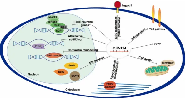
Figure 1.33 Schematic representation of miR-124 molecular targets and pathways experimentally identified. ... 16
Figure 1.44 Main nanoparticle (NP) features. ... 20
Figure 1.55 Administration of nanostructured materials to target the brain. ... 22
Figure 3.16 miRNA loaded NPs are internalized by SVZ stem/progenitor cells. ... 43
Figure 3.27 Viability studies in SVZ cells treated with NPs. ... 44
Figure 3.38 miR-124 NPs favor neuroblast proliferation. ... 46
Figure 3.49 miR-124 NPs promotes neuronal differentiation over glial differentiation. ... 47
Figure 3.510miR-124 NPs target Sox9 and Jagged1 mRNA and protein levels. ... 48
Figure 3.611miR-124 NPs activate the SAPK/JNK pathway in Tau+ axons. ... 49
Figure 3.712miR-124 NPs do not affect the number of proliferating SVZ neuroblasts both in the healthy and in a 6-OHDA-challenged mouse model of PD. ... 51
Figure 3.813miR-124 NPs induce migration of SVZ-derived neuroblasts towards the granule cell layer (GCL). ... 52
Figure 3.914miR-124 NPs induce integration of mature neurons in the granule cell layer (GCL) of the olfactory bulb (OB). ... 52
Figure 3.1015miR-124 NPs induce integration of mature neurons into the lesioned striatum of 6-OHDA-challenged mice and ameliorate the PD phenotype. ... 54
Figure 3.1116miR-124 loaded nanoparticles (miR-124 NPs) boost neuronal differentiation of neural stem/progenitors cells (NSPCs) from the subventricular zone (SVZ) in vitro and in vivo, ultimately leading to motor symptoms amelioration in a Parkinson’s disease (PD) mice model. ... 58
Figure 4.117Effect of miR-124 NPs treatment on SVZ cell cultures after OGD. ... 63
Figure 4.218miR-124 NPs do not affect infarct volume of PT mice. ... 65
Figure 4.319miR-124 does not affect neurological function after photothrombosis. ... 66
Figure 4.420Neurogenesis is not affected by miR-124 NPs treatment. ... 68
List of Tables
Abbreviations and Acronyms
6-OHDA 6-hydroxydopamine AGO Argonaute
AMPK 5' adenosine monophosphate-activated protein kinase ATP Adenosine triphosphate
Bax Bcl-2-associated X protein Bcl-2 B-cell lymphoma 2
BDNF Brain-derived neurotrophic factor Bim Bcl-2-like protein 11
BLBP Brain lipid-binding protein BrdU 5-bromo-2'-deoxyuridine BSA Bovine serum albumin
cAMP Cyclic adenosine monophosphate CD Cluster of differentiation
cdk5 Calpain/cyclin-dependent kinase 5 cDNA Complementary deoxyribonucleic acid CREB cAMP response element binding protein CSF Cerebrospinal fluid
DA Dopamine
DAB 3′-diaminobenzidine DCX Doublecortin
DGCR8 DiGeorge Syndrome Critical Region 8 Dlx2 Distal-less homeobox 2
DMEM Dulbecco's modified Eagle medium DNA Deoxyribonucleic acid
EDTA Ethylenediamine tetraacetic acid EGF Epidermal growth factor
EGFR Epidermal growth factor receptor EGTA Ethylene glycol tetraacetic acid Ezh2 Enhancer of zeste homolog 2 FDA Food and Drug Administration FGF-2 Fibroblast growth factor-2 FITC Fluorescein isothiocyanate FoxG1 Forkhead transcription factor G1
GAPDH Glyceraldehyde 3-phosphate dehydrogenase GCL Granular cell layer
G-CSF Granulocyte colony-stimulating factor GDNF Glial cell-derived neurotrophic factor GFAP Glial fibrillary acidic protein
GFP Green fluorescent protein GL Glomerular layer
HSC70 Heat shock cognate protein-70 HSP90 Heat shock protein-90
HUVEC Human umbilical vein endothelial cells i.p. Intraperitoneal injections
iASPP Inhibitory member of the apoptosis-stimulating proteins of p53 family Iba1 Ionized calcium binding adaptor molecule 1
IFNγ Interferon-gamma IgG Immunoglobulin G IL-1β Interleukin-1beta IL-6 Interleukin-6
iPSC Induced pluripotent stem cell JNK c-Jun N-terminal kinase
JSAP1 JNK pathway-specific scaffold protein 1 LNA Locked nuclei acid
LPS Lipopolysaccharide
Map-2 Microtubule-associated protein-2 MBD1 Methyl-CpG-binding domain protein 1 MCAO Middle cerebral artery occlusion MeCP2 Methyl CpG binding protein 2 MicroRNA miRNA or miR
miRISC miRNA-induced silencing complex MPP methyl phenyl pyridinium
MPTP 1-methyl-4-phenyl-1,2,3,6-tetrahydropyridine MRI Magnetic resonance imaging
mRNA Messenger ribonucleic acid mRS post-stroke modified Rankin Score mTOR Mechanistic target of rapamycin MWCO Molecular weight cut-off NeuN Neuronal nuclei
NeuroD1 Neurogenic differentiation 1 NF-κB p65 Nuclear factor-κB p65 NMR Nuclear magnetic resonance NP Nanoparticle
NSC Neural stem cell nt Nucleotide OB Olfactory bulb
OGD Oxygen and glucose deprivation Olig2 Oligodendrocyte transcription factor 2 ON Overnight
PACT protein kinase R-activating protein PAZ Piwi-Argonaute-zwille
PBS Phosphate-buffered saline
PBS-T Phosphate-buffered saline with Triton X-100 PCNA Proliferating cell nuclear antigen
PD Parkinson’s disease
PFA Paraformaldehyde PFCE Perfluoro-1,5-crown ether pHH3 Phosphohistone H3 PI Propidium iodide
PI3K Phosphoinositide 3-kinase PLA Poly(lactic acid)
PLGA Poly(lactic-co-glycolic acid) PMSF Phenylmethanesulfonyl fluoride PS Protamine sulfate
PSA-NCAM Polysialylated neural cell adhesion molecule PT Photothrombotic stroke
PTBP RNA binding polypyrimidine tract-binding protein PVA Poly(vinyl) alcohol
qPCR Quantitative real-time polymerase chain reaction RAN Ras-related nuclear protein
REST RE1 silencing transcription factor RhoG Ras homology growth-related RMS Rostral migratory stream RNA Ribonucleic acid
ROCK1 Rho-associated coiled-coil forming protein kinase 1 RT Room temperature
SCP1 Small c-terminal domain phosphatase 1 SEM Standard error of mean
SFM Serum-free medium SGZ Subgranular zone SN Substantia nigra
Sox 2 Sex determining region Y-box 2 ssRNA Single strand RNA
STAT3 Signal transducer and activator of transcription 3 SVZ Subventricular zone
TACE TNF-α converting enzyme
TBS Tris buffer saline TH Tyrosine hydroxylase TLR Toll-like receptor
Tlx Orphan nuclear receptor tailless TNF-α Tumor necrosis factor-alpha tPA Tissue plasminogen activator TRAF6 TNF receptor associated factor 6
TRBP Transactivation-response RNA-binding protein
TUNEL Terminal deoxynucleotidyl transferase dUTP nick end labeling USP Ubiquitin-specific protease
INTRODUCTION
1.1 Neural Stem Cells
Neurogenesis, the generation and development of nerve cells, was traditional viewed as a process exclusive of the embryonic and perinatal brain (Bizzozero 1894; Ramon y Cajal 1913). The acceptance of adult neurogenesis, a milestone in the field, was only made in the early 1990s (Nottebohm 2002). The first evidence of adult neurogenesis were presented by Joseph Altman and collaborators, in the 1960s (Altman 1962; Altman 1963). They showed that adult-born cells formed in the subventricular zone (SVZ) travel through the rostral migratory stream (RMS) to the olfactory bulb (OB) where they differentiate into mature neurons (Altman and Das 1965; Altman 1969). Still, the existence of post-natal new neurons in the OB and dentate gyrus of mammals was proven unequivocally only a few years later (Kaplan and Hinds 1977).Thereafter, several groups showed that adult neurogenesis was conserved from rodents (Kuhn et al. 1996; Kempermann et al. 1997) to humans (Eriksson et al. 1998; Kukekov et al. 1999), which rapidly led the scientific community to pursue the restauration of dysfunctional circuitries through the modulation of neurogenesis.
During the embryonic development, neuroepithelial cells and later radial glia cells originate neurons and glia cells based on spatial and temporal cues (Pearson and Doe 2004). Soon after birth, glial fibrillary acidic protein (GFAP)-expressing astrocyte-like cells assume the role of radial glia cells as neural stem cells (NSCs) (Doetsch et al. 1999; Garcia et al. 2004). NSCs can self-renew, proliferate and possess multipotent features, being able to differentiate into neurons, astrocytes and oligodendrocytes (Reynolds and Weiss 1992). These cells are found in discrete regions of the brain – neurogenic niches – that are mainly found in the SVZ lining the lateral ventricles and in the subgranular zone (SGZ) of the dentate gyrus of the hippocampus (Gage 2000; Ma et al. 2009). Neurogenesis in these germinal niches persist throughout life although it decreases with age (Kuhn et al. 1996). A rising number of evidence has also been pointing to the existence of other neurogenic regions in the brain, namely in the circumventricular organs located along the ventricular midline (Itokazu et al. 2006; Bennett et al. 2009), in the walls of the third and fourth ventricles (Lin et al. 2015), in the meninges of the spinal cord (Petricevic et al. 2011; Decimo et al. 2011), in the substantia nigra (SN) (Lie et al. 2002), in the amygdala(Bernier et al. 2002), in the cerebellum (Klein et al. 2005), among other regions, reviewed at (Lin 2015). All these regions share the ability to generate multipotent precursors that can be isolated and differentiated in vitro, however, they usually occur in low levels in physiological conditions being only perceptive after injury (Ming and Song 2005). These facts together with the lack of evidence for the presence of niche structures to house these cells make their role in brain homeostasis a matter of controversy.
indication of their regenerative potential (Curtis et al. 2003). Other studies have confirmed that upon lesion SVZ cells had the capability to migrate towards the lesioned area. In some cases, they could even differentiate into the respective cell phenotype of the lesioned regions, leading ultimately to functional recovery (Arvidsson et al. 2002; Parent et al. 2002; Goings et al. 2004). As so, endogenous NSCs are a virtual unlimited source of new cells that upon an injury respond by proliferating, migrating towards the lesion and differentiating into neurons, raising high expectations for regenerative medicine. Therapeutic approaches based on augmentation of the brain natural endogenous response to injury overcome limitations related with cell transplantation, such as the low number of cells typically available for therapy, low cell survival after transplantation, immune rejection of grafted cells, teratoma formation, and ethical issues (Christie and Turnley 2012). This approach seems to be particularly feasible because numerous studies have demonstrated that growth factors (e.g. glial cell-derived neurotrophic factor (GDNF), brain-derived neurotrophic factor (BDNF), granulocyte colony-stimulating factor (G-CSF)), and other molecules (e.g. erythropoietin, pramipexole, ephrin-A1) are able to induce neurogenesis leading eventually to functional recovery (Dempsey et al. 2003; Kobayashi et al. 2006; Kadota et al. 2009; Winner et al. 2009; Jing et al. 2012). However, strategies based on enhancing endogenous NSCs activity are still limited to pre-clinical studies. Developing new platforms to efficiently deliver pro-neurogenic factors to NSCs is imperative to boost the endogenous reparative capacity of the adult brain.
1.1.1 Subventricular zone
adhesion molecule (PSA-NCAM), and have a high migratory capacity travelling long distances (up to millimeters) from the SVZ to the OB. Chains of neuroblasts coming from different paths converge in the anterior SVZ to form the RMS, a tube-like structure formed of specialized astrocytes and blood vessels that confer additional physical support to the migration (Lois and Alvarez-Buylla 1994; Lois et al. 1996; Doetsch and Alvarez-Buylla 1996; Lledo et al. 2006). Neuroblasts migrate rostrally until they reach the OB; once there, they detach from the RMS chains (tangentially oriented) and move radially towards different layers of the OB. These new neurons mature and integrate mainly as GABAergic granule interneurons in the granular cell layer (GCL) and in a lower extent as GABAergic or dopaminergic (smaller amount) periglomerular interneurons in the glomerular layer (GL). There is also a very small percentage of cells that differentiate into glutamatergic juxtaglomerular neurons (Brill et al. 2009; Ming and Song 2011).
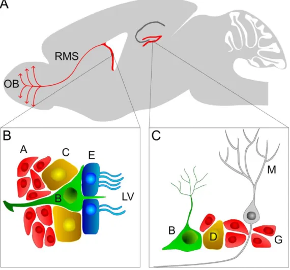
Figure 1.11 Localization and composition of neural stem cell (NSC) niches in the adult rodent brain. (A) Sagittal rodent brain slice displaying the localization of the two main neurogenic niches: the subventricular (SVZ) and subgranular (SGZ) zones. (B) At the SVZ, type B NSCs cells contact with the lateral ventricles and give rise to C progenitor cells that differentiate into neuroblasts (type A cells). Physiologically, neuroblasts migrate through the rostral migratory stream (RMS) towards the olfactory bulb (OB) where they differentiate into mature neurons; E, ependymal cells. (C) Similarly, type I (or B) stem cells at the SGZ give rise to type II (or D) progenitor cells that in turn generate neuroblasts (type III or G cells), which ultimately differentiate into mature granule neurons (M).
need to proliferate, migrate, maturate and functionally integrate into existing circuitries to trigger an efficient regenerative response. There are also limitations regarding the suitability in the type of cells generated or their survival. Hence, it is essential to understand and/or identify molecular mediators able to enhance this neurogenic response and, therefore useful for the development of novel therapeutic strategies.
Isolation and culture of NSCs in vitro were pivotal to extend our knowledge regarding adult neurogenesis (Reynolds and Weiss 1992; Kilpatrick and Bartlett 1993). Free floating three-dimensional structures of thousands of cells – neurospheres – can be obtained from harvesting the SVZ cells and cultivate them into specific conditions: serum-free medium with mitogenic growth factors, namely epidermal growth factor (EGF) and fibroblast growth factor-2 (FGF-2), into a non-adherent substrate. These conditions select cells responsive to EGF that include type C cells (around 70%) and activated type B cells, both with self-renewal and proliferative abilities. Differentiation into neurons, oligodendrocytes and astrocytes can be achieved by cultivating neurospheres in the absence of mitogenic growth factors and into an adhesive substrate. Although the limitations associated with in vitro culture, such as the non-total representation of the NSCs in vivo population, the number of passages, the loss and/or change of biological properties to name a few, it is still a solid method to appraise neural stem/progenitors cells properties in different conditions, screening potential therapeutic molecules or expand cells for replacement therapies (Doetsch et al. 2002; Gil-Perotín et al. 2013).
cells may be responsive to cues induced by cell loss. Moreover, it was recently shown, in the human brain, that precursors cells derived from the SVZ are able to migrate and differentiate into interneurons in the striatum (Ernst et al. 2014). Likewise, some pathologies, such as stroke, stimulate neurogenesis and neuroblast migration to the injured striatum (Jin et al. 2006), demonstrating the potential of adult neurogenesis in humans. Hence, SVZ NSCs hold great potential in the search for novel brain repair therapies.
1.1.2 Subgranular zone
impairments in mice can be rescued by physical exercise (Lafenêtre et al. 2010). On the other hand, stress seems to negatively correlate with neurogenesis. An example is the reduction in the proliferation of type B and type D cells in the SGZ of mice exposed to stress cause by noise (Gonzalez-Perez et al. 2011). These examples show once more the ability of NSCs to adapt to different environments. Moreover, Spalding and colleagues showed that approximately 700 new neurons are generated every day in each hippocampus in humans during adulthood. Impressively, hippocampal neurogenesis declines in a modest way (around 4-fold) during the entire human lifespan (Spalding et al. 2013). These studies confirm a relevant role of adult SGZ neurogenesis in brain function. Therefore, defects in adult neurogenesis have been associated with neurological and psychiatric diseases. Concluding, even though SGZ presents a more limited proliferative and migratory activity than the SVZ, it still holds high potential for the development of future regenerative therapies for treating neurological disorders.
1.2 Neurogenesis in pathology
The discovery of NSCs in the adult mammalian brain and the increased knowledge about the composition and function of the neurogenic niches opened new avenues in understanding brain plasticity and function. Remarkably, neurogenesis is enhanced upon brain lesion injury possibly as an attempt for brain repair. Nevertheless, the role of neurogenesis in neurodegenerative disorders is still under debate. As so, the role of neurogenesis in Parkinson’s disease (PD), which is a chronic pathology, and in stroke, an acute disorder, are discussed below.
Parkinson’s disease
Parkinson’s disease is a neurodegenerative disease that affects approximately 10 million people
throughout the world (Hermanns 2011). PD is mainly characterized by the progressive degeneration of dopaminergic neurons in the SN leading to striatal dopamine (DA) depletion and the accumulation of
alpha (α)-synuclein aggregates known as Lewy bodies (Spillantini et al. 1997; Damier 1999). The key symptoms that clinically define PD are rigidity, tremor, bradykinesia and postural instability. Currently, the standard treatment for PD is based on DA replacement (mainly using a precursor of DA, levodopa) with high efficacy in the early stages of the disorder. Nevertheless, during the time course of the disease these drugs lose effectiveness and cause side effects and severe psychiatric complications. Deep brain stimulation is also used in some patients, in more advanced stages of the disorder, with successful suppression of motor symptoms; however, it does not stop progression of the pathology. Several regenerative medicine approaches are under intense examination to address the impact of stem cells in PD. However, the low amount of data in post-mortem brain tissue from PD patients, together with contradictory experimental findings, make the role of adult neurogenesis in PD a high controversial subject within the scientific community. It was firstly shown that PD patients display impaired neurogenesis since they presented lower levels of cells expressing the marker for proliferation proliferating cell nuclear antigen (PCNA) in the SVZ, a decrease of nestin-positive cells (immature neural precursor cells) both in the OB and in the dentate gyrus of the hippocampus, as well as a reduction in
numbers while the cumulative use of L-Dopa in PD patients seems to result in increased numbers of proliferating NSCs in the SVZ (O’Sullivan et al. 2011). The SN and the ventral tegmental area (VTA) project dopaminergic fibers that innervate the neurogenic niches in a specific pattern (Höglinger et al. 2014), and they are in close proximity to EGFR-positive cells that include all type C cells and a subset of type B cells (Doetsch et al. 2002; Höglinger et al. 2004). Advanced PD patients have not only significantly less amount of EGFR-positive cells in the SVZ, but also weaker expression of EGFR
(O’Keeffe et al. 2009).In addition, EGF and EGFR levels were also found to be decreased in the striatum of PD patients (Iwakura et al. 2005). SVZ type C and A cells express both D1-like and D2-like DA receptors (Coronas et al. 1997; Höglinger et al. 2004). DA reduction in animal models leads to impairments in NSCs proliferation and EGFR expression that are D2-like receptor-mediated (Höglinger
et al. 2004; Coronas et al. 2004; O’Keeffe et al. 2009; Lao et al. 2013). Αlpha-synuclein also seems to be involved in neurogenesis impairments. Interaction between accumulated α-synuclein and p53 results in Notch1 signaling dysregulation in the SGZ of rats that potentially trigger some of the non-motor symptoms associated with the PD pathology (Crews et al. 2008; Desplats et al. 2012). Neural committed
induced pluripotent stem cells (iPSCs) obtained from fibroblasts of patients with triplication of the α -synuclein gene (SNCA; associated with early onset of PD) were unable to develop neuronal complex networks when compared with control neural committed iPSC, also showing a correlation between α -synuclein expression and neurogenesis impairment (Oliveira et al. 2015). Hypermethylation of thousands of genes has been found in brain tissue of PD patients, specifically neurogenic-related genes such as Wnt, suggesting a critical role for Wnt-associated neurogenesis in PD (Zhang et al. 2016a). Inflammation is also a major player in neurodegenerative disorders and higher expression of inflammatory molecules in PD patients, such as tumor necrosis factor-alpha (TNF-α) or other cytokines (e.g. interleukin (IL)-6), correlates with non-motor symptoms, namely anxiety and depression, which precede the motor symptoms of the pathology (Reale et al. 2009; Lindqvist et al. 2012). Similar symptomatology is found in animal models of impaired neurogenesis (Revest et al. 2009) leading some groups to defend a robust developmental component in PD onset and progression.
On the other hand, the putative role of neurogenesis impairment on PD was challenged by Hol’s
PD models are based on the administration of toxins such as 6-hydroxidopamine (6-OHDA), 1-methyl-4-phenyl-1,2,3,6-tetrahydropyridine (MPTP), rotenone, paraquat, to name a few, that cause selective death of dopaminergic neurons. Genetic models are used to model familial PD, but have also been instrumental to shed some light on PD mechanisms (Jagmag et al. 2015). The differential activation of dopamine receptors in NSCs (Höglinger et al. 2004; Coronas et al. 2004; Kippin et al. 2005) together with a high diversity of PD models (transgenic or toxin based models, acute or chronic administrations, different dosages and different spatial administration of toxins) (Dauer and Przedborski 2003) may explain the different results communicated. For example, in 6-OHDA-challenged rats it was reported either an increase (Liu et al. 2006; Arias-Carrión et al. 2006; Aponso et al. 2008) or decrease (Höglinger et al. 2004; Winner et al. 2006) of SVZ proliferation. In a 6-OHDA mouse model of PD a reduction in SVZ proliferation together with higher survival rate of newborn neurons was also reported in the OB (Baker et al. 2004; Sui et al. 2012). Recently, Fricke and colleagues reported that the 6-OHDA lesion does not affect neurogenesis (Fricke et al. 2016). These discrepancies have generated a high debate in the scientific community about the limitations of experimental models but also the design of studies including the question about the sample size, controls groups, the type of analysis performed to name a few.
Stroke
has also been reported in the adult human brain after stroke. Indeed, DCX-positive neuroblasts were found in the ischemic penumbra of cortical infarcts close to the vasculature, suggesting an important role of angiogenesis in stroke-induced neurogenesis (Jin et al. 2006). Neural stem/progenitor cells expressing nestin and musashi-1 were also observed in the ischemic human cortex during the first month after injury (Nakayama et al. 2010). An increase in the number of NSCs were seen near the SVZ of a stroke patient. Moreover, enhancement of neurogenesis and formation of novel blood vessels were also found in the peri-infarct area of this patient, indicating once more the relevance of the vasculature for neurogenesis as well as the possible migration of newborn cells into injury sites (Minger et al. 2007). Indeed, stroke patients presenting a higher microvessel density in the peri-infarct area have longer survival rates (Krupinski et al. 1994). Altogether, these reports suggest that adult NSCs are responsive to stroke injury even in the aged brain, despite the limited number of studies in the human brain after stroke. Notwithstanding, these evidence support the hypothesis that enhancing the endogenous brain repair response is correlated with improved stroke recovery.
species that can control neurogenesis (del Zoppo et al. 2007). For example, the pro-inflammatory cytokine TNF-α either increases or decreases neurogenesis in a concentration-dependent manner (Bernardino et al. 2008). Altogether, these studies show that stroke induces neurogenesis in the human brain and in animal models of stroke. SVZ seems to be the major contributor for stroke-induced neurogenesis, despite the scarce evidence for cortical neurogenesis. Nevertheless, adult NSCs, namely the SVZ NSCs, showed to be a valuable source of new neurons with brain repair potential.
1.3 MicroRNAs
MicroRNAs (miRNA or miR) were firstly discovered in the 1990s but only in the 2000s these small molecules were identified as a novel class of biologic regulators, altering the view over the central dogma of molecular biology: DNA is transcribed into RNA being then translated into protein. miRNA are small non-coding RNAs with approximately 22 nucleotides (nt) in length that were firstly identified in Caenorhabditis elegans, but were rapidly found to be ubiquitous among plants and animals. The fundamental basis of miRNA pathways is conserved among these two kingdoms, yet miRNA mode of action is primarily different (mRNA cleavage vs mRNA translational repression and/or decay) (Bartel 2004). Transcription and function of miRNA are tightly regulated and complex processes that include several steps of regulation, reviewed at (Towler et al. 2015).
miRNA can be transcribed as independent transcription units or as miRNA genes located either in introns or exons of other genes (Rodriguez et al. 2004). In mammals, intronic miRNA originate the majority of the miRNA transcripts, being controlled independently of the host genome by different promotors (Monteys et al. 2010; Godnic et al. 2013; Ramalingam et al. 2014). miRNA genes are generally transcribed by RNA polymerase II generating an imperfect stem-loop structure flanked by single strand (ss)RNA, which is capped on the 5’ end and polyadenylated in the 3’ end and it is composed of hundreds to thousands of nt – pri-miRNA (Figure 1.2) (Lee et al. 2004; Cai et al. 2004). One arm of the pri-miRNA stem-loop comprises the sequence to generate either a single or a cluster of mature miRNA (Altuvia et al. 2005; Hertel et al. 2006). The microprocessor complex is composed of two RNA-binding proteins. DiGeorge Syndrome Critical Region 8 (DGCR8) interacts with pri-miRNA and recruits the RNase III enzyme DROSHA, which is responsible for cleaving the pri-miRNA in the stem-loop, forming the precursor-miRNA (Pre-miRNA). Pre-miRNA has a 5’ phosphate group, a 2 nt overhang
machinery (Iwasaki et al. 2010). Altogether they form the miRNA-induced silencing complex (miRISC). The N-domain of AGO starts the unwind the miRNA duplex originating: a guide or mature strand, thermodynamically more stable and prevalent with higher biological activity that is retained in the miRISC complex; and a passenger strand (miR*), which is released to either be degraded or incorporated into another miRISC complex(Kwak and Tomari 2012; Noland and Doudna 2013; Meijer et al. 2014). The four AGO proteins expressed in humans (AGO-1, -2, -3 and -4) can perform non-cleavage inhibition of mRNA, but only AGO-2, the most abundantly expressed AGO protein, has nuclease activity, meaning that it is the only one able to cleave the target mRNA (Figure 1.2) (González-González et al. 2008; Valdmanis et al. 2012; Wang et al. 2012a). The majority of miRNA follows the canonical biogenesis pathway (described above); nevertheless, miRNA that are similar in structure and function but bypass some of the maturation steps of the canonical pathway have been described, the non-canonical miRNA. There are three major alternatives to synthesize these miRNA: i) Drosha/DGCR8-dependent and independent pathway; ii) Drosha/DGCR8-independent and Dicer-dependent pathway; iii) Drosha/DGCR8-inDicer-dependent and Dicer-inDicer-dependent pathway, reviewed at (Abdelfattah et al. 2014; Daugaard and Hansen 2017).
Figure 1.22 Schematic representation of the canonical pathway of microRNA (miRNA) biogenesis.
Briefly, miRNA are mostly transcribed by the RNA polymerase II into long transcripts called pri-miRNA that are processed into a miRNA precursor (pre-miRNA) of approximately 60 nucleotides (nt) long, by the microprocessor complex composed of DiGeorge Syndrome Critical Region 8 (DGCR8) and Drosha. The pre-miRNA is exported to the cytoplasm via Exportin 5 transporter, where it is further processed into a miRNA duplex (~22 nt) by Dicer and
miRNA are able to regulate in a very precise way the expression of hundreds of different genes. The same miRNA can bind to numerous different mRNA and a single mRNA can present multiple miRNA binding sites, either for the same or for different miRNA (Friedman et al. 2009). The target recognition of the miRNA is mainly based on Watson-Crick pairing of a specific region from nt 2 to 8,
seed sequence, and a match site in the 3’UTR (Bartel 2009; Wang 2014). miRNA can regulate translation of mRNA either by pairing with the target mRNA in a perfect or near perfect complementary way, mostly seen in plants, where AGO-2 cleaves the target mRNA (Figure 1.2) (Bartel 2004; Yekta et al. 2004; Karginov et al. 2010). In animals, the miRNA-mRNA interaction is mainly made through incomplete pairing with presence of mismatches and/or bulges. Herein, mRNA translational repression and mRNA destabilization/decay mechanisms prevail (Huntzinger and Izaurralde 2011; Eichhorn et al. 2014). There are still some controversies regarding the contribution of each mechanism for miRNA action. However, recent developments in the field indicate that miRNA action is time-dependent, promoting mRNA translational repression in an initial phase followed by a dominant effect of mRNA degradation in the steady-state (Djuranovic et al. 2012; Béthune et al. 2012; Meijer et al. 2013; Larsson and Nadon 2013; Eichhorn et al. 2014).
inhibitor of miR-122 (miravirsen), currently in phase II clinical trials, that showed to be highly effective in reducing the hepatitis C virus from chronic patients without causing serious side effects (Lindow and Kauppinen 2012). Our knowledge regarding miRNA increased during the past years leading to stability and nuclease resistance improvement of miRNA by chemical modifications as well as the development of some promising miRNA-based strategies currently in clinical trials. Still, the delivery of miRNA into tissues, namely into the brain, is a major limitation for the development of miRNA-based therapies. Thus, improvements on brain delivery strategies for miRNA are pivotal.
1.4 MicroRNA-124
The miR-124 was firstly identified in mice and it is transcribed from three different loci: miR-124-1 on chromosome 14, miR-124-2 on chromosome 3 and miR-124-3 on chromosome 2. The three copies have similar mature sequence and are highly conserved among species, including mice and humans. The expression levels of miR-124 increase in the prenatal period peaking at the end of the fetal development and remain elevated in the post-natal brain (Krichevsky et al. 2003). miR-124 accounts for 25% to 48% of the total miRNA in the adult brain, and it is highly expressed among all brain regions except the pituitary gland. Outside the CNS, miR-124 is 100-times less expressed than in the brain, being also considered a neuronal specific miRNA, since it is mostly expressed in neuronal cells (Lagos-Quintana et al. 2002; Mishima et al. 2007; Baroukh and Van Obberghen 2009). miR-124 is able to regulate hundreds of non-neural genes that are responsible for neural phenotype acquisition and maintenance being known as a master regulator of neurogenesis (Lim et al. 2005; Conaco et al. 2006). Expression of miR-124 is initiated during the transition of NSCs to progenitor cells and enhanced with neuronal maturation (Cheng et al. 2009; Akerblom et al. 2012). It has been shown that overexpression of miR-124 results in forced neuronal differentiation both in progenitor cells (Visvanathan et al. 2007; Yu et al. 2008) and in HeLa cells (Lim et al. 2005). Lentiviral overexpression of miR-124 precursor in combination with other factors, namely miR-9, also led to a forced differentiation of human neonatal foreskin fibroblasts into functional mature neurons (Yoo et al. 2011). In vivo, miR-124 overexpression in the SVZ increased the number of newborn neurons without affecting their migratory capability (Cheng et al. 2009; Akerblom et al. 2012), while its downregulation led to a 30% reduction of post-mitotic neurons (Cheng et al. 2009). Knockdown of miR-124-1 in mice resulted in a smaller brain size, neuronal dysfunction and axonal miss-sprouting in the dentate gyrus (Sanuki et al. 2011). miR-124 not only promotes neuronal commitment, but it also controls the choice among neuronal and astrocytic differentiation through fine-tuning the expression of a critical epigenetic regulator, Ezh2 (Neo et al. 2014), inhibition of the signal transducer and activator of transcription 3 (STAT3) signaling (Krichevsky et al. 2006), and reduction of Sox9 expression (Cheng et al. 2009) (Figure 1.3). miR-124 expression enhances axonogenesis by regulating the level of cytoskeletal proteins (Yu et al. 2008; Gu et al. 2014), and regulates dendritic differentiation by targeting the ras homology growth-related (RhoG) pathway (Figure 1.3) (Franke et al. 2012). Moreover, miR-124 is required for homeostatic plasticity, a compensatory adjustment in neuronal activity (Hou et al. 2015).
in non-neuronal cells and it is responsible for inhibiting the expression of neural genes. In neuronal cells, miR-124 keeps REST silent promoting the expression of pro-neuronal genes (Conaco et al. 2006; Visvanathan et al. 2007). The small c-terminal domain phosphatase 1 (SCP1), a critical inducer of chick and mouse embryonic neurogenesis and part of the REST signaling pathway is downregulated by miR-124 (Visvanathan et al. 2007). Other components of the REST signaling pathway including the methyl CpG binding protein 2 (MeCP2) and coREST also present binding sites for miR-124 (Wu and Xie 2006). Moreover, miR-124 controls the transition of a chromatin-remodeling complex, changing the BAF complex subunits from neural-progenitor-specific to neuron-specific, an essential step for post-mitotic neural development and dendritic outgrowth during embryogenesis (Yoo et al. 2009). Another important target of the miR-124 is the RNA binding polypyrimidine tract-binding protein 1 (PTBP1). During neuronal differentiation, miR-124 decreases PTBP1 levels increasing the amount of PTBP2 in the cells and triggering neuron-specific alternative splicing patterns (Makeyev et al. 2007). Interestingly, it was shown that downregulation of PTBP by miR-124 is sufficient to generate functional neurons from fibroblasts (Xue et al. 2013). The Notch signaling pathway is of utmost importance for the maintenance of neural stem/progenitor cells. The Notch receptor is expressed by NSCs, which in the presence of its ligand Jagged1 contributes to self-renewal and maintenance of the undifferentiated state. In rodents SVZ, overexpression of miR-124 represses Jag1 translation leading to NSCs differentiation (Cheng et al. 2009; Liu et al. 2011). Additionally, miR-124 represses the expression of the transcription factor Sox9 in the SVZ, a regulator of gliogenesis (Cheng et al. 2009). Although many aspects of the miR-124 regulatory network need to be unveiled, these data clearly support the idea that miR-124 is able to modulate both embryonic and adult neurogenesis, namely in the SVZ.
miR-124 is a brain-specific miRNA, specially expressed in neurons; nevertheless, in the past decade new evidence have pointed to the importance of this miRNA in the regulation of cell death and inflammatory processes, resulting in neuroprotection in PD (Kanagaraj et al. 2014; Wang et al. 2015a; Gong et al. 2016), alleviation of cell death in Alzheimer’s disease (Fang et al. 2012) or reduction of infarct volume in stroke (Doeppner et al. 2013; Sun et al. 2013a), among others, reviewed at (Sun et al. 2015b). Microglia cells are the first line of defense in the brain and in a physiological situation they show an active but ramified morphology (resting state). Upon injury, these cells become stimulated either in a classical way, M1 state, perpetuating inflammation, or in an alternative pattern, M2 state, with anti-inflammatory properties. Ponomarev and others showed that miR-124 was expressed in resting microglia, but it was undetectable in peripheral monocytes and macrophages. However, in a mouse model of autoimmune encephalomyelitis, characterized by activation of microglia and the infiltration of peripheral macrophages in the brain parenchyma, miR-124 expression was reduced approximately 70% in microglia and it was slightly expressed in the infiltrated macrophages during the disease onset and recovery, indicating a role of miR-124 in the maintenance of microglia quiescent state (Ponomarev et al. 2011). Moreover, administration of exogenous miR-124 into macrophages decreased pro-inflammatory markers and increased anti-pro-inflammatory ones through downregulation of the C/EBP-α -PU.1 pathway. As so, miR-124 showed to be a key regulator of microglia quiescent state and a modulator of peripheral monocytes/macrophages (Ponomarev et al. 2011; Ponomarev et al. 2013; Veremeyko et al. 2013). In human intermediate monocytes, obtained from patients with allergies and bronchial asthma, miR-124 expression levels were also elevated and cells presented M2 characteristics (Veremeyko et al. 2013). In fact, reduction of miR-124 expression levels was shown to be needed to induce microglial reactivity (Freilich et al. 2013), while its overexpression results in lower levels of
TNF-α in reactive macrophages (Sun et al. 2016). miR-124 mediates repression of ubiquitin-specific protease 2 (USP2) and USP14, and the modulation of toll-like receptor (TLR) signaling (Figure 1.3; e.g. STAT3, TNF-α converting enzyme (TACE), necrosis factor (NF)-κB p65 and TNF receptor-associated factor 6 (TRAF6)) that facilitates its anti-inflammatory properties (Sun et al. 2013b; Qiu et al. 2015).
protein and a consequent reduction of translocation of Bcl-2-associated X protein (Bax) to the mitochondria (Figure 1.3) (Wang et al. 2015a).
In stroke, miR-124 was found to be decreased in the serum of patients within the first 24 h and correlated with a worse stroke prognosis (Liu et al. 2015). Nevertheless, Ji and others reported increased levels of miR-124 in exosomes obtained from serum of a stroke patient (Ji et al. 2016). In the ischemic brain of rodents, miR-124 is decreased in neural progenitors cells of the SVZ and in the ischemic core (Liu et al. 2011; Sun et al. 2013a). There is some controversy regarding miR-124 functions in stroke. Some studies reported that the downregulation of miR-124 resulted in a reduction of infarct volumes in rodent MCAO models of stroke (Liu et al. 2013; Zhu et al. 2014). Others observed that miR-124 overexpression previous to the MCAO insult in mice resulted in neuroprotection, reduced inflammation and increased neurogenesis leading ultimately to amelioration of stroke-induced neurological deficits (Doeppner et al. 2013; Sun et al. 2013a). More recently, Hamzei Taj and colleagues reported that mice subjected to MCAO benefited from an injection of a miR-124 mimic into the striatum two days after the lesion. The miR-124 treatment resulted in lower infarct volumes and behavior deficits together with the regulation of the inflammatory response by shifting the microglia from a more pro-inflammatory state (ionized calcium binding adaptor molecule 1 and cluster of differentiation 16/36 (Iba1/CD16/32)-positive cells) to an anti-inflammatory state (Iba1/CD206-positive cells) (Hamzei Taj et al. 2016b; Hamzei Taj et al. 2016a). It is of importance to notice that miR-124 functions may vary dependent of the context. For example, in a rat model of epilepsy miR-124 led to a robust increase of microglia (CD11b-positive cells), a slight increase of astrocytes (GFAP-positive cells), and higher levels of inflammatory cytokines including IL-1β, TNF-α, and IL-6 (Brennan et al. 2016).
Altogether, these studies point towards the use of miR-124 as a broad therapeutic molecule in neurodegenerative disorders and acute brain pathologies, where it may act as neuroprotectant, anti-inflammatory mediator and enhancer of endogenous brain repair mechanisms.
1.5 Nanoparticles for drug/genetic material delivery
The unique features of miRNA make them valuable and versatile tools for clinical applications as biomarkers for diagnosis and prognosis of pathologies and/or therapeutic molecules that can act on diverse key targets or even pathways that are disease-related. miRNA are hydrophilic, negatively charged molecules with high molecular weight. These features make them extremely unstable in vivo (low half-life time in plasma and high clearance rate) and hamper the uptake of miRNA into target cells. Another important aspect to consider in miRNA-based therapeutics is dosage. High dosages of miRNA can result in saturation of the miRNA machinery leading to cellular toxicity, off-target effects that can cause toxicity in non-target tissues, and immunological response that can be harmful for the recipient (Chen et al. 2015). As so, improvement of brain delivery vehicles is of major importance.
NPs are colloidal carriers that can have a natural (e.g. Albumin, chitosan) or synthetic origin. Synthetic NPs can be polymeric (e.g. poly(ethylenimine) (PEI), poly(lactic-co-glycolic acid) (PLGA), poly(lactic acid) (PLA)) or inorganic (e.g. gold, silica). NPs can vary in size from 1 to 1000 nm and in shape from spherical, cubic, rod-like, among other forms. Moreover, NPs transport their cargo entrapped, adsorbed or covalently bound to the surface and can be positively or negatively charged or zwitterionic (Figure 1.4). Cationic NPs (positively charged) are commonly used for genetic material delivery, since they can interact with DNA and/or RNA (negatively charged) and still present a positive net charge, which allow them to bind the negatively charged plasma membrane of the target cell in a higher extent than their negative or neutral counterparts (Lorenz et al. 2006). However, positively charged NPs seem to be more toxic than neutral and anionic ones (Goodman et al. 2004; Schaeublin et al. 2011). Interestingly, NPs with high positive charges seem to cause membrane damage and, eventually toxicity to the BBB (Lockman et al. 2004). Yet, some NP formulations with moderate positive charge (up to 15 mV) have been reported as efficient vehicles to cross the BBB (Jallouli et al. 2007; Gao et al. 2014). On the other hand, negative NPs tend to cause intracellular damage. Cellular uptake is usually more efficient for spherical small NPs (from 20 nm to 50 nm), although studies suggest that small NPs are more toxic than larger ones (Jiang et al. 2008). Also for systemic delivery, a clear inverse relationship between NP size and BBB penetration has been showed. For example, low size gold NPs (15 nm) accumulate 3- and 250-times more in the mouse brain than 100 nm and 200 nm gold NPs, respectively. However, low size NPs (< 15 nm) can be disadvantaged, since 4 nm NPs are rapidly taken up by the reticuloendothelial system or transported across the endothelium of major arteries before reaching the brain (Sonavane et al. 2008).
high load of cargo, the ability to protect the cargo, a high stability both in vivo and storage, and easiness to be modulated in terms of size, shape, surface and chemistry. Thus, NPs are attractive vehicles to transport molecules into the CNS.
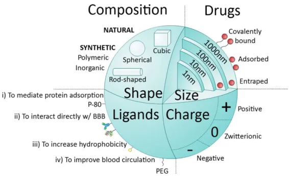
Figure 1.44Main nanoparticle (NP) features.
NPs can be classified into natural, when molecules such as proteins (albumin), polysaccharides, chitosan, among others are used, or synthetic. Synthetic NPs can be made of very common polymers such as poly(lactic-co-glycolic acid) (PLGA), poly(ethylenimine) (PEI), polyesters (poly(lactic acid) (PLA), or from inorganic agents like gold, silica or alumina. NPs can vary in size (1-1000 nm) and are able to deliver drugs into cells by entrapping, adsorbing or covalently binding them. NPs can assume different shapes (spherical, cubic, rod-like) and charges (negative, zwitterionic, positive). Another important feature of NPs is the possibility of functionalization with different types of ligands. Ligands are distributed into four major categories: i) capable of mediating protein adsorption (e.g. poly(sorbate) 80 (P-80)); ii) able to interact directly with the BBB (e.g. transferrin proteins, antibody or peptides); iii) capable of increasing hydrophobicity (e.g. amphiphilic peptides); and iv) able to improve blood circulation (e.g. poly(ethylene glycol) (PEG)).
NPs can be used not only as vehicles to deliver therapeutic agents but also as imaging agents or both. The so called theranostic agents confer diagnosis and therapy at once and normally take advantage of nanosystems that are by themselves imaging agents, such as gold, silica, iron oxide NPs, quantum dots and carbon nanotubes (Xie et al. 2010). For example, liposomes encapsulating citicoline (an inherent chemical exchange saturation transfer (CEST) magnetic resonance imaging (MRI) signal) can be detected in the brain following transient MCAO in rats in a label-free fashion, namely in the areas where BBB was disrupted (Liu et al. 2016). In fact, we have developed a NP formulation composed of material approved for other applications by the Food and Drug Administration (FDA): a polymeric agent, PLGA; a cationic peptide, protamine sulfate, which allows an efficient complexation of negatively charged molecules, namely miRNA; and perfluoro-1,5-crown ether (PFCE), a compound detectable by
19F MRI. This formulation showed to be very effective in promoting intracellular delivery of miR-132 into
endothelial cells, which subsequently exerted a pro-survival and pro-angiogenic effect in these cells when exposed to hypoxic conditions both in vitro and in vivo. Moreover, transplanted cells previously transfected with this NP formulation were tracked by 19F MRI in vivo in an ischemic limb mouse model
1.5.1 Brain delivery routes
Brain delivery of NPs can be achieved by different types of administration being the most commonly used in research the intracerebral, intranasal and/or systemic administrations (Figure 1.5). Even though, intracerebral administration is a highly invasive procedure, it guarantees delivery of high dosages of therapeutic molecules in a precise way for a long period of time. Moreover, intracerebral injections bypass issues such as immunogenicity, biodistribution, pharmacodynamics, and failure in crossing the BBB. As so, this route of administration is of major importance in pre-clinical studies specially for proof-of-principle studies. Intranasal administration has been highly studied since it is a method that circumvents the BBB. Intranasal delivery is also rapidly absorbed, non-invasive and non-destructive method of drug administration. Nevertheless, dosage, physicochemical properties of the therapeutic formulation and surface area of the nasal cavity (50% in rodents and 5% in humans) hamper the delivery of effective dosages of pharmaceuticals in most brain regions (Zhang et al. 2016c). Moreover, in pathologies such as PD, patients may present several alterations in nasal cycle, nasal mucosa pH and mucociliary clearance time (Kotan et al. 2013) that may account for reduced biodistribution and bioavailability of NPs. Systemic administration, namely intravenous injection, is much less invasive compared to intracerebral administration. Systemic delivery also allows the delivery of higher dosages of drug than the intranasal strategy, since it is a compartment with higher extent. Nevertheless, the BBB, which is a very selective barrier involved in the complex mechanisms of brain homeostasis and protection, represents the major obstacle for the passage of novel drugs targeting the brain parenchyma (Almutairi et al. 2016). Although some molecules with appropriate lipophilicity, size and charge can diffuse from blood into the brain, most molecules, both large (e.g. polypeptides, antibodies, interference RNA, miRNA) and small, are unable to overcame the BBB. As abovementioned, the use of NPs, especially when functionalized with diverse ligands can improve brain delivery through the BBB.
stem cells (neuronal repair), might enhance its potential therapeutic value. In conclusion, the development of new platforms that are able to exploit brain alterations occurring in these disorders in combination with promising therapeutic and/or imaging agents is essential to develop more efficient non-invasive and brain-directed therapies able to reach the clinic.
Figure 1.55 Administration of nanostructured materials to target the brain.
1.6 Objectives
To date, no causative therapies are available in common acute and neurodegenerative CNS disorders that can restore lost neurological function. Several regenerative medicine approaches address this issue i.e. by the use of stem cells therapies (Schapira 2009). Indeed, NSCs can be an inexhaustible source of neurons that can be recruited and/or transplanted to promote brain repair (Pluchino et al. 2003; Lindvall and Kokaia 2010). miR-124 has been described as a key neuronal fate determinant and, more recently, it has been identified as anti-apoptotic and anti-inflammatory molecule. Intracellular delivery of miRNA may be accomplished by chemical modification, liposome/microvesicle encapsulation, viral infection and electroporation. However, these strategies raise safety issues, limitations in stability and versatility, and do not allow an accurate non-invasive imaging of the
reprogramming factors’ spatial release within cells. The use of NPs constitutes a powerful platform to overcome these limitations by allowing protection, stability and spatio-temporal control of pro-neurogenic factors. To overcome these limitations, we proposed to use novel biodegradable NPs, developed by the Lino Ferreira lab that have already given proofs of an efficiently intracellular release of miRNA and can be monitored by MRI (Gomes et al. 2013).
The main goal of this project was to study the inductive effect of NPs loaded with miR-124 (miR-124 NPs) in terms of differentiation of SVZ stem/progenitor cells in vitro and to evaluate their therapeutic potential in vivo, namely in mouse models of PD and stroke. To achieve the main goal of the project we performed studies to:
1) Evaluate the effects of miR-124 NPs on SVZ NSCs in vitro, namely on cell survival, differentiation, proliferation, axonogenesis, as well as the identification of putative miR-124 molecular targets;
2) Disclose the neurogenic and functional effects of miR-124 NPs in a pre-clinical mouse model of PD;
3) Unravel the effects of miR-124 NPs on cell viability, proliferation and neurogenic potential of SVZ cultures exposed to glucose and oxygen deprivation (in vitro model of stroke);