Vol.61: e18160122, 2018
http://dx.doi.org/10.1590/1678-4324-2018160122 ISSN 1678-4324 Online Edition
BRAZILIAN ARCHIVES OF BIOLOGY AND TECHNOLOGY
A N I N T E R N A T I O N A L J O U R N A L
Solexa Profiling Identifies Differentially Expressed MiRNAs
Between Sexually Immature and Mature Equine Testis
Liangjun He
1, Shiwei Wang
2, Haifeng Deng
3, Hong Dong
1, Jingbo Chen
4*.
1Shihezi University – College of Animal Science, Shihezi, Xinjiang, China; 2Tarim University – College of Animal Science, Aral, Xinjiang, China; 3Zhaosu Horse Farm, Yili, Xinjiang, China; 4Xinjiang Academy of Animal Science –
Institute of Animal Science, Urumqi, Xinjiang, China .
ABSTRACT
MicroRNAs (miRNAs) are a class of short non-coding RNAs identified as potent regulators of gene expression. Previous studies have suggested that miRNAs are involved in mammalian spermatogenesis. Stallion fertility is an important trait for the horse breeding industry, but stallion fertility traits are largely ignored in the industry. In this study, we generated expression profiles of miRNAs in foal (immature) and stallion (mature) testes using Solexa sequencing. We identified 438 known and homologous equine miRNAs and 199 novel miRNAs which were distributed among all the chromosomes. The two developmental stages showed significant differences in miRNA expression patterns. Our result expands the horse miRNA database and provided additional information on the stallion fertility and possible spermatogenesis regulation through specific miRNAs.
Key words:horse; miRNAs; solexa; testis
INTRODUCTION
MicroRNAs (miRNAs) are short, non-coding, endogenous RNAs, 19 to 25 nucleotides (nts) in length, which have been identified as potent regulators of gene expression through post-transcriptional gene silencing. The first miRNA identified, lin-4, was found in the nematode Caenorhabditis elegans by standard positional cloning of genetic loci and is involved in developmental timing [1]. Biosynthesis of miRNAs begins with the transcription of the primary miRNA (miRNA) by RNA polymerase II. The pri-miRNA is recognized and cleaved by a microprocessor complex, Drosha and DGCR8, to produce a stem-loop RNA (pre-miRNA), which is exported from the nucleus by exportin 5 into the cytoplasm and is then cleaved by Dicer to yield a double-stranded RNA. Subsequently, the double-stranded RNA is separated into single strands, and generally one of the two strands is incorporated into the RISC complex, while the other strand is degraded [2]. Most commonly, the RISC-incorporated miRNA binds to the 3'UTR of the target mRNA by base-pairing and consequently induces either translational repression or mRNA degradation [3].
Male fertility requires that there are large numbers of normal spermatozoa in the testis, formed though a complex process known as spermatogenesis. Spermatogenesis is a precisely synchronized process, which involves mitotic cell division and propagation of spermatogonial stem cells (SSCs), meiotic division, and subsequent processing in the seminiferous tubules. The division of type A spermatogonia provides both self-renewal of SSCs and type B spermatogonia, which differentiate and divide mitotically into primary spermatocytes. During meiosis, primary spermatocytes divide into two secondary spermatocytes and then produce four haploid round spermatids, which contain half the original number of chromosomes. Finally, haploid cells undergo a morphologic transformation known as spermiogenesis to develop into mature spermatozoa.
Many studies have suggested that miRNAs are involved in spermatogenesis. The deletion of the Dicer gene (encoding an enzyme required for miRNA biogenesis) in mouse primordial germ cell results in retarded spermatogenesis, which demonstrates that this miRNA is essential for primordial germ cell and spermatogonia proliferation [4]. miRNA are also important for the late stages of spermatogenesis. The knockout of
Dicer1 in mouse germ cells causes decreasing germ cell number in the seminiferous tubules, impaired transition from round to elongated spermatids and abnormal sperm motility [5]. Immature and mature testes have different miRNA expression profiles, and many miRNAs are stage-specifically expressed in spermatogenesis. Yan et al. identified
sox5 and sox6 as presumed targets of miR-181c and rsbn1 as putative target of miR-355,
miR-181c and miR-181b [6].
Sertoli cell number [10]. Sub-fertility in stallions increases veterinary fees and management costs and ultimately diminishes the genetic contribution from prized stud horses[11]. Therefore, understanding the differential miRNA expression between mature and immature equine testes may lead to a new direction in the search for biomarkers for stallion fertility and treatments for stallion infertility.
In this study, we used Solexa deep sequencing technology to characterize and compare miRNA expression profiles between sexually mature and immature horse testis to discover miRNA biomarkers for stallion fertility. As a result, we identified 438known and homologous equine miRNAs and 199 novel miRNAs. These results show that the two testicular developmental stages have significantly different miRNA expression patterns that can be used as biomarkers of testicular maturity.
MATERIALS AND METHODS
Tissue Collection
In order to ensure the maturation of the testes, three Kazakh stallions (5–10 years old), who lived with a herd of mares (n>20) in pasture during the breeding season in previous year and the pregnancy rate of each mare herd was higher than 70%, were selected. Samples from these stallions were used as mature testes. Additional histological examination of all mature testes all showed normal spermatogenesis. Additionally, three normal Kazakh foals (immature, 5–8 months old) were castrated, and samples were collected as immature testes. The samples were immediately snap frozen in liquid nitrogen and stored in –80°C.
Small RNA Library Construction and Sequencing
Total RNA was extracted from the testes of the immature and mature testes samples using Trizol reagent (Invitrogen, USA). Subsequently, the three immature and three mature samples were pooled, respectively, to construct foal and stallion small RNA libraries. RNA quality was evaluated by an Agilent 2100 Bioanalyzer. Polyacrylamide gel electrophoresis (PAGE) was used to isolate the fraction containing RNA species of 10–40nt in length, which were then ligated with 3'- and 5'-adaptors. The adaptor-ligated RNAs were converted into single-stranded cDNAs and amplified by RT-PCR. Finally, the purified cDNA was sequenced on a Genome Analyzer according to the
manufacturer’s instructions.
Expression Estimation and Differential Expression Analysis of Mirna
homologs. We used DEGseq version 1.20.0 package in R 3.1.1.
(http://bioinfo.au.tsinghua.edu.cn/software/degseg) and set the threshold p value<0.001 under MARS (MA-plot-based method with random sampling model) method to define differentially expressed miRNAs.
Mirna Target Gene Prediction and Functional Enrichment Analysis
Given the fact many E. caballus miRNAs have not been experimentally validated, we choose to identify reliable miRNA target genes using the strategy of searching for homologous miRNAs with other species. Equine miRNA sequences were searched in BLAST to find homologous miRNAs in the humangenome. Validated targets of these homologous miRNAs were downloaded from miRTarbase (http://mirtarbase.mbc.nctu.edu.tw/), targets were retained if the support type is not
“weak”. Gene Ontology (GO) analysis of the targets of differentially expressed miRNAs
was conducted using the topGO package, and KEGG enrichment analysis was performed using the pathview package.
Quantitative Real-Time Rt-Pcr to Validate Mirna Expression
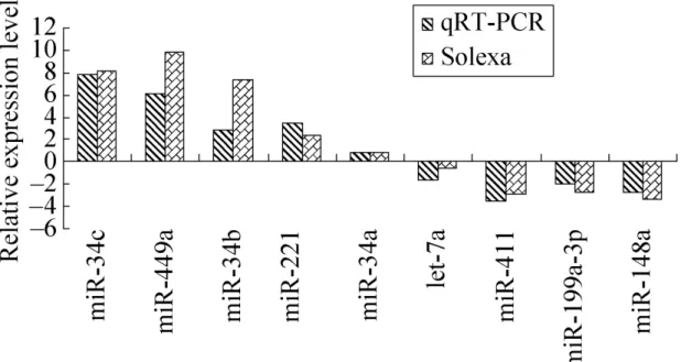
Quantitative real-time RT-PCR (qRT-PCR) was conducted to validate the miRNA expression changes, as previously described [12]. We selected nine miRNAs, including
five up-regulated miRNAs (miR-34c, miR-449a, miR-34b-5p, miR-34a and miR-221) and four down-regulated miRNAs (miR-411, miR-199a-3p, miR-let-7a and miR-148a) and examined their expression changes in the foal and stallion testis samples. miRNA-specific stem-loop RT primers, miRNA-miRNA-specific PCR forward primers and universal reverse primers were designed as previously described [13 ](Table S1). GAPDH was
used as the internal control for miRNA detection [14,15 ].
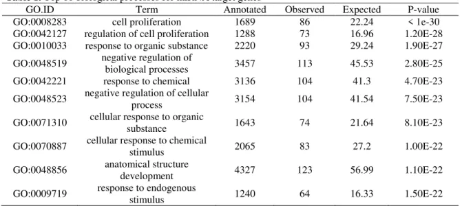
Table 1. Top 10 biological processes for miRNA target genes
GO.ID Term Annotated Observed Expected P-value
GO:0008283 cell proliferation 1689 86 22.24 < 1e-30
GO:0042127 regulation of cell proliferation 1288 73 16.96 1.20E-28 GO:0010033 response to organic substance 2220 93 29.24 1.90E-27
GO:0048519 negative regulation of
biological processes 3457 113 45.53 2.80E-25
GO:0042221 response to chemical 3136 104 41.3 4.70E-23
GO:0048523 negative regulation of cellular
process 3154 104 41.54 7.50E-23
GO:0071310 cellular response to organic
substance 1643 74 21.64 8.10E-23
GO:0070887 cellular response to chemical
stimulus 2065 83 27.2 1.00E-22
GO:0048856 anatomical structure
development 4327 123 56.99 1.10E-22
GO:0009719 response to endogenous
stimulus 1240 64 16.33 1.50E-22
One microgram total RNA was reverse-transcribed into cDNA using reverse
analysis and agarose gel electrophoresis were used to confirm the specific PCR products. The qPCR validation was carried out for three biological replicates. The 2
-△△Ct method was used to determine the expression level differences of the miRNAs for samples.
RESULTS
Small RNA Composition of the Horse Testis Libraries
We performed Solexa deep sequencing of two small RNA libraries derived from three pooled foal samples and three pooled stallion samples. We obtained 9,250,474 and 9,813,836 raw reads, respectively, from each library. After quality filtering, we had 9,176,703 clean reads in the foal library (Table S2) and 9,683,245 clean reads in the stallion library(Table S3). RNAs of 21–23 nts in length comprised 79.43% of all small RNAs in the foal library, while the stallion library showed a bimodal distribution with the major peak at 22–23 nts and a secondary peak at 25–27 nts (Fig 1).
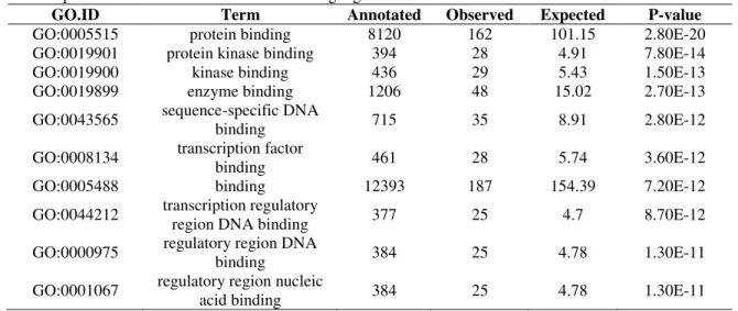
Table 2. Top 10 molecular functions for miRNA target genes
GO.ID Term Annotated Observed Expected P-value
GO:0005515 protein binding 8120 162 101.15 2.80E-20
GO:0019901 protein kinase binding 394 28 4.91 7.80E-14
GO:0019900 kinase binding 436 29 5.43 1.50E-13
GO:0019899 enzyme binding 1206 48 15.02 2.70E-13
GO:0043565 sequence-specific DNA
binding 715 35 8.91 2.80E-12
GO:0008134 transcription factor
binding 461 28 5.74 3.60E-12
GO:0005488 binding 12393 187 154.39 7.20E-12
GO:0044212 transcription regulatory
region DNA binding 377 25 4.7 8.70E-12
GO:0000975 regulatory region DNA
binding 384 25 4.78 1.30E-11
GO:0001067 regulatory region nucleic
acid binding 384 25 4.78 1.30E-11
Table 3. Top 10 enriched pathways for miRNA target genes
Pathways Annotated Significant Expected P-value
Pathways in cancer 326 40 6.83 3.60E-21
Cell cycle 124 19 2.60 5.72E-12
Chronic myeloid leukemia 73 15 1.53 1.45E-11
Bladder cancer 42 12 0.88 2.71E-11
Pancreatic cancer 70 14 1.47 1.07E-10
Small cell lung cancer 85 15 1.78 1.46E-10
Melanoma 71 13 1.49 1.65E-09
Colorectal cancer 62 12 1.30 3.72E-09
p53 signaling pathway 68 12 1.42 1.13E-08
Figure1. Small RNA length distribution and abundance in libraries of foal and stallion
Conserved Mirnas and Novel Mirnas
The stallion library had a higher percent of unannotated small RNAs (49.01%) than that of the foal (25.75%) (Tables S2 and S3).
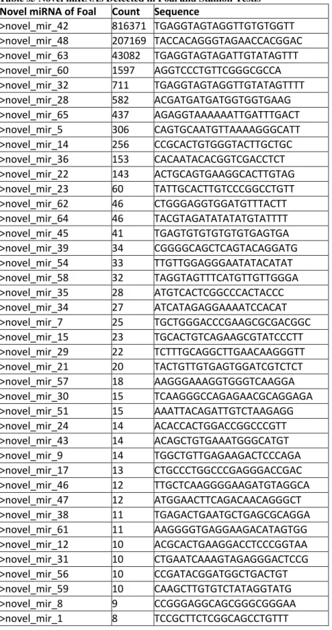
We detected438 known and conserved miRNAs in our study (374 miRNAs in the foal library and 387 miRNAs in the stallion library) (Table S4). In total, 323 miRNAs were shared between the two libraries, but we also identified 135novel candidate miRNAs (Table S5), of which 65 were found in the foals and 99 were found in the stallions. We detected expression of nine members of the let-7 family, including let-7a, let-7a-5p, let-7b-5p, let-7c, let-7d, let-7e, let-7f, let-7f-5p and let-7g. Let-7a, let-7f and let-7c
were the most abundant miRNAs in the two libraries. Total read counts of the let-7
family were 4,864,334 and 2,546,959 in the foal and stallion libraries, respectively, which accounted for 82.67% and 83.45% of total known and conserved miRNA sequences in the corresponding libraries. This is consistent with a previous report that the members of the let-7 family are highly abundant and conserved miRNAs among both plants and animals [16 ].
Analysis and Validation of Differentially Expressed Mirnas
We found 180 differentially expressed miRNAs between foal and stallion testes. Of these, 105 miRNAs were significantly down-regulated and 75 miRNAs were up-regulated in stallion testes compared with foal testes (Figure 2 and Table S6).
Figure 2. The volcano plot of differentially expressed miRNAs. The X-axis shows the log2-tran
Figure 3. Validation of the sequencing results by qRT-PCR. The x-axis represents the miRN
Mirnas Preferentially Expressed in Equine Testis
we identified five preferentially expressed miRNAs in testis, which are eca-let-7a, eca-let-7f, eca-miR-432, eca-miR-503 and eca-miR-125a-5p. These miRNAs could be further used as biomarkers of horse testes.
Functional Analysis of Differentially Expressed Mirnas
We selected 15 abundant and differentially expressed miRNAs that had homologs in the human genome to find validated target genes and perform KEGG pathway analysis. As a result, 417 high confident validated miRNA-mRNA interactions were identified and further functional analysis are based on the validated genes (Table S8).
Interestingly, GO enrichment analysis showed that the target genes were highly involved in developmental and regulatory roles (Table 1 and Table 2). Two essential genes for self-renewal (SOX2 and NANOG) and three important genes involved in apoptosis (MYC, BCL2 and BBC3) were identified, which is consistent with results from mouse testes [6].
Consistent with the predicted target genes of differentially expressed miRNAs between porcine mature and immature testes [17], the KEGG pathway annotation also showed that the most enriched pathways were for cancer. This is not surprising considering the role of these miRNAs in development as seen in the GO analysis (TableS12).
DISCUSSION
The foal library has a length distribution peaked at 22 nt, a common length for miRNAs. The stallion library showed a bimodal distribution peaked at 21-23nt and at 25-27nt. Both agree with previous studies of immature and mature pig [17] and human [18] testis. According to studies of human RNAs, the second represents piRNAs, which are another class of small RNAs, mostly found in germ cells and required for spermatogenesis [19]. Since there is no known horse piRNA data bank, so many miRNA at the secondary peak
didn’t matched to piRNAs and was classified as unannotated small RNAs. It explains
why the stallion library had higher percent of unannotated small RNAs than the foal library.
The difference in miRNA profiles between immature and mature testes has been studied in mouse [6], pig [17] and dog [20] using various techniques, including qRT-PCR, microarrays and Solexa sequencing. We compared our results with the miRNA expression seen in mouse and pig testes. In a study comparing miRNA expression between sexually mature and immature porcine testes, 96 significantly up-regulated miRNAs and 26 significantly down-regulated miRNAs were identified in the sexually mature porcine testes. We found six overlapping up-regulated miRNAs (miR-449a,
miR-449b, miR-34c-5p, miR-34c, miR-34b-3p, miR-34b-5p and miR-184) and three overlapping down-regulated miRNAs (miR-411, miR-487b and miR-485-5p) between porcine and equine mature testes. Interestingly, five of the six overlapping up-regulated miRNAs were among the top ten up-regulated miRNAs and belong to two miRNA families, the miR-34 family and the miR-449 family. However, the overlapping down-regulated miRNAs showed no specific pattern among the fold-change ranking. Using a microarray approach, another study compared the miRNA expression between immature and mature murine testes [6] and revealed that miR-34b-5p, miR-34c-5p and
In these studies, the miR-34b, miR-34c and miR-449 families were the top up-regulated families in sexually mature testis. In the present study, miR-34b-3p, miR-34b-5p, miR-34c, miR-449a and miR-449b were the most up-regulated miRNAs in stallion testis. One previous study has reported that miR-449 was preferentially expressed in murine testis, with the highest levels in spermatocytes and spermatids [21]. In mouse testis, miR-34c was expressed at very low levels before p12 (the prepubertal period), after which its expression increased sharply and persisted until adulthood in the mouse. Furthermore, this miRNA was localized in pachytene spermatocytes and was highly expressed in spermatids [22]. Bouhallier [23] found that miR-34c highly expressed in pachytene spermatocytes and round spermatids, indicating that miR-34c was meiosis-specific. In human seminal plasma, miR-34c-5p was markedly decreased in azoopermia and increased in asthenopermia [24]. Additionally, miR-34b-3p, miR-34b-5p and
miR-34c-5p were found to be significantly decreased in subfertile and nonobstructive azoopermatic males. Administration of a CYP26B1 inhibiter, which intervened in retinoic acid-mediated spermatogenesis, resulted in significantly up-regulated miR-34b
and miR-34c-5p in cultured canine testicular parenchyma [25]. MiR-34b, miR-34c and
miR-449 family are similar in nucleotide sequence and have identical seed sequence [21]. In mouse testis, miR-449 deletion up-regulated miR-34b and miR-34c expression but mouse spermatogenesis is not affected, which suggested miR-34b, miR-34c and miR-449 family is functionally redundant in murine testis [21].
Among the predicted miRNA target genes, CDK4, CCNE2, BCL2, and NOTCH1 were targeted by multiple differentially expressed miRNAs. CDK4 is an important regulator for the G1 to S phase cell cycle transition. The majority of Cdk4-/-male mice displayed infertility [26], and Cdk4
-/-male mice showed age-dependent testes block in spermatogenesis [27]. CCNE2 is a G1 cyclin that associates with Cdk2 and is required for driving cells to enter the S phase of the cell cycle and for cell proliferation. CCNE2 -
male mice displayed reduced fertility, testicular size and sperm count and frequent abnormal meiosis in spermatocytes, suggesting that CCNE2 is essential for normal spermatogenesis [28]. BCL2 is a member of the Bcl-2 family, which regulates apoptosis. Bcl-2 has been shown to be targeted by has-miR-34b, has-miR-34c and has-miR-449a. Yamamoto reported that overexpression of Bcl-2 in transgenic mice testis impaired spermatogenesis [29]. The expression of exogenous Bcl-2 in spermatogonia resulted in abnormal accumulation of spermatogonia and degeneration of germ cells, indicating that apoptosis is essential for normal spermatogenesis [30]. NOTCH1 is one of the validated target genes for has-miR-34b, has-miR-34c and has-miR-449a in miRTar base. It has been reported that NOTCH1 is critical for germ cell development and differentiation in rat testis, and NOTCH1 is not detected in the testes of patients with spermatogenic maturation arrest [31]. In summary, the GO term and KEGG pathway annotation for differentially expressed miRNAs suggests likely roles for these miRNAs in spermatogenesis.
In this study, there were 77 miRNAs derived from genes on the X chromosome, of which 29 were up-regulated and 18 were down-regulated in the stallion compared to the
foal. Using Fisher’s test, we observed that significantly more differentially expressed
miRNA is present on the X chromosome (P = 0.0051).
a considerable percent of all up-regulated miRNAs in that sample, which suggests that the miR-8908 family may be essential for equine spermatogenesis.
Besides the 10 up-regulated miR-8908 family members,there were 29 up-regulated X-linked miRNAs, which accounted for 39% of all up-regulated miRNAs (29/75) in stallion testis, a significantly high ratio compared to the whole genome. It has been reported that some miRNAs from the X chromosome escape meiotic sex chromosome inactivation during spermatogenesis [32]. Those miRNAs were found in spermatocytes from the mid- to late pachytene stage of spermatogenesis. Our finding of a high percentage of up-regulated X-linked miRNAs is consistent with this report.
CONCLUSIONS
We compared the expression profiles of miRNAs in foal (immature) and stallion (mature) testes using Solexa sequencing. These differentially expressed miRNAs will help us to better understand the mechanisms involved in spermatogenesis and may lay the foundation for research into stallion infertility.
REFERENCES
1. Lee RC, Feinbaum RL, Ambros V. The C. elegans heterochronic gene lin-4 encodes small RNAs with antisense complementarity to lin-14. Cell. 1993;75:843–54.
2. Lee Y, Ahn C, Han J, Choi H, Kim J, Yim J, et al.The nuclear RNase III drosha initiates microRNA processing. Nature. 2003; 425:415–9.
3. Kim VN.MicroRNA biogenesis: coordinated cropping and dicing. Nat Rev Mol Cell Biol. 2005; 6: 376-85.
4. Hayashi K, Sousa-Lopes SMC, Kaneda M, Tang F, Hajkova P, Lao K, et al. MicroRNA biogenesis is required for mouse primordial germ cell development and spermatogenesis. PLoS One. 2008; 3: e1738.
5. Maatouk DM, Loveland KL, McManus MT, Moore K, Harfe BD. Dicer is required for differentiation of the mouse male germline. Biol Reprod. 2008;79:696–703.
6. Yan NH, Lu YL, Sun HQ, Tao DC, Zhang SZ, Liu WY, et al. A microarray for microRNA profiling in mouse testistissues. Reproduction. 2007; 134: 73-9.
7. Zhou M. In silico detection and characteristics of novel microRNA genes in the Equus caballus genome using anintegrated ab initio and comparative genomic approach. Genomics. 2009; 94:125-31.
8. Kim MC, Lee SW, Ryu DY, Cui FJ, Bhak J, Kimc Y. Identification and characterization of microRNAs in normal Equine tissues by next generation sequencing. PLoS ONE. 2014; 9:e93662.
9. Das PJ, Mccarthy F, Vishnoi M, Paria N, Gresham C, Li G, et al. Stallion sperm transcriptome comprises functionally coherent coding and regulatory RNAs as revealed by microarray analysis and RNA-seq. PLoS ONE. 2013;8:e56535.
10. Johnson L, Varner DD, Thompson DL Jr. Effect of age and season on the establishment of spermatogenesis in the horse. J Reprod Fertil. 1991; 44(Suppl):87-97.
11. Turner R. Oristaglio. Pathogenesis, diagnosis, and management of testicular degeneration in stallions. Clin TechEquine Pract. 2007; 6:278-84.
12. Varkonyi-Gasic E, Wu R, Wood M, Walton EF, Hellens RP. Protocol: a highly sensitive RT-PCR method for detection and quantification of microRNAs. Plant Methods. 2007; 3:12. 13. Chen C, Ridzon DA, Broomer AJ, Zhou Z, Lee DH, J. Nguyen T, et al. Real-time
quantification of microRNAs by stem–loop RT–PCR. Nucleic Acids Res. 2005; 33:e179. 14. Ing N H, Laughlin A M, Varner D D, et al. Gene expression in the spermatogenically inactive
15. Shields J E, Kochan K J, Jeong J, et al. Initial characterization of a gene abundantly expressed in stallion testis. J Equine Vet Sci, 2009, 29(5): 324-5.
16. Rouch S, Slack FJ.The let-7 family of microRNAs. Trends Cell Biol. 2008; 18: 505-516. 17. Lian CJ, Sun BX, Niu SL, Yang RJ, Liu BY, Lu CY, et al.A comparative profile of the
microRNA transcriptome in immature and mature porcine testes using solexa deep sequencing. FEBS Joural. 2012;279:964-75.
18. Yang QL, Hua J, Wang L, Xu B, Zhang H, Ye N, et al. MicroRNA and piRNA profiles in normal human testis detected by next generation sequencing. Plos ONE.2013; 6:e66809. 19. Kim VN. Small RNAs just got bigger: piwi-interacting RNAs (piRNAs) in mammalian
testes. Gene Dev. 2006; 20:1993-1997.
20. Kasimanickam V R, Kasimanickam RK. Differential expression of microRNAs in sexually immature and mature canine testes. Theriogenology. 2015; 83:394-8.
21. Bao JQ, Li D, Wang L, Wu JW, Hu YQ, Wang ZG, et al.449 and MicroRNA-34b/c Function Redundantly in Murine Testes by Targeting E2F Transcription Factor-Retinoblastoma Protein (E2F-pRb) Pathway. J Biol Chem. 2012; 287: 21686-98.
22. Liang X, Zhou D, Wei C, Luo H, Liu J, Fu R, et al. MicroRNA-34c enhances murine male germ cell apoptosis through targeting ATF1. PLoS ONE. 2012; 7: e33861.
23. Bouhallier F, Allioli N, Lavial F. Role of miR-34c microRNA in the late steps of spermatogenesis. RNA. 2010; 16:720-731.
24. Abu-Halima M, Hammadeh M, Schmitt J, Leidinger P, Keller A, Meese E, et al. Altered microRNA expression profiles of human spermatozoa in patients with different spermatogenic impairments. Fertil Steril. 2013; 99:1249-55.
25. Kasimanickam VR, Kasimanickam RK. Dysregulated microRNA clusters in response to retinoic acid and CYP26B1 inhibitor induced testicular function in dogs. PLoS ONE. 2014; 9:e99433.
26. Rane SG, Dubus P, Mettus RV, Galbreath EJ, Boden G, Reddy EP, et al. Loss of Cdk4 expression causes insulin-deficient diabetes and Cdk4 activation results in beta-islet cell hyperplasia. Nat Genet. 1999; 22:44–52.
27. Mettus RV, Rane SG. Characterization of the abnormal pancreatic development, reduced growth and infertility in Cdk4 mutant mice. Oncogene. 2003; 22:8413–21.
28. Geng Y, Yu Q, Sicinska E, Das M, Schneider JE, Bhattacharya S, et al.Cyclin e ablation in the mouse. Cell. 2003; 114:431-43.
29. Yamamoto CM, Hikim AP, Lue Y, Portugal AM, Guo TB, Hsu SY, et al.Impairment of spermatogenesis in transgenic mice with selective overexpression of Bcl-2 in the somatic cells of the testis. J Androl. 2001; 22: 981–91.
30. Furuchi T, Masuko K, Nishimune Y, Obinata M, Matsui Y. Inhibition of testicular germ cell apoptosis and differentiation in mice misexpressing Bcl-2 in spermatogonia. Development. 1996; 122:1703-9.
31. Hayashi T, Kageyama Y, Ishizaka K, Xia G, Kihara K, Oshima H. Requirement of Notch 1 and its ligand jagged 2 expressions for spermatogenesis in rat and human testes. J Androl. 2001; 22:999–1011.
32. Song R, Ro S, Michaels JD, Park C, McCarrey JR. Many X-linked microRNAs escape meiotic sex chromosome inactivation. Nat Genet. 2009; 41:488 – 93.
Table s5 Novel miRNAs Detected in Foal and Stallion Testis
Novel miRNA of Foal Count Sequence
>novel_mir_42 816371 TGAGGTAGTAGGTTGTGTGGTT
>novel_mir_48 207169 TACCACAGGGTAGAACCACGGAC
>novel_mir_63 43082 TGAGGTAGTAGATTGTATAGTTT
>novel_mir_60 1597 AGGTCCCTGTTCGGGCGCCA
>novel_mir_32 711 TGAGGTAGTAGGTTGTATAGTTTT
>novel_mir_28 582 ACGATGATGATGGTGGTGAAG
>novel_mir_65 437 AGAGGTAAAAAATTGATTTGACT
>novel_mir_5 306 CAGTGCAATGTTAAAAGGGCATT
>novel_mir_14 256 CCGCACTGTGGGTACTTGCTGC
>novel_mir_36 153 CACAATACACGGTCGACCTCT
>novel_mir_22 143 ACTGCAGTGAAGGCACTTGTAG
>novel_mir_23 60 TATTGCACTTGTCCCGGCCTGTT
>novel_mir_62 46 CTGGGAGGTGGATGTTTACTT
>novel_mir_64 46 TACGTAGATATATATGTATTTT
>novel_mir_45 41 TGAGTGTGTGTGTGTGAGTGA
>novel_mir_39 34 CGGGGCAGCTCAGTACAGGATG
>novel_mir_54 33 TTGTTGGAGGGAATATACATAT
>novel_mir_58 32 TAGGTAGTTTCATGTTGTTGGGA
>novel_mir_35 28 ATGTCACTCGGCCCACTACCC
>novel_mir_34 27 ATCATAGAGGAAAATCCACAT
>novel_mir_7 25 TGCTGGGACCCGAAGCGCGACGGC
>novel_mir_15 23 TGCACTGTCAGAAGCGTATCCCTT
>novel_mir_29 22 TCTTTGCAGGCTTGAACAAGGGTT
>novel_mir_21 20 TACTGTTGTGAGTGGATCGTCTCT
>novel_mir_57 18 AAGGGAAAGGTGGGTCAAGGA
>novel_mir_30 15 TCAAGGGCCAGAGAACGCAGGAGA
>novel_mir_51 15 AAATTACAGATTGTCTAAGAGG
>novel_mir_24 14 ACACCACTGGACCGGCCCGTT
>novel_mir_43 14 ACAGCTGTGAAATGGGCATGT
>novel_mir_9 14 TGGCTGTTGAGAAGACTCCCAGA
>novel_mir_17 13 CTGCCCTGGCCCGAGGGACCGAC
>novel_mir_46 12 TTGCTCAAGGGGAAGATGTAGGCA
>novel_mir_47 12 ATGGAACTTCAGACAACAGGGCT
>novel_mir_38 11 TGAGACTGAATGCTGAGCGCAGGA
>novel_mir_61 11 AAGGGGTGAGGAAGACATAGTGG
>novel_mir_12 10 ACGCACTGAAGGACCTCCCGGTAA
>novel_mir_31 10 CTGAATCAAAGTAGAGGGACTCCG
>novel_mir_56 10 CCGATACGGATGGCTGACTGT
>novel_mir_59 10 CAAGCTTGTGTCTATAGGTATG
>novel_mir_8 9 CCGGGAGGCAGCGGGCGGGAA
>novel_mir_11 8 TGGAGGCAGCGAGGAGCACGT
>novel_mir_16 8 TCAGGCTCAGTCCCCTCCCGAT
>novel_mir_20 8 TATATATATATATGTACGTAT
>novel_mir_52 8 TACAGTACTGTGATAACTGAAGGA
>novel_mir_18 7 AGGGGAGAAGGTAAGCAGAGA
>novel_mir_19 7 ACTAGGACAAGGAAGCTGGCAG
>novel_mir_33 7 CACAGAACTTTTAACCAGTAGGCC
>novel_mir_37 7 TGATGAACTTATGAGGAGCTGCTA
>novel_mir_44 7 CAGGAGGAAGACTGAGGTGGAA
>novel_mir_53 7 TCTCAGTATAGAATTCCGCGGGC
>novel_mir_55 7 CGTGGTGCGGGACGGAGCGGA
>novel_mir_25 6 TCAATGTCTGTGAGTCGGCAGCTA
>novel_mir_26 6 ACTGACTGGGAATGAAAGGCTG
>novel_mir_27 6 TCACGCGGTGAGAGGAAGGACC
>novel_mir_3 6 ACCTTGGCTCTAGACTGCTTACT
>novel_mir_4 6 TGATAGAATGTTTGCACCAGTGAC
>novel_mir_49 6 ACCCGGGCTCTGTGGGCAGGCGG
>novel_mir_10 5 AGGAATTGCAGGGGCATCTTTATC
>novel_mir_13 5 TTGGCTCTGTGAGGTCGGCTCA
>novel_mir_2 5 TCGGAGTCAGGAACGGCGTCTGGC
>novel_mir_40 5 AAAGTTGGCAGATGGTGAGTGAA
>novel_mir_41 5 AGGAGTCTTGTTTCTACTTT
>novel_mir_50 5 TCCTTGATCTGGGTTGGCTGAG
>novel_mir_6 5 TCTGTGTAGATGACATCGTGTTC
Novel miRNA of Stallion Count Sequence
>novel_mir_42 582547 TGAGGTAGTAGGTTGTGTGGTT
>novel_mir_48 66380 TACCACAGGGTAGAACCACGGAC
>novel_mir_63 33011 TGAGGTAGTAGATTGTATAGTTT
>novel_mir_32 2420 TGAGGTAGTAGGTTGTATAGTTTT
>novel_mir_60 1951 AGGTCCCTGTTCGGGCGCCA
>novel_mir_36 572 CACAATACACGGTCGACCTCT
>novel_mir_28 541 ACGATGATGATGGTGGTGAAG
>novel_mir_65 219 AGAGGTAAAAAATTGATTTGACT
>novel_mir_1 123 TCCGCTTCTCGGCAGCCTGTTT
>novel_mir_120 99 TTTGTTCGTTCGGCTCGCGTGA
>novel_mir_14 82 CCGCACTGTGGGTACTTGCTGC
>novel_mir_122 69 TTCATTGTAGGAAGTTCAGGAGTCAC
>novel_mir_5 69 CAGTGCAATGTTAAAAGGGCATT
>novel_mir_23 60 TATTGCACTTGTCCCGGCCTGTT
>novel_mir_133 52 TAGGAACTTGTGCGGCAGGGATTT
>novel_mir_112 50 AAAGGAACATGTGGCAGCCAGGTG
>novel_mir_22 49 ACTGCAGTGAAGGCACTTGTAG
>novel_mir_83 47 TGGGTAAACTGATTAGCTGGCATC
>novel_mir_64 42 TACGTAGATATATATGTATTTT
>novel_mir_76 42 GCGGACGCGATGGCAGGCAGCAGG
>novel_mir_41 41 AGGAGTCTTGTTTCTACTTT
>novel_mir_107 39 TGAAGCAGAGCGCACGAACTCAA
>novel_mir_101 38 TGTAGTTGGAAGGATGCCCCTGGA
>novel_mir_130 38 TGGAAAACTGAGGACTGTCCGGCA
>novel_mir_111 29 TGGACAAGCTGGATTTCAAGT
>novel_mir_121 25 TACAAAATGGAGGAAGATCGGCAC
>novel_mir_62 25 CTGGGAGGTGGATGTTTACTT
>novel_mir_126 23 TGTGTGTAAGTGGGGAAGCTGG
>novel_mir_66 23 TTTGAAGACAAGCATAGCCTCATT
>novel_mir_105 22 TCTGGGTTAGACTGACGGCTTCG
>novel_mir_71 21 GCGCGCCGGCGTCCCGGGGGG
>novel_mir_92 20 TTCTAGTTGTGGCATATGGGAA
>novel_mir_94 20 TGCAGAGCACCGGCCACTGTGGGT
>novel_mir_128 19 CTGAGGAGATGTGGGAGAAGT
>novel_mir_35 19 ATGTCACTCGGCCCACTACCC
>novel_mir_81 19 AGGAAGCTGAGGCACATGGAAGTT
>novel_mir_8 17 CCGGGAGGCAGCGGGCGGGAA
>novel_mir_87 17 TCCAGGAAGGCGGGCACCAGGTG
>novel_mir_118 16 TGGCTGTTAGTTGAAATCCCGGTG
>novel_mir_54 16 TTGTTGGAGGGAATATACATAT
>novel_mir_116 15 AGGGTTTGTAGAGTGCAGCCGGC
>novel_mir_70 15 TGCAAGTAGGAAGACAGTGGGCTC
Braz. Arch. Biol. Technol. v.61: e18160122 2018
>novel_mir_88 15 TGTAAAATGAGAGGGTTGGACTAA
>novel_mir_119 13 TGATTGTTCAGAGGGGATGAAGC
>novel_mir_39 13 CGGGGCAGCTCAGTACAGGATG
>novel_mir_58 13 TAGGTAGTTTCATGTTGTTGGGA
>novel_mir_95 13 TAAGACGTATAGGCTGGTTCGGTA
>novel_mir_108 12 TGCAAAGCAGGAACGCTGGCCTC
>novel_mir_11 12 TGGAGGCAGCGAGGAGCACGT
>novel_mir_131 12 TGAATGTTGTGCAGCACTCCAA
>novel_mir_16 12 TCAGGCTCAGTCCCCTCCCGAT
>novel_mir_91 12 TGCCCCACAGGGAGATCCCGGA
>novel_mir_134 11 TCAGAGCAAGTAGTAATTCGAGA
>novel_mir_29 11 TCTTTGCAGGCTTGAACAAGGGTT
>novel_mir_82 11 TACTGTTGTGAGTGGATCGTCTC
>novel_mir_93 11 TCTCTTAGGGTGAAGTCTCGGGC
>novel_mir_102 10 TGCAGATGTCGGCACGAAGGATTT
>novel_mir_103 10 TGATAGACAGCGAGGAACCT
>novel_mir_124 10 TCCCATGAGATTCTGAGGCCAGGT
>novel_mir_51 10 AAATTACAGATTGTCTAAGAGG
>novel_mir_80 10 TCCAGGACTGTGGGGGCGCC
>novel_mir_89 10 TCCTAAGAGACTGAACAGAGGGA
>novel_mir_98 10 AGGGGCTGGGATTGGGGCAGGG
>novel_mir_106 9 TGGATGCAGTTTCTGGCAGAGCTC
>novel_mir_125 9 GTGGAGCTCTGGATCCAGGT
>novel_mir_127 9 TCTGATCACCCTGGGGCCCGACT
>novel_mir_132 9 AAGGGGTGAGGAAGACATAGTG
>novel_mir_135 9 TTCCTGTGGACATGGTGGGTGGG
>novel_mir_69 9 TCTCAGAAGCAGACCAAGGAATTC
>novel_mir_78 9 TGGGAAGCACCGGGGGACATCT
>novel_mir_110 8 TAGGGCTCGGTAACTAGGAGTGGA
>novel_mir_113 8 TTACAAGTAGGAGCATCTGTGTTT
>novel_mir_20 8 TATATATATATATGTACGTAT
>novel_mir_75 8 AGCAGATTTAGGGGATCAGGA
>novel_mir_84 8 TTACGTTCAAGCTCATCTCAGGTA
>novel_mir_100 7 AGCGTGGTTTGATGAGCAGG
>novel_mir_114 7 TAAGATGAGGAGAAAGGATTTGA
>novel_mir_67 7 TCGTGAAGTCGCTGGAGATGAGGC
>novel_mir_68 7 TGAACTCAGAAGTAATTATGGACC
>novel_mir_73 7 TCCTAGGGGTTGAATGATTGGCT
>novel_mir_74 7 TCACAAGATGTTGGGATGGCGGGT
>novel_mir_77 7 TGGGGACGGCCTGGAGATAAAGG
>novel_mir_86 7 TGACATGGAGCAGCACGCCGAGGC
>novel_mir_99 7 TGTAAGTCAAGCTCTCTGAGGC
>novel_mir_104 6 TGAATGGACAGATGGATGGATG
>novel_mir_129 6 TGGAATAATCAGATGTGTAGGGGC
>novel_mir_96 6 TTGCAGTGATGACTTGAGTCTGT
>novel_mir_115 5 TGGCAGCTTGGATCACTTGAGTCT
>novel_mir_117 5 TGCTTGTCAGTGGTCTCAGGAAA
>novel_mir_123 5 CCGGAAGTAAGGCGTCTCTTCCTG
>novel_mir_3 5 ACCTTGGCTCTAGACTGCTTACT
>novel_mir_34 5 ATCATAGAGGAAAATCCACAT
>novel_mir_43 5 ACAGCTGTGAAATGGGCATGT
>novel_mir_72 5 TAACAGTCTCCAGTCACGGCCA
>novel_mir_79 5 TTGGCTCTGTGAGGTCGGCTCAA
>novel_mir_90 5 TGAGAGACGGTGGCAGGAAGTGT
>novel_mir_97 5 TGGGGTGGGGCTGGGGAGGGC