Março de 2014
The role of Hedgehog signaling during zebrafish larvae
fin fold regeneration
Dissertação para obtenção do Grau de Mestre em Genética Molecular e Biomedicina
Orientadora: Susana Alexandra Rodrigues Pascoal, Doutora, Instituto de Medicina
Molecular, Faculdade de Medicina da Universidade de Lisboa
Rita Joana Soares Serrano
Licenciada em Biologia
Júri:
Presidente: Prof. Doutora Paula Maria Theriaga Mendes Bernardo Gonçalves Arguente: Prof. Doutora Susana Santos Lopes
Março de 2014
The role of Hedgehog signaling during zebrafish larvae
fin fold regeneration
Dissertação para obtenção do Grau de Mestre em Genética Molecular e Biomedicina
Orientadora: Susana Alexandra Rodrigues Pascoal, Doutora, Instituto de Medicina
Molecular, Faculdade de Medicina da Universidade de Lisboa
Rita Joana Soares Serrano
Licenciada em Biologia
Júri:
Presidente: Prof. Doutora Paula Maria Theriaga Mendes Bernardo Gonçalves Arguente: Doutora Susana Santos Lopes
The role of Hedgehog signaling during zebrafish larvae fin fold regeneration
Copyright Rita Joana Soares Serrano, FCT/UNL, UNL
i
Acknowledgements/Agradecimentos
Primeiro, gostaria de agradecer à minha orientadora, Dra. Susana Pascoal, pela disponibilidade para me ensinar durante este ano e por todo o apoio, paciência e encorajamento transmitidos que foram sem dúvida fundamentais para este trabalho. Muito Obrigada!
Gostaria, também, de agradecer à Dra. Leonor Saúde pela simpatia com que sempre me recebeu, pelos conselhos e acompanhamento ao longo deste projeto. Obrigado pela oportunidade de integrar um grupo tão dinâmico com quem pude aprender e crescer.
Aos meus colegas de laboratório: Margarida Figueira, Rita, Raquel, Margarida Pereira, Sara, Ana e Zé, um muito obrigado pelo companheirismo, pela preocupação, pela motivação e ajuda quando precisa. Ao João Pereira, um obrigado por acreditar em mim, por ser sempre motivador e estar sempre lá! Obrigado por tudo o que me ensinaste. À Rita Fior, um muito obrigado por me ter mostrado pela primeira vez o que era a Biologia do Desenvolvimento e que era este o meu caminho…
Gostaria, também, de agradecer a todas as pessoas que contribuíram para este trabalho: Aida, Lara e Sara Matos, obrigado por cuidarem dos meus peixes e por vezes também de mim. Obrigado pelos sorrisos e simpatia nos bons e maus momentos. Ao António Temudo e à Ana Nascimento, agradeço todos os ensinamentos sobre microscopia, Photoshop, e muito mais, que foram essenciais para este trabalho. Obrigado pela paciência e disponibilidade. À Andreia e Margarida Pinto, agradeço
nunca terem desistido dos meus cortes histológicos…foram meses e meses de tentativas e estiveram sempre disponíveis para tentar algo diferente.
Um especial obrigado a toda a minha família por sempre ter acreditado em mim e no meu empenho, por me ter permitido vir estudar para Lisboa e seguir o meu caminho. Mami, obrigado por seres sempre compreensiva e por toda a motivação. Guga, a tia agora já pode brincar contigo!
Finalmente, um muito obrigado ao Pedro. Não há palavras suficientes para expressar o quanto significas para mim. Obrigado por sempre me apoiares e nunca me deixares cair.
“Embrace the mystery of it all…”
iii
Abstract
Several studies have demonstrated that although the structure of the adult and larval zebrafish caudal fin is different, there are similarities at the cellular and molecular level that turn larval zebrafish fin fold a useful model to study the basic principles of regeneration. In this process, while the essential role for Hedgehog (Hh) signaling is well established in the adult zebrafish caudal fin system, its involvement in juvenile tissue regeneration is still unknown. The aim of this Master thesis was therefore to evaluate the contribution of the Hh signaling pathway to the larval zebrafish fin fold regeneration process. Accordingly, we analyzed the expression of several Hh signaling components through in situ hybridization. Here, we showed that several of these genes are effectively expressed in
the larval regenerating fin tissue, suggesting a role for Hh signaling also during larval regeneration. However, divergence in the regulation of few Hh signaling components appears to exist between the adult and larval zebrafish fin regeneration processes. Nevertheless, similarly to adult caudal fin regeneration, when Hh signaling was blocked, by using cyclopamine, the larval fin fold regenerative outgrowth is severely impaired.
Since larval zebrafish fin fold is ciliated, and primary cilia are closely related to Hh signaling regulation in vertebrate systems, we further addressed the role of primary cilia during larval fin fold regeneration process. To this end, we used the zebrafish iguana mutant, in which primary cilia are not
formed, to study the modulation of Hh signaling expression during larval fin fold regeneration in the absence of primary cilia. Here, we found that several genes were expressed with a delay, coincident with the delay in the mutant fin fold regeneration observed in previous work.
We show that Hh signaling in the fin fold is crucial to promote cell proliferation. When Hh signaling is blocked using cyclopamine there is a strong blockage of cell proliferation and regeneration is also blocked. Surprisingly, in iguana mutants where Hh signaling is impaired but not totally
blocked, cell proliferation is not detected but regeneration still occurs. This raises the question about the requirement of cell proliferation in larvae fin fold regeneration. By blocking the cell cycle using aphidicolin we demonstrate that cell proliferation is not necessary for zebrafish larvae fin fold regeneration.
.
v
Resumo
Vários estudos têm demonstrado que, apesar da estrutura da barbatana caudal do peixe-zebra adulto e da larva ser diferente, existem semelhanças ao nível celular e molecular entre o processo regenerativo de ambos, que tornam a barbatana caudal da larva de peixe-zebra um bom modelo para estudar os princípios básicos da regeneração. Neste processo, enquanto o papel da sinalização Hedgehog (Hh) na barbatana caudal do peixe-zebra adulto está bem estabelecido, o seu envolvimento na regeneração da barbatana caudal da larva é desconhecido. O principal objetivo desta tese de Mestrado foi, assim, avaliar o contributo da via de sinalização Hh para o processo regenerativo da barbatana caudal da larva de peixe-zebra. Nesse sentido, analisámos a expressão de vários componentes da sinalização Hh através de hibridação in situ. Esta análise permitiu verificar que vários
componentes da via Hh são efetivamente expressos no tecido regenerado da barbatana caudal da larva, sugerindo que a via Hh tem um papel importante durante este processo. No entanto, parecem existir divergências na regulação de alguns componentes da via de sinalização Hh entre os processos regenerativos do peixe-zebra adulto e da larva. Contudo, através do estudo da regeneração na ausência de sinalização Hh demonstramos, ainda, que a via Hh é essencial para regular o crescimento regenerativo da barbatana caudal de larvas de peixe-zebra tal como no adulto.
Dado que a barbatana caudal da larva de peixe-zebra é ciliada, e os cílios primários estão relacionados com a transdução do sinal da via Hh em sistemas de vertebrados, o nosso segundo objetivo foi tentar compreender o papel dos cílios primários durante o processo de regeneração da barbatana caudal de larvas de peixe-zebra. Para tal, utilizamos o mutante iguana, nos quais os cílios
primários não se formam, para estudar a modulação da expressão da via durante este processo. Este estudo permitiu verificar que vários genes da via Hh são expressos com um atraso, que coincide com o atraso observado durante o processo de regeneração do mutante iguana em estudos anteriores.
Finalmente, neste trabalho é sugerido pela primeira vez que a proliferação celular não é necessariamente obrigatória para o processo regenerativo da barbatana caudal da larva de peixe-zebra.
vii
Table of Contents
Acknowledgements/Agradecimentos ... i
Abstract ... iii
Resumo ... v
Table of Contents ... vii
List of Figures ... ix
List of Tables ... xi
List of Abbreviations ... xiii
Chapter 1: Introduction ... 1
1.1 Regeneration in the zebrafish model ... 1
1.1.1 Adult zebrafish fin regeneration ... 2
1.1.2 Molecular signaling involved in fin regeneration ... 2
1.1.3 Early life stage zebrafish model ... 3
1.2 The Hedgehog signaling pathway ... 5
1.2.1 Mechanism of vertebrate Hedgehog signaling ... 5
1.2.2 Hedgehog signaling through primary cilia ... 7
1.3 The zebrafish iguana mutant ... 12
1.4 Aims... 13
Chapter 2: Materials and Methods ... 15
2.1 Zebrafish lines and husbandry ... 15
2.2 Fin fold amputation ... 15
2.3 Drug treatments ... 16
2.3.1 Cyclopamine treatment ... 16
2.3.2 Aphidicolin treatment ... 17
2.4 Detection of mitotic nuclei with Bromodeoxyuridine ... 17
2.4.1 Bromodeoxyuridine (BrdU) incorporation ... 17
2.4.2 Immunohistochemical detection of BrdU ... 17
2.5 In situ hybridization ... 18
2.5.1 RNA probes synthesis ... 18
2.5.2 Prevention of pigmentation development ... 20
2.5.3 Whole mount in situ hybridization ... 20
2.6 Histological analysis ... 21
2.7 Measurement of the regenerated area ... 21
2.8 Data analysis and statistics ... 22
2.9 Image acquisition and processing ... 22
2.10 Solutions and buffers ... 22
viii
3.1 Characterization of the expression pattern of Hedgehog pathway components during zebrafish larvae fin
fold regeneration ... 25
3.2 Impact of Hedgehog signaling inhibition on the larvae fin fold regeneration ... 32
3.2.1 The fin fold regenerative process in smoothened mutants ... 32
3.2.2 The fin fold regenerative process in cyclopamine treated larvae ... 34
3.2.2.1Cyclopamine interferes with Hedgehog signaling expression in the novo tissue ... 37
3.2.2.2Cell proliferation is severely reduced following the cyclopamine treatment... 41
3.3 The iguana mutant regenerative process ... 43
3.3.1 Hedgehog signaling is impaired during iguana mutant larvae fin fold regeneration ... 43
3.3.2 Iguana mutant fin fold regenerative process requires no cell proliferation ... 49
3.4 Impact of cell proliferation arrest on zebrafish larvae fin fold regeneration ... 51
Chapter 4: Discussion ... 57
4.1 Hedgehog signaling and the regenerative process ... .57
4.1.1 Hedgehog signaling reactivation during zebrafish larvae fin fold regeneration process……….….57
4.1.2 Hedgehog signaling regulates the regenerative outgrowth of the zebrafish larvae fin fold ………....59
4.2 Hedgehog signaling in the zebrafish iguana mutant ... 60
4.3 The role of cell proliferation during zebrafish larvae fin fold regeneration ... 62
Chapter 5: Conclusion ... 67
References…. ... .69
Appendix A – Supplementary Methods ... 75
ix
List of Figures
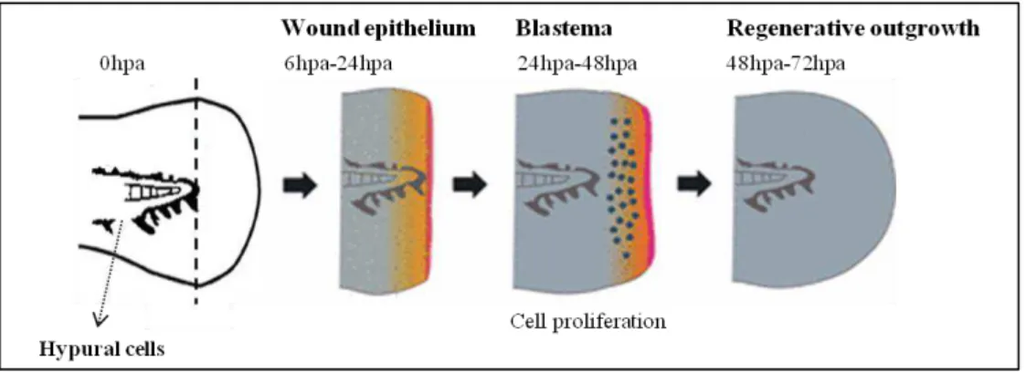
Figure 1.1 –Schematic representation of the zebrafish larvae fin fold regeneration process……….4
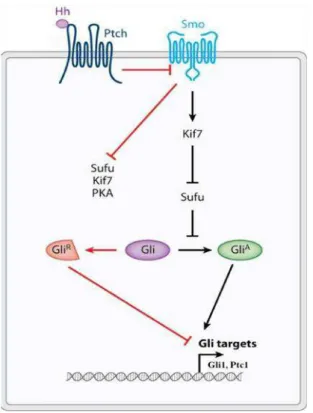
Figure 1.2 –The main components of theHedgehog signaling pathway………....7
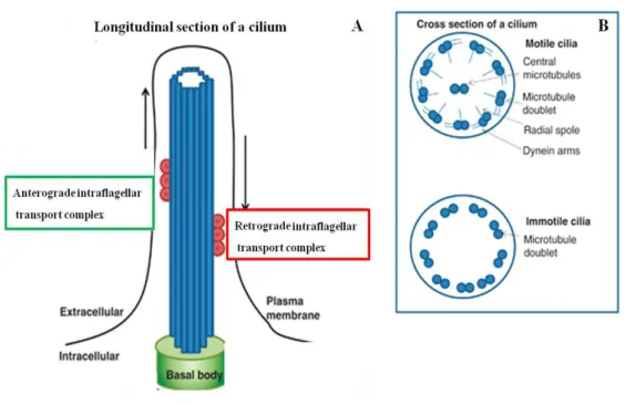
Figure 1.3 –Schematic representation of the cilia structure and microtubule arrangement…………...……8
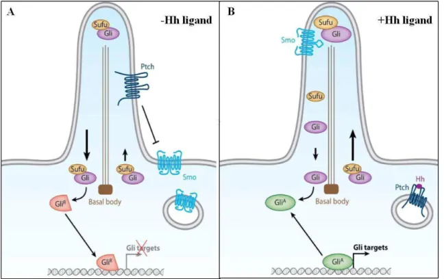
Figure 1.4 –The vertebrates Hedgehog signaling pathway………..……….11
Figure 2.1 –Methodology adopted to study the process of zebrafish larvae fin fold regeneration………..16
Figure 2.2– Representation of the area measurement performed in ImageJ software………..23
Figure 3.1 –Expression pattern of the Hedgehog signaling pathway genes during zebrafish larvae fin fold regeneration……….…...32
Figure 3.2 –Expression levels of thedifferent Hedgehog signaling pathway components during zebrafish larvae fin fold regeneration………...…………..33
Figure 3.3 – Smoothened mutant and sibling zebrafish larvae at 2 days post
fertilization………..34
Figure 3.4 – Zebrafish smu mutant larvae are not able to recover their fin fold upon an amputation………...35
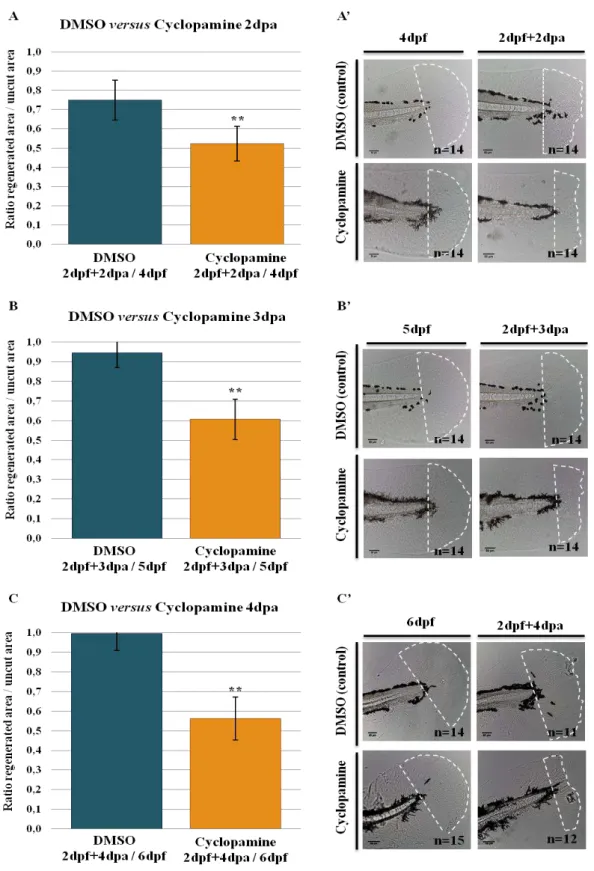
Figure 3.5 –Cyclopamine inhibits the regeneration of the zebrafish fin primordia………..38
Figure 3.6 – Expression pattern of the Hh signaling pathway ligands and receptors during DMSO and cyclopamine treated larvae fin fold regeneration……….………..41
Figure 3.7 – Expression pattern of Hh signaling pathway transducer, repressor and transcription factors DMSO and cyclopamine treated larvae fin fold regeneration…………....42
Figure 3.8 –Cell proliferation is inhibited in response to the cyclopamine treatment………..44
Figure 3.9 –Expression pattern of the Hh signaling pathway ligands and receptors during iguana mutant and iguana sibling larvae fin fold regeneration…………..48
Figure 3.10 – Expression pattern of the Hh signaling pathway transducer, repressor and transcription factors during iguana mutant and iguana sibling larvae fin fold regeneration………..49
Figure 3.11 –Comparison of the expression levels of the Hedgehog signaling pathway genes in iguana mutant versusiguana sibling larvae during fin fold regeneration……….………….…50
Figure 3.12 –Cell proliferation is absent from iguana mutant larvae fin fold………..52 Figure 3.13 – The aphidicolin treatment completely inhibited cell proliferation throughout the larvae fin
fold……….……….…....55
Figure 3.14 – Cell proliferation is not required to the process of zebrafish larvae fin fold regeneration
x
Figure B1 – Histological sections of wild type zebrafish larvae fin fold following whole mount in situ hybridization for ihhb and patched1 genes...75
xi
List of Tables
Table 2.1 - Constructs used as DNA templates for mRNA probe synthesis……….19
Table 3.1 – Efficiency of the aphidicolin treatment to inhibit cell proliferation in the three experimental
xiii
List of Abbreviations
ACFP Adult caudal fin primordia
BCIP Bromo Chloro Indolyl Phosphate
BrdU Bromodeoxyuridine
BSA Bovine Serum Albumine
cDNA Complementary Deoxyribonucleic Acid
dpa days post amputation
dpf days post fertilization
Dhh Desert hedgehog
DIG Digoxigenin
DMSO Dimethyl sulphoxide
DNA Deoxyribonucleic acid
E. coli Escherichia coli
FGF Fibroblast growth factor
Gli Glioblastoma
GliA Gli activator isoform
GliR Gli repressor isoform
Hh Hedgehog
hpa hour post amputation
IFT Intraflagellar Transport
igu iguana
Ihh Indian hedgehog
ISH In Situ Hybridization
μm micrometer
μM microMolar
ml millilitre
mM milliMolar
NBT Nitro Blue Tetrazolium
PBS Phosphate Buffered Saline solution
PBST Phosphate Buffer Saline/ % TritonX-100
PBT Phosphate Buffer Saline/ 0.1% Tween-20
PDGF Platelet-derived growth factor
PFA Paraformaldehyde
Ptc1 Patched1
Ptc2 Patched2
% Percentage
PTU Phenyl-1-thiourea
RA Retinoic Acid
RNA Ribonucleic Acid
rpm revolutions per minute
Shh Sonic hedgehog
Smo Smoothened
SuFu Supressor of fused
Chapter 1:
Introduction
1.1
Regeneration in the zebrafish model
Regeneration is an event that can be found in certain organisms that permits the complete reconstitution of lost or damaged tissues and organs (Sanchez Alvarado, 2000). Depending on the context, regeneration can follow an injury, acting as a mechanism of repair, or it can be a constitutive event involved in maintaining the physiological integrity of the organism. There are two main organ/tissue regenerative strategies within different organisms: morphallaxis and epimorphic regeneration. Morphallaxis is defined as the reconstruction of the organism form by remodeling of pre-existing tissue after severe damage, such as the type of regeneration occurring in hydra (Bosch, 2007). On the other hand, epimorphic regeneration depends on cell proliferation and on the formation of a regeneration-specific structure, the blastema, which comprises proliferative cells that differentiate and lead to a complete recovery of the lost tissue. This type of regeneration is seen, for example, during amphibian limb and tail regeneration, and zebrafish (Danio rerio) fin regeneration (Galliot and
Ghila, 2010).
Mammals have only a limited capacity to regenerate their tissues and organs during adult life. Their regeneration capacity includes the limited replacement of certain cell types in a physiological manner, which is transversal to all animals (Stoick-Cooper et al., 2007). Cells of the skin,
gastrointestinal epithelium, bone, skeletal muscle and hematopoietic tissue are regularly replaced upon apoptosis and aging through the activity of self-renewing stem cells. However, most adult tissues/organs, in mammals, have lost their potential for further growth and differentiation (Stoick-Cooper et al., 2007). In mammals, regeneration is usually applied to processes such as liver growth
after partial resection, a process that consists of compensatory increase of the mass of the organ rather than true regeneration (Michalopoulos, 2007). Consequently, damage of a tissue or organ usually produces a permanent damage from scarring to disability. Conversely, some non-mammalian vertebrate models, such as the zebrafish, retain enormous regenerative potential in tissues as diverse as cardiac muscle, retina, liver, spinal cord axons, cerebellum, and the fins (Poss et al., 2003). Unlike the
situation in mammals, zebrafish tissues with regenerative abilities never form scars after injury since they undergo a complete tissue reconstitution process, and do not seem to have a decline in their regenerative capabilities throughout adult life (Azevedo et al., 2011; Shao et al., 2011). Taken
1.1.1 Adult zebrafish fin regeneration
The zebrafish caudal fin is an established model to study regeneration that presents many advantages in comparison to other organs: is easily accessible, amputation does not compromise the survival of the fish, completely regenerates in a short time frame (2 weeks at 28ºC) and it has a relatively simple architecture composed of 18 bony rays attached to the axial skeleton by muscles (Poss et al., 2003). The regeneration in the zebrafish caudal fin, termed “epimorphic regeneration”,
involves three stereotypic successive steps, starting from the wound healing in the first 12 hours post amputation (hpa), followed by wound epidermis and the blastema formation at 24-48hpa and regenerative outgrowth after 48hpa (Poss et al., 2003). Wound healing comprises the migration of
epithelial cells that cover the wound and form an epithelial cap (or wound epidermis) at the amputation site within 12hpa. Bromodeoxyuridine (BrdU) incorporation assays demonstrated that this response does not involve cell proliferation (Poleo et al., 2001). From 12hpa until 48hpa, the wound
epidermis becomes thicken in several layers, and completely covers the amputation site (Poleo et al.,
2001). This process is a critical event, since the regeneration does not proceed without it, and comprises migration events from the mesenchymal tissue underneath the wound epidermis and once again cell proliferation is not involved (Poss et al., 2003). Meanwhile, cell types in the mesenchymal
tissue, such as osteoblasts (the bone forming cells), undergo dedifferentiation and migrate distally (Tu and Johnson, 2011). These cells form the blastema, which is a group of undifferentiated cells, distal to the amputation plane within 48hpa. These blastema cells proliferate and re-differentiate, being responsible for replacing the lost tissue in a process called regenerative outgrowth (Poleo et al., 2001;
Poss et al., 2003). The blastema is divided in two compartments: the proximal blastema that is
composed of highly proliferative cells and the distal blastema that is composed of non-or slow-proliferative cells (Poleo et al., 2001; Poss et al., 2003). As the regenerative outgrowth proceeds, the
blastema grows distally and cells in the proximal proliferating zone differentiate into new structures which replace the amputated part of the fin. Finally, regenerative outgrowth is completed around 14dpa (Poleo et al., 2001; Poss et al., 2003).
1.1.2 Molecular signaling involved in fin regeneration
In the adult zebrafish, FGF has been described as necessary to blastema formation and subsequent regenerative outgrowth. It has been demonstrated that inhibition of FGF signaling, through an Fgfr1 (Fibroblast Growth Factor receptor 1) inhibitor (SU5402) or a transgenic line ( hsp70:dn-fgfr1) that expresses the dominant negative Fgfr1 protein upon heat shock, causes an aberrant
regenerative epithelium and consequently impedes blastema formation (Poss et al., 2000b; Lee et al.,
2005). In addition, a genetic zebrafish mutant study revealed that fgf20a (a gene of the FGF signaling)
is absolutely required for the initiation and formation of the blastema (Whitehead et al., 2005) and
referred fgf20a as an initiator of regeneration. On the other hand, a proper balance of Wnt/β-catenin
signaling is critical for the formation and proliferation of blastema cells that is required to complete regeneration (Kawakami et al., 2006). Hh signaling has been shown to play a role not only in the
caudal fin re-patterning but also in the regenerative outgrowth through the coordination of cell proliferation events (Quint et al., 2002). It has been demonstrated that amputation of the caudal fin of
zebrafish stimulated regeneration of the dermal skeleton and re-expression of Hh signaling pathway genes (Quint et al., 2002). Moreover, disruption of the Hh signaling using cyclopamine, a steroidal
compound that blocks Hh signaling (Chen et al., 2002), has been shown to cause a reduction in cell
proliferation within the blastema and arrest the caudal fin outgrowth (Quint et al., 2002). Conversely,
the ectopic expression of the ligand Shh leads to additional bone deposition, suggesting a role in proliferation and differentiation of osteoblasts (Quint et al., 2002).
1.1.3 Early life stage zebrafish model
Recently, the zebrafish embryonic caudal fin (“the fin fold”) of the 2 days post-fertilization (dpf) larvae has become appreciated as a new model to study the regenerative process (Kawakami et al., 2004). The larval zebrafish fin fold is a simple structure, composed of mesenchymal cells with no
cartilage enfolded in two epithelial layers, which is non-homologous to the adult caudal fin (Yoshinari and Kawakami, 2011). It consists of a transient structure that is replaced by the adult zebrafish caudal fin during a later larval development stage through the proliferation of a pool of cells localized in the ventral gap of the melanophores streak at the larvae fin fold (Figure 1.1). This pool of cells, termed hypural cells or adult caudal fin primordia (ACFP), is essential to the development of the adult caudal fin and is dependent on Hh signaling (Hadzhiev et al., 2007).
Although an embryonic structure, the larval fin fold is capable of tissue regeneration in a process similar to that observed in the adults (Kawakami et al., 2004). The larval fin fold regeneration
process involves the healing of the wound around 1dpa, the formation of a blastema from 1dpa-2dpa and regenerative outgrowth from 2dpa. Within 3dpa the larvae retain the complete form and structure of the lost part of the fin fold (Kawakami et al., 2004) (Figure 1.1). The wound healing occurs through
it has been shown that blastema (msxc, msxe) and regenerative epithelium (dlx4) markers expression
occurs in the regenerating fin fold (Kawakami et al., 2004). Currently, though, it is unknown whether
the larval fin fold blastema has a specific function for proliferation as in the adult zebrafish caudal fin regeneration. Kawakami et al. (2004) reported that upon amputation of the larvae fin fold, cell
proliferation occurs in a spatial restricted manner in the blastema. Actively proliferating cells denoted as blastema-like cells are detected in the area adjacent to the amputation plane from1dpa-2dpa. After the blastema formation, both the adult and larvae regenerating fins exhibit a common cell proliferation profile with the proliferation starting at the distal area. In addition, the distal-most cells do not proliferate during the late phase of repair and drastic cell proliferation occurs in the proximal region (Kawakami et al., 2004). Contrasting to these data, Mateus et al. (2012) reported that there is a
significant increase in cell proliferation in response to fin fold amputation at 11-16hpa, but cell proliferation does not appears spatially restricted to a blastema region. Instead, cell proliferation occurs in a broad area of the fin fold and not only in the most distal region of the tissue, implicating
that the larval fin fold blastema is not a “classical blastema” (Mateus et al., 2012). The authors
propose that the difference between cell proliferation patterns might be due to the distinct protocols used in both papers to determine cell proliferation (Mateus et al., 2012). Even so, it is demonstrated
that, like the adult regeneration, cell proliferation is activated in the larval fin fold in response to injury (Kawakami et al., 2004; Mateus et al., 2012). Since the larvae fin fold presents no fully differentiated
cell types, except for actinotrichia (Yoshinari and Kawakami, 2011), almost no cell differentiation occurs during larval fin fold regenerative outgrowth until the complete recovery of the amputated tissue around 3dpa.
Since the caudal fin regenerative process appears to be conserved in the larval fin fold system, it presents as a powerful new way to study tissue regeneration and identify its intrinsic regulatory mechanisms. In addition, the larval fin fold has some advantages in comparison to the adult system, namely the speed of regeneration, the structural simplicity of this non-vascularized appendage and the possibility to amputate hundreds of fin folds within an hour gathering a higher experimental number. Further, zebrafish larvae do not feed for up to 5dpf, minimizing the environmental effect on the regeneration assay, and are easier to manipulate and perform chemical and molecular assays (Yoshinari and Kawakami, 2011).
It has been previously reported that, in addition to similar regenerative events between the adult and the larval zebrafish systems, several signaling pathways required during adult caudal fin regeneration are also activated during larval fin fold regeneration. This suggests that larval and adult zebrafish caudal fin regeneration also share a common molecular mechanism. Indeed, microarray studies have shown that a large number of up-regulated expression markers are coincident during adult and larval fin regeneration (Mathew et al., 2009; Yoshinari et al., 2009). Moreover, previous larval
regeneration studies demonstrated that, similar to the adult, inhibition of Fgfr1 with SU5402 arrests larvae fin regeneration by blocking the blastema formation (Kawakami et al., 2004). Also, fgf20a that
was identified as an initiator of blastema formation in the adult regenerating fin (Whitehead et al.,
2005) is also highly induced in the larval fin fold tissue. When canonical Wnt signaling is blocked, the blastema and wound epithelium formation is also blocked in the larval model (Mathew et al., 2008).
However, the functional requirement for Hh signaling in regeneration of the larval fin fold is currently unknown.
1.2
The Hedgehog signaling pathway
The Hh signaling pathway is a well conserved signaling pathway in multicellular organisms that regulates cell growth and tissue patterning during embryonic development and adult tissue homeostasis (Ingham and McMahon, 2001). During development, it regulates the growth and patterning of tissues, such as the limb, the heart, motorneurons, muscle and bone (Ingham and McMahon, 2001). In the adult organism, Hh signaling is involved in the regulation of cell proliferation and differentiation (Ingham and McMahon, 2001), such as in regeneration of the hematopoietic tissue. Therefore, misregulation of the Hh pathway has been implicated in several congenital malformations and tumor formation (Ingham and McMahon, 2001).
1.2.1 Mechanism of vertebrate Hedgehog signaling
In vertebrates, there are mainly three hh genes, encoding extracellular signaling proteins of the
hedgehog (Shh) (Bumcrot et al., 1995) the best characterized and widely expressed Hh protein. These
Hh proteins (also called Hh ligands or Hh signals) are secreted from Hh-producing cells and transferred to adjacent cells. Once released from the cells, Hh bind to a complex of proteins on an Hh-receiving cell. This complex includes the Hh-binding protein Patched (Ptc), which has high affinity for all Hh ligands. Ptc is a 12-pass transmembrane protein that in the absence of Hh inhibits the action of the downstream signaling component Smoothened (Smo). Upon binding of Hh to Ptc, inhibition of Smo is relieved. Smo is a 7-pass transmembrane G-protein and the Hh pathway activator. Smo promotes the downstream signal transduction through interaction with the Gli (glioma-associated genes) family of zinc finger transcription factors (Fig. 1.2). The Gli transcription factors are mediators of the Hh response and have been found to function as transcriptional activators, repressors or both (Eggenschwiler and Anderson, 2007; Briscoe and Therond, 2013).
In vertebrates, there are three Gli transcription factors: Gli1, Gli2 and Gli3. All three Gli transcription factors have five highly conserved zinc finger DNA binding domains and C-terminal activation domains, while Gli2 and Gli3 also have N-terminal repressor domains (Sasaki et al., 1999).
Hence, Gli1 functions solely as a transcriptional activator whereas Gli2 and Gli3 can act both as activators and repressors. Their bi-functionality is determined by the presence of Hh signaling. In the absence of Hh and Smo activation, full length Gli2 and Gli3 are proteolitically processed resulting in the removal of the carboxyl-terminal activation domain. In this form (GliR), these transcription factors are translocated to the nucleus and act to repress transcription of Hh target genes. Disruption of this processing by Smo, allows full length Gli2 and Gli3 to translocate to the nucleus and act as transcriptional activators (GliA). The ratio of GliR and GliA forms varies as the concentration of Hh changes. The balance of these forms within the nucleus of a given cell ultimately determines the activation or repression of Hh target genes. Among the Hh pathway target genes are Gli1 that further enforces the Hh pathway activation, and Ptc1 that establishes a negative feedback regulation of the pathway by repressing Smo (Eggenschwiler and Anderson, 2007; Briscoe and Therond, 2013) (Fig. 1.2).
Regulation of Gli transcription factors processing and nuclear translocation has an essential role in the modulation of the Hh target gene expression. This process is mediated through a number of interacting proteins including Smo, Supressor of Fused (Sufu), Kif7 and Protein kinase A (PKA). Sufu acts as negative regulator of the Hh signaling through the formation of a complex with full length Gli proteins and promoting their processing into GliR via PKA (Merchant et al., 2004). In addition, Sufu
has also been shown to play a positive role on Hh signaling by maintaining sufficient full-length Gli levels in the cytoplasm required for their activation by Smo (Humke et al., 2010; Tukachinsky et al.,
Due to whole genome duplication and further rearrangements, zebrafish have two ihh genes
(ihha and ihhb), two shh genes (shha and shhb) (Gensure et al., 2004). Moreover, four Glis have been
identified in the zebrafish: Gli1 is a major activator with expression that only partially depends on Hh signaling (Karlstrom et al., 2003; Ninkovic et al., 2008), Gli2a and Gli2b have been described as
minor repressors (Ke et al., 2008) and Gli3 has both activator and repressor functions (Tyurina et al.,
2005).
1.2.2
Hedgehog signaling through primary cilia
In vertebrates, intracellular Hh signaling is highly dependent on the structural cellular component, the primary cilium (Huangfu and Anderson, 2005; Kim et al., 2010; Roy, 2012). Primary
cilia are organelles stabilized by microtubules that project from the cell surface of most vertebrate cells. Cilia are formed during the interphase of the cell cycle from the basal body, a modified centrosome, which associates to the plasma membrane (Wissam et al., 2009). Since no protein
the anterograde movement of IFT particles from the base to the tip of the cilia, while Dynein regulates the retrograde movement from the tip to the base of the cilia (Rosenbaum and Witman, 2002; Scholey, 2008). The IFT proteins are composed of approximately 17 proteins arranged into two complexes, IFT-A and IFT-B, which are required, respectively, for the retrograde and anterograde movement within cilia. Therefore, the disruption of the IFT system can lead to a complete absence of cilia, or their stunted growth, with repercussions to their functionalities.
The core of the cilia, the axoneme, elongates from the basal body and extends towards the extracellular environment to form the cilium (Fig. 1.3). Based on structural composition and motility, there are three types of cilia: motile, nodal and primary cilia. Motile cilia consist of nine doublet microtubules and a central pair (9+2), which move relative to each other and cause the cilium to bend and therefore move. Motile cilia are commonly present in large number of cells and beat to cause fluid flow. Nodal cilia are present on cells of the embryonic node during development and can also beat but lack of the central pair of microtubules (9+0). Nodal cilia are responsible to cause preferential morphogen gradients helping to establish left-right body axis asymmetry during embryonic development. On the other hand, primary cilia, present in almost nearly all vertebrate cells, consist of nine doublet microtubules only (9+0) and are therefore non-motile (Wissam et al., 2009).
Primary cilia, once considered vestigial structures, have been in recent years discovered as sensory organelles (Wissam et al., 2009). Primary cilia main function is to act as chemo and
mechano-sensors to sense fluids and mechanical stress, such as in the kidney cells (Wissam et al., 2009).
However, there are examples of primary cilia that detect osmolarity, temperature, and gravity (Wissam
et al., 2009). In addition, in some specialized cells, such as the photoreceptors and olfactory neurons,
primary cilia have evolved to specialized functions in sensory perceptions (Wissam et al., 2009).
Primary cilia are therefore thought to be essential to concentrate signals and promote an efficient and rapid signal transduction within the cell, and allow specific protein interactions or modifications to occur (Wissam et al., 2009). According to its capacity to sense extracellular signals, it is not
surprising that this active organelle was recently described as essential to the coordination of several signaling pathways involved in both embryonic development and tissue homeostasis. The signaling pathways known to depend on primary cilia include: Platelet Derived Growth Factor Receptor α
(PDGFRα) growth factor signaling, epidermal growth factor signaling, Wnt signaling, and Hh signaling (Eggenschwiler and Anderson, 2007; Goetz and Anderson, 2010).
Many components of the Hh pathway are enriched in the primary cilium and, in response to Hh stimulus, dynamically shuttle in or out this organelle. For example, in mouse, it has been shown that Smo is absent from primary cilia in limb bud resting cells, although in response to Shh stimulation Smo accumulates in the cilia tip (Corbit et al., 2005). Smo translocation, which requires the IFT
system, is essential to Hh signaling transduction (Corbit et al., 2005). In addition, Ptc1 has been
detected in primary cilia of mouse embryonic fibroblasts (MEFs) cultures and demonstrated to undergo internalization, moving out of the cilium, when bound to Hh (Rohatgi et al., 2007). Also,
Sufu, negative regulator of Hh signaling, has been shown to localize in primary cilia distal tip in the absence of Hh ligands (Haycraft et al., 2005). Gli1, Gli2, Gli3A and Gli3R have as well been detected
in primary cilia, indicating that Hh signaling transduction is reliant on primary cilium (May et al.,
2005; Eggenschwiler and Anderson, 2007). Furthermore, genetic screens in the mouse have revealed that mutations in components of the IFT machinery result in absence of cilia and consequently defective Hh signaling (Haycraft et al., 2005). Ift88 mutant mouse (also referred to as Tg737)
demonstrated patterning defects in Hh-dependent tissues, including failure of ventral neurons specification and polydactyl limbs (Huangfu et al., 2003; Huangfu and Anderson, 2005; Liu et al.,
2005), accompanied by reduction or absence of downstream Hh target genes expression. In the limb bud, it has been shown that Shh mainly functions to derepress Hh target genes expression through the decrease of Gli-R levels (Eggenschwiler and Anderson, 2007; Goetz and Anderson, 2010). Thus, a reduction of these levels would lead to a gain-of-function of Hh signaling phenotype. Accordingly,
ift88 mutant mice demonstrated an increase in the levels of full-length Gli3 protein, which would
derived from ift88 mutant mouse limb bud lacked of the responsiveness to Shh (Haycraft et al., 2005;
Eggenschwiler and Anderson, 2007). Therefore, cilia are not only essential to the processing of Gli transcription factors into their repressor form but to activation of Hh target genes transcription in response to Hh ligands (May et al., 2005). Moreover, cilia are also essential to the activation of Gli
full-length proteins. In ift88 mutant mouse, a reduction of Gli2 activating form in the neural tube leads
to a failure in the ventral neurons specification in a similar pattern to those with shh mutations
(Huangfu et al., 2003). Therefore, depending on the tissue and their dependence on GliA or GliR
function, the loss of primary cilia can result in low or high Hh signaling activity as primary cilia are required to generate both activator and repressor forms of Gli transcription factors (Huangfu and Anderson, 2005; Liu et al., 2005; May et al., 2005).
The current view is that primary cilia are essential for normal Hh signaling transduction in all systems studied, including the mouse, Xenopus, chicken and zebrafish (Roy, 2012). In zebrafish, it has
been shown that Smo, like in mouse, translocates to the primary cilium in response to Hh stimulation and that mutations that result in the disruption of Smo localization to this organelle cause a decrease in Hh pathway activity in zebrafish embryos (Aanstad et al., 2009). Also, it has been demonstrated that
Gli2 activating form translocates to the primary cilium of cells in the zebrafish embryo being this localization mediated through Hh pathway activity (Kim et al., 2010). In ptc1/ptc2 double mutant
zebrafish, the levels of Gli2a are increased at the tip of the cilia, while in smo mutant zebrafish the
levels are increased in the cilia basal bodies (Kim et al., 2010). This implicates that in the absence of
Hh signaling Gli2a is translocated from the tip of the cilia to the basal body and that Hh signaling regulation is therefore dependent on primary cilia.
Further studies have demonstrated that there is as well a ciliary defect associated with the deregulation of Hh signaling in the zebrafish (Huang and Schier, 2009). Huang and Schier (2009), using a maternal-zygotic zebrafish that lack Ift88 gene (MZovl), revealed that zebrafish cilia mutants
demonstrate defective neural patterning and somites formation, reflecting a disturbed Hh signaling activity (Huang and Schier, 2009). Nevertheless, while in mouse cilia mutants the expression of ptc1,
a downstream Hh signaling target, is reduced or absent (Huangfu et al., 2003), in zebrafish cilia
mutants the expression of ptc1 is reduced, and at the same time expanded, in different tissues
depending on the tissue requirements for Hh signaling (Huang and Schier, 2009). As a result, cell types that require maximal Hh signaling activity are absent in both zebrafish and mouse cilia mutants, while cell types that depend on lower Hh signaling threshold levels are expanded in zebrafish but are absent in mouse cilia mutants. As an example, the motorneurons are specified in zebrafish cilia mutants (Huang and Schier, 2009) and absent in mouse ift88 mutants (Huangfu et al., 2003),
signaling independent expression than mouse gli1, which results in greater levels of Hh
signaling-independent pathway activation (Huang and Schier, 2009). As mentioned above, gli1 transcription is
not fully dependent on Hh signaling in the zebrafish (Karlstrom et al., 2003; Ninkovic et al., 2008),
and therefore is expressed even in the absence of primary cilia, while gli1 is inhibited in mouse cilia
mutants (Eggenschwiler and Anderson, 2007). In particular, it has been demonstrated that zebrafish cilia mutants treated with gli1 morpholinos show a reduced expression of ptc1 compared to untreated
zebrafish cilia mutants (Huang and Schier, 2009), suggesting that the expanded Hh pathway activity in zebrafish cilia mutants is indeed dependent on gli1 basal expression. Hence, there is a conserved
requirement of cilia for “normal” Hh signaling in the zebrafish; although it appears that only maximal Hh pathway activation is reduced in zebrafish cilia mutants.
Therefore, in vertebrates, it has been postulated that in absence of Hh, Ptc localizes on the cell membrane of cilia and inhibits Smo from entering into to the primary cilium. Smo is trapped in intracellular vesicles. Gli proteins, at the tip of the cilium are then processed into repressor forms by Sufu. The Gli repressors associate with the IFT proteins and are transported in retrograde direction from the ciliary tip to the basal body. Upon stimulation of Hh, Ptc moves from the cilia. Smo is derepressed, associated with IFT proteins and transported in anterograde direction from the basal body towards the cilia tip. At the cilia tip interacts with Sufu and Gli proteins to regulate the Gli processing and promotes the formation of Gli activating forms. The Gli activating forms are then transported in retrograde direction and enter the nucleus to regulate the transcription of the downstream targets (Fig.1.4) (Eggenschwiler and Anderson, 2007; Goetz and Anderson, 2010).
1.3
The zebrafish
iguana
mutant
Iguana is a novel zinc finger protein, also referred to as the zebrafish homolog of Dzip1 (Daz interacting protein 1, mammalian protein) (Sekimizu et al., 2004; Wolff et al., 2004), that is required
for normal Hh signaling transduction in the zebrafish embryo (Odenthal et al., 2000; Sekimizu et al.,
2004; Wolff et al., 2004).
The iguana (igu) gene was first associated to a role in Hh signaling regarding the several
phenotypical traits that it shared with the zebrafish midline mutants (Brand et al., 1996; Odenthal et al., 2000), including ventral neural tube defects, U-shaped somites and malformations in the midline,
and a characteristic ventral curved body phenotype (Brand et al., 1996; Van Eeden et al., 1996).
Zebrafish midline mutants have been shown to encode components of the Hh signaling cascade, including shha (syu), gli1 (dtr), smo (smu) and gli2 (yot) (Brand et al., 1996; Van Eeden et al., 1996). Iguana mutant phenotype is unique among the midline mutants in that it demonstrates a reduction in
downstream Hh signaling target genes expression in the ventral neural tube and expanded expression of Hh target genes in the somites (Sekimizu et al., 2004), contrasting with the absence of Hh target
genes expression in both neural tube and in the somites in zebrafish midline mutants. Reflecting this altered pattern of ptc1 and gli1 expression, the specification of Hh-dependent muscle cell types is also
impaired in iguana mutant zebrafish (Wolff et al., 2004). In zebrafish iguana mutant, cell types that
require maximal levels of Hh signaling like the muscle pioneers are lost or reduced, while cells that require sub-maximal signaling activation like medial fast fibers (MFFs) are over produced (Wolff et al., 2004). Therefore, iguana mutants exhibit both a gain and a loss-of-function of Hh signaling
activity highly reminiscent to MZovl mutant zebrafish (Huang and Schier, 2009). The loss of Hh-dependent cell types could be explained by a reduction of Hh signaling activity in iguana embryos or
has been shown that Iguana functions downstream of Smo and upstream of the transcription mediators Gli in the Hh signaling cascade (Sekimizu et al., 2004; Wolff et al., 2004). In the absence of Shh and
Smo activity, the expansion in numbers of MFFs is unaffected in iguana mutant embryos, implicating
that Iguana acts downstream of Smo. In addition, depletion of Sufu causes a further expansion in the number of MFFs, whereas over-expression of Sufu eliminates all Hh-dependent muscle cell types from iguana mutant embryos (Wolff et al., 2004). Sufu acts to modulate the Gli protein function
acting to inhibit their transcriptional activation (Merchant et al., 2004). Therefore, it was proposed that
GliA and GliR ratio was disrupted in the absence of Iguana activity and Iguana had a role in the modulation of the Gli protein function (Sekimizu et al., 2004; Wolff et al., 2004).
More recently, a role for the Iguana protein on primary ciliogenesis was discovered (Glazer et al., 2010; Tay et al., 2010). In particular, it has been demonstrated that the Iguana protein cell
sub-localization is the cilia basal body and that Iguana is not required for the docking of the basal bodies but essential for the ciliary axoneme growth. Consequently, iguana mutant zebrafish lacks of the
primary cilia (Glazer et al., 2010; Tay et al., 2010). Since the mechanism by which the Iguana protein
regulates the Gli activator and repressor activities is unknown, it raised the possibility that it is a cilia-related role that explains its requirement for Hh signaling in the zebrafish and not a direct role as a component of the Hh signaling cascade (Glazer et al., 2010; Tay et al., 2010).
1.4
Aims
Previous work from our lab has shown that zebrafish larvae fin fold is ciliated and that upon amputation there is a ciliogenic event associated with the regenerative response (Lima, 2010). Based on the recent connection between primary cilia and Hh signal transduction (Huang and Schier, 2009), and since no one has previously characterized whether Hh signaling is induced upon amputation of the larval zebrafish fin fold and its functional requirement during its regeneration process, the main objective of this work is to determine the contribution of the Hh signaling pathway to the larval fin fold regenerative process. In a broad perspective, we want to assess whether the adult and larval zebrafish fin systems share the same requirement for Hh signaling during regeneration. In addition, we want to assess the importance of primary cilia as regulators of Hh signaling during the larval zebrafish fin fold regeneration process by analyzing the modulation of Hh signaling genes expression in the presence and absence of primary cilia, using the iguana mutant. Finally, our third objective is to
Chapter 2:
Materials and Methods
This chapter describes the methodologies adopted to achieve the aims and objectives stated in section 1.4 of Chapter 1.
2.1
Zebrafish lines and husbandry
Zebrafish (Danio rerio) embryos were obtained by natural spawning of AB wild type fish or by
identified heterozygous carriers for iguana (igutm79a) (Brand et al., 1996) or smoothened (smub641)
(Varga et al., 2001) mutation. Igutm79ais a point mutation coding for a premature stop codon, causing
a non-functional truncated Iguana/Dzip1 protein (Brand et al., 1996). As stated in section 1.2 of
Chapter 1, Iguana/Dzip1 localizes to the basal body of the primary cilium and is required for its proper formation (Kim et al., 2010). Therefore, homozygous igu mutant embryos lack of the primary cilia.
On the other hand, smub641is point mutation that changes a glycine to an arginine residue in the second
transmembrane domain of the Smoothened (Smo) protein, which severely affects the protein structure and function (Varga et al., 2001). Smo is the downstream transducer of Hh signaling and consequently
essential for activation of Hh signaling downstream targets transcription. Thus, homozygous smu
mutant embryos have the Hh signaling pathway inhibited.
All zebrafish lines were maintained and raised according to standard protocols (Westerfield, 2000). Zebrafish embryos were staged by days post fertilization (dpf) at 28ºC and confirmed according to morphological criteria provided in Kimmel et al. (1995). Homozygous igu and smu mutant embryos
were identified at 2dpf based on published descriptions (Brand et al., 1996; Varga et al., 2001) and
Mendelian inheritance. In a heterozygous progeny pool of embryos, 25% should be wild type, 50% should be heterozygous for the specific mutation and 25% should be homozygous.
2.2
Fin fold amputation
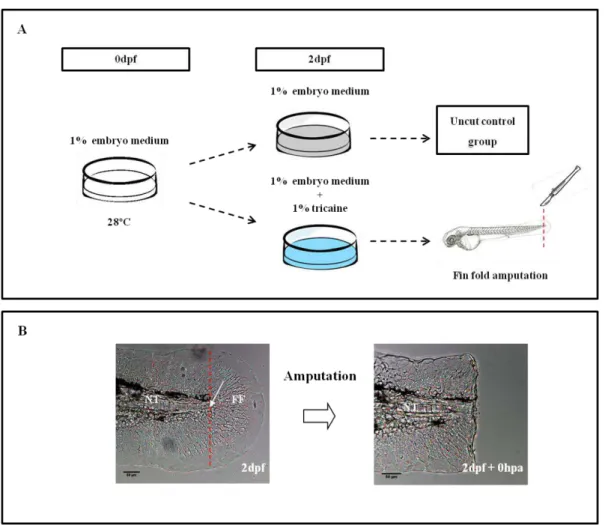
Embryos were maintained in 1% embryo medium at 28ºC until amputation of the fin fold at 2dpf. Zebrafish embryos were then randomly selected as an uncut control group or to undergo amputation of the fin fold (experimental group). Prior to amputation, the chorions were removed and embryos were anesthetized in embryo media supplemented with 1% Tricaine (160 mg/ml) (MS222, Sigma Aldrich) for approximately 5 minutes. This allows immobilization of the embryo and reduces any possible discomfort caused by the procedure. Using a scalpel, the amputation of the fin fold was performed just posterior to the end of the notochord as previously described (Kawakami et al., 2004)
or respective vehicles at 28ºC and allowed to regenerate their fin folds for several time points. Larvae were then collected and used for the following procedures.
2.3
Drug treatments
2.3.1 Cyclopamine treatment
Cyclopamine, a small molecule that interferes with Smo receptor (Chen et al., 2002), was used
to inhibit Hh signaling during larval zebrafish fin fold regeneration. Cyclopamine (cat: C4116, Sigma-Aldrich) was resuspended in DMSO (dimethyl sulfoxide; cat: F515, Fynnzymes) to a 12mM stock solution based on published protocols (Hadzhiev et al., 2007) and stored at -20ºC. Prior to treatment
with cyclopamine, a solution of cyclopamine at the final concentration of 200μM was freshly prepared
Figure 2.1–Methodology adopted to study the process of zebrafish larvae fin fold regeneration. (A)Embryos were maintained in 1% embryo medium until the fin fold amputation stage (2dpf), when they were transferred to 1% embryo medium supplemented with 1% tricaine. (B) The fin fold was surgically ablated just posterior to the end of the notochord. The amputation plane is indicated by the red dotted line and the arrow indicates the most distal end of the notochord. NT –
from the stock solution through dilution in 1% embryo medium. The cyclopamine treatment was initiated following the amputation of wild type larvae fin fold at 2dpf and continued until 1-4 days post amputation (dpa). As vehicle control of the cyclopamine experiments, a group of larvae was treated with an equivalent amount of DMSO in embryo medium and analyzed at the same regenerative stages. Cyclopamine and DMSO treated larvae then processed for in situ hybridization (1-3dpa) and/or
immunohistochemical detection of BrdU (2-4dpa).
2.3.2 Aphidicolin treatment
Aphidicolin, a pharmacological inhibitor of DNA polymerase α (Ikegami et al., 1978), was used
in this work to arrest cell proliferation during larval zebrafish fin fold regeneration. Aphidicolin (cat: A0781, Sigma Aldrich) was resuspended in DMSO (cat: F515, Fynnzymes) to a 3mM stock solution and stored at -20ºC. After the treatment with aphidicolin, a solution of aphidicolin at the final concentration of 30 or 50 50μM was prepared from the stock solution by dilution in 1% embryo medium. The aphidicolin treatment was initiated after amputation of wild type larvae fin fold at 2dpf and continued for 2, 3 and 4dpa. As positive control, a group of larvae was treated with an equivalent amount of DMSO in 1% embryo medium. Aphidicolin and DMSO treated larvae were processed for immunohistochemical detection of BrdU.
2.4
Detection of mitotic nuclei with Bromodeoxyuridine
2.4.1 Bromodeoxyuridine (BrdU) incorporation
Bromodeoxyuridine (BrdU) is an analogue of the DNA precursor thymidine. In proliferating cells the DNA has to be replicated before division can take place, which occurs during the S phase. If BrdU is administrated at this stage, cells will incorporate it into their DNA just like they would incorporate thymidine. Consequently, the number of cells incorporating BrdU is dependent on the frequency of cell proliferation of the tissue. The amount of BrdU incorporated can be detected using specific anti-BrdU antibodies immunohistochemically.
BrdU (cat: B5002, Sigma-Aldrich) was diluted in 1% embryo medium to a 2mM solution from a 10mg/ml stock solution dissolved in 1% PBS with 7mM NaOH. The patterns of cell proliferation, during the steps of the regeneration process, were analyzed following BrdU incubation for 6 hours preceding larvae fixation at specific regenerative endpoints per Kawakami et al. (2004).
2.4.2 Immunohistochemical detection of BrdU
through a series of methanol-PBS (from 25%, 50%, 75% and 100% methanol) gradient and storage at -20°C until processed. Immunohistochemistry experiments were performed according to the protocol presented in Appendix A.
2.5
In situ
hybridization
In situ hybridization is a powerful technique for visualizing gene expression patterns in specific
tissues. This technique allowed us to study the expression of several Hh signaling pathway genes at different stages of larvae fin fold regeneration (1dpa-3dpa) using specific digoxigenin-labeled (DIG) RNA probes.
2.5.1 RNA probes synthesis
The antisense mRNA probes used in this research were synthesized from the constructs listed at Table 2.1
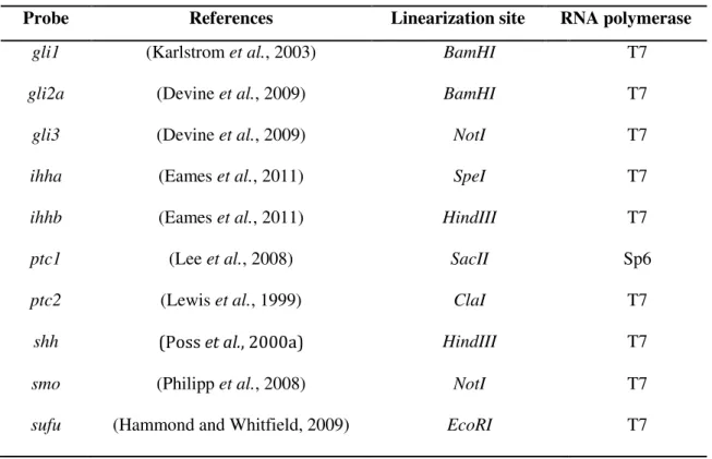
Table 2.1 - Constructs used as DNA templates for mRNA probe synthesis.
Probe References Linearization site RNA polymerase
gli1 (Karlstrom et al., 2003) BamHI T7 gli2a (Devine et al., 2009) BamHI T7 gli3 (Devine et al., 2009) NotI T7 ihha (Eames et al., 2011) SpeI T7 ihhb (Eames et al., 2011) HindIII T7 ptc1 (Lee et al., 2008) SacII Sp6 ptc2 (Lewis et al., 1999) ClaI T7
shh (Poss et al., 2000a) HindIII T7
smo (Philipp et al., 2008) NotI T7
sufu (Hammond and Whitfield, 2009) EcoRI T7
Bacteria transformation
Each plasmid DNA was transformed into the Escherichia coli (E. coli) strain DH5α for further
(1μl) was incubated with 100 μl of cells on ice for 30 minutes. A heat shock was applied at 42 ºC
during 45 seconds (sec), followed by cooling on ice for 2 minutes. Next, 1ml of LB medium was added to the bacteria solution and the mixture was incubated for 1 hour with agitation at 37ºC.
Bacteria were plated in solid medium (LB) containing ampicilin (100μg/μl) to select the transformed
bacteria and grown overnight in an incubator at 37ºC, with the plates inverted.
Plasmid DNA Amplification
For plasmid DNA amplification, a single colony of transformed bacteria was collected using a micropipette tip and inoculated in a 15ml falcon containing 2ml of pre-warmed LB medium
supplemented with ampicilin (100μg/μl). The mixture was grown in an incubator-shaker during 6 hours at 37ºC. Following the 6 hours incubation, 1,5ml of culture was transferred into a 1000ml Erlenmeyer containing 100ml of pre-warmed LB medium with 100μl of ampicilin and incubated
overnight in a shaker- incubator at 37ºC.
Plasmid DNA Isolation
For small scale preparation of plasmid DNA, 2ml of a grown overnight bacterial culture of transformed competent cells was processed using the Wizard® Plus SV Minipreps DNA Purification System (Promega) according to the protocol suggested by the manufacturer (Appendix A). For large scale preparation of plasmid DNA, 100ml of a grown overnight bacterial culture of transformed competent cells was processed using the Roche Genopure Plasmid Midi Kit (Applied Science), according to the manufacturer’s instructions (Appendix A). The extracted plasmid DNA was run on 1% Agarose (Sigma) gel to ensure the DNA integrity and quantified using the Nanodrop ®ND-1000
Spectrophotometer (Thermo Scientific).
Plasmid DNA Linearization
The plasmid DNA was linearized by digesting 5’ of the probe sequence with an appropriate
restriction enzyme (Promega/Roche) (Table 2.1) under optimal conditions specified by the
manufacturers. In order to digest the plasmid DNA, the following reagents were mixed: 5μg of
template DNA, 5μl of 10X NE Buffer, 5μl 10x BSA (100mg/ml), 1μl of restriction enzyme in a final volume of 50μl. The restriction mix was incubated for 3 hours at 37ºC. The restriction products were then validated by gel electrophoresis in 1% Agarose (Sigma) gel to confirm a single digestion by restriction enzyme. Afterwards, the Wizard SV Gel and PCR Clean-Up System Kit (Promega) was
used to purify the restriction reaction products following the manufacturer’s protocol. Plasmid DNA
concentration was assessed against RNAse-free water using Nanodrop ®ND-1000 Spectrophotometer
Antisense mRNA Probe Transcription
The DIG labeled mRNA probes were synthesized with an appropriate RNA polymerase (T7 or SP6, Roche or Promega) (Table 2.1) following the manufacturer’s guidelines. Briefly, approximately 1μg linearized plasmid DNA was added to 0.75M DTT, 10x DIG-NTP mix (Promega/Roche),
RNAsin (Promega/Roche), 10x Transcription Buffer (Promega/Roche) in a final volume of 25μl with
RNase free water. Finally, 20U of RNA polymerase was added and the reaction was incubated at 37ºC
for 3 hours. RNA was purified by adding 20.5μl of RNAse free water, 2μl of 0.5M EDTA (pH 0.8), 2.5μl of 8M LiCl and 150μl of 100% cold ethanol and incubated at -20ºC for 2 hours. Subsequently, RNA was precipitated by centrifugation at 14680rpm for 30 minutes at 4ºC. The supernatant was removed and the pellet was washed with 1ml 70% cold ethanol. The precipitate was for a second time centrifuged at 14680 rpm during 15 minutes at 4ºC and the supernatant was removed. The pellet was
then briefly air dried and resuspended in 30μl of 10 mM EDTA and stored at -20ºC. The probes were
visualized by electrophoresis of 1μl on a 1% Agarose (Sigma) gel in order to validate its integrity and to ensure the amount of synthesized RNA probe. The RNA concentration was assessed against RNAse-free water using the Nanodrop®ND-1000 Spectrophotometer (Thermo Scientific).
2.5.2 Prevention of pigmentation development
In order to prevent pigment formation the embryos were treated with 1-phenyl-2-thiourea (PTU, 0.03mg/ml) (cat: P3755, Sigma-Aldrich) before the onset of pigmentation at 1dpf. 1% PTU was added to the embryo medium from a stock solution of 25% PTU. PTU inhibits melanogenesis by blocking all tyrosinase-dependent steps in the melanin pathway (Karlsson et al., 2001) and the embryos remain
transparent as long as the PTU treatment is continued. Therefore, the PTU solution was changed daily as needed.
2.5.3 Whole mount in situ hybridization
For whole mount in situ hybridization, larvae were fixed as previously described and stored at
-20°C until processed. Whole mount in situ hybridizations were performed as described in Thisse and
Thisse (2008) (Appendix A).
To validate our results, the in situ hybridization studies were performed at least three times with
2.6
Histological analysis
Wild type larvae submitted to whole mount in situ hybridization were processed for sectioning
and further microscopy analysis of the Hh signaling expressing cells. Larvae were rehydrated successively in 50% glycerol/PBS and 25% glycerol/PBS solutions for 5 minutes each and washed several times in PBS. The samples were then passed through a 15% sucrose/PBS solution allowing larvae to settle and incubated with a 30% sucrose/PBS solution at 4ºC overnight for cryoprotection. Afterwards, the samples were embedded in melted 15% gelatin (Sigma)/30% sucrose/PBS solution for an hour at 37ºC and further incubated at 4ºC on the appropriate support for solidification. Once the embedding blocks were solidified, they were trimmed into shape ready for sectioning. The trimmed blocks were frozen in isopenthane/liquid nitrogen and stored at -80ºC until sectioning. Longitudinal
sections of 20μm stained fin folds of uncut controls and fin folds subjected to amputation were
obtained using a Leica CM3050 S Cryostat and mounted on slides using Mowiol (Sigma).
2.7
Measurement of the regenerated area
To determine the regenerative outgrowth of smu mutants, cyclopamine and aphidicolin treated
larvae in comparison to control conditions (smu sibling or DMSO, respectively), two measurements
were collected: the fin fold regenerated area and the uncut fin fold area of larvae at the same developmental stages in each situation. To track the regenerative state until the end of the larvae fin fold regenerative process, the larvae were fixed in 4% paraformaldehyde (PFA) overnight and the fin folds were imaged on days 2, 3 and 4 post amputation using a Leica DM2500 bright field microscope with a 20x objective lens and a Leica DFC420 digital camera. The images were then processed in Image J (NIH) software.
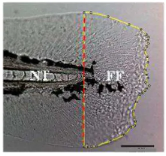
Each image was set to scale with a calibration graticule under 20 x magnifications. The area measurements were obtained using the notochord as reference point to trace a line along the amputation plane and outlining the whole fin fold as depicted in Figure 2.2. The area of tissue posterior to this amputation plane presented the amount of regenerated tissue. Subsequently, the area measurements were acquired using the Measure tool of ImageJ (NIH) software. Images were adjusted for overall contrast and brightness in ImageJ software (NIH).
2.8
Data analysis and statistics
Since normal fin fold growth also occurs during regeneration of the larvae fin fold, the regenerated area was estimated at each stage as the ratio of the regenerated fin fold area related to the uncut area of larvae at the same developmental stage, in both control and experimental conditions. Subsequently, the mean and standard deviation were estimated for each ratio and plotted in graphs. Finally, the differences in control versus experimental conditions were analyzed using a two tailed
Student t-test. Significance was established for p-values less than 0.05.
Calculation of descriptive statistics, plot design and t-tests were performed using Excell Microsoft Office 2007.
2.9
Image acquisition and processing
In situ hybridization stained larvae were cleared and mounted in 100% glycerol. Imaging was
performed using a Leica Z6APO stereomicroscope equipped with a Leica DFC490 digital camera under a 36x objective lens. Detailed analysis of histological sections was obtained using a Leica DM2500 bright field microscope equipped with a Leica DFC429 digital camera under a 20x objective lens. High-resolution imaging of BrdU labeling was performed under a confocal scanning system (Zeiss LSM 510 Meta confocal microscope) equipped with an argon (Ar) laser with peak outputs of 488nm using a dry 20x objective lens. Confocal z-stacks were captured at 1.2 μm intervals and
analysed with ImageJ (NIH) software. Figures were processed using Adobe Photoshop® CS6 Extended
software (Adobe Systems) and/or ImageJ software (NIH).
2.10
Solutions and buffers
The following overview comprises general buffers and solutions. Other solutions are mentioned together with the methods.
1% Embryo Medium (10L)
50x 14,69g NaCl; 0.63g KCl; 2.43g CaCl2.2H2O; 4.07g MgSO4.7H2O; 1ml Methylene Blue
10x PBS (1L)
137mM NaCl; 2.7mM KCl; 4.3mM Na2HPO4.7H2O; 1.4mM KH2PO4
PBT
0.1% Tween20 in PBS
4% PFA
Hybmix buffer solution
50% Formamide; 5x SSC; 0.1% Tween20; Citric Acid (pH=6.0); Heparin 50μg/ml; tRNA 500 μg/ml.
Blocking solution (ISH)
2% Goat serum, 2mg/ml BSA (in PBT solution)
NTMT buffer solution
0.1M Tris-HCl pH 9.5, 50mM MgCl2, 0.1M NaCl, 0.1% Tween 20
Staining Solution
50mg/ml NBT, 50mg/ml BCIP. (in NTMT solution)
20x SSC