UNIVERSIDADE DE BRASÍLIA
INSTITUTO DE GEOCIÊNCIAS
ÁREA DE CONCENTRAÇÃO: GEOLOGIA REGIONAL
DISSERTAÇÃO DE MESTRADO N
O312
UM NOVO PODOCNEMÍDEO FÓSSIL DE GRANDE PORTE DA
FORMAÇÃO SOLIMÕES (MIOCENO-PLIOCENO), ACRE,
BRASIL E AS RELAÇÕES FILOGENÉTICAS ENTRE OS
PODOCNEMIDAE
Marcos Vitor Dumont Júnior
Orientador: Prof. Dr. Rodrigo Miloni Santucci Co-orientadora: Profa. Dra. Caroline Thaís Martinho
Banca Examinadora:
Orientador: Rodrigo Miloni Santucci
Examinadores Internos: Ricardo Lourenço Pinto – IG/UnB Renato Caparroz – IB/UnB (Suplente)
Examinadores Externos:
Agradecimentos
Agradeço ao CNPq, pela concessão da bolsa de mestrado para o desenvolvimento deste trabalho.
Ao programa de pós-graduação em Geologia do Instituto de Geologia da UnB e ao Laboratório de Pesquisas Paleontológicas da UFAC pela estrutura e material para a execução deste trabalho.
Ao meu orientador Dr. Rodrigo Miloni Santucci, pelo apoio, incentivo, disponibilização de material bibliográfico, tempo e paciência, desde a graduação, assim como pelas contribuições e críticas essenciais para esse trabalho.
À Dra. Caroline Thaís Martinho, pelo apoio, em especial durante meu período de seleção e início na pós-graduação da Geologia da UnB.
Ao Dr. Édson Guilherme da Silva, pelo auxílio logístico em Rio Branco, bem como suas inúmeras contribuições durante as visitas ao LPP-UFAC. Bem como pela amizade.
Ao Dr. Jonas Pereira de Souza Filho, por abrir as portas do LPP-UFAC para mim. Bem como à equipe do laboratório.
Ao Dr. Ricardo Lourenço Pinto, pelo apoio e pelo espaço cedido para as reuniões com meu orientador.
Agradeço também a meus pais, Marcos e Déa, por compreenderem minha ausência durante esse período, bem como pelo apoio financeiro.
Aos meus sogros, Ana Maria e Célio, pelo incentivo à busca de meus sonhos acadêmicos.
Aos amigos, pelos momentos de descontração, ainda que infelizmente raros durante esse período.
ÍNDICE
1. Resumo...1
2. Introdução...2
3. Artigo ...5
4. Conclusões ...59
5. Referências Bibliográficas...61
1. Resumo
A família Podocnemidae é representada hoje por oito espécies viventes distribuídas na América do Sul e em Madagascar. A família também possui um rico registro fóssil, encontrado em quase todo o hemisfério sul, com uma grande variedade de formas e tamanhos, assim como uma história paleobiogeográfica complexa. Aqui nós descrevemos uma nova espécie de Podocnemidae fóssil do Mioceno-Plioceno da Formação Solimões do Brasil, Podocnemis manchineri sp. nov., baseada em um casco quase completo, uma carapaça e plastrão fragmentários e uma carapaça fragmentária, compreendendo três diferentes indivíduos. Nós analisamos esse novo táxon e outros cinco táxons conhecidos apenas por material pós-craniano em uma análise de parcimônia usando uma matriz de caracteres extraída da literatura, além de novos caracteres criados nesse estudo baseados na observação da carapaça e plastrão de vários podocnemídeos viventes e extintos. Os resultados indicam que P. manchineri está aninhada dentro do gênero Podocnemis. Nossa análise também valida a posição de outros táxons baseados em carapaças como membros do gênero Podocnemis (e. g. P. negrii¸ P. medemi e P. pritchardi), que têm sido referidos como incertae sedis dentro de Podocnemidae em outros estudos. Além disso, essa análise resolveu o táxon
2. Introdução
A Formação Solimões (Mioceno-Plioceno) têm revelado muitos fósseis de vertebrados ao longo dos anos, representando uma grande variedade de peixes, crocodilianos, tartarugas e mamíferos (Barbosa-Rodrigues 1892; Price1964; Sill 1970; Campos 1977; Campos & Broin 1981; Gasparini 1985; Bocquentin & Rancy 1987; Bocquentin & Santos 1989; Bocquentin & Souza-Filho 1989, 1990; Souza-Filho & Bocquentin 1989, 1991; Broin et al. 1993; Souza-Filho et al. 1993; Latrubesse et al. 1997; Bocquentin-Villanueva et al. 1997; Gaffney et al. 1998; Souza-Filho 1998; Bocquentin & Guilherme 1999; Negri & Ferigolo 1999; Bocquentin et al. 2001; Carvalho et al. 2002; Bocquentin & Melo 2006; Kay & Cozzuol 2006; Hsiou et al. 2007; Meylan et al.2009; Hsiou2010; Riff et al.2010).
Os Testudines encontrados na Formação Solimões, em sua maioria, pertencem ao grupo Pleurodira, típico do hemisfério sul, facilmente diferenciados do subgrupo Cryptodira pelo modo de retração da cabeça. Em Pleurodira a retração é lateral e em Cryptodira ela ocorre para trás, com a cabeça se encaixando entre os ombros (Figura 9). Os Pleurodira são atualmente representados por três famílias: Podocnemidae, da América do Sul e Madagascar, com 8 espécies de água doce, de tamanho pequeno até o maior Pleurodira atual, Podocnemis expansa, que alcança 90cm de comprimento; Pelomedusidae, da África, Madagascar e Ilhas Seychelles, pequenos, com cerca de 18 espécies de água doce; e Chelidae, de porte pequeno a grande, com cerca de 40 espécies de água doce, presentes na América do Sul, Austrália e Nova Guiné (Rueda-Almonacid
et al. 2007; Pough et al., 2008). Embora hoje apenas ocorram em ambiente dulcícola nos continentes do Hemisfério Sul, os Pleurodira ocuparam também ambientes de água salobra e ambientes próximos à costa da maioria dos continentes desde o Eocretáceo, sendo bem mais diversificado no registro fóssil (Gaffney et al., 2006).
Até agora, os Podocnemidae conhecidos da Formação Solimões incluemCaninemys tridentata (Meylan et al. 2009), “Stupendemys” souzai (Bocquentin & Melo 2006), e
Podocnemis negrii (Carvalho et al.2002), sendo C. tridentata o único táxon bem aceito. A atribuição de S. souzai ao gênero referido é criticada e S. souzai pode não representar um único táxon (Meylan et al. 2009; Gaffney et al. 2011) e P. negrii é considerada
em estudos cladísticos provavelmente ocorre devido ao comportamento ambíguo que espécies conhecidas apenas por carapaças e plastrões apresentam nesse tipo de análise. A maior parte dos caracteres usados na filogenia de podocnemídeos são retirados do crânio e mandíbula, enquanto os caracteres pós cranianos são escassos (e. g. Meylan et al.2009; Cadena et al. 2010, 2012; Gaffney et al., 2011). Interessantemente, devido à complexidade do casco dos Testudines, onde tanto o plastrão quanto a carapaça sendo compostos por uma associação de ossos superpostos por escudos dérmicos, pode se esperar que vários caracteres possam ser retirados dessa associação. Ao invés disso, os caracteres de carapaça geralmente representam menos de 25% dos caracteres usados em estudos cladísticos referentes a Podocnemidae fósseis (Tabela 6). Isso contrasta fortemente com o grande número de espécies de cágados fósseis conhecidos apenas pela carapaça. Aqui nós descrevemos uma nova espécie de podocnemídeo, proveniente da Formação Solimões, Acre, Brasil (Fig. 2), e a comparamos a outras espécies relacionadas, fósseis e atuais.
Adicionalmente, nós executamos uma análise cladística, incluindo espécies fósseis e viventes de podocnemídeos, usando uma compilação de caracteres da literatura e novos caracteres do casco. Que forneceram uma hipótese filogenética que inclui alguns táxons baseados apenas em cascos, comumente deixados de fora desse tipo de estudo (e. g.
outras palavras, a presença de sinapomorfias é a única evidência de uma origem evolutiva comum entre dois ou mais táxons.
3. Artigo
A new large podocnemid turtle from the upper Miocene Solimões Formation, Acre, Brazil and the phylogenetic relationships within Podocnemidae
Marcos Vitor Dumont Júniora, Rodrigo Miloni Santuccib*, Caroline Thaís Martinhoc, Édson Guilherme da Silvad
aInstituto de Geociências, Universidade de Brasília, Campus Universitário Darcy Ribeiro, Asa Norte,
70910-900, Brasilia, DF, Brazil; bFaculdade UnB Planaltina, Universidade de Brasília, Área
Universitária 1, Vila Nossa Senhora de Fátima, 73345-010, Brasilia, DF, Brazil; cPontifícia Universidade Católica do Rio Grande do Sul, Instituto do Meio Ambiente e Recursos Naturais, Av. Ipiranga, 6681, Prédio 96J, Partenon, 90619-900, Porto Alegre, RS, Brazil; dUniversidade Federal do Acre, Laboratório de Pesquisas Paleontológicas, CCBN, BR-364, KM 4, Distrito Industrial, 69915-900,
Rio Branco, AC, Brazil
*
Abstract
The family Podocnemidae is represented today by eight moderate-sized extant species distributed in South America and Madagascar. They also have a rich fossil record, which is found over almost all the southern hemisphere, with a wide variety of forms and sizes, as well as a complex palaeobiogeographical history. Here we describe a new species of fossil Podocnemidae from the Miocene-Pliocene Solimões Formation of Brazil, Podocnemis manchineri sp. nov., based on an almost complete large shell, a fragmentary carapace and plastron, and a fragmentary carapace, comprising three different individuals. We analysed this new taxon and other five fossil taxa known only by shell material in a parsimony analysis using a morphological character matrix with characters extracted from the literature, as well as new characters created in this study based on observation of the carapace and plastron of several extinct and extant podocnemid taxa. The results indicate that P. manchineri is nested within the genus
Podocnemis. Our analysis also validated the position of several shell taxa to the genus
Podocnemis (e. g. P. negrii, P. medemi,and P. pritchardi), which have been referred as
incertae sedis within Podocnemidae in previous studies. Furthermore, this analysis resolved Stupendemys geographicus as more closed related to the clade that includes
Bairdemys than to the clade that includes Peltocephalus within Erymnochelyinae. Additionally, for the first time it is recovered the presence of fossil taxa (namely
Kenyemys (also shell based) and Turkanemys ) between the common association of
Erymnochelys and Peltocephalus, commonly recovered in morphological phylogenetic approaches. Morphological comparisons of fragmentary material from the Solimões Formation also suggest that at least a third taxon of a large sized fossil podocnemid could have been present in South-western Amazonia.
Key-words: Podocnemidae, Acre, Solimões Formation, Podocnemis, Phylogenetic Analysis.
The Brazilian Solimões Formation (Miocene-Pliocene) has yielded many vertebrate fossils over the years, representing a great variety of fishes, crocodilians, turtles, and mammals (Barbosa-Rodrigues 1892; Price 1964; Sill 1970; Campos 1977; Campos & Broin 1981; Gasparini 1985; Bocquentin & Rancy 1987; Bocquentin & Santos1989; Bocquentin & Souza-Filho 1989, 1990; Souza-Filho & Bocquentin 1989, 1991; Broin et al. 1993; Souza-Filho et al. 1993; Latrubesse et al. 1997; Bocquentin-Villanueva et al. 1997; Gaffney et al. 1998; Souza-Filho 1998; Bocquentin & Guilherme1999; Negri & Ferigolo1999; Bocquentin et al.2001; Carvalho et al.2002; Bocquentin & Melo2006; Kay & Cozzuol2006; Hsiou et al.2007; Meylan et al.2009; Hsiou 2010; Riff et al. 2010). Concerning turtles, so far, the known record of Podocnemidae from the Solimões Formation includesCaninemys tridentata (Meylan et al. 2009), “Stupendemys” souzai (Bocquentin & Melo 2006), and Podocnemis negrii
(Carvalho et al.2002), with the only well accepted taxon being C. tridentata. S. souzai
is criticized and may not represent a single taxon (Meylan et al. 2009; Gaffney et al. 2011) and P. negrii is considered to be incertae sedis within the Podocnemidae (Meylan
Stupendemys geographicus, Podocnemis negrii, Podocnemis medemi, Podocnemis pritchardi, and Kenyemys williamsi).
Abbreviations
LACM – Natural History Museum of Los Angeles County; UFAC – Universidade Federal do Acre, Laboratório de Pesquisas Paleontológicas; UCMP – University of California, Museum of Paleontology, Berkeley, California.
Geological setting
The Solimões Formation, located in north-western South America, consists of claystones, sandstones and siltstones intercalated with a few lignite and limestone layers (Radambrasil 1977; Hoorn 1993). The Solimões Formation has lateral continuity on neighbour basins, receiving different names. In Pastaza, Marañón and Madre de Dios basins from Peru the Solimões Formation is known as Pebas, Ipururo, and Nauta formations. In Colombia, on Amazonas and Putumayo basins, it receives the denominations of Amazonic Tertiary and La Tagua layers (Hoorn 1993, 1994a; Campbell et al.2001; Roddaz et al.2005; Rebata et al. 2006).
Palynological data suggest an age range from Miocene to Pliocene for the Solimões Formation in the Amazonas State of Brazil (Cruz 1984). Many biostratigraphic works based on palynomorphs were carried out in central and north-western Amazonia (Hoorn 1993, 1994a, b, 1995). However, most vertebrate fossils from the Solimões Formation come from the south-western of Amazonia and no relation between the north and south sediments has been established yet (Campbell et al.2000, 2001, 2006).
Other biostratigraphic propositions have been made, using mainly fossil mammals. To the south-western Brazilian Amazonia a Huayquerian, possibly reaching Montehermosian age has been suggested (upper Miocene to Pliocene) (Campbell et al. 1985; Frailey1986; Latrubesse1992; Latrubesse et al.1997, 2007).
fluvial megafan complex (Latrubesse et al. 1997, 2007); a marginal marine influence area (Hoorn1993, 1994a, b, 1995); or an internal sea (Räsänen et al.1995; Hovikoski et al.2005).
Fossil reptiles are abundant, with a great variety of extinct crocodilians, being represented by Crocodylidae (Charactosuchus), Alligatoridae (Caiman, Purussaurus,
Mourasuchus), and Gavialidae (Brasilosuchus, Gryposuchus, and Hesperogavialis) (Barbosa-Rodrigues1892; Price1964; Sill1970; Gasparini1985; Bocquentin & Souza-Filho 1989, 1990; Souza-Filho 1998; Souza-Filho & Bocquentin 1989, 1991; Souza-Filho et al.1993; Riff et al.2010). The chelonians are represented by the Podocnemidae (Podocnemis and Stupendemys), Chelidae (Chelus), and Testudinidae (Chelonoidis) (Barbosa-Rodrigues 1892; Campos 1977; Campos & Broin 1981; Broin et al. 1993; Bocquentin & Rancy 1987; Bocquentin & Santos 1989; Gaffney et al. 1998; Bocquentin et al. 2001; Carvalho et al. 2002; Bocquentin & Melo 2006; Riff et al. 2010). There are also records of Squamata, represented by snakes (Aniliidae, Boidae and Colubridae) and lizards (Teiidae) (Hsiou et al. 2007; Hsiou 2010). All these findings tend to support a continental water body palaeoenvironmental interpretation.
Historical background
In past years various phylogenetic analyses, with both molecular and morphological data, have been contributing to the understanding of the Podocnemidae inter-relationships (Georges et al. 1998; França & Langer 2006; Gaffney et al. 2006; Noonan & Chippindale2006; Vargas-Ramírez et al.2008; Meylan et al.2009; Cadena
et al. 2010, 2012; Gaffney et al. 2011). However, there are still many problems to be solved in order to understand podocnemid phylogeny.
The morphological data results conflict with the molecular ones. The relationships among the living genera Podocnemis, Peltocephalus, and Erymnochelys
those results may be of great palaeobiogeographic importance, since Erymnochelys is from Madagascar while the other two genera are from South America.
The Podocnemidae shell is known as very conservative, with many shell based taxa being first referred initially as Podocnemis and later, with skull discoveries, revised and reassigned to other genera (Gaffney et al.2006, 2011). This led to a major focus on bringing new cranial characters for phylogenetic analyses, while new postcranial characters have been much rarer. Usually more than 75% of the characters in a phylogenetic analysis are from the skull and jaws (Meylan et al. 2009; Cadena et al. 2010, 2012; Gaffney et al.2011). Consequently, shell based taxa, such as Stupendemys
and Kenyemys, hardly produce well resolved cladograms in this context.
So far, the only Solimões Formation podocnemid included in a phylogenetic analysis is the skull based taxon Caninemys tridentate, whereas the shell based taxa from the Solimões Formation (“Stupendemys” souzai and Podocnemis negrii) have never been included in a cladistics study. Both shell taxa are controversial. “Stupendemys” souzai assignment to the genus Stupendemys has been criticized and the referred material may not represent a single taxon (Meylan et al. 2009; Gaffney et al. 2011). Podocnemis negrii has been considered as incertae sedis within the Podocnemidae, due to the lack of synapomorphies to support its suggested relationship as a sister-taxon to the extant P. sextuberculata (Carvalho et al. 2002; Gaffney et al. 2011).
There are many other indeterminate Podocnemidae fossil remains from the Solimões Formation in the UFAC collection. Among them, there is UFAC-1000 and UFAC-1001, considered until now to be an indeterminate podocnemid (Rancy & Bocquentin-Villanueva, 1987). Here we review these specimens, in addition to a third non described specimen (UFAC-1559) in order to clarify their status within the Podocnemidae.
Systematic palaeontology Testudines Linnaeus, 1758
Pleurodira Cope,1864
Pelomedusoides Cope, 1868
Podocnemidinura Cope, 1868
Podocnemis Wagler, 1830
Type species: Emys expansa Schweigger, 1812.
Included species: Podocnemis expansa, P. vogli, P. unifilis, P. erythrocephala, P. lewyana, P. sextuberculata, and P. bassleri.
Diagnosis: As in Gaffney et al. (2011).
Podocnemis manchineri sp. nov.
(Figs. 2 and 3)
Etymology: Named after the indigenous Manchineri tribe, living near the collection site of the holotype.
Holotype: UFAC-1000, a nearly complete carapace and plastron.
Referred materials: UFAC-1001, incomplete carapace and plastron; UFAC-1559, a fragmentary carapace.
Locality and horizon: UFAC-1000, Acre River, Seringal Bélgica, Assis Brasil, Acre State, Brazil; 1001, Juruá River, Cruzeiro do Sul, Acre State, Brazil; UFAC-1559, Upper Purus River, Purus 6 UFAC locality, AM (Fig. 1). Solimões Formation, upper Miocene (Huayqueriense) (Campbell et al.1985; Frailey1986; Latrubesse1992; Latrubesse et al.1997, 2007). All materials are deposited in the UFAC fossil vertebrate collection.
Description
UFAC-1000 Carapace
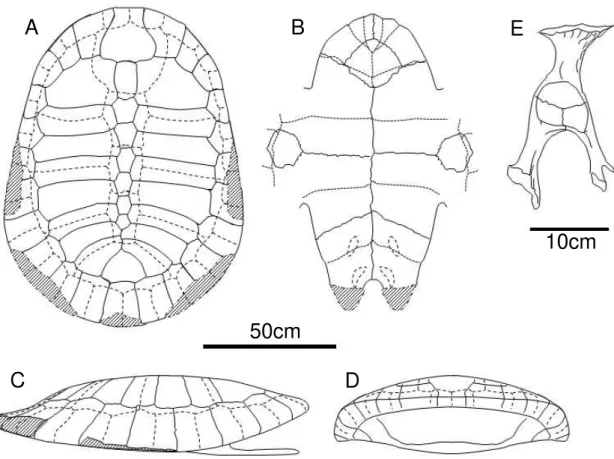
The UFAC-1000 (Figs. 2A and 3A, C, and D) specimen is represented by an almost complete carapace and plastron. The carapace of the UFAC-1000 is uniformly arched, without any irregularities, ornamentations, keels or depressions. The carapace bones are regularly articulated, not presenting significant alterations due to diagenetic processes. This indicates that the dorsoventral flattening of the shell represents a feature present in the living animal, more easily seen in frontal view. The plastral bridge goes outwards, almost parallel to the dorsoventral axis. The scars left by the dermic scutes are easily seen and identifiable. The shell interior is still filled with the matrix and, therefore, not accessible. The presence or absence of ducts, such as axillary musk duct is uncertain, due to the poor preservation state of the regions where they may have been placed, such as the joining region between the plastral bridge and the peripheral bones.
Nuchal bone. It is slightly wider than long. Its widest portion is the region where it meets the first costal and the first peripherals, reaching the minimum width at the end of the first cranial quarter of the bone. The lateral edges are sinuous, while the cranial edge is curved without any embayment or notch. This gives the bone a general bulbous shape from a dorsal view.
Neural series. The carapace has seven neural bones. The first neural is oval and elongated, connected only to costals one, neural two, and the nuchal bone. All other neurals connect to two pairs of costals, the ones with the same number of the neural and the proceeding ones. The neurals two to five have six sides, with an elongated hexagonal shape, resembling a coffin. The neural six is shorter, showing a more regular hexagonal shape, and the neural seven has five sides, resembling a gem.
Suprapygal. It is wider than long and is skirt shaped. It is caudally formed by three arches, where it articulates to the pygal and the peripherals eleven. The suprapygal do not contact the neural bones.
Pygal. The caudal edge is broken, but the bone is trapezoidal in shape and wider
caudally.
Costals. There are eight pairs of costals. Only the seventh and eighth pairs connect each
neural bone. The first one is longer than the others, with a rounded anterior edge, connected to the nuchal and the peripheral bones one to four. The costals two and three are slightly arched caudally. The costal four is almost straight, and the remaining ones are sequentially more arched cranially than the anterior costal bone.
Peripherals. From the eleven pairs of peripheral bones, only the second, third and fourth are completely preserved on the right side of the shell. The others show different degrees of preservation. However, it is possible to infer the probable shell outline by the combination of the information on both sides of the shell. It would have been oval in shape, with the maximum width at the seventh or eighth peripheral. The peripherals strongly vary in shape with the peripheral one being larger than the others. The size of peripherals decreases progressively until the peripheral six, which is the smallest. From the peripheral seven onward the size gradually increases until the last bone of the series.
Vertebral scutes. The first vertebral scute is clearly wider than the others, with a different and more rounded shape. The second and third scutes are very similar, showing a vase-like shape. All the six edges are curved, arched towards the center, with the cranial edge being larger than the caudal one. The maximum width is reached where the cranial and caudal lateral edges meet. The fourth scute is similar to vertebrals two and three, being only thinner than these elements and with the caudal end arched slightly caudally, while in vertebrals two and three it is arched cranially. The fifth and last vertebral scute is wider than the others, being roughly triangular in shape, resembling a fish tail.
Pleural and marginal scutes. There are four pairs of pleural scutes and twelve pairs of
marginal scutes. None of the marginal scutes reach any of the costal bones. The first marginal pair is proportionally small and roughly rectangular in shape.
Plastron
The plastron of UFAC-1000 (Fig. 3B) is almost complete. The cranial and caudal extremities are broken. The epiplastra is also broken, almost not preserved.
Entoplastra.The maximum width is reached about in the middle of the bone, despite the broken cranial edge, the different shortening of the cranial and caudal portions of the bone suggest a shorter caudal and a longer cranial portion.
Scales. The region that would be covered by the gular and intergular scales is not preserved. The humeral scales contact each other in the midline, covering the entoplastra, what is left from the epiplastra almost completely, and a small portion of the hioplastra. The pectoral scales covers a big portion of the hioplastra, cranially it reaches the epiplastra and the entoplastra. The abdominal scales cover portions of the hioplastra, the hipoplastra, and the mesoplastra almost completely, which is also covered only by marginal scutes.
Pelvic girdle
Only the right pelvic girdle is accessible for study (Figs. 2D and 3E). It is not completely preserved, with the pubic and ischiatic processes broken. Furthermore, the ilium is broken, so that the pelvic girdle can be detached from the carapace, where the other half of the ilium is still attached to the shell. However, the pelvic girdle bones are still articulated to each other. They have a slender structure, being considerably flat. The ilium is slightly more robust than the other bones, but flattens anterocaudally, forming a small crest in its lateral portion. The acetabulum has the shape of a slightly arched drop, with the tip placed in the pubis.
UFAC-1001
The UFAC-1001 specimen (Fig. 2B) preserves the cranial right portion of the carapace, articulated to the plastron, which keeps its cranial lobe at right side. The scute scars are easily seen. Additionally, as in UFAC-1000, the region where the axillary musk ducts may have existed is not well preserved.
In the carapace of the UFAC-1001 the nuchal and costals one to three are partially preserved. Peripherals one to four are completely preserved and the fifth is almost completely preserved. The nuchal has a bulbous contour where it meets the peripheral one. The axillary buttress is preserved in the visceral portion of the costal one reaching the peripheral three. The costal two is also present but only partially.
The plastron of UFAC-1001 is represented only by its cranial portion.
Epiplastra. Only the right epiplastra is completely preserved, the left is partially preserved. They arch smoothly, delimitating the cranial edge of the plastron.
smaller than the cranial one. Both right hioplastra and mesoplastra are partially preserved.
Scales. The intergular scale completely separates the gulars, covering the cranial margin of the entoplastra. The gulars reach the entoplastra.
The humeral scales meet each other in the midline, covering the entoplastra almost completely, a large portion of the epiplastra, and a small portion of the hioplastra. The pectoral scales cover a large portion of what is preserved from the hioplastra, and reaches the epiplastra, and the entoplastra. The abdominal scale covers the rest of the preserved portion of the hioplastra and mesoplastra.
UFAC-1559
UFAC-1559 (Fig.2 C) consists of a carapace fragment. None of the bones is completely preserved, however it is possible to identify and delimitate them. The neural one is broken, but its caudal portion has an elongated oval shape. The neurals two to five are coffin-shaped. The axillary buttress is partially preserved in costal one. The costals two and three are partially preserved. The second vertebral scute is easily identifiable and has a vase-like shape as in UFAC-1000. It is only slightly more arched medially on the contact with pleural one than in the vertebral two of the UFAC-1000. This slight difference is the only feature that is not identical when comparing the overlapping parts of UFAC-1000, UFAC-1001, and UFAC-1559.
Comparison
Here we highlight the main differences seen in the P. manchineri when compared to other closely related fossil taxa. Podocnemis manchineri differs from P. pritchardi by having seven rather than six neurals, as well as by having a less extreme dorsoventral shell flattening and by not possessing an almost rectangular mesoplastra. It differs from P. medemi in the shape of the entoplastra, which is proportionally shorter caudally and by the shape of the plastron lobes which are shorter and more rounded in
has no connection between the last pairs of vertebrals. It is also much larger in size than
P. manchineri, the shell has a nuchal notch, and is medially depressed. Because these differences, we rule out the possibility that the materials described here and S. geographicus belong to the same taxon. P. manchineri can also be compared to
Cerrejonemys wayuunaiki, from the Palaeocene of Colombia, the shell of C. wayuunaiki
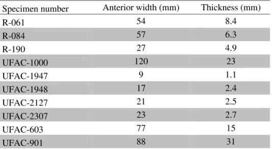
is thicker, despite the preserved bones having a similar length and width when compared to P. manchineri shells. Considering that, and also their different ages, it does not seem reasonable to consider both as the same taxa (Table 1).
According to Meylan et al. (2009), using skull-shell ratios of recent podocnemid species, it is hypothesized that the shell of Caninemys tridentata would be less than 1.2-1.5 meters, compatible with the P. manchineri shell size (1.2 meters). The skull of P. bassleri is slightly shorter (15.7 cm) than that of C. tridentata (16.5 – 17.0 cm) (Meylan
et al. 2009) and the Carbonemys cofrinii holotype skull is even larger (21 cm) than C. tridentata (Cadena et al.2012). Therefore all these taxa could also be compatible in size with the shell of P. manchineri.
The extant species of Podocnemis are much smaller than P. manchineri. The larger extant member of the genus P. expansa can reach a maximum total length of about 90 cm (Rueda-Almonacid et al. 2007), while the shell of P. manchineri is much larger (1.2 meters). Due to the massive difference in size, as well as some morphological differences, such as the axillary buttress reaching peripheral 3 in P. manchineri, instead of peripheral 2 in all other Podocnemis extant species except in P. vogli, we discard the idea of P. manchineri belonging to any extant species (Table 2).
Phylogenetic analysis
To examine the phylogenetic relationships of P. manchineri, , we included several podocnemid taxa for which the skull is known, as well as some shell based taxa, commonly excluded from the analyses due to missing data, such as Stupendemys geographicus, P. medemi, P. negrii, P. pritchardi, and Kenyemys williamsi.
2006, 2011; Meylan et al.2009; Cadena et al.2010; Aquentin 2012), and 13 are new. These new characters regard carapace and plastron and where based on first-hand observation of some taxa, as well as from literature review of fossil and recent podocnemid species.
All taxa were coded on species level except by the outgroups Chelidae (Chelus fimbriatus, Phrynops geoffroanus), Pelomedusidae (Pelomedusa, Pelusios), Bothremydidae (Kurmademys, Cearachelys, Foxemys) and the podocnemids Neochelys
(N. fajumensis and N. zamorensis), and Stereogenys (S. cromeri and S. libyca). The coding was based on direct observation and on photographs of fossil and recent species of podocnemids, as well as from literature (Wood & Díaz de Gamero 1971; Wood 1976, 1983, 1997, 2003; Lapparent de Broin 2000; De la Fuente 2003; Gaffney & Forster2003; França & Langer2006; Meylan2006; Gaffney et al.2006, 2011; Meylan
et al.2009; Cadena et al.2010, 2012). The outgroups were chosen based on Gaffney et al. (2006).
The character matrix was constructed using NEXUS Data Editor and analyzed using PAUP 4.0 beta 10 (Swofford 2000). The following protocol was used in the analyses: random addition sequence with 100,000 replicates as addition sequence method, Three Bisection and Reconnection (TBR) as swapping algorithm. The branches were also collapsed if the minimum branch length is zero, and synapomorphies for the nodes follow ACCTRAN character optimization. No topological constrains were used.
A second analysis without P. bassleri was conducted, since this taxon has been constantly referred as problematic or a wildcard in phylogenetic analyses, being commonly excluded (e. g. Meylan et al.2009; Gaffney et al.2011; Cadena et al.2012). Additionally, it is the only taxa assigned to the genus Podocnemis known only by skull material. We also conducted a test by coding the postcranial characters of P. manchineri
in two skull only taxa, P. bassleri and Caninemys tridentata, to compare changes in the first cladogram and look for possible relations.
Results
(Appendix 2). None of the new characters were uninformative or constant. The analysis of 45 taxa yielded 134 most parsimonious trees of 331 steps (CI excluding uninformative characters = 0.5266; RI = 0.7615; RC = 0.4141). The analysis without P. bassleri (44 taxa) resulted in 12 most parsimonious trees of 331 steps (CI excluding uninformative characters = 0.5266; RI = 0.7588; RC = 0.4126). The strict consensus in both cases is very similar (Figs. 4 and 5). The only difference is within the Podocnemis
genus, which is less resolved when P. bassleri is included in the analysis (Fig. 4). The results using only the holotype of P. manchineri or the information available from all referred specimens were the same. The combination of P. manchineri and P. bassleri
data does not change the position of any other taxa in the analysis, and this combination of taxa takes the position of P. manchineri in the cladogram without P. bassleri. On the other hand, the combination of P. manchineri and C. tridentata resulted in a large polytomy within the subfamily Erymnochelyinae.
Our analysis yielded trees with similar topologies to the trees from other works, which are also based on morphology (e. g. Meylan et al. 2009; Cadena et al. 2010, 2012; Gaffney et al.2011). Among these previous works, the tree found in our study is more similar to that of Gaffney et al. (2011). The topology for the taxa outside Podocnemidae is the same (Chelidae (Pelomedusidae + Araripemys) (Euraxemydidae (Bothremydidae (Brasilemys (Hamadachelys + Portezueloemys + Podocnemidae)))))), as well as the topology of the clade that includes Cretaceous taxa, such as: (Bauruemys
(Peiropemys (Pricemys + Lapparentemys))). The position of Cerrejonemys is the same as in Cadena et al. (2010, 2012), being the sister-taxa of the genus Podocnemis.
Discussion
Taxonomy and phylogenetic relationships of P. manchineri
the late Miocene of Amazonia, and Podocnemis bassleri, from the Mio-Pliocene of Peru. All comparable taxa are different from the P. manchineri.
In any case, our analysis support a Podocnemis monophyly that includes all the recent species (Podocnemis expansa, P. vogli, P. unifilis, P. erythrocephala, P. lewyana, and P. sextuberculata) plus the fossil species, both skull(P. bassleri) and shell based (P. manchineri, P. negrii, P. pritchardi ,and P. medemi), which is supported by the following unambiguous synapomorphies: prefrontal with an interorbital sulcus at the sutural area between both prefrontals (character 7) and dentary with accessory ridges (character 77) (Fig. 4).
Phylogenetic analysis
Here we resolved the position of three fossil Podocnemidae previously regarded as incertae sedis (P. negrii, P. medemi, and P. pritchardi) (Fig. 4). Since the most basal taxon within the genus Podocnemis in our analysis (excluding P. bassleri) is the recent
P. erythrocephala, we assume that the incertae sedis taxa analysed here actually belong to the genus Podocnemis (Fig. 5). From those, P. negrii is the sister-taxon of P. sextuberculata, as has been already suggested by Carvalho et al. 2002. This close relationship is supported by the following unambiguous synapomorphies: carapace with keeled neurals, 92 (01) and carapace with the second vertebral scute hexagonal in shape, 103 (21). P. pritchardi and P. medemi relations are less cleared, but both belong to the Podocnemis clade (Fig. 4).
In the subfamily Erymnochelyinae our topology for Caninemys, Dacquemys and the unnamed taxon UCMP-42008 is the same as in Gaffney et al. (2011). An interesting new result is the relationships of Peltocephalus, Turkanemys, Kenyemys, and
Erymnochelys, which comprise a monophyletic clade, (Peltocephalus (Kenyemys
(Turkanemys + Erymnochelys))). In all morphological cladistic analyses Peltocephalus
and Erymnochelys are depicted as sister-taxa (e. g. França & Langer2006; Meylan et al. 2009; Cadena et al. 2010, 2012; Gaffney et al. 2011), this more common topology would represent a huge gap in the known fossil record, since both species are living representatives and considering that Peltocephalus is from South America and
up of the Gondwana, with fossil specimens yet to be found, either on continental Africa or Antarctica. Anyhow, both explanations would need more evidence to be adequately supported (like saltwater tolerance in recent species, or new fossil findings). In our analysis, however, the presence of African species between Peltocephalus and
Erymnochelys furnishes new information to subsequent studies on biogeographical history of these two extant taxa (Fig. 4).
Although we had a polytomy comprising Stupendemys, all the Bairdemys
species, Cordichelys, Latentemys, Mogharemys, Papoulemys, Neochelys and
Brontochelys (Lemurchelys (Shweboemys (Stereogenys))), we provide for the first time a phylogenetic proposition to Stupendemys. However, the relationships among those taxa are not well established. In any case, our topology within the genus Bairdemys, and the topology of the clade comprising Brontochelys, Lemurchelys, Shweboemys, and
Stereogenys agree with that of Gaffney et al. (2011).
In our study we observed a clear division within Podocnemidae with one clade leading to the genus Podocnemis and the other one leading to the clade that includes the other extant Podonemidae and Bairdemys. These two clades have been previously regarded as the subfamilies Podocneminae and Erymnochelyinae. Here we propose the phylogenetic definitions for the two clades as follows: Erymnochelyinae would be the stem-based clade defined as all Podocnemidae more related to
Erymnochelys madagascariensis than to Podocnemis expansa. However, due to the conflict between morphological and molecular datasets we propose a new name to the Erymnochelyinae subfamily, Peltocephalinae. The morphological data yield a clear division within Podocnemidae, where Erymnochelys madagascariensis is more related to Peltocephalus dumeriliana than to Podocnemis expansa. However, the molecular data shows a closer relationship between E. madagascariensis and P. expansa. That would leave P. dumeriliana in a third subfamily, not supported by morphological data. Therefore, we prefer to use P. dumeriliana as the type species for the subfemily, Peltocephalinae. Being the stem-based clade defined as all Podocnemidae more related to Peltocephalus dumerilianus than to Podocnemis expansa and Podocneminae is the stem-based clade defined as all Podocnemidae more related to Podocnemis expansa
than to Peltocephalus dumerilianus.
which is depicted as more related to Podocnemis expansa than to Peltocephalus dumerilianus in molecular dataset cladograms, that would leave Peltocephalus dumerilianus in a third subfamily, not well recognizable in morphological analyses, if
E. madagascariensis is used to define a clade. So, this division works well with both current molecular and morphological datasets.
Large podocnemid fossil taxa in western Amazonia
This new species confirms that there is at least two very large podocnemids in the late Miocene of Amazonia (Lapparent de Broin et al. 1993; Gaffney et al. 1998; Meylan et al.2009). Since Stupendemys is a shell-based taxon, it could not be compared to the skull based taxa like Caninemys and P. bassleri. Our analysis suggests that P. manchineri and Stupendemys are two distinct large podocnemid shell based taxa from the late Miocene of Acre mainly based on the differences observed in overall shell morphology.
Additionally, we consider the possibility that there are more than two species of large Podocnemidae in the late Miocene of Amazonia. There are some odd bones such as UFAC-901, UFAC-637, and UFAC-933, as well as the possibility that the fossils assigned to “Stupendemys” souzai may actually belong to more than one taxa (Meylan
et al.2009).
similar in length and width, or with the overall shell thickness. Those are also incompatible with the testudinids already known from the Solimões Formation. Because of that, they could represent another large taxon of either a testudinid or a podocnemid with a thicker shell, like Cerrejonemys (Fig. 6, 7 and Tables 4, 5).
There are some comparable fossil cervical vertebrae showing again that there may have been more than two large podocnemids in the Miocene of south-western Amazonia. Unfortunately, one of them is missing from the collection of University of Acre. Only a cast from the UFAC-1542 has left. However, it still can provide some valuable information. The vertebra is typically podocnemid in shape, with a saddle shaped centra. It strongly resembles the cervical vertebrae of the living Podocnemis
species, except for its greater size. This vertebra is clearly different from the other large sized vertebrae assigned to “Stupendemys” souzai (e. g. UFAC-1163, UFAC-1553, UFAC-1554, and UFAC-5275), being smaller and more elongated. They likely represent the same taxon as the LACM-131949, also from the late Miocene of Amazonia (Gaffney et al.1998). All the vertebrae assigned to S. souzai are considered to be from the caudal part of the cervical series, each one being probably the eighth or seventh vertebra of the series (Negri & Bocquentin 1998; Bocquentin & Melo 2006). From these, we highlight UFAC-5275 where the ventral surface is much more arched and the neural channel is considerably smaller than all the other vertebrae. Also, the UFAC-5275 has a more robust constitution. These different features indicate that the UFAC-5275 might represent a third taxon, different from S. souzai and UFAC-1542 (Fig. 8).
Conclusions
Here we could differentiate it from all other species with overlapping parts, as well as include P. manchineri and several shell based taxa in a phylogenetic analysis. Moreover, it is very likely that a third large sized podocnemid lived in the Miocene-Pliocene of South-western Amazonia.
We also conclude that the presence of fossil species between the common association of Peltocephalus and Erymnochelys showed here could help to explain the relationship and biogeographic history of those extant taxa.
We also provide phylogenetic definitions for the subfamilies Erymnochelyinae and Podocneminae. Erymnochelyinae is the stem-based clade defined as all Podocnemidae more related to Peltocephalus dumerilianus than to Podocnemis expansa
and Podocneminae is the stem-based clade defined as all Podocnemidae more related to
Podocnemis expansa than to Peltocephalus dumerilianus.
After the inclusion of new characters regarding carapace and plastron in the data matrix and according to our analysis, we conclude that although the podocnemid shell morphology is generally referred as conservative it can provide important phylogenetic information. Our phylogenetic analysis supports the assignment of P. negrii, P. medemi, and P. pritchardi to the genus Podocnemis. The analysis also depicted a better idea of the phylogenetic position of Stupendemys geographicus, regarded as a member of Erymnochelyinae. Also, one of the main problematic taxa in several phylogenetic analyses, P. bassleri, known only from skull material is recovered as a Podocnemis as well. Interestingly, in contrast with other phylogenetic analyses based on morphological data, the resolution of the analysis within Podocnemis decreased with the inclusion of P. bassleri, while the analysis with shell based fossil taxa resulted in a more resolved topology. Since we included new shell character in the analysis, it may suggest that they can be as important as skull characters in cladistics studies concerning chelonians.
Acknowledgements
References
Anquetin, J. 2012. Reassessment of the phylogenetic interrelationships of basal turtles (Testudinata). Journal of Systematic Palaeontology, 10, 3–45.
Barbosa-Rodrigues, J. B. 1892. Les reptiles de la vallée de l’Amazone. Vellosia, 2, 41–60.
Bocquentin, J. & E. Guilherme. 1999. As preguiças Mylodontinae (Mammalia, Xenarthra, Mylodontidae) do Neógeno do Sítio Niterói, Acre, Brasil. Acta Geologica Leopoldina, 22, 57–67.
Bocquetin-Villanueva, J., Jégu, M. & Brito, P. M. 1997. “An extinct
Phractocephalus species (Siluriformes, Pimelodidae) from the Mio-Pliocene Solimões Formation of Acre State, Brazil”. In Abstracts of International Symposium on Phylogeny and Classification of Neotropical Fishes, Museu de Ciências e Tecnologia. PUCRS, 1997. p. 56. Porto Alegre.
Bocquentin, J. V. & Rancy, A. 1987. “Presença de Chelus lewisi Wood, 1976 (Testudinata, Pleurodira) no Neógeno do Estado do Acre, Brasil”. In 4th Congresso Latino-Americano de Paleontologia, Santa Cruz de la Sierra, 1987, Edited by: Asociacion Boliviana de Paleontologia. 566–573. Santa Cruz de la Sierra.
Bocquentin, J. V., Santos, J. C. R. 1989. “Ocorrência de Chelus colombianus
(Chelonii, Chelidae) no Mioceno Superior do Acre, Brasil”. In Resumos do 11th Congresso Brasileiro de Paleontologia, Curitiba, 1989, Edited by: Sociedade Brasileira de Paleontologia. 104–105. Curitiba.
Bocquentin, J. V. & Souza-Filho, J. P. 1989. “Nova interpretação do gênero
Purussaurus (Crocodylia, Alligatoridae)”. In Resumos do 11th Congresso Brasileiro de Paleontologia, Curitiba, 1989, Edited by: Sociedade Brasileira de Paleontologia. 427–
Bocquentin, J. V.., Souza-Filho, J. P. 1990. O crocodiliano Sul-Americano
Carandaisuchus como sinonímia de Mourasuchus (Nettosuchidae). Revista Brasileira de Geociências, 20, 230–233.
Bocquentin, J. V., Guilherme, E. & Negri, F. R. 2001. Duas espécies do gênero
Chelus (Pleurodira, Chelidae) no Mioceno Superior-Plioceno Inferior da Amazônia Sul-Ocidental. Revista Universidade Guarulhos, 6, 50–55.
Bocquentin, J. V. & Melo, J. 2006. Stupendemys souzai sp. nov. (Pleurodira, Podocnemididae) from the Miocene-Pliocene of the Solimões Formation, Brazil.
Revista Brasileira de Paleontologia, 9, 187–192.
Broin, F., Bocquentin, J. & Negri, F. R. 1993. Gigant turtles (Pleurodira, Podocnemididae) from the late Miocene-early Pliocene of South-Western Amazon. Bulletin de l’Institut Français d’Études Andines, 22, 657–670.
Cadena, E. A., Bloch, J. I. & Jaramillo, C. A. 2010. New podocnemidid turtle (Testudines: Pleurodira) from the Middle-Upper Paleocene of South America. Journal of Vertebrate Paleontology, 30, 367–382.
Cadena, E. A., Ksepka, D. T., Jaramillo, C. A. & Bloch, J. I. 2012. New pelomedusoid turtles from the late Palaeocene Cerrejón Formation of Colombia and their implications for phylogeny and body size evolution. Journal of Systematic Paleontology, 10, 313–331.
Campbell Jr., K. E. & Frailey, C. D. 1984. Holecene flooding and species diversity in southwestern Amazônia. Quaternary Research, 21, 369–375.
Campbell Jr., K. E., Heizler, M., Frailey, C. D., Romero-Pitman, L. & Prothero, D. R. 2001. Upper Cenozoic chronostratigraphy of the southwestern Amazon Basin.
Geology, 29, 595–598.
Campbell Jr., K. E., Frailey, C. D. & Romero-Pitman, L. 2000. The Late Miocene gomphothere Amahuacatherium peruvium (Proboscidea: Gomphotheriidae) from Amazonian Peru: implications for the Great American Faunal Interchange. Instituto Geológico Minero y Metalúrgico, Série D, Estudios Regionales, 23, 1–152.
Campbell Jr., K .E., Frailey, C. D., Romero-Pitman, L. 2006. The Pan-Amazonian Ucayali Peneplain, late Neogene sedimentation in Amazônia, and the birth of the modern Amazon River system. Palaeogeography, Palaeoclimatology, Palaeoecology,
239, 166–219.
Campos, D. A. 1977. Tartarugas fósseis do Brasil. Unpublished MSC Dissertation, Universidade Federal do Rio de Janeiro, 101 pp.
Campos, D. A. & Broin, F. 1981. Tartarugas fósseis do Brasil. Anais da Academia Brasileira de Ciências, 53, 210–211.
Carvalho, P., Bocquentin, J. & Broin, F. L. 2002. Une nouvelle espèce de
Podocnemis (Pleurodira, Podocnemididae) provenant du Néogène de la Formação Solimões, Acre, Brésil. Geobios, 35, 677–686.
Cruz. N. M. C. 1984. “Palinologia do linhito do Solimões no Estado do Amazonas”. In
Anais do 2nd Simpósio de Geologia da Amazônia, Manaus, 1984. 473–480. Manaus.
Frailey, C. D. 1986. Late Miocene and Holocene Mammals, exclusive of the Notoungulata, of the rio Acre region, Western Amazonia. Contributions in Science, Natural History Museum, 374, 1–46.
Frailey, C. D., Lavina, E., Rancy, A. & Souza-Filho, J. A. 1988. Proposed Pleistocene/Holocene lake in the Amazon Basin and its significance to Amazonian geology and biogeography. Acta Amazonica, 18, 119–143.
Gaffney, E. S., Campbel, K. E. & Wood, R. C. 1998. Pelomedusoid side-necked turtles from Late Miocene sediments in South-western Amazonia. American Museum Novitates, 3245, 1–11.
Gaffney, E. S. & R. Wood. 2002. Bairdemys, a new side-necked turtle (Pelomedusoides: Podocnemididae) from the Miocene of the Caribbean. American Museum Novitates, 3359, 1–28.
Gaffney, E. S. & Forster, C. A. 2003. Side-necked turtle lower jaws (Podocnemididae, Bothremydidae) from the Late Cretaceous Maevarano Formation of Madagascar.
American Museum Novitates, 3397, 1–13.
Gaffney, E. S., Tong, H. & Meylan, P.A. 2006. Evolution of the side-necked turtles: the families Bothremydidae, Euraxemydidae, and Araripemydidae. Bulletin of the American Museum of Natural History, 300, 1–698.
Gaffney, E. S., Meylan, P. A., Wood, R. C., Simons, E. & Campos, D. A. 2011. Evolution of the side-necked turtles: the family Podocnemididae. Bulletin of the American Museum of Natural History, 350, 1–37.
Gasparini, Z. 1985. Un nuevo cocodrilo (Eusuchia) Cenozóico de América del Sur.
Coletânea de Trabalhos Paleontológicos, Série Geologia, 27, 51–53.
nuclear gene sequence variation. Biological Journal of the Linnean Society, 67, 213– 246.
Hoorn, C. 1993. Marine incursions and the influence of Andean tectonics on the Miocene depositional history of northwestern Amazônia: results of a palynostratigraphic study. Palaeogeography Palaeoclimatology Palaeoecology, 105, 267–309.
Hoorn, C. 1994a. Fluvial palaeoenvironments in the Amazonas Basin (Early Miocene - early Middle Miocene, Colombia). Palaeogeography, Palaeoclimatology, Palaeoecology, 109, 1–54.
Hoorn, C. 1994b. An environmental reconstruction of the palaeo-Amazon river system (Middle–Late Miocene, NW Amazonia). Palaeogeography, Palaeoclimatology, Palaeoecology, 112, 187–238.
Hoorn, C. 1995.Comment on Late Miocene tidal deposits in the Amazonian foreland basin by Räsänen, M., Linna, A.M., Santos, J.C.R., Negri, F.R. Science, 273, 122–123.
Hovikoski, J., Räsänen, M., Roddaz, M., Brusset, S., Hermoza, W., Pittman, L. & Lertola, K. 2005. Miocene semidiurnal tidal rhythmites in Madre de Dios, Peru.
Geology, 33, 177–180.
Hsiou, A. S. 2010. Os lagartos e serpentes (Lepidosauria, Squamata) do Mioceno médio-superior da região norte da América do Sul. PhD Thesis, Universidade Federal do Rio Grande do Sul, 236 pp.
Hsiou, A.S., Ferigolo, J. & Albino, A. 2007. Sobre os Squamata (Lepidosauria) da Formação Solimões, Mioceno da Amazônia Sul-Ocidental, Brasil. Ameghiniana
(Supplement), 44, 23R.
Lapparent de Broin, F. 2000. The oldest pre-podocnemidid turtle (Chelonii, Pleurodira), from the Early Cretaceous, Ceará State, Brasil, and its environment.
Treeballs del Museu de Geología de Barcelona, 9, 43–95.
Lapparent de Broin, F., Bocquentin, J. & Negri, F. R. 1993. Gigantic turtles (Pleurodira, Podocnemididae) from the late Miocene–early Pliocene of south western Amazon. Bulletin de l’Institut Francais d’Etudes Andines, 22, 657–670.
Latrubesse, E. M. 1992. El cuaternario fuvial de la cuenca del Purus en el estado de Acre, Brasil. Unpublished PhD Thesis, Universidad Nacional de San Luis, 214 pp.
Latrubesse, E. M., Bocquentin, J., Santos, C. R. & Ramonell, C. G. 1997. Paleoenvironmental model for the late Cenozoic southwestern Amazonia: paleontology and geology. Acta Amazonica, 27, 103–118.
Latrubesse, E. M., Silva, S. A. F., Cozzuol, M. A. & Absy, M. L. 2007. Late Miocene continental sedimentation in southwestern Amazônia and its regional significance: Biotic and geological evidence. Journal of South American Earth Sciences, 23, 61–80.
Meylan, P. A. 1996. Skeletal morphology and relationships of the Early Cretaceous sidenecked turtle, Araripemys barretoi (Testudines: Pelomedusoides: Araripemydidae), from the Santana Formation of Brazil. Journal of Vertebrate Paleontology, 16, 20–33.
Meylan, P. A., Gaffney, E. S. & Campos, D. A. 2009. Caninemys, a new side-necked turtle (Pelomedusoides: Podocnemididae) from the Miocene of Brazil. American Museum Novitates, 3639, 1–26.
Negri, F. R. & Bocquentin, J. 1998. Vértebras cervicais e xifiplastrão de Stupendemys
sp. (Chelonii, Podocnemididae, Podocnemidinae) no Mio-Plioceno do Estado do Acre e da região frontereiriça Brasil-Peru. Boletim do Museu Paraense Emilio Goeldi, 10, 17–
Negri, F. R. & Ferigolo, J. 1999. Anatomia craniana de Neoepiblemaambrosettianus
(Ameghino, 1889) (Rodentia, Caviomorpha, Neoepiblemidae) do Mioceno Superior-Plioceno, Estado do Acre, Brasil, e revisão das espécies do gênero. Boletim do Museu Paraense Emílio Goeldi Série Ciências da Terra, 11, 1–80.
Noonan, B. P. & Chippindale, P. T. 2006. Vicariant origin of Malagasy reptiles supports Late Cretaceous Antarctic land bridge. American Naturalist, 168, 730–741.
Price, L. I. 1964. Sobre o crânio de um grande crocodilídeo extinto do Alto Rio Juruá, Estado do Acre. Anais da Academia Brasiliera de Ciências, 36, 59–66.
Radambrasil. 1977. Geologia. Levantamento de recursos naturais. Ministério de Minas e Energia, DNPM, 14, 49–66.
Rancy, A. & Bocquentin, J. V. 1987. “Dois quelônios do Neógeno do Acre, Brasil”. In
Anais do 10th Congresso Brasileiro de Paleontologia, Rio de Janeiro, 1987, Edited by: Sociedade Brasileira de Paleontologia. 181–187. Rio de Janeiro.
Räsänen, M. E., Linna, M. A., Santos, J. C. & Negri, F. R. 1995. Late Miocene tidal deposits in the Amazonian Foreland Basin. Science, 269, 386–390.
Rebata-H., L. A., Gingras, M. Y. K., Räsänen, M. E. & Barberi, M. 2006. Tidal channel deposits on a delta plain from the Upper Miocene Nauta Formation, Marañón Foreland Sub-basin, Peru. Sedimentology, 53, 971–1013.
Rueda-Almonacid, J. V., Carr, J. L., Mittermeier, R. A., Rodríguez-M., J. V., Mast, R. B., Vogt, R. C., Rhodin, A. G. J., De La Ossa-Velázquez, J., Rueda, J. N. & Mittermeier, C. G. 2007. Las tortugas y los cocodrilianos de los países andinos del trópico. Serie de guías tropicales de campo Nº 6. Editorial Panamericana, Bogotá, 538 pp.
P.Wesselingh, (eds) Amazonia: landscape and species evolution - a look into the past. Blackwell Publishing Ltd., Chichester.
Roddaz, M., Baby, P., Brusset, S., Hermoza, W. & Darrozes, J. M. 2005. Forebulge dynamics and environmental control in western Amazônia: the case study of the Arch of Iquitos (Peru). Tectonophysics, 399, 87–108.
Souza-Filho, J. P. 1998. Novas formas fósseis de Crocodylia (Alligatoridae e Gavialidae) da Formação Solimões, Cenozóico do Estado do Acre-Brasil, representadas por materiais cranianos e mandibulares. Unpublished PhD Thesis, Universidade Federal do Rio Grande do Sul, 194 pp.
Souza-Filho, J. P. & Bocquentin, J. 1989. “Brasilosuchusmendesi, n.g. n.sp. um novo representante da Família Gavialidae do Neógeno do Estado do Acre, Brasil”. In 11th Congresso Brasileiro de Paleontologia, Curitiba, 1989, Edited by: Sociedade Brasileira de Paleontologia. p. 139. Curitiba.
Souza-Filho, J. P. & Bocquentin, J. 1991. “Caiman niteroiensis sp. nov. (Alligatoridae, Crocodylia) do Neógeno do Estado do Acre, Brasil”. In 12th Congresso Brasileiro de Paleontologia, São Paulo, 1991, Edited by: Sociedade Brasileira de Paleontologia. p. 126. São Paulo.
Sill, W. D. 1970. Nota preliminar sobre un nuevo gavial del Plioceno de Venezuela y una discusión de los gaviales sud-americanos. Ameghiniana, 7, 151–159.
Swofford, D. 2002. PAUP ∗. Phylogenetic Analysis Using Parsimony (∗and other methods). Version 4.0b10. Sinauer Associates, Sunderland.
Vonhof, H. B., Wesseling, F. P. & Ganssen, G. M. 1998. Reconstruction of the Miocene western Amazonian aquatic system using molluscan isotopic signature.
Vargas-Ramírez, M., O. V. Castaño-Mora, & U. Fritz. 2008. Molecular phylogeny and divergence times of ancient South American and Malagasy river turtles (Testudines: Pleurodira: Podocnemididae). Organisms, Diversity, and Evolution, 8, 388–398.
Wesselingh, F. P., Räsänen, M. E., Irion, G., Vonhof, H. B., Kaandorp, R., Renema, W., Romero-Pittman, L. & Gingras, M. 2002. Lake Pebas: a palaeo-ecological reconstruction of a Miocene long-lived lake complex in Western Amazônia.
Cainozoic Research, 1, 35– 81.
Wood, R. C. 1976. Stupendemys geographicus, the world’s largest turtle. Breviora,
436, 1–31.
Wood, R. C. 1983. Kenyemys williamsi, a fossil pelomedusid turtle from the Pliocene of Kenya. Pp. 74–85 in G. J. Rhodin & K. Miyata (eds) Advances in herpetology and evolutionary biology. Cambridge, Massachusetts.
Wood, R. C. 1997. Turtles. Pp. 155–170 in R. F. Kay, R. H. Madden, R. L. Cifelli & J. J. Flynn (eds) Vertebrate paleontology in the Neotropics. Smithsonian Institution Press, Washington.
Wood, R. C. 2003. Fossil turtles from Lothagam. Pp. 115–136 in M.G. Leakey & J. M. Harris (eds) Lothagam: the dawn of humanity in eastern Africa. Columbia University Press, New York.
Appendix 1: Character list
1. Nasals: (0) present; (1) absent. (Gaffney et al., 2006).
2. Prefrontals meet on midline: (0) absent; (1) present. (Gaffney et al., 2006).
3. Prefrontal, anterior overhang onto apertura narium externa: (0) shaped by the nasals ; (1) by the prefrontals, covering a small portion of the posterior part of the apertura, ending in acute medial tip; (2) by the prefrontals, completely covering the apertura, ending in a straight to convex edge. (Cadena et al., 2010).
4. Frontal, orbital position: (0) Facing laterally/anterolaterally; (1) dorsolaterally; (2) dorsally. (Cadena et al., 2010).
5. Frontal, interorbital groove: (0) absent; (1) present. (Gaffney et al., 2011).
6. Prefrontal/frontal: (0) flat or slight convex; (1) strongly convex dorsally. (Gaffney et al., 2011).
7. Prefrontal, interorbital sulcus at the sutural area between both prefrontals: (0) absent; (1) present. (Broin, 2000).
8. Prefrontal at the interorbital space: (0) wide; (1) narrow. (Cadena et al., 2010).
9. Parietal, quadratojugal-parietal contact: (0) absent; (1) short contact; (2) long contact. (Gaffney et al., 2011).
10. Parietal, parietal-pterygoid contact in septum orbitotemporale: (0) absent; (1) present and wider; (2) present and narrower. (Gaffney et al., 2011).
12. Temporal emargination, secondary roofing of the fossa temporalis in dorsal view: (0) not advanced and highly concave allowing the complete exposure of the otic chamber roof; (1) medially advanced with posteriorly expanded posterolateral temporal emargination of the parietals and quadratojugal with concave margins, covering partially or almost totally the otic chamber roof; (2) very advanced with convex to straight tapering margins completely covering the roof of the otic chamber. (Broin, 2000).
13. Parietal, interparietal scale: (0) absent; (1) equilateral triangle; (2) elongate triangle; (3) parallel sided; (4) broad posteriorly. (Gaffney et al., 2011).
14. Parietal, interparietal scale, anterior margin: (0) anterior to the frontal parietal suture; (1) posterior to the frontal parietal suture. (Cadena et al., 2010).
15. Jugal-parietal contact: (0) absent; (1) present. (De la Fuente, 2003).
16. Jugal-quadrate contact: (0) absent; (1) present. (Gaffney et al., 2011).
17. Jugal, cheek emargination: (0) slight; (1) reaches level of orbit; (2) reaches above level of orbit; (3) reaches above quadrate. (Gaffney et al., 2011).
18. Squamosal, ventral vertical flangea: (0) absent; (1) present. (Gaffney et al., 2011).
19. Squamosal-parietal contact: (0) present; (1) absent. (Gaffney et al., 2006).
20. Postorbital, size: (0) equal to orbit; (1) smaller than orbit. (Gaffney et al., 2011).
22. Premaxillae, pinched snout: (0) absent; (1) concave outline near premaxilla-maxilla contact, snout not elongated; (2) concave outline posterior to prepremaxilla-maxilla-premaxilla-maxilla contact, snout elongated. (Gaffney et al., 2011).
23. Premaxillae, one or two accessory ridges on the ventral surface of the premaxilla: (0) absent; (1) present. (Cadena et al., 2010).
24. Premaxillae, foramen prepalatinum in suture with maxilla (Meylan et al, 2009): (0) in premaxilla only; (1) in premaxillamaxillary suture; (2) absent. (Meylan et al., 2009).
25. Premaxillae, foramen prepalatinum relative to triturating ridge: (0) on flat surface; (1) under triturating ridge; (2) absent. (Meylan et al., 2009).
26. Maxilla, medial expansion of triturating surface (Gaffney et al, 2011): (0) absent; (1) present, forming median maxillary ridge; (2) secondary palate with midline cleft. (Gaffney et al., 2011).
27. Maxilla, secondary palate long: (0) no; (1) yes. (Gaffney et al., 2011).
28. Maxilla, triturating surface convexity: (0) absent or shallow; (1) deep. (Gaffney
et al., 2011).
29. Maxilla, labial ridge: (0) high and narrow; (1) low and thick. (Gaffney et al., 2011).
30. Maxilla, accessory ridges: (0) absent; (1) one or two. (Gaffney et al., 2011).
31. Maxilla, meet broadly on midline: (0) no; (1) yes. (Gaffney et al., 2011).
32. Maxilla, median maxillary ridge: (0) absent; (1) present. (Meylan et al., 2009).
34. Palatine, medial edges of palatal cleft: (0) absent; (1) parallel; (2) curved. (Gaffney et al., 2011).
35. Palatine, palatine extent in triturating surface: (0) narrow or absent; (1) moderate, but much less than maxilla extent; (2) large, equal or slightly less than maxilla extent. (Gaffney et al., 2011).
36. Palatine, dorsal process of palatine contacts parietal in septum orbitotemporale: (0) no; (1) yes. (Gaffney et al., 2011).
37. Palatine, dorsal process reaches frontal: (0) no; (1) yes. (Gaffney et al., 2011).
38. Palatine, fossa orbitalis posterior pocket: (0) absent; (1) present in septum orbitotemporale. (Gaffney et al., 2011).
39. Palatine-basisphenoid contact separates pterygoids: (0) no; (1) yes. (Gaffney et al., 2011).
40. Palatine, second palate: (0) absent ; (1) present. (Gaffney et al., 2006).
41. Palatine, foramen palatinum posterius: (0) present; (1) absent. (Meylan et al., 2009).
42. Quadrate, antrum postoticum: (0) large; (1) smaller; (2) smallest and slitlike. (Gaffney et al., 2011).
43. Quadrate, fossa precolumellaris: (0) very small to absent; (1) present but shallow; (2) deep and well defined. (Gaffney et al., 2006).
45. Quadrate, incisura columellae auris: (0) no posterior bony restrictions; (1) eustachian tube separated from stapes by bone or narrow fissure; (2) eustachian tube and stapes enclosed or nearly enclosed by bone. (Gaffney et al., 2011).
46. Quadrate, quadrate-basioccipital contact: (0) absent ; (1) present. (Gaffney et al., 2006).
47. Quadrate, medial process reaches braincase: (0) absent ; (1) present. (Gaffney et al., 2011).
48. Quadrate, ventral projection: (0) very short, condylus mandibularis very close to the cavum tympani region; (1) short, condylus mandibularis slightly separated from the cavum tympani region; (2) long, condylus mandibularis considerably separated from the cavum tympani region. (Cadena et al., 2010).
49. Quadrate, condylus mandibularis shape: (0) much wider than long, with anterior and posterior edges straight to concave making it shorter at midline ; (1) slightly wider than long, kidney shaped, with anterior edge straight to concave and posterior edge convex. (Cadena et al., 2010).
50. Quadratojugal: (0) absent; (1) present. (Gaffney et al., 2006).
51. Pterygoid, cavum pterygoidei: (0) absent ; (1) partial ; (2) complete. (Gaffney et al., 2011).
52. Pterygoid, anterior opening of cavum pterygoidei: (0) absent; (1) small opening; (2) moderate opening; (3) large opening with foramen cavernosum inroof. (Gaffney et al., 2011).