João Paulo Ribeiro Proença Santana
Optimization of a sperm cryopreservation protocol for spotted
wolffish (Anarhichas minor Olafsen, 1772)
João Paulo Ribeiro Proença Santana
Optimization of a sperm cryopreservation protocol for spotted
wolffish (Anarhichas minor Olafsen, 1772)
Thesis for master’s degree in Aquaculture and Fisheries
Under the supervision of:
Dr José Beirão dos Santos, Nord University
Dr Elsa Cabrita, University of Algarve
Declaração de autoria de trabalho
Declaro ser o autor deste trabalho, que é original e inédito. Autores e trabalhos consultados estão devidamente citados no texto e constam da listagem de referências incluída.
(João Paulo Ribeiro Proença Santana)
Optimization of a sperm cryopreservation protocol for spotted
wolffish (Anarhichas minor Olafsen, 1772)
Direitos de cópia ou Copyright
© Copyright: João Paulo Ribeiro Proença Santana
A Universidade do Algarve tem o direito, perpétuo e sem limites geográficos, de arquivar e publicitar este trabalho através de exemplares impressos reproduzidos em papel ou de forma digital, ou por qualquer outro meio conhecido ou que venha a ser inventado, de o divulgar através de repositórios científicos e de admitir a sua cópia e distribuição com objetivos educacionais ou de investigação, não comerciais, desde que seja dado crédito ao autor e editor.
iii
Acknowledgments
I would like to sincerely thank all the people that directly or indirectly, contributed to this work, or that supported me through this not-always-so-easy journey.
First of all, to the best supervisors. Doctor Elsa Cabrita for supporting me from the beginning until the end, and for making sure that this experience would be as expected. To doctor José Beirão that received me like a friend from day one and never failed to support and push me to go on even when I was not able to keep on track of the demanded tasks.
To Willy Sandaa, CEO of AMINOR AS for providing us their broodstock.
To Bjørnar Eggen, and Fredrik Strand for helping on the samplings and instructing me on the procedures related to the species.
To Ana Cambeiro that used her free time helping me, overnight in the lab, instead of going out and explore a new country.
To the “aquagroupies”, Miguel Cabano, Madalena Mendes and, Filipa Beça with whom, all research related (and unrelated) questions were discussed.
To my parents and grandparents that have always incited me to leave my comfort zone and explore the world.
This work was supported by the WOLFSTORE project (AF0078) funded by the marine biotechnology research program from northern Norway, MABIT, by a Cost STSM, COST Office (Food and Agriculture COST Action FA1205: AQUAGAMETE), and by an ERASMUS+ grant.
v
Index
Acknowledgments ... iii Abstract ... 11 Resumo ... 13 1. Introduction ... 17 1.1. Wolffishes ... 171.2. Spotted wolffish aquaculture ... 19
1.3. Reproduction ... 20
1.4. Cryopreservation ... 22
1.5. Sperm quality assessment ... 23
1.6. Cryopreservation on spotted wolffish sperm ... 25
1.7. Design of a cryopreservation protocol for fish sperm ... 26
2. Objectives ... 31
3. Materials and methods ... 35
3.1. Broodstock ... 35 3.2. Sperm collection ... 35 3.3. Sperm analyses ... 36 3.3.1. Sperm motility ... 36 3.3.2. Sperm viability ... 37 3.4. Preliminary trials ... 38 3.4.1. Cell stickiness ... 38 3.4.2. Extender solution ... 38 3.5. Experimental design ... 38
3.5.1. Cryoprotectant solution and concentration ... 38
vi 3.5.3. Thawing rates ... 41 3.5.4. Sperm Viability ... 41 3.5.5. Fertilization trials ... 42 3.6. Statistical analyses ... 44 4. Results ... 47 4.1. Preliminary analyses ... 47 4.1.1. Cell stickiness ... 47 4.1.2. Extender solution ... 49 4.2. Cryoprotectants toxicity ... 51 4.3. Freezing rates ... 53 4.4. Thawing rates ... 56 4.5. Sperm viability ... 59 4.6. Fertilization ratios ... 59 5. Discussion... 63 5.1. Preliminary analyses ... 63 5.2. Cryopreservation ... 64 5.3. Fertilization ratios ... 65 6. Conclusions ... 69 7. References ... 73
7
Index of figures
Figure 1 Classification of Anarhichas minor ... 18
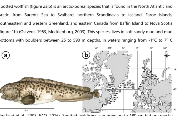
Figure 2(a) Schematic draw of spotted wolffish. Adapted from” COSEWIC Assessment and Status Report on the Spotted Wolffish, Anarhichas minor, in Canada”, by Committee on the Status of Endangered Wildlife in Canada, 2012, Ottawa (b) Spotted wolffish distribution. ... 18
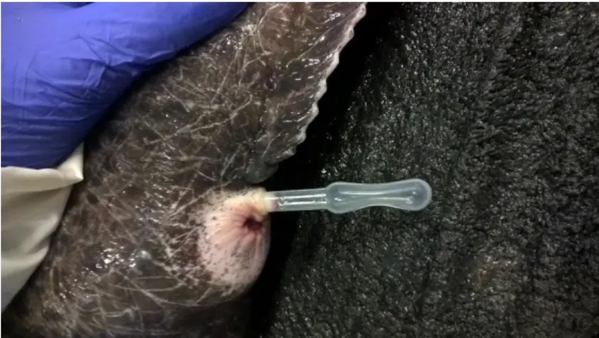
Figure 3 Sperm collection in spotted wolffish... 36
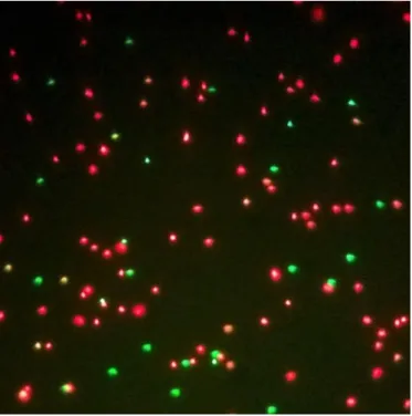
Figure 4 Sperm cells stained with SYBR-14 and propidium iodide, magnified x400 in fluorescence microscope ... 37
Figure 5 Loading sperm into cryopreservation straws ... 40
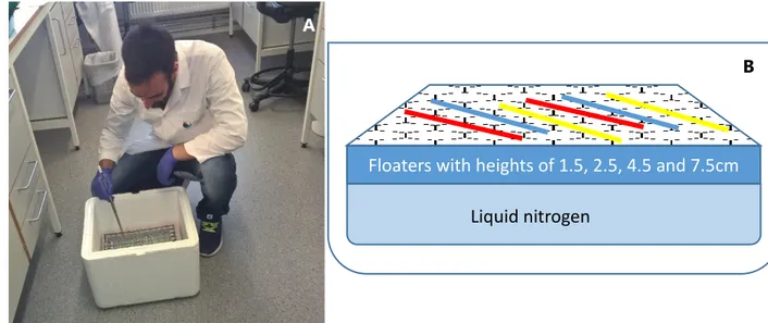
Figure 6 (A) Placing the straws in the cryopreservation box (B) Schematics of the cryopreservation box ... 40
Figure 7 Thawing the cryopreserved sperm in a water bath ... 41
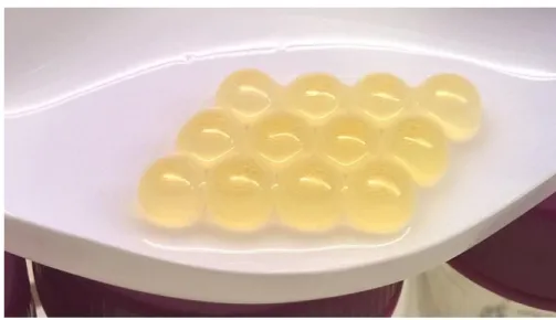
Figure 8 Spotted wolffish eggs ... 42
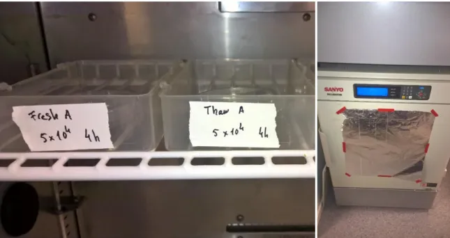
Figure 9 Eggs placed in the incubating boxes ... 43
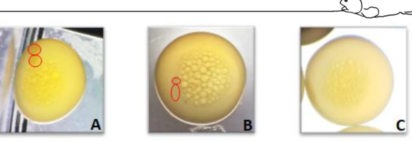
Figure 10 Egg development at 20 to 24 hours after fertilization. Developed egg with normal cell division (A); Developed egg with abnormal division (B); Non-developed egg (C). Red circles represent divided cells. ... 44
Figure 11 Sperm motility parameters comparing sperm diluted in a solution containing 1% BSA (white bars) and a solution without BSA (WO) (blue bars). (A) Percentage of motile cells (Mot); (B) Wobbliness (WOB); (C); Lateral head displacement (ALH); (D) Beat cross frequency (BCF); (E) Curvilinear velocity. (n=2; mean)... 48
Figure 12 Sperm motility parameters comparing well slides (white bars) and regular microscopy slides (blue bars). (A) Percentage of motile cells (Mot); (B) Wobbliness
8
(WOB); (C); Lateral head displacement (ALH); (D) Beat cross frequency (BCF); (E) Curvilinear velocity. (n=2; mean) ... 49
Figure 13 Sperm motility parameters of sperm after exposure to different extender solutions (Kime and Tveiten (KT) (blue bars); Kime and Tveiten with glucose (KT+G) (orange bars); and Smith and Ryan (SR) (grey bars)) at different times (0 hours (CTRL); 1 hour; 3 hours; and at 24 hours. (A) Percentage of motile cells (Mot); (B) Wobbliness (WOB); (C); Lateral head displacement (ALH); (D) Beat cross frequency (BCF); (E) Curvilinear velocity. (n=2; mean) ... 50
Figure 14 Sperm motility parameters comparing the toxic effect of DMSO (blue bars), propanediol (PROP) (orange bars) or methanol (METH) (grey bars) on cryopreserved sperm and, compared with fresh sperm (white bars). (A) Percentage of motile cells (Mot); (B) Wobbliness (WOB); (C); Lateral head displacement (ALH); (D) Beat cross frequency (BCF); (E) Curvilinear velocity (VCL). (n=5; mean ± SEM; p<0.05). Different letters refer to significantly different results obtained with Tukey test for each sperm motility parameter. ... 52
Figure 15 Sperm motility parameters comparing four freezing rates for sperm cryopreserved with 10% DMSO (blue bars), propanediol (PROP) (orange bars) or methanol (grey bars), with fresh sperm (white bars). (A) Percentage of motile cells (Mot); (B) Wobbliness (WOB); (C); Lateral head displacement (ALH); (D) Beat cross frequency (BCF); (E) Curvilinear velocity. (n=5; mean ± SEM; repeated measures MANOVA; p<0.05). Different letters refer to significantly different results. ... 54
Figure 16 Freezing curves for DMSO (A); propanediol (B); and methanol (C); with different distances from liquid nitrogen surface (1.5, 2.5, 4.5 and 7.5 cm), along 10 minutes. 55
Figure 17 Sperm motility parameters comparing two thawing rates for sperm cryopreserved with DMSO (blue bars) or propanediol (orange bars), compared with fresh sperm (white bars). (A) Percentage of motile cells (Mot); (B) Wobbliness (WOB); (C); Lateral head displacement (ALH); (D) Beat cross frequency (BCF); (E) Curvilinear velocity. n= 5; mean ± SEM; repeated measures MANOVA; p<0.05). Different letters refer to significantly different results. ... 57
9
Figure 18 Thawing curves for DMSO (A); and propanediol (B), with two different bath temperatures (5ºC for 1 min; and 10ºC for 25s) ... 58
Figure 19 Sperm viability comparison between fresh and cryopreserved sperm. (n=6; mean±SEM; Student’s t-test; p<0.05) Different letters refer to significantly different results ... 59
Figure 20 Percentage of fertilized eggs with normal division (colored bars) and abnormal divisions (dash colored bars) using fresh sperm (white bars), thawed sperm (blue bars), and washed sperm (orange bars). Fertilizations were performed using 5x10⁴ spz/egg,
5x10⁵ spz/egg and 5x10⁵ spz/egg with addition of 10% DMSO. (n=5; mean ± SEM;
two-way ANOVA, p<0.05) Different letters refer to significantly different results. Uppercase letters refer to comparison within treatments and lowercase letters refer to comparison within concentrations. ... 60
11
Abstract
Spotted wolffish is a potential candidate for cold water aquaculture, presenting high growth rates in captivity, good fillet yielding and a valuable and tasty meat. Reproduction in captivity is dependent on in vitro fertilization, however, low sperm volume with relatively low cell concentration and the lack of gametes synchronization (simultaneous availability of mature eggs and sperm) represent a challenge for the aquaculture industry. In this context, it is crucial the development of protocols, such as sperm cryopreservation, that allow sperm storage and maximization of its use. Sperm was diluted in a
solution described by Kime and Tveiten
(2002)
, and four sequential experiments were conducted. First, three different cryoprotectants (DMSO; 1, 2-propanediol; and methanol) at different concentrations (5, 10, and 20%) were tested for their toxicity. Second, sperm was cryopreserved in 0.5ml straws, at different distances from the liquid nitrogen (1.5, 2.5, 4.5, and 7.5cm) that correspond to different freezing rates. Afterwards, two different thawing rates (1min at 5ºC; and 25s at 10ºC) were tested. Finally, the fertilization capacity of cryopreserved sperm was tested against fresh, and cryopreserved and then washed sperm. The ratio of eggs with normal cell divisions, abnormal divisions or undeveloped were counted at the two-cell division stage. The trial on cryoprotectants toxicity revealed no differences between the control samples and cryoprotectants at concentration up to DMSO 10%, 1, 2-propanediol 10%, and methanol 20%, thus, these were chosen to use on the freezing trial. Motility values after freezing decreased for all the cryoprotectants, however, methanol revealed to have the lowest protective capacity and DMSO the highest throughout all the freezing rates, while propanediol presented the best results when freeze at 4.5 cm from liquid nitrogen. The thawing trial was carried using only DMSO 10% and 1, 2-propanediol 10%, revealing slight differences between the two temperatures. However, the best results were obtained using DMSO 10%. For the fertilization trial we selected the aforementioned cryoprotectant and thawing at 5ºC for 1min. Cryopreserved sperm showed lower fertilization capacity at a concentration of 5x10⁴ spz/egg compared with fresh sperm. Nevertheless, at aconcentration of 5x10⁵ spz/egg, similar fertilizations rates to the fresh sperm were obtained.
There was no improvement in the fertilization rates after washing the sperm to remove the cryoprotectant. An over dosage of DMSO revealed minor effects in abnormal divisions and
12
lower number of developed eggs. To cryopreserve spotted wolffish sperm it is recommended that sperm is diluted on the extender described by Kime and Tveiten (2002), containing 10% DMSO, loaded on 0.5ml straws, freeze at a height of 4.5 or 7.5cm from liquid nitrogen for 10min and thawed for 1min at 5ºC. To obtain fertilization ratios equivalent to those obtained with fresh sperm, in vitro fertilization should be performed with a concentration of 5x10⁵
cryopreserved sperm cells per egg. DMSO did not affect the eggs in the tested conditions during the contact time of 4 hours.
13
Resumo
O peixe-lobo pintado (Anarhichas minor Olafsen, 1772), é um potencial candidato para a diversificação da aquacultura em águas frias. Esta espécie apresenta uma taxa de crescimento elevada em cativeiro, bom rendimento de carcaça, e uma carne bastante valorizada pelo seu sabor. A sua reprodução em cativeiro está dependente da fertilização in vitro, no entanto, o baixo volume de sémen com relativa baixa concentração de células, associado à frequente falta de sincronização entre a produção de sémen por parte dos machos e a libertação ovos por parte das fêmeas representa um desafio para as empresas de aquacultura. Tendo estes factos em consideração, o desenvolvimento de protocolos de criopreservação, que permitem o armazenamento de sémen por tempo indeterminado e permitem também a maximização do seu uso e a melhor gestão de stock.
As amostras de sémen de peixe-lobo pintado foram recolhidas usando a técnica de stripping, ao longo da época reprodutiva decorrida entre novembro e janeiro. A qualidade do sémen foi avaliada através dos parâmetros de mobilidade recolhidos pelo sistema CASA SCA 6.2. O sémen foi diluído na solução descrita por Kime e Tveiten (2012) que foi baseada nas análises ao plasma seminal desta espécie e é composta por 154 mM NaCl, 4,55 mM CaCl2, 2,37 mM
MgSO4, 4,83 mM KHCO3, e 1 mM de glucose. Foram feitas quatro experiências sequenciais
para examinar a resposta das células espermáticas a diferentes tratamentos. Primeiro, foi testada toxicidade de três crioprotectores diferentes, (DMSO, 1, 2-propanediol e metanol) a concentrações distintas (5, 10 e 20%). De seguida, o sémen foi congelado em palhinhas de criopreservação com um volume de 0,5ml, colocadas em caixas de criopreservação, a diferentes alturas em relação à superfície do azoto líquido (1,5, 2,5, 4,5 e 7,5 cm) para que fossem obtidas taxas de congelação distintas. De seguida, foram testadas duas taxas de descongelação (1 min a 5ºC, e 25 s a 10ºC). Finalmente, a capacidade de fertilização do sémen criopreservado foi testada contra a mesma do sémen fresco, e sémen criopreservado e posteriormente “lavado” para remover o crioprotector e reduzir a possível toxicidade inerente ao mesmo devido ao longo tempo de quatro horas de contacto entre os ovos e o sémen. O rácio de ovos desenvolvidos com divisões normais, divisões anormais, e não-desenvolvidos foi obtido no estado de duas divisões celulares, 20 a 24 horas após o início da fertilização. A experiência sobre a toxicidade dos crioprotectores não revelou diferenças significativas entre o controlo com sémen fresco e o sémen com os crioprotectores DMSO e 1, 2-propanediol até
14
uma concentração de 10%, e metanol até 20%, por essa razão, estes foram os tratamentos escolhidos para proceder às seguintes experiências. Os valores de motilidade para o sémen criopreservado decresceram em relação ao controlo, para todos os tratamentos, no entanto, o sémen criopreservado utilizando metanol revelou uma motilidade bastante baixa, enquanto que a preservação feita com DMSO apresentou os valores mais elevados. O ensaio sobre a descongelação procedeu apenas utilizando os tratamentos com DMSO 10% e 1, 2-propanediol 10%. Foram reveladas diferenças mínimas entre as duas temperaturas de descongelação, no entanto, considerando os crioprotectores, o tratamento com DMSO apresentou os melhores valores de motilidade. O teste à capacidade de fertilização foi feito utilizando apenas este crioprotector, e uma descongelação por 1 minuto a 5ºC. O esperma criopreservado apresentou uma capacidade de fertilização mais baixa em relação ao controlo à concentração de 5x10⁴ spz/ovo, no entanto, utilizando uma concentração de 5x10⁵ spz/ovo
foram obtidas taxas de fertilização similares às obtidas utlizando sémen fresco. Não foi observada nenhuma mais valia em relação à remoção do crioprotector por lavagem do sémen. A sobredosagem de DMSO revelou alguns efeitos no número crescente de divisões anormais, e na redução no número de ovos desenvolvidos.
Para criopreservar sémen de peixe-lobo pintado é recomendado que o sémen seja diluído no extender descrito por Kime e Tveiten (2012), contendo DMSO a 10%, sendo de seguida inserido em palhinhas de criopreservação com a capacidade de 0,5 ml e congelado a 4,5 ou 7,5 cm da superfície do azoto líquido e descongelado a 5ºC por 1 minuto.
De forma a serem obtidos os mesmo valores de fertilização equivalentes aos obtidos com sémen fresco, a fertilização in vitro deve ser feita utilizando uma concentração de 5x10⁵
spz/ovo.
Palavras-chave:
Peixe-lobo pintado ∙ Anarhichas minor ∙ Sémen ∙ Criopreservação ∙ Motilidade15
17 1. Introduction
European’s aquaculture is dominated by five major species: three marine species in southern countries (seabream, Sparus aurata L., seabass, Dicentrarchus labrax L. and turbot,
Scophthalmus maximus L.), and two anadromous species (Atlantic Salmon, Salmo salar L. and
Rainbow trout, Oncorhynchus mykiss Walbaum) in northern countries (Moksness et al., 2004). In particular, Norwegian aquaculture is a well implemented activity with demonstrated revenue, however, it is based in the two pelagic species: atlantic salmon and rainbow trout (Norwegian Ministry of Trade, 2014). Due to its importance in Norwegian fisheries, cod, Gadus
morhua L. got the biggest focus from the producers and researchers when the wild stocks
collapsed, leaving behind many other important species for aquaculture that were being investigated at the time. For example, the production of bottom-living species such as turbot and Atlantic halibut, Hippoglossus hippoglossus L., is restrained and produced in a relatively low quantity (Norwegian Ministry of Trade, 2014). When compared with salmonid cage aquaculture, this type of production is labour-intensive, frequently presents logistic constrains, and is very space demanding (Foss et al., 2004). In recent years, there has been a great effort to increase the diversification in aquaculture production in Europe, and projects like DIVERSIFY have identified some species with prized qualities to be produced in aquaculture. In addition, Engelsen et al. (2007) pointed some cold-water species for aquaculture such as cod, haddock
Melanogrammus aeglefinnus, european hake, Merlluccius merluccius, halibut, turbot, sole Solea solea Quensel and, wollfish Anarhichas spp.
1.1. Wolffishes
Anarhicadidae family comprises, the wolffishes, with two genus, (Anarhichas and
Anarrhichthys) and five species (Atlantic wolffish Anarhichas lupus L; spotted wolffish A. minor
Olafsen 1772; Bering wolffish A. orientalis Pallas 1814; Northern wolffish A. denticulatus Kroyer 1845; and Wolf eel, Anarrhichthys ocellatus Ayres 1855) (Mecklenburg, 2003). Wolffishes were named due to its canine-like teeth. They are cold-water fish, living in shallow to moderately deep bottom dwelling areas, around North Atlantic and North Pacific Oceans. These fish have an elongated body and can grow up to 20 kg or 1.2-2.4 m. They are characterized by a big heavy head, robust jaws and strong canines and molars, related to its carnivorous behaviour, and used for digging out and preying on hard shell or thick-skinned animals like lumpsucker fish, clams, crabs, or sea urchins (FAO, 2016).
18
Only the Atlantic wolffish (A. lupus) and the spotted wolffish (A. minor) have commercial interest for fisheries, however, from the aquaculture perspective, A. minor presents superior traits such as higher growth rates, later maturity, better husbandry behaviour, higher egg volume per female and better fillet yielding (Moksness, 1994, Foss et al., 2004)
Figure 1 Classification of Anarhichas minor
Spotted wolffish (figure 2a,b) is an arctic-boreal species that is found in the North Atlantic and Arctic, from Barents Sea to Svalbard, northern Scandinavia to Iceland, Faroe Islands, southeastern and western Greenland, and eastern Canada from Baffin Island to Nova Scotia (figure 1b) (Østvedt, 1963, Mecklenburg, 2003). This species, lives in soft sandy mud and mud bottoms with boulders between 25 to 590 m depths, in waters ranging from -1ºC to 7º C
(Imsland et al., 2008, FAO, 2016). Spotted wolffishes can grow up to 180 cm but are mostly
found in sizes up to 120 cm. Wild fish grow approximately 10 cm per year until gonadal
Kingdom Animalia Phylum Chordata Class Actinopterygii Order Perciformes Family Anarhichadidae Genus Anarhichas Species A. minor
a
b
Figure 2(a) Schematic draw of spotted wolffish. Adapted from” COSEWIC Assessment and Status Report on the Spotted Wolffish, Anarhichas minor, in Canada”, by Committee on the Status of Endangered Wildlife in Canada, 2012, Ottawa (b) Spotted wolffish distribution.
19
maturation, that occurs 1 to 2 years earlier for females than males, at 7 to 9 years, or 60 to 90 cm (Falk-Petersen et al., 1999). It presents the characteristics of the Anarhicadidae family, with an elongate body, large head, rounded snout, and a strong jaw with canines and molar teeth (Albikovskaya, 1983, FAO, 2016).
1.2. Spotted wolffish aquaculture
First artificially fertilized spotted wolffish eggs hatched in 1994. Through the following 10 years, production was well established, with a harvest reaching more than 100 mt/year, yet, production never reached an acceptable market quota. One explanation may come from the overlapping interest in the fast-expanding cod aquaculture in early 2000’s, on which most attentions and available funds were focused, stalling the production and research in other species of interest (Foss and Sparboe, 2009). After cod industries collapsed, the attention on new aquaculture species resurfaced.
Nowadays, rearing conditions for spotted wolffish are well established, being possible to produce this species in land-based facilities or in flat-bottom sea cages (Foss et al., 2004, Mortensen et al., 2007, Foss and Sparboe, 2009). Although flat-bottom sea cages are a good alternative to land-based facilities, with knowingly lower costs for implementation, this type of production is prone to temperature variation throughout the year. This situation can raise some production problems considering that Hansen and Falk-Petersen (2001b), Hansen and Falk-Petersen (2002), and Imsland et al. (2006) observed that the optimal temperature for growth, feed conversion ratio, and survival is rather strict at 6 to 10ºC. Shallow raceways in land-based facilities, despite the great establishment costs when compared to sea cage aquaculture, are pointed as the most favourable to successful production. This type of facilities allows a great control over environmental conditions, easy management, implementation of specialized automatization, and can support higher densities (Foss et al., 2004, Foss and Sparboe, 2009). This last characteristic should be taken into consideration given that Imsland et al. (2009) and Tremblay-Bourgeois et al. (2010) stated that high rearing densities (40 kg/m2
for juveniles, and 90 kg/m2 for adults) lead to higher growth rates, feed conversion,
productivity and lower stress levels.
In Norway, cultured spotted wolffish meat was considered by consumers to be of better quality when compared to wild-caught, with market values of 7-8€/kg for whole fish and 11-12€/kg for fillet, rating this species right above Atlantic salmon (Foss and Sparboe, 2009).
20
In view of all the favourable characteristics and economic importance of spotted wolffish for aquaculture, it should be a desired species for new fish farmers, nevertheless, some bottlenecks regarding broodstock management and reproduction are still to be solved, ergo, this could be considered a risky business.
1.3. Reproduction
Observation of natural reproductive behaviour on this species is scarce due to the inaccessible nature of the spawning sites. Nonetheless, studies on reproductive behaviour for Atlantic wolffish and ocean pout (Macrozoarces americanus L.) suggest that mating pairs are formed several weeks before spawning. The spawning season culminates with the release of a gelatinous mass of eggs by females, that are thereafter guarded by the male in the forthcoming months (Keats et al., 1985, Pavlov and Novikov, 1986). Albeit the absence of favourable proof, fertilization in this species is believed to be internal, as it presents similar reproductive attributes to close species such as Atlantic wolffish and ocean pout, on which this type of reproduction was observed before (Johannessen et al., 1993, Pavlov and Moksness, 1994, Yao and Crim, 1995). Nevertheless, Kime and Tveiten (2002) stated that males may lack the required anatomical structures to guarantee internal fertilization, and considered unlikely that spermatozoa are able to penetrate the egg mass within the ovarian cavity due to the presence of a viscous ovarian fluid.
Females are easily recognized during the reproduction season by their swollen abdomens (Foss et al., 2004). Some hours before spawning, females present an opening of the genital pore that gradually increases until approximately 1 cm. At spawn, females release, in a single event, a large volume (c. 0.7-3.0 l) of pale yellow eggs (65-70%), of 5.4 to 6.5 mm, surrounded by thick ovarian fluid (Falk-Petersen et al., 1999, Kime and Tveiten, 2002, Falk-Petersen and Hansen, 2003, Gunnarsson et al., 2008). The egg size ranges from 5.4 to 6.5 mm. Incubation lasts around 800 to 1000 daydegrees (Dº), along which, much emphasis should be dedicated on keeping the right temperature regimes to secure the best egg survival rates and high-quality fry after hatching. A temperature range between 6 and 8ºC presented the best survival and hatching rates, allied to best early development (Falk-Petersen et al., 1999, Hansen and Falk-Petersen, 2001b, Hansen and Falk-Petersen, 2001a). This long egg-incubation period is in fact space demanding and, being highly susceptible to microorganism contaminations it requires repeated disinfections during the process, increasing costs and effort (Pavlov and
21
Moksness, 1993, Pavlov, 1995, Falk-Petersen et al., 1999, Hansen and Falk-Petersen, 2001a). Nevertheless, newly hatched larvae are relatively large and well developed, with 22-25 mm long and weighing 80 to 110 mg, readily accepting commercial feeds (Hansen and Falk-Petersen, 2002). These traits allow to dodge the usual bottlenecks related to most marine larvae production like the need of live-feeding regimes and further weaning that usually leads to high mortalities and is laborious.
Spotted wolffish males produce small volumes (1-8 ml) of low concentration sperm comparatively to its size (Falk-Petersen et al., 1999, Kime and Tveiten, 2002). Sperm cells are motile on stripping and can keep its motility for up to 48 h, yet, motility decreases when these cells are placed in solutions out of 300-500 mOsm/Kg range, even ceasing movement when in contact with seawater (c. 990 mOsm/Kg) (Kime and Tveiten, 2002). This characteristic is quite unusual when compared to other cultured species since a general rule for sperm activation in marine species is an increase in osmolality, that naturally occurs when the gametes are released in seawater to fertilize the eggs. After activation, spermatozoa should reach the egg before motility ceases, which in the case of external fertilizers happens within a short-time period (turbot Scophtalmus maximus 600 s; cod Gadus morhua 7 to 800 s; halibut Hippoglossus
hippoglossus 110-120 s; sea bass Dicentrarchus labrax 60 s, grouper Epinephelus marginatus
35 min) (Cosson et al., 2008, Cabrita et al., 2009).
Kime and Tveiten (2002) stated that males do not exhibit mating behaviour, even when in the presence of females about to spawn, suggesting the absence of behavioural and environmental signals in captivity. All things considered, reproduction in captivity for this species is dependent of in vitro fertilization (Falk-Petersen et al., 1999). Due to the variable quality of sperm along the reproductive season (Kime and Tveiten, 2002), not always is possible to collect the required volume for the fertilizations (Le François et al., 2008, Gunnarsson et al., 2009). Therefore, to assure sperm availability along with spawns it is required to store it making use of sperm conservation techniques.
Sperm short-term storage or refrigeration (2 to 6ºC) is cheap, does not rely on specialized equipment, nor requires specialized skills, therefore can be performed easily in a fish farm, and allows sperm viability to be kept for one to two weeks depending on the species (Bobe and Labbe, 2008). However, due to spotted wolffish sperm characteristics, and since it is still not possible to temporarily deactivate spermatozoa motility for this species, the time limit for
22
conservation is related to the period on which sperm are motile. Therefore, long-term storage or cryopreservation is an alternative that should be considered.
1.4. Cryopreservation
Polge et al. (1949) accidentally discovered that fowl spermatozoa when frozen in a 40% glycerol solution could withstand temperatures below -70ºC. Since then, cryopreservation techniques have been developed and applied to different types of cells, tissues and organs, and on different areas like human health, ecology and animal production. Cryopreservation had a huge role in the rapid development of dairy and cattle production around the world since it enabled better management of broodstock, drastically improving genetics and germplasm distribution between farmers (Bailey et al., 2000).
Cryopreservation can be important in fish production and conservation, and like in livestock management, it contributed for the aquaculture industry development and on the implementation of different species. Nowadays, sperm cryopreservation is used with different purposes related to the simplification of broodstock management in fish and shellfish aquaculture such as the synchronization of gamete availability between males and females; sperm management and rationalization; transport of gametes amongst fish farmers, and germplasm storage for genetic selection programs or conservation of species (Cabrita et al., 2010).
Long term storage or cryopreservation uses liquid nitrogen to reach such low temperatures that allows theoretically the preservation of cells indefinitely. Hence, it is frequently the chosen technique for cell banking and commercial uses. During the cryopreservation process, cells need to withstand extreme temperatures, and this usually leads to a decrease in sperm viability due to cryoinjury. In this context, specific protocols need to be developed for the different species (Bobe and Labbe, 2008, Cabrita et al., 2010).
When an aqueous solution is exposed to temperatures under its freezing point, ice crystals are formed and increase in size. Since the crystals are composed only by water molecules, the concentration of the remaining components in solution increase. Due to its composition, cellular plasma freezes on slower rate than the solution on which cells are diluted, therefore, due to the increasing solutes concentration outside the cell during the freezing process, and to osmoregulatory mechanisms, the cells start to dehydrate. If the temperature decreases too fast (fast freezing) the formation of ice crystals occurs both in the surrounding solution and in
23
cellular plasma, leading to crystal formation in both sides, which causes plasma membrane rupture and cell death. On the other hand, slow freezing creates ice crystals of bigger size in the extracellular space. In this case, cells will dehydrate slowly, increasing its internal concentration and freezing point. During the thawing process the cells will suffer the inverse process. The free water surrounding the cells will migrate into the dehydrated cells. If this volume increment is too quick, the cells may not be able to adapt, and it leads to cell rupture. Along these processes, the cells are exposed to variations in pH, osmotic stress and enzymatic denaturation that affect their viability. Considering this, the freezing and thawing should be adjusted to decrease these factors. To protect the cells from the issues described before, during the cryopreservation process, substances called cryoprotectants are added to the extender solution. Permeable cryoprotectants such as glycerol, DMSO, methanol, or propanediol, enter the cells, replacing a portion of water, this way reducing the effects of fast dehydration. Non-permeable cryoprotectants, on the other hand, dehydrate the cells at a safe pace and increase the viscosity of the solution, diminishing the formation of ice crystals. Nevertheless, cryoprotectants can also be toxic to cells, thus their concentration should be adjusted carefully.
The development of a cryopreservation protocol should bear in mind that regardless of how optimized the method is, the reduction in viability or function of the spermatozoa is always affected. This is due to the necessary manipulation; possible biological threats like contamination; conservation mechanisms; lack of optimization of the methods; or simply due to the cell biological processes (Robles et al., 2008, Herráez, 2009).
1.5. Sperm quality assessment
Spermatozoa should be capable of going through different events: reach the egg; cross its envelopes or pass the micropyle; recognize the oolema and fuse with egg’s plasma membranes; activate egg’s metabolic pathways; and contribute genetically to embryos’ genome, in summary, it must be able to successfully fertilize an egg. Though, sperm can be affected by different parameters and its fertilization capacity may change. Cultured species are very susceptible to husbandry conditions like management, stress, disease, nutrition disorders, or water quality. These conditions that affect fish welfare will consequently disturb spermatogenesis and sperm quality, therefore motility and viability values will widely vary
24
between groups of fish, individuals, or along reproductive seasons for the same individuals (Rurangwa et al., 2004, Robles et al., 2008).
Different methods can be used to give an estimate of sperm quality such as: volume and concentration, determination of seminal plasma characteristics, or individual cell attributes such as morphology, ATP, plasma membrane integrity (viability), and motility.
Sperm motility is frequently used since it provides, in most cases, a good correlation to fertilization. Initially, and for a long time, these analyses relied on the visual evaluation by a technician, yet, while this method is simple and inexpensive, it is subjective and hardly replicated, therefore it is not considered reliable. Newer methods based on computer-assisted sperm analysis (CASA) are widely used since they can provide results with great consistency between samples. Initially designed to analyse sperm of mammals, especially humans, and birds, this method was then adapted to be used in fish sperm, turning it into a powerful tool in research (Rurangwa et al., 2004).
CASA system refers to the physical equipment used to visualize and computerize static and dynamic sperm images, and to the methods used to process and analyse them. CASA systems are able to assess different motility parameters of interest, including several that could not be measured before (Rurangwa et al., 2004)
A standard CASA system has a microscope coupled with a camera, connected to a computer loaded with software designed to analyse the captured videos. Even though the software output provides different parameters, the most relevant and commonly used are percent motile sperm, percentage of progressive sperm, curvilinear velocity (VCL), angular path velocity (VAP), straight line velocity (VSL), linearity (LIN), straightness (STR), wobble (WOB), lateral head displacement (ALH), and beat-cross frequency (BCF). The percentage of motile sperm cells is calculated according to the predetermined parameters and distinguishes between mobile and immobile cells. From the motile sperm, different velocity parameters are calculated. VCL gives the point to point velocity along the trajectory, while VAP is given by a smooth average path of these points and, VSL is assumed by the velocity between the first and last point of the trajectory. Other parameters more related with the sperm trajectory are WOB that is the ratio between VCL and VAP, estimating the sperm wiggle, the ALH that is the deviation from the angular path, that occurs at each point to point movement and, the BCF, that is obtained by the number of intersections with the angular path (Rurangwa et al., 2004, Amann and Waberski, 2014)
25 1.6. Cryopreservation on spotted wolffish sperm
Two previous works attempted to establish a cryopreservation protocol for spotted wolffish sperm (Le François et al., 2008, Gunnarsson et al., 2009). Both works identified the same reproductive concerns that were previously described and suggested that cryopreservation could solve some of the problems associated with sperm availability and quality. The first study about this topic was conducted by Le François et al. (2008) to identify an adequate cryopreservation medium for wolffishes (A. minor, A. lupus), but also to evaluate the presence of antifreeze protein (AFPs) in their seminal fluid. These authors studied the toxicity of two cryoprotectants (10% DMSO and Methanol) and the commercial diluent Cryo-Fish on sperm motility. According to Le François et al. (2008) after the freeze-thawing process undiluted sperm and sperm diluted on the Cryo-Fish commercial solution (consisting of a diluent with DMSO and egg yolk emulsion), kept motility values of 25% and 60%, respectively. Nonetheless, the sperm motility was assessed visually under a microscope, which is highly variable according to the observer and hardly replicated. In addition, analyses were conducted at 15ºC, and this may have led to decreased motility, given that this temperature is close to 18ºC, at which temperature was described by Kime and Tveiten (2002) that sperm would cease to move. In the work by Gunnarsson et al. (2009) the effects of different concentrations of DMSO, different freezing rates and different sizes of freezing straws on the post-thaw motility of spotted wolffish were analysed. In comparison with the previous study, more variables were tested. Trials for DMSO concentration and freezing rates, showed no differences in post-thaw motility. According to the authors, the straw size (0.5 and 1.0 ml) did not affect sperm survival. Unfortunately, the methods for motility evaluation in this article were poorly described. The observation of the motile vs non motile spermatozoa adopted the same method as described in the previous study, therefore, the same problems may be accounted.
Despite this, the two studies prove that cryopreservation is a viable tool to be applied to improve sperm management in this species and further development of aquaculture industry, since decent sperm survival was obtained. Nevertheless, both authors fail to deliver a full description of the executed experiments, this way, preventing the adequate replication of the experiment. Indeed, both Le François et al. (2008) and Gunnarsson et al. (2009) recognize that further experiments are required to assess the optimum conditions in order to improve the technique.
26
1.7. Design of a cryopreservation protocol for fish sperm
To develop a cryopreservation protocol, different steps must be previously defined: 1) the method to obtain the sperm samples; 2) selection of appropriate samples; 3) extender and cryoprotectants selection; 4) the type of storage containers; 5) freezing and thawing curves, 6) removal, if needed, of the cryoprotectants after thawing; and 7) fertilization methodology using cryopreserved sperm (Herráez, 2009).
The most common method to obtain fish sperm is the abdominal massage, or stripping. First, and to avoid contaminations, pressure is applied in the bladder area, to remove faeces and urine. Before sperm collection, the genital area should be carefully cleaned and dried. The anatomy of some species allows the collection of sperm directly from the testicles using cannulation, however, for those species on which this practice is impossible, sperm can be collected from the genital papillae using a needless syringe or a Pasteur pipette. Some species cannot be striped, and the sacrifice of the fish is necessary to collect the testicles and the sperm (Gwo, 2008).
Sperm samples containing faeces, urine, or blood, should be discarded since sperm quality might be affected by contaminants. Different methods can be used to assess sperm quality, however, we should consider that this step must be done in the least time possible, to avoid sample degradation. Considering this, and the fact that it provides a good prediction of fertilization capacity, motility is usually the chosen method. After selection of the best samples the sperm is diluted in the extender (Rurangwa et al., 2004, Gwo, 2008).
Extenders have different purposes, one of them is to dilute the sperm in order to increase the capacity of incorporation of the cryoprotectants by all the cells. The extender is also intended to keep the sperm immotile while keeping it at the best conditions. The cryoprotectant, responsible for reduction of cryoinjury, is diluted in the extender solution. This needs to provide the best protection during the freezing process, yet it must be of low toxicity for the cells (Herráez, 2009).
Sperm can be frozen into pellets, that is, drops of sperm, however, it is usually loaded into cryopreservation straws or cryovials, available at different volumes. Freezing is usually done in polystyrene boxes, filled with liquid nitrogen, where the straws or cryovials are placed at specific heights, defined for each species, providing a quicker (at lower heights) or slower (at higher heights) freezing rate. This can also be done using programable freezing devices, that allow different freezing rates to be programmed (Herráez, 2009).
27
To thaw the sperm solution, the straws or cryovials are placed in a water bath. The temperature and time of this bath should be defined before and must be enough so that all the solution is liquified, while not warming the cells (Herráez, 2009)
Even though cryopreservation is successful, there are inevitable damage to the cells. Therefore, the fertilization capacity may not be the same as the one using fresh sperm. This way, the sperm concentration must be adjusted according to the motility parameters of cryopreserved sperm. Fertilization, depending on the species and type of sperm activation, is done using dry or wet methods (Herráez, 2009).
29
31 2. Objectives
The main purpose of this experiment was to develop a detailed and optimized cryopreservation protocol for Anarhichas minor sperm. Therefore, to successfully accomplish this objective, different steps were completed:
• Select an extender, or dilution media, in which A. minor sperm keep its characteristics.
• Choose the cryoprotectant and concentration, with lower toxicity, that provides the best protection to the sperm cells against cryoinjury.
• Optimize a freezing and thawing rate that minimize the cellular damages of the freezing-thawing procedures.
• Develop a fertilization protocol with cryopreserved sperm.
With the obtained results, a step-by-step protocol was developed, thus, aiding in reproductive management of this species in aquaculture industry, as well as in scientific research and species conservation.
33
35 3. Materials and methods
3.1. Broodstock
The experiments were carried in Mørkvedbukta research station (Nord University, Bodø, Norway) using a farmed origin A. minor broodstock from AMINOR AS (Halsa, Norway). Fish were kept indoors, in 5 square fiberglass tanks of 2 by 2 m, and water depth of 0.4 m (1600 l), in a semi-closed system. Breeders (n=32) were kept at a density of approximately 15 kg/m2
and a sex ratio between 1:2 to 2:1 (female:male). Fish were exposed to natural photoperiod and ranging temperature and salinity (33 ppm) according to Mørkvedbukta bay natural patterns. Oxygen was kept higher than 80% in all tanks.
Only males that produced milt in last reproductive season were chosen to be used in the experiment. All males rested at least one month between sperm collections.
Experiments were carried within the reproductive season from October to February.
3.2. Sperm collection
Males were anaesthetized with Tricaine (MS-222) (500 ppm) in seawater. After anaesthesia, the head was covered with a wet towel, and fish repeatedly massaged in the abdominal area to release urine and faeces. Only after, the urogenital papilla was carefully cleansed with paper towels to remove faeces, urine and seawater. Afterwards, a Pasteur pipette (previously kept cold) was applied with slight suction (figure 3) and the semen collected by massage in the lateral area of the abdomen where the testis are located. When compared with samples collected by Kime and Tveiten (2002), this technique allowed the collection of higher sperm concentration with lower urine contamination (Beirão and Ottesen, 2018). In order to reduce contamination, the pipettes were replaced frequently, and the urogenital papillae cleaned as many times as needed. Pipettes containing collected milt were kept on ice until sperm analysis. Semen characteristics, such as volume, cell concentration, and percentage of motile sperm were recorded for each individual male for further tracking. Only samples with motility higher than 60% were used on the trials. In all instances, pooled semen from at least two males was used. A total of 26 pools were performed.
36 3.3. Sperm analyses
3.3.1. Sperm motility
Sperm motility parameters were analysed with the CASA (Computer Assisted Sperm Analysis) system SCA 6.2 – Motility module (Microptic, Barcelona, Spain). Images were recorded using a digital camera (Basler acA1300-200uc, Ahrensburg, Germany) attached to an optical phase-contrast microscope (Nikon Eclipse Ci, Tokyo, Japan) with x10 negative phase contrast objective, with a stage temperature controller set to 6ºC (Linkam T95-PE, Tadworth, United Kingdom).
Sperm analyses started within 3 h after sperm collection. In spotted wolffish the sperm is already motile on stripping, therefore there was no need to use an activation solution. Instead, sperm was pre-diluted in an extender solution to adjust the number the cells concentration, three CASA videos were captured right after drift movement stopped.
CASA software settings were adjusted for this species sperm analyses: 50 frames/s, 1 s acquisition time, 10-50 µm2 for head area.
From all the parameters analysed by the software, only the percentage of motile sperm, curvilinear velocity (VCL), beat cross-frequency (BCF), wobbliness (WOB), and amplitude of lateral head displacement (ALH) were considered. These were the parameters chosen by Kime and Tveiten (2002) as the most valuable for this species sperm analysis, and that allow a better analysis of the erratic movement of this species’ sperm.
37 3.3.2. Sperm viability
In order to assess sperm viability of cryopreserved sperm, the fluorescent dyes, SYBR-14 and propidium iodide (PI) were used. SYBR-SYBR-14, a permeable dye, stains with green, cells with the plasma membrane functional; by contrast, PI, a non-permeable dye, stains with red, damaged or dead cells that lost membrane integrity (Garner and Johnson, 1995, Robles et al., 2008).
Using the LIVE/DEAD™ Sperm Viability Kit (TermoFisher), SYBR-14 1mM was diluted 1:10 in the extender solution designed by Kime and Tveiten (2002). Sperm was diluted 1:10 in an Eppendorf to the final volume of 200 µl. To this suspension, 1 µl of the previously prepared SYBR-14 solution was added, then incubated for 10 minutes, after that, 1.5 µl of PI 2.4mM stock solution was added and let incubate for 5 minutes. Two µl of this solution were loaded in a microscopy slide and observed in a fluorescence microscope with 400x magnification.
At least 100 cells were counted per slide, and three slides were evaluated per sample. The number of green and red stained cells (figure 4) was counted and the percentage of viable cells was calculated as the ratio of viable cells/total number of cells (viable + non-viable cells).
38 3.4. Preliminary trials
3.4.1. Cell stickiness
A preliminary trial was conducted to assess the necessity of adding BSA solution in the extender solution to decrease cell stickiness and to test the best observation slide. Regular microscope slides and well slides (10 wells) (Marienfeld, Germany) were tested. Sub-samples from the same pool (n=2) were diluted 1:20 in the extender solution developed by Kime and Tveiten (2002) containing either 1% (w/v) bovine serum albumin (BSA) or without BSA (Harrison et al., 1978). Different slides were observed under the microscope to examine cells. The cell stickiness was analysed and the time it took for the cell drift to stop was observed. The best sample volume to use between the slide and the cover slip was also adjusted in this trial.
3.4.2. Extender solution
In order to assess the influence of the extender on sperm quality, three different extender solutions were tested. The first extender, based on spotted wolffish seminal fluid analysis by Kime and Tveiten (2002), contained 145 mM NaCl, 4.55 mM CaCl2, 4.83
mM KHCO3, 2.37 Mm MgSO4, and 1.00 Mm glucose (KT+G); the second extender had
the same composition but without glucose (KT); and the third one was based on Smith and Ryan (2010) analysis of Xiphophorus nigrensis sperm, a species with known internal fertilization characteristics. This extender was composed by 207 mM NaCl, 5.4 mM KCl, 1.3 mM CaCl2, 0.49 mM MgCl2, 0.41 mM MgSO4, and 10 mM Tris (SR).
Sperm from different pools (n=2) was incubated in the three extenders with a dilution of 1:1 and kept at 4ºC. Sperm motility parameters were analysed before dilution on the extenders and then, at 1 h, 3 h, and 24 h after incubation.
3.5. Experimental design
3.5.1. Cryoprotectant solution and concentration
Three cryoprotectants at different concentrations were tested. The cryoprotectants were selected based on earlier studies: 1) DMSO was the chosen cryoprotectant in previous studies in ocean pout (Yao et al., 2000), Atlantic wolffish (Le François et al., 2008) and spotted wolffish (Gunnarsson et al., 2009); 2) 1, 2-propanediol which presented the second best results for ocean pout sperm (Yao et al., 2000); and 3)
39
methanol that is recommended for other cold-water species such as salmonids (Lahnsteiner et al., 1997, Jodun et al., 2007), and Atlantic halibut (Babiak et al., 2008). Each cryoprotectant was tested at three different concentrations (5%, 10% and 20%, final concentration in the semen extender KT+glucose based on the results in trial 3.4.2). A sub-sample with no addition of cryoprotectant was used as control. In total, 10 different treatments were tested (3 cryoprotectants x 3 concentrations + control). Fifty µl of pooled semen were incubated in 0.5 ml Eppendorf tubes containing the different cryoprotectants at the different concentrations diluted 1:1 in the extender solution. The solutions were incubated for 2 min at 4ºC, after which, sperm motility parameters were evaluated.
The experience was replicated with five different pools. To reduce the effect of natural decrease of motility along the trial period, the order by which the treatments were tested was randomized in each pool.
The three treatments with best results were used in trial 3.5.2.
3.5.2. Freezing rates
Different freezing rates were tested for sperm cryopreservation in 0.5 ml straws (MiniTube, Germany). The freezing rates were obtained using styrofoam floating devices to create different distances from liquid nitrogen surface (1.5, 2.5, 4.5 and 7.5 cm) (figure 6b). Each 0.5 ml straw was filled (figure 5) with a solution containing 400 µl sperm diluted 1:1 in the extender and chosen cryoprotectant based on trial 3.5.1, 10% DMSO, 10% Propanediol and, 20% Methanol. After 2 min equilibration time, straws were placed during 10 min in the floating device, before being submerged in liquid nitrogen (figure 6a). The freezing curves were record using a thermocouple (Hanna Instruments, Italy). The straws were thawed at 10ºC for 25 s, and sperm motility parameters immediately analysed, as described before.
A non-cryopreserved sub-sample was used as control. In total, 13 different treatments were tested (3 cryoprotectants x 4 heights + control).
40
Figure 5 Loading sperm into cryopreservation straws
For each treatment, three straws were used, and the experiment was repeated with five different pools.
According to trial 3.5.1, 10% DMSO, 10% propanediol and 20% methanol were selected to test these different distances, considering that these were the highest cryoprotectants concentrations that did not present toxic effects for the cells.
Figure 6 (A) Placing the straws in the cryopreservation box (B) Schematics of the cryopreservation box
Floaters with heights of 1.5, 2.5, 4.5 and 7.5cm
Liquid nitrogen
B
41 3.5.3. Thawing rates
In order to evaluate the impact of thawing rates in sperm quality, cryopreserved sperm using the two treatments with best results in trial 3.5.2 (10% DMSO and 10% Propanediol were chosen as cryoprotectants and the distance from the LN2 of 4.5 cm
as freezing rate). Straws were thawed at either 5ºC for 1 min, or at 10ºC for 25 s (figure 7). The time for each thawing temperature was the shortest needed to completely thaw the sample under the selected temperature conditions. The temperature during thawing was recorded using a thermocouple (Hanna Instruments, Italy). After thawing the sperm motility parameters were immediately analysed.
Similar to the previous trial, a non-cryopreserved sub-sample was used as control. In total, 5 different treatments were tested (2 cryoprotectants x 2 temperatures + control) For each treatment, three straws were used, and the experiment repeated with five different pools.
Figure 7 Thawing the cryopreserved sperm in a water bath
3.5.4. Sperm Viability
To test sperm viability after cryopreservation, sperm was frozen using the best protocol from earlier trials. A non-cryopreserved sub-sample was used as control. In total, 2 treatments were tested (1 frozen-thawed + control).
42 3.5.5. Fertilization trials
To evaluate fertilization rates after cryopreservation, groups of 50 eggs were fertilized either with fresh or cryopreserved sperm with the best treatments in trials 3.5.1, 3.5.2, and 3.5.3. A third treatment was included to evaluate the fertilization capacity of “washed sperm”, centrifuged sperm to remove the cryoprotectant (Yang et al., 2006). For this treatment frozen-thawed sperm was centrifuged at 300 g during 10 min at 4ºC. The supernatant was removed and replaced by extender solution to resuspend the sperm cells. The centrifuge speed was selected according to the results for ocean pout by Yao et al. (2000).
The 50 eggs (figure 8) per treatment were placed in incubation boxes in a dark room (to diminish light effect on the eggs), and fertilized with a specific sperm volume adjusted to obtain the two sperm:egg ratios of 5 x 10⁴:1or 5 x 10⁵:1. Higher ratios
could mask an effect of the sperm fertilizing ability in spotted wolffish, according to Beirão and Ottesen (2018). To test the potential toxic effect of the cryoprotectant in the eggs when a large volume of cryopreserved sperm is used for fertilization, a third treatment was conducted using the proportional volume of DMSO, equivalent to a fertilization using a sperm:egg ratio of 5 x 10⁶. Due to limited amount of male gametes,
the eggs were fertilized with a sperm concentration of 5 x 10⁵ spz : egg, and right after,
it was added 10 times the volume of DMSO that would be needed to preserve this
volume of sperm.
43
Based on the works of Le Francois and Archer (2007), eggs were mixed every thirty minutes during the contact time of 4 h. After that, seawater was added, and the eggs left incubating at 6ºC (figure 9).
Fertilization rate was evaluated 22 h after the beginning of the contact time, between the 2 and 4 cell stage, and the ratio between fertilized eggs and non-fertilized eggs was assessed stages. Three pools of eggs incubations run in duplicate (two groups of 50 eggs), for each one of the three treatments, at three different concentrations were used.
Eggs were classified into fertilized with normal cell cleavage (2 or 4 cells of similar size), fertilized with abnormal cleavage, and undeveloped (Pavlov and Moksness, 1996). As represented, respectively, on figure 10.
44
Figure 10 Egg development at 20 to 24 hours after fertilization. Developed egg with normal cell division (A); Developed egg with abnormal division (B); Non-developed egg (C). Red circles represent divided cells morphology.
3.6. Statistical analyses
Statistical analyses were conducted with the R statistical software (R Core Team, 2017). A significance level (α) of 0.05 was used throughout all the statistical analysis. Data was tested for normality with Shapiro-Wilk tests and observation of quantile-quantile plots (Q-Q plots) and, for homogeneity of variances with the Bartlett’s test. These tests revealed that the data fits the normal distribution, however, on trials 3.5.1 to 3.5.3, parameters WOB and BCF cannot be considered normally distributed. Using an arcsine of the square root transformation, this data could be normally distributed at α=0.01. Although this would be rejected to the chosen significance level (α=0.05), this can be considered a minor violation to the test and normality was accepted to all the parameters (Zar, 2010).
Differences between the treatments on trials 3.5.1 to 3.5.3 were detected by MANOVA for the dependent variables VCL, ALH, WOB and BCF, whereas for percentage of motile cells it was used a single way ANOVA. In the event of significant differences in MANOVA, it was computed ANOVA for each of the dependent variables. When significant differences were obtained on the single way ANOVA, a multiple comparisons post-hoc Tukey test was used to specify the different treatments.
A Tuckey test was also used to describe the differences on the viability trial (3.5.4).
Fertilization rate (trial 3.5.5) results were analysed with a two-way ANOVA. Sperm:egg ratio and treatment (control, cryopreserved and washed sperm) were considered as independent variables. Multiple comparisons for this trial were made using a post hoc Tuckey test.
45
47 4. Results
4.1. Preliminary analyses 4.1.1. Cell stickiness
Due to the little sample size (n=2) it was not possible to run statistical analysis; therefore this trial was evaluated qualitatively. Average values for the parameters assessed with CASA software for motility (%), WOB (%), ALH (µm), BCF (Hz), and VCL (µm/s) are displayed in figure 11.
The values for Mot (%) were approximately 25% higher when the sperm was diluted in the extender 1% BSA compared to the observations made without BSA. Moreover, during the analysis it was observed that the number of active cells stuck to glass, was higher on the treatment without BSA.
Despite the observed similar mean values (figure 12), though not recorded, it was also observed during the analyses that using the well slides, the cell drift took longer to cease. Considering that sperm analyses should be done in the less time possible, a lower period of cell drift is important. Therefore, the regular slides were chosen for the next trials. The amount of solution in the observation slide was also adjusted in this trial, and it was
concluded that using a 2 µl drop was enough to obtain the correct observation whilst keeping the cell drift as low as possible.
48
Figure 11 Sperm motility parameters comparing sperm diluted in a solution containing 1% BSA (white bars) and a solution without BSA (WO) (blue bars). (A) Percentage of motile cells (Mot); (B) Wobbliness (WOB); (C); Lateral head displacement (ALH); (D) Beat cross frequency (BCF); (E) Curvilinear velocity. (n=2; mean)
49
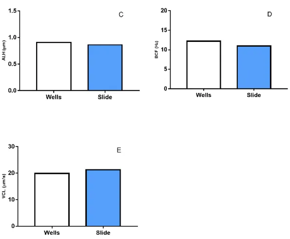
Figure 12 Sperm motility parameters comparing well slides (white bars) and regular microscopy slides (blue bars). (A) Percentage of motile cells (Mot); (B) Wobbliness (WOB); (C); Lateral head displacement (ALH); (D) Beat cross frequency (BCF); (E) Curvilinear velocity. (n=2; mean)
4.1.2. Extender solution
There were no visible differences concerning the percentage of motile cells after the first hour between the different extenders, with values around 64% of motile cells for the KT, 63% for KT+G and 55% motile cells for the SR (figure 13). Three hours after the sperm was diluted in the extender solutions, the percentage of motile cells decreased for the treatments KT and KT+G (≈ 47% and 48% motile cells, respectively), while for SR treatment this percentage was circa 63% motile cells. At the end of the trial, 24 hours, all the treatments showed decreased motility in comparison with the fresh sperm at the beginning of the trials (0h). Motility decreased from approximately 58 (fresh sperm) to values between 26% and 38% (KT+G and SR, respectively). Since for cryopreservation protocols, active sperm is expected to be in contact with the extender solution for short
50
periods of time, it was decided to use the solution KT+G, since this one was developed specifically for this species
Figure 13 Sperm motility parameters of sperm after exposure to different extender solutions (Kime and Tveiten (KT) (blue bars); Kime and Tveiten with glucose (KT+G) (orange bars); and Smith and Ryan (SR) (grey bars)) at different times (0 hours (CTRL); 1 hour; 3 hours; and at 24 hours. (A) Percentage of motile cells (Mot); (B) Wobbliness (WOB); (C); Lateral head displacement (ALH); (D) Beat cross frequency (BCF); (E) Curvilinear velocity. (n=2; mean)
51 4.2. Cryoprotectants toxicity
The toxicity effect of three different cryoprotectants, at three different concentrations was assessed in this trial using CASA software, and the results for motility (%), WOB (%), ALH (µm), BCF (Hz), and VCL (µm/s) are described in figure 14.
The null hypothesis that the sperm swimming parameters (WOB, ALH, BCF, AND VCL) are not affected by cryoprotectants toxicity was rejected (Pillai’s=1.666, F9,36=2.856, P<0.001).
ANOVA test revealed that these tested parameters presented significant differences (F1,9>33.8, P<0.001). The analysis of variance for the motility parameter (Mot) revealed
significant differences (F9,290=20.9,p≈0).
According to multiple comparisons using Tukey test, it revealed that there were no significant differences between 5 and 10% for both DMSO and propanediol, with sperm keeping it’s motility between 54.88±11.68% and 60.13±8%, and between 49.99±13.97% and 53.6±8.37%, respectively. Sperm cryopreserved using DMSO and propanediol solutions at concentrations of 20% showed decreased values (29.3±14.48% and 28.09±9.84%, respectively) in comparison to fresh sperm (≈55.91±14.7%). For methanol, no significant differences were found using any of the three concentrations. Sperm motility was kept between 54.44±8.59% and 62.08±10.17% for this treatment. For all the remaining parameters (WOB, ALH, BCF and VCL) it was observed the statistical lower values at the concentration of 20% for both DMSO and propanediol.
52
Figure 14 Sperm motility parameters comparing the toxic effect of DMSO (blue bars), propanediol (PROP) (orange bars) or methanol (METH) (grey bars) on cryopreserved sperm and, compared with fresh sperm (white bars). (A) Percentage of motile cells (Mot); (B) Wobbliness (WOB); (C); Lateral head displacement (ALH); (D) Beat cross frequency (BCF); (E) Curvilinear velocity (VCL). (n=5; mean ± SEM; p<0.05). Different letters refer to significantly different results obtained with Tukey test for each sperm motility parameter.
53 4.3. Freezing rates
Different freezing rates were tested to assess the best pace to freeze this species’ sperm. Four different distances from liquid nitrogen surface (1.5 cm; 2.5 cm; 4.5 cm; 7.5 cm) were used. Results are presented in figure 15 and freezing curves on figure 16.
The null hypothesis that sperm swimming parameters (WOB, ALH, BCF, and VCL) are not affected by the freezing rate was rejected by MANOVA (Pillai’s=1.018, F12,48=3.327,
P<0.01). Computing ANOVA, all the parameters presented significant differences (F12,48>21.16, p<0.01). The percentage of motile cells, analysed with ANOVA, presented
significant differences (F12,48=19.69, p≈0). A post hoc Tukey test was performed for each of the five parameters,
For all treatments, there was a decrease in the percentage of motile cells (between 0.98±3.09% for METH at 2.5 cm, and 39.36±16.93% for DMSO at 7.5cm) when compared to the control, using fresh sperm (61.35±7.67%). The best results after freezing were obtained using 10% DMSO at the four different distances from the LN2 (from
25.95±15.18% to 39.36±16.93%, or propanediol 10% at 4.5 cm (26.63±15.48%). Methanol presented the lowest values of motile cells using any of the distances (maximum of 7.58±11.57%). Methanol was also the only cryoprotectant that affected WOB, ALH, BCF, and VCL (maximum of 31.86±9.6%, 0.43±0.38 µm, 4.9±1.66 Hz, and 9.37±8.56% µm/s, respectively) in comparison with fresh sperm swimming parameters (61.86±7.23%, 0.86±0.11 µm, 12.42±2.08 Hz, 21.43±3.93 µm/s, respectively).
As expected, straws at different heights froze at different rates, nevertheless, cryoprotectants also affect these rates. Methanol shows the highest freezing rates throughout the trial (between 20.1 and 48.34 ºC/min) whilst DMSO presented a slower rate (from 10.67 to 32.88 ºC/min).
54
Figure 15 Sperm motility parameters comparing four freezing rates for sperm cryopreserved with 10% DMSO (blue bars), propanediol (PROP) (orange bars) or methanol (grey bars), with fresh sperm (white bars). (A) Percentage of motile cells (Mot); (B) Wobbliness (WOB); (C); Lateral head displacement (ALH); (D) Beat cross frequency (BCF); (E) Curvilinear velocity. (n=5; mean ± SEM; repeated measures MANOVA; p<0.05). Different letters refer to significantly different results.
55
Freezing rates (ºC/min) DMSO Distance from LIN (cm) 4ºC to -20ºC -20ºC to Tf 1.5 32.8 14.34 2.5 29.9 10.03 4.5 18.68 8.58 7.5 10.67 6.89
Freezing rates (ºC/min) Propanediol Distance from LIN (cm) 4ºC to -20ºC -20ºC to Tf 1.5 37.87 14.23 2.5 23.55 11.83 4.5 18.68 8.58 7.5 10.67 12.42
Freezing rates (ºC/min) Methanol Distance from LIN (cm) 4ºC to -20ºC -20ºC to Tf 1.5 48.34 13.29 2.5 36.9 11.47 4.5 26.1 8.56 7.5 20.1 7.23
A
B
C
Figure 16 Freezing curves for DMSO (A); propanediol (B); and methanol (C); with different distances from liquid nitrogen surface (1.5, 2.5, 4.5 and 7.5 cm), along 10 minutes.