Article
Printed in Brazil - ©2018 Sociedade Brasileira de Química*e-mail: eaffontes@ufv.br
Applicability of a Voltammetric Assay Based on the Electroreduction of Oxygen to
Evaluate the Antioxidant Capacity of Pequi (
Caryocar
brasiliense
Camb.) Pulp
FláviaA.Pinheiro,a,bLeonardoL.Okumura,cAstréaF.S.Silva,cJúnioG.Silva,c LetíciaR.Ferreira,bEdiltonS.BarcelloscandEdimarA.F.Fontes*,b
aInstituto Federal de Educação, Ciência e Tecnologia do Espírito Santo, Campus Venda Nova do Imigrante, 29375-000 Venda Nova do Imigrante-ES, Brazil
bDepartamento de Tecnologia de Alimentos, Universidade Federal de Viçosa, 36570-000 Viçosa-MG, Brazil
cDepartamento de Química, Universidade Federal de Viçosa, 36570-000 Viçosa-MG, Brazil
This study aimed to use a differential pulse voltammetric (DPV) assay, based on the electroreduction of oxygen, to evaluate the antioxidant capacity of pequi pulp. The results were expressed in coefficient of antioxidant capacity (K), at different pH values and in trolox equivalent antioxidant capacity (TEAC), and were compared to the 2,2’-azinobis(3-ethylbenzothiazoline-6-sulfonic acid) (ABTS) and 2,2-diphenyl-1-picrylhydrazyl (DPPH) spectrophotometric assays. The principal component analysis showed higher K for pequi pulp extract and pH tending to 7.15. The antioxidant capacity was related to the presence of gallic acid in its composition. Regarding the TEAC value, the best result was obtained by the DPV test (53.43 ± 5.14 µmol trolox g-1 pulp).
The pequi pulp showed antioxidant capacity, compared to the standard antioxidant compounds evaluated. It was concluded that the voltammetric protocol was efficient to assess the total antioxidant capacity of pequi pulp and can be an alternative to conventional spectrophotometric assays, for the analysis of food matrices.
Keywords: electrochemical techniques, food analysis, antioxidant capacity, bioactive compounds, pequi
Introduction
The assessment of the antioxidant potential of foods is carried out by the determination of their antioxidant capacity. Various methodologies are employed for this purpose, especially the spectrophotometric assays, including the 2,2’-azinobis(3-ethylbenzothiazoline-6-sulfonic acid) (ABTS) and 2,2-diphenyl-1-picrylhydrazyl
(DPPH).1 However, these colorimetric methods have some
limitations.2,3 For example, the samples must undergo
a pretreatment to eliminate interfering agents, such as
reducing sugars and organic acids.4 In fact, each method
has its own characteristics, such as the reaction mechanism, types of oxidation substrates, reaction medium (basic, acid or neutral), compound proof of reaction and expression of
results.1 This hinders the comparison of results obtained by
different authors that used different methodologies. The differences between the reactive species and their forms of activity hinder the development of a simple
and universal method to accurately and quantitatively
determine the total antioxidant capacity.5 Therefore, the
use of electrochemical techniques becomes an alternative
for this analysis.6 The processes of formation and/or
stabilization of free radicals are related to
oxidation-reduction reactions.7 Therefore, several studies have used
voltammetric electrochemical techniques to assess the antioxidant capacity of foods and isolated compounds, since they improve methodology standardization and increase the amount of information about the compound of interest.6,8-12
Voltammetry presents the following advantages over traditional spectrophotometric techniques: low operation and instrumental cost; use of less organic solvents, consequently decreasing waste generation; the analysis can be performed in the sample without pre-treatment for separation/cleaning of the compounds that affect other methods, colored materials or other dispersed solid
particles; fast data acquisition, sensitivity and portability.13-17
capacity of food matrices. However, no study using pequi pulp for this purpose has been found so far.
Pequi (Caryocar brasiliense Camb.) is a typical fruit
of the Brazilian cerrado that has not been the subject of scientific investigation. It is an important source
of nutrients, whose pulp has about 3.73 ± 0.01 g per
100 g of proteins; 26.15 ± 0.85 g per 100 g of lipids;
58.83 ± 2.06 mg per 100 g of calcium; 1.40 ± 0.59 mg per
100 g of iron and 2.81 ± 0.70 mg per 100 g of zinc.18 It also
contains bioactive compounds with antioxidant properties,
such as vitamin C (6.63 ± 1.02 mg per 100 g),19 phenolic
compounds (1.80 ± 0.04 to 3.34 ± 0.07 mg GAE g-1) and
total carotenoids (37.08 ± 1.84 to 187.00 ± 12.43 µg g-1).20
Violaxanthin, lutein and zeaxanthin are among the
predominant carotenoids observed.21
Therefore, this study aimed to verify the applicability of an experimental protocol, based on voltammetry, to evaluate the antioxidant capacity of pequi pulp.
Experimental
Reagents and solutions
All reagents used were of analytical grade of purity (P.A.). Acetone, acetic acid, boric acid, phosphoric acid, ethanol, sodium hydroxide and methanol were purchased from Vetec (Duque de Caxias, RJ, Brazil); ABTS, DPPH, potassium persulfate, L-ascorbic acid, citric acid,
p-coumaric acid, gallic acid, α-tocopherol, 2(3)-t
-butyl-4-hydroxyanisole (BHA), 2,6-di-tert-butyl-4-methylphenol
(BHT) and 6-hydroxy-2,5,7,8-tetramethylchroman-2-carboxylic acid (trolox) were acquired from Sigma-Aldrich Chemical Company (St. Louis, MO, USA).
The 0.1 mol L-1 Britton-Robinson buffer was used as
support electrolyte in the voltammetric analyses, at pH
2.20, 7.15 and 10.05. It was prepared from 0.1 mol L-1
solutions of acetic acid, boric acid and phosphoric acid. The pH was adjusted with sodium hydroxide solution,
8.0 mol L-1, in a pH Meter, model 827 pH lab (Metrohm,
Herisau, Switzerland). The solutions of the antioxidant standards used in the electrochemical test were prepared in ethanol, 95%, on the day of the analysis.
Preparation of the pequi pulp extract
The pequi (Caryocar brasiliense Camb.) pulp was
obtained through the processing of fresh fruits from the December 2015 harvest of the municipality of Santana de Pirapama, MG, Brazil, located at 19°00’21’’S (latitude), 44°02’35’’W (longitude). The pulp was obtained with the aid of a depulping machine (depulping machine (Itametal/
NPC Equipamentos, model Bonina 0.25 df, Itabuna, Brazil) and used in the production of the extract, as recommended
by Rufino et al.22 The extract was stored in an amber flask
and maintained under freezing (–18 °C) for later defrosting and use.
Instrumentation of the voltammetric electrochemical assay
The voltammetric measurements were performed using a potentiostat/galvanostat, Autolab (Metrohm Pensalab, Utrecht, The Netherlands), in interface with a computer through the General Purpose Electrochemical System (GPES), version 4.9, with a 50 mL electrochemical cell provided with three electrodes, one gold working electrode
having a geometric area of 0.196 cm2; one platinum wire
auxiliary electrode and one Ag|AgCl, 3.0 mol L-1 KCl
reference electrode (Metrohm, Herisau, Switzerland). Following each voltammetric reading, the surface of the working electrode was polished for approximately 1 min, with alumina 0.3 and 0.05 µm (Buehler, Lake Bluff, IL, USA) on an alumina polishing pad, rinsed with ultrapure
water (resistivity of 18.2 MΩ cm at 25 °C) from a Milli-Q
purification system (Millipore, Bedford, MA, USA).
Voltammetric characterization of pequi pulp and antioxidant standards
The voltammetric profile of the compounds with antioxidant capacity was characterized by cyclic voltammetry (CV), with scanning of anodic direction,
ranging from –0.6 to +1.4 V, at scan rate of 100 mV s-1 and
step potential of 2 mV. For such, 10.0 mL of the 0.1 mol L-1
Britton-Robinson buffer, pH 2.20, were transferred to the electrochemical cell. Five minutes after the nitrogen
gas injection (N2, 99.9% purity) (White Martins Gases
Industriais Ltda, Juiz de Fora, MG, Brazil), it was added an aliquot of 300 µL of the antioxidant standard solution
33.3 mmol L-1 (L-ascorbic acid, citric acid, p-coumaric
acid, gallic acid, α-tocopherol, BHA, BHT and trolox) for
the deoxygenation of the solution. Its final concentration
in the cell was around 1.0 mmol L-1. Then, N
2 was injected
for 1 min to homogenize the mixture.
The pequi pulp was assessed with the addition of 300 µL of the extract in replacement of the antioxidant standard solution. It was also performed analysis of the supporting electrolyte solution to verify the presence of some peak.
In addition to the evaluation of the voltammetric profile
of the antioxidant compounds at pH 2.20, the 0.1 mol L-1
Assessment of the total antioxidant capacity through the electroreduction of oxygen
The methodology used in this study was developed through the application of the differential pulse voltammetric (DPV) technique, based on a research carried out by
Korotkova et al.23 In order to develop the experimental
protocol, preliminary tests were carried out to determine
the time corresponding to O2 injection demands to promote
the saturation of the supporting electrolyte and its selection. It was also determined the concentration range of some antioxidant standards with improved linearity (not shown in this article). The experimental conditions adopted for the DPV readings were: cathodic direction measurements, ranging from 0 to –0.6 V, pulse amplitude of 100 mV, scan
rate of 20 mV s-1 and modulation time of 2 ms.
Molecular oxygen (O2) was used as oxidant for being
a strong oxidizing agent (Eº = 1.229 V)24 and one of the
most important radicals found in our biological system. At
ground state, O2 is a diradical with one unpaired electron in
each oxygen. As a radical, it can abstract hydrogen atoms, similarly to the combustion and autoxidation reactions, or
work as an electron receptor in biological systems.25
10.0 mL of 0.1 mol L-1 Britton-Robinson buffer (pH 2.20,
7.15 and 10.05) were transferred to the electrochemical cell.
Following saturation with O2 for 5 min (industrial oxygen,
purity > 90%) (White Martins Gases Industriais Ltda, Juiz de Fora, MG, Brazil), the cathodic peak current of the oxygen (oxidizing agent) was read, without antioxidant addition (control). Then, this solution received the addition
of aliquots of 50 µL of 0.02 mol L-1 antioxidant standard
solution (solution for the use of L-ascorbic acid, citric acid,
p-coumaric acid, gallic acid, α-tocopherol, BHA, BHT
and trolox), so that its concentration in the cell ranged
between 0.1 and 0.6 mmol L-1. A manual homogenization
was conducted and after the solution remained at rest for 1 min, the voltammetric measurement was performed. A similar procedure was adopted in the analysis of the pequi pulp extract, with the addition of successive 50 µL aliquots of the undiluted extract, which replaced the standard antioxidant solution.
The residual current, inherent to the supporting electrolyte, was determined through voltammetric reading
of the 0.1 mol L-1 Britton-Robinson buffer (pH 2.20,
7.15 and 10.05), after an injection of N2 for 10 min. The
voltammetric measurements were carried out in duplicate, and the experiment was conducted with three replications. The baseline of the voltammograms was corrected through the moving average, using the tool available in the GPES software system, version 4.9. The voltammograms were
plotted with the aid of the OriginPro 8® software.26
A graphic of (Ipc – Ires)/(Ip0 – Ires) = f (volume of
the antioxidant standard or volume of pequi extract) was developed based on the averages of the cathodic oxygen current obtained for each concentration of the
antioxidant standard or pequi extract, where: Ipc is the
peak cathodic current of O2, in the presence of compound
with antioxidant capacity; Ires is the peak cathodic current
of the deoxygenated supporting electrolyte, and Ip0 is the
initial peak cathodic current of O2, without the addition
of antioxidant compound. The current decay curve was also observed. The antioxidant capacity of the pequi pulp extract and the antioxidant standards were expressed as antioxidant capacity coefficient (K), which is the angular coefficient of the curve.
In addition to the K value, the antioxidant capacity of the pequi pulp was expressed as trolox equivalent antioxidant
capacity, given by the TEAC value (µmol trolox g-1 pequi
pulp). For such, we used the results of cathodic current of oxygen, pH 7.15, obtained by the voltammetric protocol previously described, with pequi pulp concentration
ranging from 5 to 30 mL L-1 and trolox (antioxidant
standard) ranging from 100 to 600 µmol L-1.
Spectrophotometric assay of the radical cation ABTS•+
The determination of the antioxidant capacity by the ABTS test was performed according to the methodology
described by Re et al.,27 with modifications. In an amber
container, 5 mL of 7.0 mmol L-1 ABTS solution were added
to 5 mL of 2.45 mmol L-1 potassium persulfate solution,
which remained protected from light for 12 to 16 h for
the generation of ABTS•+ cation chromophore. Then, the
radical solution was diluted in 80% ethanol until reaching an absorbance of 0.700 ± 0.005, at wavelength of 734 nm, in a spectrophotometer (BEL Photonics UV-M51, Piracicaba, Brazil) previously calibrated with 80% ethanol. 0.5 mL of different dilutions of the pequi pulp extracts and 3.5 mL of
the ABTS•+ radical solution were placed in test tubes and
homogenized in a tube shaker. After 6 min of reaction, the absorbance of the samples was read at 734 nm. 80% ethanol was used as control, in replacement of the pequi extracts. The analysis was completely performed in low light environment. The antioxidant capacity was expressed by
the TEAC value (µmol trolox g-1 pequi pulp), according to
Rufino et al.,22 calculated from the trolox analytical curve,
prepared with standard solutions, with concentrations
ranging between 10 and 100 µmol L-1.
Spectrophotometric assay of the DPPH• radical
test, according to the methodology described by
Brand-Williams et al.,28 with modifications.
In a low light environment, aliquots of 0.1 mL of different dilutions of pequi extracts were transferred to
test tubes, which were added to 2.9 mL of 60 µmol L-1
methanolic solution of the DPPH• radical previously
prepared. The mixture was then homogenized in a tube shaker. The tubes were placed to rest, in the dark, for 25 min. Next, absorbance was read at 515 nm in a spectrophotometer (BEL Photonics UV-M51, Piracicaba, Brazil) previously calibrated with methanol. Methanol was used as the control, in replacement of the pequi extracts.
Similarly to the ABTS test, the results were expressed
based on the TEAC value (µmol trolox g-1 pequi pulp),
according to Rufino et al.22 Therefore, a trolox analytical
curve was developed with standard solutions, with
concentrations ranging from 30 to 600 µmol L-1.
Statistical analysis
The results for antioxidant capacity, expressed in TEAC and antioxidant capacity coefficient (K) were presented as
the mean (± standard deviation) of the values obtained from three replicates. The statistical analyses were performed using analysis of variance (ANOVA), linear regression with no adjustment and Tukey test at 5% probability,
using the Statistical Analysis System (SAS), version 9.3,29
licensed for the Universidade Federal de Viçosa (Viçosa, MG, Brazil). A principal component analysis (PCA) was applied to produce graphics for better explaining the results found for the antioxidant compounds, according to K and
pH, using the Matlab platform.30
Results and Discussion
Characterization of the voltammetric profile of the compounds with antioxidant capacity
The voltammetric profile of the pequi pulp extract and antioxidant standards was assessed using the cyclic voltammetry technique, prior to the application of the experimental protocol, to evaluate the behavior of the oxygen electroreduction. Figure 1 shows the cyclic voltammograms for the pequi extract, L-ascorbic acid, BHA
Figure1. Cyclic voltammograms of pequi pulp extract and antioxidant standards L-ascorbic acid, BHA and trolox in 0.1 mol L-1 Britton-Robinson buffer,
and trolox (the other figures are presented in the Figure S1, Supplementary Information).
Cathodic peaks with greater current intensity were observed, close to the potential of 0.810, 0.440 and 0.250 V, at pH 2.20, 7.15 and 10.05, respectively, in all
compounds analyzed. These peaks are from the 0.1 mol L-1
Britton-Robinson buffer, as their intensity was similar to that of peaks obtained after the addition of the antioxidant standards and pequi pulp extract. Only BHA and trolox presented cathodic peaks that are not from the supporting electrolyte. The BHA showed better defined peaks, close to the potential –0.200 V at pH 7.15 and 10.05.
Regarding the reading in the oxidation direction, all the analyzed compounds presented some anodic peak. Trolox (at pH 2.20, 7.15 and 10.05) and BHA (at pH 2.20 and 10.05) presented more defined peaks, with increased current intensity, which indicates increased sensitivity of the technique used for these compounds, since all antioxidant standards were analyzed in the same
concentration (1.0 mmol L-1). It was also observed for these
peaks that the pH affected the displacement of the potential for values closer to 0 V, which improves the tendency of the antioxidant standard to oxidize, in the following order: pH 10.05 > 7.15 > 2.20.
The potential variation can also be understood according to the Nernst equation (equation 1):
(1)
where, E is the real potential (V) under nonstandard conditions; E° is the tabulated standard potential (V);
R is the universal gas constant (8.314 J K-1 mol-1); T is
the absolute temperature (K); n is the number of electrons involved on the reaction stoichiometry; and F is the
Faraday constant (96,485 C mol-1). It demonstrated that the
potential depends on the concentration of the species in the solution and that it can be changed in case of variation in the concentration of the products formed, depending on the medium pH. The same initial concentration has been used for all the antioxidant standards, but the structural change of the molecules varies differently for each compound
(according to the pKa, for instance), depending on the
medium pH. It can also make the redox reaction more thermodynamically favorable for certain substances.
In a study for the assessment of the antioxidant capacity
of natural compounds, Arteaga et al.17 also verified that,
as pH increased, the oxidation potentials changed to less positive values. It implies that oxidation becomes easier and may be related to the deprotonation of compounds.
Alberto et al.31 also reported that the thermochemical
viability of the electron transfer process (SET mechanism) of trolox increased with the degree of deprotonation of the antioxidant standard, for different free radicals.
The CV technique showed that all the evaluated compounds, except for BHA and trolox, were not electroactive at the range of 0 to –0.6 V, a potential window used for subsequent monitoring of the cathodic current of O2.
In vitro total antioxidant capacity assessment expressed by
the antioxidant capacity coefficient (K)
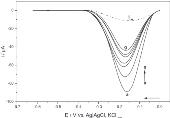
Figure 2 shows the voltammograms of oxygen
electroreduction in 0.1 mol L-1 Britton-Robinson buffer,
pH 7.15, with the addition of the pequi pulp extract. This
Figure shows that the O2 current decreased as the pequi
extract concentration (a → g) increased. This behavior was
also verified for the antioxidant standards for all pH values analyzed. The proportion of oxygen species in the solution decreases when it reacts with the antioxidant, according
to the voltammetric readings, since less O2 molecules are
available to receive electrons from the working electrode. Therefore, the intensity of the cathodic current of the oxidant decreases.
The effect of the increased antioxidant concentration was assessed (depending on the added volume) and the medium
pH on the O2 current decay rate [(Ipc – Ires)/(Ip0 – Ires)].
No effect of the pH versus volume interaction (p≥ 0.05)
was observed. Therefore, the factors pH and volume worked independently and were studied separately for each sample with antioxidant capacity. Based on the averages of the current values obtained by voltammetric readings, a regression analysis was applied to the volume factor
Figure2. Differential pulse voltammograms of the oxygen electroreduction in 0.1 mol L-1 Britton-Robinson buffer, pH 7.15; in the absence of extract of
the pequi pulp (a) and with the addition of the extract (b to g), being (b) 50; (c) 100; (d) 150; (e) 200, (f) 250 and (g) 300 µL. Ires: residual current of the
[(Ipc – Ires)/(Ip0 – Ires) × compound volume added]. The K
was determined for the evaluated compounds (Table 1) through the angular coefficient of each curve obtained in this analysis. Figure 3 presents the linear regression curves for the pequi extract, gallic acid, L-ascorbic acid and trolox. Table 1 shows that the mean values of K ranged between
0.768 ± 0.112 mL-1 and 2.222 ± 0.172 mL-1, respectively
for α-tocopherol (pH 2.20) and L-ascorbic acid (pH 10.05).
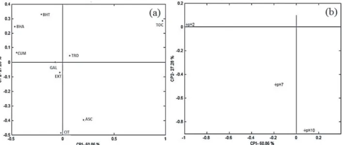
A principal component analysis of the antioxidant compounds depending on K and pH was applied (Figure 4) for better understanding of the results described in Table 1. The two first components explained 88.14%.
Table1. Antioxidant capacity coefficient (K) for the pequi pulp extract and several antioxidant patterns (found in pequi and antioxidants recognized as reference standards), at different pH values
Compound Coefficient of antioxidant capacity / mL
-1
pH 2.20 pH 7.15 pH 10.05
Extract of pequi pulp 1.719 ± 0.202 1.805 ± 0.234 1.704 ± 0.139
L-Ascorbic acid 1.577 ± 0.266 1.542 ± 0.121 2.222 ± 0.172
Citric acid 1.676 ± 0.476 2.120 ± 0.239 2.006 ± 0.297
p-Coumaric acid 2.149 ± 0.084 1.756 ± 0.344 1.587 ± 0.241
Gallic acid 1.826 ± 0.249 1.523 ± 0.315 1.784 ± 0.251
α-Tocopherol 0.768 ± 0.112 1.355 ± 0.399 1.527 ± 0.448
BHA 2.214 ± 0.271 1.486 ± 0.109 1.534 ± 0.224
BHT 1.910 ± 0.379 1.620 ± 0.210 1.355 ± 0.396
Trolox 1.616 ± 0.292 1.773 ± 0.378 1.587 ± 0.122
Mean values ± standard deviation (n = 3). BHA: 2(3)-t-butyl-4-hydroxyanisole; BHT: 2,6-di-tert-butyl-4-methylphenol.
Figure3. Variation curves of cathodic oxygen current rate as a function of the volume of pequi pulp extract and 0.02 mol L-1 solution of compounds with
Figure 4 shows the distribution of the antioxidants in relation to the principal components (a) and a projection of the groups in relation to pH (b). The simultaneous assessment of Figures 4a and 4b reveals a distribution of the antioxidant compounds, according to the K and pH values. Therefore, it is possible to infer that the
compounds p-coumaric acid, BHA and BHT presented
better antioxidant behavior at pH 2.20; L-ascorbic acid, at pH 10.05; and pequi extract and citric acid, at pH 7.15. The other pH values seem to affect the pequi extract, which was also verified for citric acid, in relation to pH 10.05. The antioxidant capacity of gallic acid was affected by pH 2.20 and 7.15.
The increased antioxidant capacity observed at more alkaline pH values is directly related to the deprotonation of certain compounds, and may involve different mechanisms of antioxidant capacity, such as the donation of hydrogen
atom or electrons, or the coexistence of both mechanisms.32
In this work, this justification can be attributed to the ascorbic and citric acids, since the greater antioxidant tendency of these compounds was verified at pH values in which both were deprotonated.
Figure 4 shows that the compound α-tocopherol
presented the greatest difference among those analyzed, which may be explained by its lower solubility in the supporting electrolyte, compared to the other compounds under study.
The antioxidant capacity of trolox and α-tocopherol
standards was not affected by pH under the experimental conditions of the present study. It can be explained by the fact that the K value is directly related to the reaction stoichiometry (how much oxygen reacts with an antioxidant
molecule), which is not necessarily altered by the medium pH variation (differently from that described for the potential), since K also depends on the reaction kinetics.
It must be highlighted that the distribution of the antioxidant standards may be related to the similarity of the chemical structure of the compounds. Among the compounds assessed, greater structural similarity was
found between trolox and α-tocopherol, and between
BHA and BHT.
Figure 4 shows that BHA, BHT and p-coumaric acid
seem to present similar characteristics for the antioxidant capacity, which may be related to the presence of a phenol group in the chemical structure of these compounds.
Ahmed and Shakeel33 state that the behavior and the
intensity of the reduction of the oxidizing agent vary according to the structure of the antioxidant compounds. These authors attributed the variation in the mode and magnitude of elimination of the superoxide radical to the structure-activity relationship of different flavonoids: the presence of phenol groups in the chemical structure of the molecule, which increases its antioxidant capacity; as well as the presence of a planar structure and unsaturations, since they enhance the stability of the phenoxyl radical formed.
Figure 4 shows that the pequi pulp extract and the gallic acid presented similar distribution. Therefore, based on the values of the antioxidant capacity coefficient obtained in this study, the antioxidant capacity of the pequi extract can be strongly attributed to the presence of gallic acid in its composition, compared to the other antioxidant standards assessed. Gallic acid was described as the predominant compound among the phenolic compounds
evaluated by Machado et al.34 in the aqueous extract
of pequi pulp (from the region of Montes Claros, MG,
Brazil). Almeida et al.35 identified gallic acid, by using
HPLC-DAD-MS (high-performance liquid chromatography with diode array detection coupled to mass spectrometry), as the major phenolic compound observed in the lyophilized pequi pulp, corresponding to 31% of the total phenolics.
The antioxidant potential of gallic acid was also observed
in a study carried out by Arteaga et al.,17 which highlighted
the presence of adjacent phenolic groups, prone to oxidation, and a carboxyl group that favors the stabilization of the species resulting from the oxidative process.
In vitro evaluation of the total antioxidant capacity expressed
in DPVTEAC, ABTSTEAC and DPPHTEAC
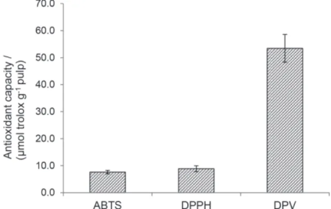
Figure 5 presents the results of the antioxidant capacity of pequi pulp, evaluated by the DPV, ABTS and DPPH tests, expressed in trolox standard, TEAC value
(µmol of trolox g-1 pequi pulp). This Figure shows that
the greatest antioxidant capacity was obtained by the
DPV electrochemical test (53.43 ± 5.14 µmol trolox g-1
pulp), in comparison with the ABTS spectrophotometric
tests (7.62 ± 0.66 µmol trolox g-1 pulp) and DPPH
(8.88 ± 1.08 µmol trolox g-1 pulp).
Nascimento et al.36 assessed pequi from São Miguel
do Araguaia, GO, Brazil, by the DPPH test and found
12.0 µmol trolox g-1 of lyophilized pequi pulp. Rufino et al.37
conducted a study to evaluate the antioxidant capacity of tropical nontraditional Brazilian fruits and found values
ranging from 6.3 ± 0.2 to 153.0 ± 2.6 µmol trolox g-1 of
fruit, respectively, for umbu and camu-camu, using the ABTS test.
It is known that the total antioxidant capacity of a food matrix is affected by the synergistic activity of its
bioactive components.32 Therefore, the best result was
found by the DPV test, possibly because this technique allows the joint evaluation of the reducing power of pequi bioactive components (phenolic compounds, ascorbic acid and carotenoids). On the other hand, the ABTS and DPPH tests are more specific for polyphenols, and may underestimate the value obtained. The higher sensitivity of the voltammetric electrochemical test applied in this study can also be attributed to the thermodynamic enhancement of the reaction mechanism involved between the antioxidant compounds of pequi and oxygen (oxidizing agent).
Sawai et al.38 state that the DPPH• radical test is not the
most appropriate to evaluate the antioxidant potential of lipophilic compounds, including carotenoids. Therefore, this trial is more efficient to evaluate the antioxidant capacity of phenolic compounds, and it underestimates the antioxidant capacity of pequi pulp.
Schaich et al.39 mentioned several factors that limit
the use of the ABTS and DPPH tests, such as disregard for chemical aspects, including the effects of solvents, concentration, medium pH, radical solubility and, particularly, reaction kinetics; use of stable radicals with stereochemical with hindrance instead of easy access and
short duration small radicals (example: OH• and O
2-•),
for better simulation of in vivo occurrence; and lack of
standardization of the experimental procedures, especially the expression of the results, which prevents comparing the results from different researchers.
Besides, the spectrophotometric assays ABTS and DPPH are affected by the activity of interfering agents
present in the food matrix. Oliveira et al.4 described the
ascorbic acid as an interfering agent in the evaluation of the antioxidant capacity of anthocyanins in foods.
Schaich et al.39 cited the interference caused by the oxygen
dissolved in the medium and the use of organic solvents (for instance, methanol in the DPPH test).
The antioxidant capacity of a matrix is affected by several factors, such as solubility and the chemical structure of the bioactive compounds, chemical quality of the oxidizing agent, reaction mechanism, specific experimental conditions, among others. Therefore, it is difficult to compare the TEAC values obtained by different tests, since the different results can be attributed to the characteristics
of each technique.40
Arteaga et al.17 report that the electrochemical tests
are more advantageous than the DPPH test, since they present greater precision and speed of analysis; the tests can be carried out at different pH values, and on different reaction media, which allows the comparison between the antioxidant capacities of molecules under different experimental conditions; they are less expensive, mainly
Figure5. Antioxidant capacity of pequi pulp expressed in trolox equivalent (wet basis), according to electrochemical differential pulse voltammetry (DPV) and spectrophotometric assays of the radical cation ABTS•+ and
due to the use of less organic solvents, which also reduces the waste generated.
Therefore, the voltammetric test is a viable alternative for the evaluation of the antioxidant capacity of food matrices.
Conclusions
The results obtained in this study lead to the inference that the pequi pulp has potential to work as a reducing agent when compared to the antioxidant standards evaluated. Its antioxidant capacity was strongly attributed to the presence of gallic acid in its composition. Besides, it was observed that the pH of solution affects the activity of most antioxidant compounds and cannot be neglected in such analyses.
Among the three in vitro assays applied, ABTS, DPPH
and DPV, the best result for antioxidant capacity of the pequi pulp was obtained by the voltammetric test and expressed in trolox standard equivalent. Therefore, this protocol was efficient to evaluate the antioxidant capacity. It is also an alternative methodology to carry out this analysis, mainly for providing relevant information for the redox process that cannot be obtained from the traditional spectrophotometric assays.
Supplementary Information
Supplementary information is available free of charge at http://jbcs.org.br as a PDF file.
Acknowledgments
The authors are grateful to financial support from the Conselho Nacional de Desenvolvimento Científico e Tecnológico (CNPq, Brazil), Fundação de Amparo à Pesquisa do Estado de Minas Gerais (FAPEMIG, Brazil) and Instituto Federal de Educação, Ciência e Tecnologia do Espírito Santo (IFES, Brazil).
References
1. Shahidi, F.; Zhong, Y.; J. Funct. Foods2015, 18, 757. 2. Apak, R.; Özyürek, M.; Güçlü, K.; Çapanoğlu, E.; J. Agric.
Food Chem.2016, 64, 997.
3. Apak, R.; Özyürek, M.; Güçlü, K.; Çapanoğlu, E.; J. Agric.
Food Chem.2016, 64, 1028.
4. Oliveira, I. R. N.; Teófilo, R. F.; de Oliveira, E. B.; Ramos, A. M.; Barros, F. A. R.; Maia, M. P.; Stringheta, P. C.; Int. J. Food Sci. Technol.2017, 52, 511.
5. Alves, C. Q.; David, J. M.; David, J. P.; Bahia, M. V.; Aguiar, R. M.; Quim. Nova2010, 33, 2202.
6. Dar, R. A.; Brahman, P. K.; Khurana, N.; Wagay, J. A.; Lone, Z. A.; Ganaie, M. A.; Pitre, K. S.; Arab. J. Chem.2017, 10, S1119.
7. Halliwell, B.; Lancet2000, 355, 1179.
8. de Macêdo, I. Y. L.; Garcia, L. F.; Oliveira-Neto, J. R.; Leite, K. C. S.; Ferreira, V. S.; Ghedini, P. C.; Gil, E. S.; Food Chem.
2017, 217, 326.
9. Peixoto, C. R. M.; Fraga, S.; Justim, J. R.; Gomes, M. S.; Carvalho, D. G.; Jarenkow, J. A.; Moura, N. F.; J. Electroanal.
Chem.2017, 799, 519.
10. Cruz, D.; Barroso, M. F.; Ramalhosa, M. J.; Coelho, A.; da Silva, H.; Duarte, A. J.; González-García, M. B.; Carvalho, A. P.; Delerue-Matos, C.; J. Electroanal. Chem.2016, 763, 110. 11. Falcão, S. I.; Tomás, A.; Freire, C.; Vilas-Boas, M.; Eur. Food
Res. Technol.2016, 242, 1393.
12. Ferreira, R. D. Q.; Greco, S. J.; Delarmelina, M.; Weber, K. C.;
Electrochim. Acta2015, 163, 161.
13. Lugonja, N. M.; Stanković, D. M.; Miličić, B.; Spasić, S. D.; Marinković, V.; Vrvić, M. M.; Food Chem. 2018, 240, 567. 14. Hoyos-Arbeláez, J.; Vázquez, M.; Contreras-Calderón, J.; Food
Chem.2017, 221, 1371.
15. Oliveira-Neto, J. R.; Rezende, S. G.; Reis, C. F.; Benjamin, S. R.; Rocha, M. L.; Gil, E. S.; Food Chem.2016, 190, 506. 16. David, I. G.; Bizgan, A. M. C.; Popa, D. E.; Buleandra, M.;
Moldovan, Z.; Badea, I. A.; Tekiner, T. A.; Basaga, H.; Ciucu, A. A.; Food Chem.2015, 173, 1059.
17. Arteaga, J. F.; Ruiz-Montoya, M.; Palma, A.; Alonso-Garrido, G.; Pintado, S.; Rodríguez-Mellado, J. M.; Molecules2012, 17, 5126.
18. Alves, A. M.; Fernades, D. C.; Sousa, A. G. O.; Naves, R. V.; Naves, M. M. V.; Braz. J. Food Technol.2014, 17, 198. 19. Machado, M. T. C.; Mello, B. C. B. S.; Hubinger, M. D.; J. Food
Eng.2013, 117, 450.
20. Ribeiro, D. M.; Fernandes, D. C.; Alves, A. M.; Naves, M. M. V.; Food Sci. Technol.2014, 34, 507.
21. Azevedo-Meleiro, C. H.; Rodriguez-Amaya, D. B.; J. Food Compos. Anal.2004, 17, 385.
22. Rufino, M. S. M.; Alves, R. E.; de Brito, E. S.; Morais, S. M.; Sampaio, C. G.; Pérez-Jiménez, J.; Saura-Calixto, F. D.;
Comun. Tec. - EMBRAPA, Cent. Pesqui. Agropec. Trop. Umido
2007, 128, 1. Available at https://www.infoteca.cnptia.embrapa. br/infoteca/bitstream/doc/426954/1/Cot128.pdf, accessed in February 2018.
23. Korotkova, E. I.; Karbainov, Y. A.; Shevchuk, A. V.;
J. Electroanal. Chem.2002, 518, 56.
24. Zoski, C. G.; Handbook of Electrochemistry, 4th ed.; Elsevier:
Amsterdam, Netherlands, 2007.
25. Solomons, T. W. G.; Fryhle, C. B.; Química Orgânica, vol. 1, 10a ed.; LTC: Rio de Janeiro, Brasil, 2016.
27. Re, R.; Pellegrini, N.; Proteggente, A.; Pannala, A.; Yang, M.; Rice-Evans, C.; Free Radic. Biol. Med.1999, 26, 1231. 28. Brand-Williams, W.; Cuvelier, M. E.; Berset, C.; LWT - Food
Sci. Technol.1995, 28, 25.
29. Statistical Analysis System, SAS Procedures Guide, version 9.3; SAS Institute Inc., Cary, NC, USA, 2011.
30. Matlab R2016a, version 9.0; The MathWorks Inc., Natick, MA, USA, 2016.
31. Alberto, M. E.; Russo, N.; Grand, A.; Galano, A.; Phys. Chem.
Chem. Phys.2013, 15, 4642.
32. Altunkaya, A.; Gökmen, V.; Skibsted, L. H.; Food Chem.2016,
190, 25.
33. Ahmed, S.; Shakeel, F.; Czech J. Food Sci.2012, 30, 153. 34. Machado, M. T. C.; Mello, B. C. B. S.; Hubinger, M. D.; Food
Bioprod. Process.2015, 95, 304.
35. Almeida, M. R.; Darin, J. D. C.; Hernandes, L. C.; Aissa, A. F.; Chisté, R. C.; Mercadante, A. Z.; Antunes, L. M. G.; Bianchi, M. L. P.; Plant Foods Hum. Nutr.2012, 67, 171.
36. do Nascimento, N. R. R.; Alves, A. M.; Silva, M. R.; Naves, M. M. V.; Rev. Bras. Frutic.2017, 39, 1.
37. Rufino, M. S. M.; Alves, R. E.; de Brito, E. S.; Pérez-Jiménez, J.; Saura-Calixto, F.; Mancini-Filho, J.; Food Chem.2010, 121, 996.
38. Sawai, Y.; Moon, J. H.; Sakata, K.; Watanabe, N.; J. Agric. Food
Chem.2005, 53, 3598.
39. Schaich, K. M.; Tian, X.; Xie, J.; J. Funct. Foods2015, 14, 111. 40. Cárdenas, A.; Gómez, M.; Frontana, C.; J. Electroanal. Chem.
2014, 729, 116.
Submitted: November 9, 2017
Published online: March 6, 2018


