UNIVERSIDADE DO ALGARVE
Development of gene therapy to deliver the
receptor-specific rhTRAIL DHER variant to induce
apoptosis in activated Hepatic Stellate Cells for the
treatment of Liver Fibrosis
Ana Raquel Pato Santa Maria
Dissertation
Integrated Master in Biological Engineering
Dissertation directed by:
External Supervisor: Prof. Dr. Hidde Haisma Internal Supervisor: Prof. Drª Gabriela Silva
ii
Development of gene therapy to deliver the
receptor-specific rhTRAIL DHER variant to induce
apoptosis in activated Hepatic Stellate Cells for the
treatment of Liver Fibrosis
Declaração da Autoria do Trabalho
Eu, ___________________________________________________declaro ser a autora deste trabalho, que é original e inédito. Autores e trabalhos consultados estão devidamente citados no texto e constam da listagem de referências incluída.
A Universidade do Algarve tem o direito, perpétuo e sem limites geográficos, de arquivar e publicitar este trabalho através de exemplares impressos reproduzidos em papel ou de forma digital, ou por qualquer outro meio conhecido ou que venha a ser inventado, de o divulgar através de repositórios científicos e de admitir a sua cópia e distribuição com objetivos educacionais ou de investigação, não comerciais, desde que seja dado crédito ao autor e editor.
iii
Acknowledgments
First of all I want to thank to Professor Hidde Haisma, my supervisor at the University of Groningen for accepting me to do my internship in the PGM laboratory. I appreciate all the support, understanding, teaching, criticism and for having encouraged me to want to know more.
Second, I want to thank to Petra Ettema for the confidence, support, commitment and friendship especially during the 6 months of internship.
I would also like to thank Marilena Ourailidou, Nick Eleftheriadis, Niek Leus, Thea, Hanna and Marta for creating not only our friendship over these six months, but also for all the support and companionship in the lab. It was easier going work every day with colleagues like them.
I would also like to thank Robbert Cool for all the support, criticism and ideas, Janine for all the sympathy and concern with me and also to Professor Frank Dekker for the words of encouragement.
I would like to thank to Professor Gabriela Silva, my supervisor in Portugal, for all the help in the bureaucratic process in the University of Algarve, the criticism, the teaching and the words of support. To the Algarve University, my second home during the last six years, where I learned, I cried, I laughed, I give up and at the same time continue, but during those years I was happy and I graduated in the course in which I dreamed and I'll be happy to work. To the academic association and for all management positions that I took, because it was through this experience that I acquired many skills that cannot be learned in books.
To my friends in Portugal for all the support, but mostly the friends that I made in the Netherlands. Ina, Karen, my housemates and the members of ESN for every moment of fun and adventures in the Netherlands.
Last but not the least, my family, my mother, my father, brother, grandparents and boyfriend for all the support, love, and affection for believing in me and that I would be able to arrive to the end. In addition to a personal achievement, all this work was developed with effort, dedication and above all commitment on my part. But the same could not have happened without the unconditional support of my parents.
iv
“Do the best you can until you know better.
Then when you know better do better. “
v
Index
Acknowledgments ... iii
Index ... v
Index for Figures ... viii
List of Abbreviations ... x
Abstract ... xii
Resumo ... xiv
Chapter 1-Introduction ... 1
1.1.Healthy and fibrotic Liver ... 1
1.1.1.Hepatic Stellate Cells ... 2
1.2.Tumor Necrosis Factor–related apoptosis inducing ligand ... 5
1.3.Gene therapy using adenovirus ... 8
1.4.Histone deacetylation and histone acetyltransferase inhibitors ... 11
1.5.Objectives ... 13
Chapter 2-Materials and Methods ... 15
2.1.Cell culture and cell lines ... 15
2.2.Antibodies and rhTRAIL proteins ... 15
2.3.Materials ... 16
2.4.Construction and production of the vector pADTRACK-CMV-IGK-HA- ---rhTRAIL DHER ... 17
2.4.1.Cloning process to create the vector ... 17
2.4.2.Analysis of the cloned vector ... 19
2.4.3.Expression of the protein in HEK-293 cells ... 19
2.5.Analysis of the expression of the rhTRAIL DHER protein in HEK-293 cells ... 21
2.5.1.Analysis of the expression of the protein through western blotting analysis .. 21
2.5.2.Analysis of the function of protein through survival assay in SW-948 cells .. 22
2.6.Generation of adenovirus plasmid by homologous recombination -in bacterial ---cells ... 24
2.6.1.Preparation of electrocompetent cells... 24
2.6.2.Generation of recombinant adenoviral plasmid in BJ5183 ... 25
vi
2.7.Analysis and evaluation of the activity of receptor rhTRAIL mutants in- LX2
---cells and HepG2 cells. ... 27
2.7.1.Activity of specific proteins rhTRAIL DHER, 4C7 and WT in LX2 cells ... 27
2.7.2.Activity of specific proteins rhTRAIL DHER, 4C7 and WT in HepG2 ---cells ... 28
2.8.Evaluation of the effects of the combination of rhTRAIL DHER, 4C7 and ---WT with HDACs and HATs inhibitors ... 29
2.9.Statistical Analysis ... 30
Chapter 3-Results ... 31
3.1.Construction and production of the vector pDTRACK-CMV-IGK-HA- --- ---rhTRAIL DHER ... 31
3.2.Expression and analysis of the protein in HEK293 cells ... 35
3.3.Generation of adenovirus plasmid by homologous recombination in bacteria ... 40
3.4.Evaluation of the activity of rhTRAIL DHER in LX2 cells and HepG2 cells. ... 44
3.5.Evaluation of the effects with the combination of rhTRAIL variants with ---HDACs and HATs inhibitors ... 46
3.5.1.Combination of SAHA, Entinostat, MG149 and C646 with rhTRAIL--- ---(DHER, 4C7 and WT) induces apoptosis in SW948 ... 47
3.5.2.SAHA with rhTRAIL (DHER, 4C7 and WT) promote apoptosis while ---Entinostat, C646 and MG149 with rhTRAIL (DHER, 4C7 and WT) --- have no combination effect in H460 cells... 51
3.5.3.SAHA combined with rhTRAIL (DHER, 4C7 and WT), promotes ---apoptosis more efficiently compared with the combination of ---Entinostat, C646 and MG149 in HUH-7 cells ... 56
3.5.4.SAHA combined with rhTRAIL variants promotes apoptosis more ---efficiently compared with the combination of Entinostat, C646 and ---MG149 in HepG2 cells ... 60
Chapter 4-Discussion ... 65
Chapter 5-Conclusion and Future Perspectives ... 72
References... 74
Appendix ... 82
A.1. Sequencing ... 82
A.2. Constituents of enzymatic reactions ... 83
vii
A.4. Protocols used for molecular techniques ... 86
A.4.1. Electroporation protocol ... 86
A.4.2. FuGENE® HD transfection reagent protocol ... 86
A.4.3. MTS assay protocol ... 87
A.4.4. Western Blotting protocol... 88
A.4.5. Purification of miniprep products ... 89
A.4.6. Purification of PCR products ... 90
A.4.7. Protocol to create electrocompetent bacteria ... 91
viii
Index for Figures
Figure 1.1. Components of a healthy liver.. ... 1
Figure 1.2..Hepatic liver cells in a normal and injured liver. ... 3
Figure 1.3. Pathways of activation of hepatic stellate cells.. ... 3
Figure 1.4. The receptors express on the surface of the cells that can bind to TRAIL. .. 6
Figure 1.5. Pathways of induced apoptosis by TRAIL.. ... 7
Figure 1.6. The replication cycle of an adenovirus vector ... 9
Figure 1.7. The dynamic equilibrium of HATs and HDACs in the cells.. ... 13
Figure 2.1. Schematic diagram of the cloning process to create the --- pADTRACK-CMV-IgK-HA-rhTRAIL DHER vector. ... 18
Figure 2.2. The crystal violet structure ... 23
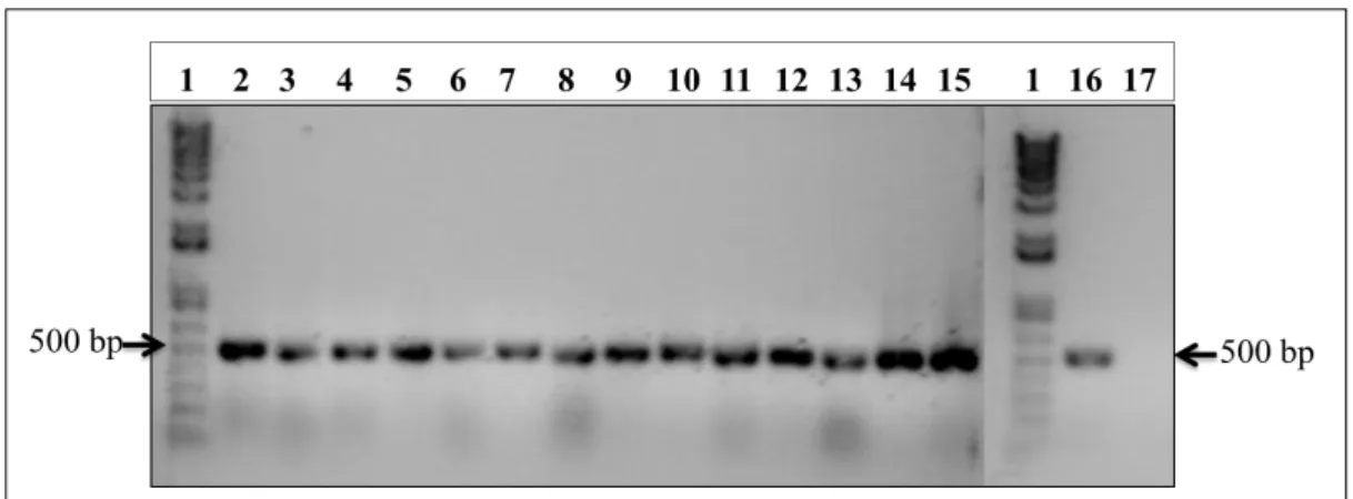
Figure 3.1. Amplification of the rhTRAIL DHER gene by a PCR reaction.. ... Figure 3.2. The constructed vector pADTRACK-CMV-IGK-HA-rhTRAIL DHER. .. 32
Figure 3.3. PCR reaction for the analysis of the new constructed vector. ... 33
Figure 3.4. The plasmid constructed and the original shuttle plasmid. ... 33
Figure 3.5. Restriction endonuclease digestions for the generated clones of ---pADTRACK-CMV-IGK-HA-rhTRAIL DHER.. ... 34
Figure 3.6. Transfected HEK-293 cells after 72hours of incubation ... 36
Figure 3.7. Screening of the rhTRAIL DHER protein through western blotting ... 37
Figure 3.8. Analysis of the function of the rhTRAIL DHER in SW948. ... 39
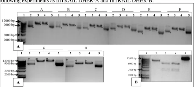
Figure 3.9. Recombination of pADTRACK-CMV-IGK-HA-rhTRAIL ---DHER into pAdEasy-1. ... Figure 3.10. Linearization of shuttle vector with PmeI restriction endonuclease.. ... Figure 3.11. Endonuclease digestions of the recombinant plasmid and the controls.. .. 43
Figure 3.12. RhTRAIL DHER, 4C7 and WT combined in LX2 cells and ---HepG2 cells ... 45
Figure 3.13. Combination of SAHA, Entinostat, MG149 and C646 with ---rhTRAIL DHER in SW948 cells induces apoptosis ... 48
Figure 3.14. rhTRAIL 4C7 combined with SAHA, Entinostat, MG149 and ---C646 induces apoptosis in SW948. ... 49
Figure 3.15. SAHA, Entinostat, MG149 and C646 combined with ---rhTRAIL WT induces in SW948... 50
Figure 3.16. Combination of SAHA, Entinostat, C646 and MG149 and ---rhTRAIL DHER increase the apoptosis in H460. ... 53
Figure 3.17. Combination of SAHA, Entinostat, C646 and MG149 with ---rhTRAIL 4C7 in H460 promote a higher apoptosis. ... 54
Figure 3.18. SAHA, Entinostat, C646, MG149 and rhTRAIL WT in H460 ---cells induces apoptosis ... 55
Figure 3.19. Combination of SAHA, Entinostat, C646, MG149 and ---rhTRAIL DHER in HUH-7 ... 57
Figure 3.20. Combination of C646, MG149, Entinostat and SAHA with ---rhTRAIL 4C7 in HUH-7 ... 58
ix Figure 3.21. SAHA, Entinostat, C646 and MG49 with rhTRAIL WT in
---HUH-7 cells ... 59
Figure 3.22. SAHA with rhTRAIL DHER in HepG2 cells inducesapoptosis in a
synergistic manner and the combination of MG149, C646 andEntinostat and
---C646 have no combination effect in the cells ... 62
Figure 3.23. SAHA with rhTRAIL 4C7 in HepG2 cells induces apoptosis and---
MG149, C646 and Entinostat promote a slight apoptosis in the cells ... 63
Figure 3.24. The effect of the combination of SAHA, Entinostat, MG149 and
x
List of Abbreviations
Acetyl-CoA Acetyl Coenzyme A
Ad5 Human Adenovirus
APS Ammonium PerSulfate
BSA Bovine Serum Albumin
CIP Calf Intestinal Alkaline Phosphatase
CLD Chronic liver diseases
DcR 1 or 2 Decoy Receptor 1 or 2
DISC Death-Inducing Signaling Complex
DMEM Dulbecco’s minimum essential medium
DRs Death receptors
EASL European Association for the Study of the Liver
ECM Extracellular Matrix
EDTA Ethylenediamine Tetraacetic Acid
EGF Epidermal Growth Factor
ET-1 Endothelin-1
FADD Fas-Associated Death Domain
FBS Fetal Bovine Serum
HA Human Influenza Hemagglutinin
HAT Histone AcetylTransferase
HDACS Histones Deacetylases
HMG High Mobility Group
HSC Hepatic Stellate Cells
xi
IGK Immunoglobulin K light chain
LB Luria- Bertani broth medium
MFs Myofibroblast
NAD Nicotinamide Adenine Dinucleotide
NF-KB Nuclear Factor Kappa B
OPG Osteoprotegerin
PBS Phosphate Buffered Saline
PCR Polymerase Chain Reaction
PDGF Platelet-Derived Growth Factor
PMS Phenazine Methyl Sulfate
RER Rough Endoplasmic Reticulum
RhTRAIL 4C7 Recombinant human TRAIL Variant 4C7 RhTRAIL DHER Recombinant human TRAIL Variant DHER RhTRAIL WT Recombinant humam TRAIL Wild Type
RT Room Temperature
SER Smooth Endoplasmic Reticulum
TAE Tris-acetate-EDTA
TEMED Tetramethylethylenediamine
TGF-β Transforming Growth Factor Beta
TNF Tumor Necrosis Factor
TRAIL Tumor Related Apoptosis Inducing Ligand VEGF Vascular Endothelial Growth Factor
xii
Abstract
Liver fibrosis is caused by excessive accumulation of extra-cellular matrix, produced by the hepatic stellate cells (HSC), when they evolve from the quiescent state to an activated state. These cells express on their surface receptors for Tumor Necrosis Factor- Related Apoptosis Inducing Ligand (TRAIL), a member of the TNF family. Five TRAIL receptors have been identified to date: the pro-apoptotic DR4 and DR5, and the decoy receptors DcR1, DcR2 and OPG. It is known that activated hepatic stellate cells overexpress the DR5 receptor. Soluble human recombinant TRAIL (rhTRAIL) is an interesting protein for the treatment of liver fibrosis, due to the efficacy of death induction. In this project, we constructed a vector capable of producing the DR5-specific variant rhTRAIL DHER in mammalian cells, for specific binding to the TRAIL DR5 receptor, and to efficiently induce apoptosis in activated stellate cells. We verified that the protein is functional and active when added to a cell line (SW948) sensitive for the rhTRAIL proteins. Moreover, we evaluated the effects of the protein rhTRAIL DHER, produced in E.Coli bacteria, in LX2 cells and HepG2 cells. Our results demonstrate that the rhTRAIL DHER protein induces apoptosis in LX2 cells, without producing any effect in the HepG2 cells. However, to develop a therapy capable to induce a continuous production and a more efficient apoptosis in activated HSC, we used an adenoviral vector (pAdEasy-1) to recombine with our constructed vector, in other to produce an adenovirus. However, the recombination of both plasmid were not achieved, due to the complexity of the vectors used to recombine and create a unique vector.
As previously demonstrated, the normal cells in the liver express the TRAIL DR5 receptor, to a lesser extent than the activated cells. For this reason, it was necessary to create a treatment that promotes apoptosis specifically in the target cells, but does not affect the other cells. We aimed to investigate the effects of combinations between HDAC (SAHA and Entinostat) and HAT (C646 and MG149) inhibitors with rhTRAIL variants. Our results confirm the synergistic effect of combinations of SAHA and rhTRAIL variants in H460, HUH-7 and HepG2, while the effect of Entinostat with rhTRAIL
xiii
variants did not promote a combination effect. For the HAT inhibitors we demonstrated a protective effect in MG149 with rhTRAIL variants, which was even more visible in HUH-7 and HepG2 cells. For C646 the combination effect was low in HepG2, HUH-7 and H460 cells.
In conclusion, we constructed a vector capable of producing the DR5-receptor specific protein rhTRAIL DHER in mammalian cells, and we have shown the synergistic effect of SAHA with rhTRAIL variants and a possible HAT inhibitor that may protect the normal cells in a fibrotic liver.
Keywords: TRAIL, Hepatic Stellate Cells, Histone acetylasetransferase, Histone
xiv
Resumo
O fígado é um dos maiores órgãos do corpo humano e é responsável por funções vitais para o organismo, compreendendo o suporte e o apoio a outros órgãos. Este órgão é responsável pela síntese de componentes sanguíneos, regulação dos níveis de glucose, desintoxicação, produção de bílis e síntese de componentes essenciais à função de outros órgãos. O fígado é constituído por quatro lobos com diferentes tamanhos e formas, e por dois principais vasos sanguíneos, a artéria hepática e as veias-porta. A artéria hepática transporta o sangue da artéria aorta, enquanto as veias-porta transportam o sangue rico em nutrientes da digestão e também do pâncreas e baço. Os vasos sanguíneos subdividem-se em capilares, que subdividem-se ligam a um lóbulo, a unidade funcional do fígado. Cada lóbulo contém milhões de células hepáticas (~80% de todo o fígado), as responsáveis pela função metabólica. As restantes células que constituem o fígado são células endoteliais, estreladas, linfócitos, células de Kupffer e células biliares epiteliais.
As doenças hepáticas crónicas são caracterizadas por uma repetida lesão nas células hepáticas, devido a infeção por vírus (hepatite B e C), doenças autoimunes, excesso de álcool e drogas, doenças inflamatórias, doenças genéticas ou fatores esporádicos. Contudo, a constante lesão destas células leva a acumulação de ferimentos no fígado induzindo este órgão a um estado fibrótico, podendo evoluir para cirrose ou cancro. Como resposta aos ferimentos causados pelas mais diversas lesões, inicia-se um processo de acumulação de matriz extracelular. Este processo deve-se sobretudo às células estreladas do fígado, que evoluem para um estado ativo, devido ao aumento da sua resposta fenotípica, sintetizando em excesso diversos fatores de crescimento que são responsáveis não só pela fibrogénese como também por respostas inflamatórias crónicas.
A ativação das células estreladas do fígado é realizada em três fases: a iniciação ou fase pré-inflamatória, onde se inicia a alteração do fenótipo e a expressão dos genes, devido sobretudo à estimulação pancreática graças ao excesso de produção de fatores de crescimento; a fase de perpetuação é devida a numerosas reações distintas, tais como, a proliferação, a contractilidade, a fibrogénese, a degradação da matriz, a perda de
xv
retinóides e de células inflamatórias; Por fim, a fase de reversão ou apoptose, é a fase em que nas células estreladas é desencadeada a apoptose, e, é nesta fase que podem interferir determinados fatores que levam à reversão da lesão das células e, por conseguinte, a reversão da fibrose.
O Tumor necrosis factor-related apoptosis inducing ligand (TRAIL) é um dos fatores que está associado à promoção da apoptose das células estreladas ativadas, pois estas células expressam em excesso o recetor para este fator. Sendo um membro da família Tumor Necrosis Factor (TNF) é uma proteína transmembranar tipo 2 e é expressa nos mais variados tecidos. O TRAIL interage com os recetores na superfície membranar das células e induz a apoptose. Até ao momento foram descritos cinco recetores: TRAIL DR4, DR5, DcR1, DcR2 e osteoprotegerin (OPG). O recetor TRAIL DR4 e DR5 têm um domínio e sinais de apoptose. Apesar de os DcR1 e DcR2 terem homologia com domínios de apoptose, o DcR1 tem um domínio de morte citoplasmática truncada e não funcional, enquanto o DcR2 falta uma região citosólica e está ancorado na membrana plasmática.
O TRAIL recombinante solúvel é interessante para terapias do cancro e fibrose no fígado, uma vez que é mais específico na indução da apoptose, e a produção de um TRAIL solúvel recombinante específico para os recetores DR5 e DR4 é permitirá o desenvolvimento de terapias mais específicas. Um dos objetivos do nosso trabalho é a construção de um vector que contenha o gene para o TRAIL recombinante variante D269H e E195R, promovendo uma ligação mais específica ao recetor TRAIL DR5, logo uma apoptose mais rápida e eficaz.
Contudo, a administração de proteínas diretamente ao paciente não é tão eficaz como quando a proteína é produzida diretamente nas células alvo. Por esta razão, nós pretendemos construir um vector adenoviral, que contenha o gene de interesse para a produção da proteína rhTRAIL DHER.
Os nossos resultados demonstraram que o vector construído, pADTRACK-CMV-IGK-HÁ-rhTRAIL DHER, tem capacidade de expressar aquando transfectado em células, HEK-293. Além disso, a proteína produzida tem atividade biológica e exerce as funções esperadas, quando o mesmo é adicionado a uma linha celular bastante sensível
xvi
para o TRAIL recombinante solúvel, onde promove a apoptose das células, como esperado. A utilização de vectores adenovirais é de grande importância em terapia genética, devido à rápida infeção do vírus nas células alvo, à baixa patogenicidade em seres humanos e ao facto de o genoma do vírus não sofrer rearranjo nas células hospedeiras. Porém, a construção do vector adenoviral não correu como esperado, possivelmente pela baixa possibilidade de recombinação de um vector digerido com outro não digerido por enzimas de restrição.
No entanto, sendo um dos objetivos a promoção da apoptose em células estreladas ativadas, a proteína rhTRAIL DHER foi adicionada a uma linha celular fibrótica do fígado (LX2) e também a uma linha celular hepática não fibrótica (HepG2). A proteína, rhTRAIL DHER, de origem bacteriana, foi utilizada em vez da proteína produzida em células humanas devido à quantidade disponível. Os nossos resultados demonstraram um efeito promotor de apoptose nas células LX2 e a ausência de efeito nas HepG2. Este efeito, embora reduzido, permitiu concluir que a indução de apoptose foi específica para as células que expressam o recetor TRAIL DR5 (LX2). Terapeuticamente, este resultado é importante porque as células saudáveis expressam o recetor TRAIL DR5 em quantidades reduzidas, comparando com células fibróticas. Como descrito na literatura, a administração conjunta de inibidores HDACs com as proteínas rhTRAIL DHER, 4C7 ou WT poderá promover maior indução de apoptose e ao mesmo tempo aumentar a especificidade celular.
Assim, o último objetivo do nosso trabalho consistiu em confirmar uma maior indução de apoptose aquando da administração conjunta da proteína rhTRAIL DHER,4C7 ou WT com inibidores HDAC (histonas deacetilases) como, SAHA e Entinostat. Simultaneamente, pretendemos verificar se inibidores HAT (histona acetiltransferase), MG149 e C646 exercem um efeito de proteção aquando da conjugação com as proteínas rhTRAIL DHER, 4C7 ou WT. Os inibidores HAT, histona acetiltransferase, transferem um grupo acetil da acetil co-enzima A para o grupo ε-amino num resíduo específico de lisina existente nas caudas amino-terminal das histonas, resultando de uma neutralização de carga positiva. Porém a reação inversa é catalisada por as histonas deacetilases. A nossa hipótese é que os HAT, combinados com as
xvii
proteínas rhTRAIL DHER, 4C7 e WT poderiam ter um efeito protetor nas células cancerígenas. Os nossos resultados mostram que, MG149 não exerce qualquer efeito em combinação com as proteínas.
Os nossos resultados comprovaram o efeito sinergético da combinação de SAHA com as proteínas rhTRAIL DHER, 4C7 e WT, quando adicionado em diferentes linhas celulares, HepG2, HUH-7 e H460. Contudo, ao contrário do que já foi demonstrado por outros autores, a combinação do Entinostat com as proteínas rhTRAIL DHER,4C7 e WT demonstrou efeito sinérgico reduzido, o que poderá ser considerado um efeito de proteção, pois não existe promoção de apoptose das células. Relativamente, aos inibidores HAT, C646 e MG149, os resultados demonstram que a combinação do MG149 com a proteína rhTRAIL DHER não exerce qualquer efeito de apoptose, o que pode indicar um efeito de proteção. O mesmo se verificou para o inibidor C646, apesar de o efeito de proteção ser menor, pois promove uma pequena percentagem de apoptose nas células.
Palavras-chave: Apoptose, fibrose no fígado, TRAIL, HDAC, HAT, proteína rhTRAIL
1
-Chapter 1-
Introduction
1.1. Healthy and fibrotic Liver
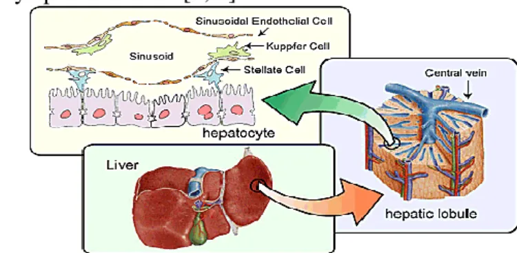
The liver is the largest organ in the human body, providing essential metabolic, exocrine and endocrine functions [1, 2]. These functions include metabolism of dietary compounds, regulation of glucose levels, detoxification, production of bile and blood homeostasis [3]. The liver is a brown organ with four lobes of different shape and size. This organ is connected with two large blood vessels, the hepatic artery and the portal vein [1, 2]. The blood vessels subdivide into capillaries, which lead to a lobule (the functional units of the liver). Each lobule contains millions of hepatic cells which are the basic metabolic cells [2] (see Figure 1.1).
The hepatic cells are the largest part of the liver (~80% of the mass) in the adult organ. These cells are responsible for the secretion of essential serum proteins and for the synthesis of different proteins. These important cells have the capacity for detoxify, metabolize, and inactivate drugs toxins and endogenous compounds [3]. The other 20% of cells consist of endothelial cells, stellate cells, lymphocytes, Kupffer cells, the hepatic sinusoids and biliary epithelial cells [2, 3].
Figure 1.1. Components of a healthy liver. The image shows a healthy liver (in the center), the
composition of a hepatic lobule (in the right), and the constituent cells of the liver (in the top). Adapted from [4].
2
The combination of all the cells that forms liver tissue, and the combination of functions gives a complexity to the organ becoming vulnerable to many diseases. Chronic liver diseases (CLD) are characterised by repetitive injury in the hepatocyte cells, induced by chronic viral hepatitis (hepatitis B and C viruses), autoimmune injury (neonatal liver disease, Wilson´s disease), alcohol abuse, drugs toxicity, chronic inflammatory conditions and vascular derangements, either congenital or acquire [5, 6]. When the liver is constantly suffering injuries the evolution of these damage can induce this organ to fibrosis, cirrhosis or cancer, depending on the damage this organ suffered.
In response to the injury, an extracellular matrix or scar is produced, and its accumulation in the liver is called hepatic fibrosis. An end stage of fibrosis is denominated cirrhosis, resulting in nodule formation that can outcome in alteration of hepatic functions and blood flow [6]. The primary effector of liver fibrosis are hepatic stellate cells (HSC). Due to the injuries the cells became activated into a myofibroblast (MFs) phenotype [7] and contributing to CLD progression because of their highly phenotypic responses, such as: an excess deposition of extracellular matrix (ECM), and a synthesis and delivery of several critical growth factor capable of supporting not only fibrogenesis but also chronic inflammatory responses and neo-angiogenesis [5].
According to the European association for the study of the liver (EASL) , until 2013, 59% of patients that needs a liver transplants in Europe suffer from cirrhosis which in majority is caused by virus infection and alcohol misuse [8].
1.1.1 Hepatic Stellate Cells
The extracellular matrix normally produced by the liver is essential for maintaining the different functions of resident liver cells, like hepatocytes, stellate cells and sinusoidal endothelium [6]. This interstitial ECM is limited to the capsule around large vessels, and in the portal areas. When the liver becomes fibrotic, the collagen and non-collagenous constituents growths three- to fivefold followed by a change in the subendothelial space from a low-density basement to an interstitial type matrix [6].
3
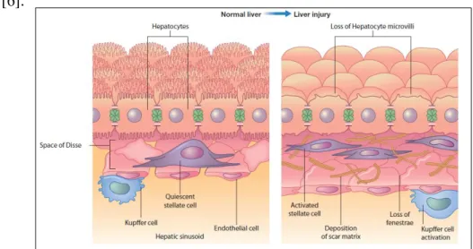
Following this change in the matrix, the loss of hepatocyte microvilli and the disappearance of endothelial fenestration occur when the liver becomes fibrotic (Figure 1.2.) [6].
Figure 1.2. Hepatic liver cells in a normal and injured liver. On the left is shown the normal liver cells,
the stellate cells are situated in the subendothelial space of Disse; on the right is shown the liver cells after injury, and the effects are visible: the stellate cells become activated, the hepatocytes lose the microvilli and the activated HSC produce a deposition of an extra cellular matrix. Adapted from [6].
When the liver becomes fibrotic this is due to the activation of stellate cells in which follows in three phases (Figure 1.3.): the initiation phase, the perpetuation phase and the resolution phase [9].
Figure 1.3. Pathways of activation of hepatic stellate cells. The different phases can be distinguished in
the Figure, namely the initiation, perpetuation and resolution or reversion phases. The initiation is provoked by oxidant stress, apoptotic bodies and paracrine stimuli from the other cell types; the perpetuation is characterized by specific changes; and in the resolution or reversion phase, the cells can induce the apoptosis or revert the injury. Adapted from [10].
4
I. Initiation Phase:
The response of the quiescent hepatic stellate cells to paracrine stimulation by all neighboring cell types, like sinusoidal endothelium, Kupffer cells, hepatocytes and platelets, leads the HSC to activation [9]. The Kupffer cells stimulate matrix synthesis, cell proliferation, and release of retinoid by stellate cells through the actions of cytokines (TGF-β) and reactive oxygen intermediates/lipid peroxides [10, 12]. In case of injury, the hepatocytes also promote HSC initiation with a process mediated by FAS, and can involve the Tumor Necrosis Factor (TNF)-related apoptosis inducing ligand (TRAIL) [10, 13, 14]. Apoptotic fragments liberated from hepatocytes are fibrogenic and may contribute to the activation of HSC [6, 15]. Platelets are the most important paracrine stimuli, generating different important mediators, like platelet-derived growth factor (PDGF), TGF-β1 and epidermal growth factor (EGF) [6].
II. Perpetuation Phase:
To accumulate the ECM, the hepatic stellate cells undergo a series of phenotypic changes, including proliferation, contractility, fibrogenesis, chemotaxis, matrix degradation, pro-inflammatory responses and cytokine release [6]. In proliferation, a lot of mitogenic factors, but also cognate tyrosine kinases receptors, are unregulated. PDGF is one of the principal mitogenic factors that increases their expression. Other mitogens factors are involved in the proliferation of the hepatic stellate cells [6]. The contraction by activated hepatic stellate cells obstructs hepatic blood flow by constricting individual sinusoids and by contracting the cirrhotic liver [6]. A principal contractile stimulus to HSC is ET-1, and due to the activation of stellate cells there are increase of expression of smooth muscle actin (α-SMA) [6]. The fibrogenesis is the key of the stellate cells to contribute for liver fibrosis. TGF- β1 is an important fibrogenic factor, because several mechanisms surrounding this factor contribute for the increase of activation of ESC, another important is TNF factor [6, 16]. Sinusoidal endothelial cells and hepatic cells are inflammatory effectors, they can rapidly lose their fenestrations up on injury and express pro-inflammatory molecules, ICAM-1, VEGF and adhesion molecules [6, 18, 19].
5
III. Resolution/Reversion Phase:
The reversion of stellate cell activation, or selective removal of activated HSC by apoptosis, can explain the loss of activated cells in the resolving phase in liver injury [6]. The apoptosis can occur due to soluble factors and matrix components that are present during injury. Moreover, cell death ligands, such as TRAIL and FAS, are expressed in liver injury, and activated HSC are more vulnerable to TRAIL mediated apoptosis [6, 14, 20, 21]. The resolution/reversion phase in the activation of the ESC is one important phase that can be used to promote the apoptosis of HSC cells through tumor-specific TRAIL receptor binding. The HSC have a central role in the liver fibrosis, when they are activated overexpress the TRAIL receptor in their surface becoming an ideal targets for TRAIL agonists [20].
1.2. Tumor
Necrosis
Factor–related
apoptosis
inducing ligand
Tumor necrosis factor (TNF) related apoptosis inducing ligand (TRAIL) belongs to a family of cytokines that function as protuberant mediators of immune regulation and inflammatory responses [21]. TRAIL can bind to two pro-apoptotic receptors, DR4 and DR5 [22, 23]. With HSC activation there is an increase in DR4 and DR5 expression levels, while that of DR5 remains 103 times higher than that of DR4 [19]. At the same time the expression of anti-apoptotic receptors DcR1, DcR2 and osteoprotegerin (OPG) also increases (Figure 1.4) [23, 27–30]. TRAIL protein is expressed in a wide range of tissues and interacts with TRAIL receptors to induce the apoptosis of the cells [23,25,26].
6
Figure 1.4. The receptors expressed on the surface of the cells that can bind to TRAIL protein.
Receptor DR4 and DR5 have a death domain that can bind to TRAIL protein and induce apoptosis. The other hand, DcR1, DcR2 and OPG compete with receptor DR4 and DR5 for biding to the TRAIL protein, and these receptors do not promote the apoptosis. Adapted from [22].
The death receptors TRAIL DR4 and DR5 have an intracellular death domain motif via which the apoptotic signal is initiated. In contrast, DcR2 has a truncated and nonfunctional cytoplasmic death domain, whereas DcR1 lacks a cytosolic region and it is anchored to the plasma membrane, and OPG is a soluble decoy receptor [22].
When TRAIL protein binds to TRAIL receptor the extrinsic pathways are activated inducing apoptosis of mammalian cells [29]. The binding of TRAIL to its receptors results in trimerization of the receptor and clustering of the receptors intracellular death domain, allowing formation of the death-inducing signaling complex (DISC) [22] via binding of the adaptor molecule, Fas-associated death domain (FADD). This leads to binding and activation of caspase-8 and caspase-10. When caspase-8 or caspase-10 is activated, this cleaves caspase-3, leading to apoptosis (Figure 1.5.) [22].
7
Figure 1.5. Pathways of induced apoptosis by TRAIL. The TRAIL binds to the receptor, leading to a
recruitment of FADD and this molecule activates the caspase-8. Type I pathway is sufficient to induce apoptosis. However in other cell types, the type II pathway, through mitochondria, is necessary to induce the apoptosis. Adapted from [22].
In response to DNA damage, cell cycle checkpoint defects, loss of survival factors and other stressed factors, induce the intrinsic pathway of apoptosis in the cells. In this pathway, the activation of proapoptotic Bcl-2 gene induces the mitochondria to release apoptotic factors, such as cytochrome-c and SMAC/DIABLO, to the cytoplasm [23, 32– 34]. The cytochrome-c connects with the APAF-1 adaptor, creating an apoptosome, activating the protease caspase-9. On the other hand, SMAC/DIABLO induces apoptosis binding to inhibitor of apoptosis (IAP), and preventing the caspase activation [23, 35, 36]. The intrinsic and extrinsic pathways can works together, and the capase-8 cleaves the proapoptotic Bcl-2 family member Bid. The cleaved product is translocated to the mitochondria to promote cytochrome-c release due to the interaction with Bax and Bak [23, 33].
TRAIL protein can bind to the five TRAIL receptors, however a soluble recombinant TRAIL can be an interesting treatment for cancer therapies and liver fibrosis. The soluble recombinant human TRAIL (rhTRAIL) is specific in inducing apoptosis in cancer cells and does not affect healthy cells. However, the capability of wild-type rhTRAIL to bind to all five receptors can reduce the levels of apoptosis [23, 37]. In order
8
to overcome this, receptor-specific variants of rhTRAIL were constructed: DHER and 4C7 that can bind specifically to DR5 and to DR4 receptors, respectively. The rhTRAIL DHER variant comprises mutations D269H and E195R, and the rhTRAIL 4C7 variant carries mutations G131R, R149I, S159R, N199R, K201H and S215D, using the rhTRAIL denominated WT (Wild Type) [37, 38]. Both variants, rhTRAIL DHER and 4C7, have a higher affinity to the receptor DR5 and DR4, respectively. Especially mutant rhTRAIL DHER can be used to develop new therapies to treat liver fibrosis, due to the higher affinity to DR5 receptor and the induced expression of DR5 receptor in HSC.
1.3. Gene therapy using adenovirus
Genes are the units of heredity and their specific sequences encode different proteins. When genes suffer alterations, the production of proteins cannot occur, resulting in genetic disorders. Gene therapy is fundamental to replace defective genes, which are responsible for diseases [39–41]. This therapy can replaced a mutated gene that causes disease by a healthy copy, it can inactivate the mutated gene or it can introduce new gene to fight the disease. One of the most difficult problems to achieve this goal is to replace the gene into the patient’s target cells. For this reason it can be used a vector to replace the defective gene [39]. To deliver the vectors there are two techniques, ex vivo and in
vivo [41, 42]. With the in vivo therapy the vector is directly injected into the patient´s
blood to bind to the target cells [39]. For gene therapy, the vectors used include: viral vector or non-viral methods. The viral method uses retrovirus, adenovirus, adeno-associated viruses and herpes simplex virus [39].
The characteristics of the adenovirus, they are best suited for gene therapy: the ubiquitous of the vector, because of the isolation from the different species, the most common used in this therapy is the serotypes 2 and 5; the rapid infection of the adenovirus and the high levels of gene transfer in a range of human cells; the low pathogenicity of the vector in humans; the capacity of the vector to accept large sequences of DNA and transduce these transgenes; the genome of the adenovirus do not suffer rearrangement
9
and the DNA of the insert is maintained without alterations; lastly, the adenovirus are easily to manipulate using recombinant techniques [41].
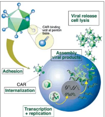
The adenovirus has a double stranded DNA. When the infection occurs the DNA enters the nucleus, and the genes from the early region 1 (E1a and E1b) are transcribed. During the infection, the region from E1 to E4 is expressed, and this region is the principal for the process of viral gene expression [41] . The adenovirus infects the host cells, introducing the genome inside of the cells. The DNA of the adenovirus is not introduced into the genome of the cells, but is transcribed as the other genetic material of the cells (Figure 1.6.)[39].
Figure 1.6. The replication cycle of an adenovirus vector. The image depicts the replication of an
adenovirus vector in the cells, encoding the desired proteins. Adapted from [41].
The recently constructed adenoviral vectors have deletions in E1, E2 and E4 genes, due to the capability of these sequences to produce viral proteins, responsible to induce the immune response in the host and decrease the toxicity in vivo [41]. However, with the use of the adenovirus it is difficult to obtain efficient gene transfer after a second administration, due to the neutralizing antibodies, and the viral DNA disappears over time, so that in chronic diseases it is necessary repeat the infection of the adenovirus from
10
time to time [41]. The use of adenovirus are more advantageous than the use of protein added directly to the patients, due to the capability of the adenovirus to produce the protein in the target cells and maintaining constant levels of protein over a long period, so the efficacy will be larger. For this reason, the use of a adenovirus vector carrying the gene of rhTRAIL DHER for delivery in the liver is an interesting therapy for the treatment of liver fibrosis.
In a preclinical perspective, it was demonstrated that adenoviral DNA is expressed in liver, skeletal muscle, heart, brain, lung, pancreas and tumor tissue, once the vector is administrated intravenously. However, most of the virus accumulates in the liver [43, 44]. Gene therapy is a relatively new procedure, and the scientists do not yet understand all the associated to this therapy. Potential problems include: short lived nature of gene therapy, immune response, problems with viral vectors, multigenic disorders and insertional mutagenesis [39].
In an ethical perspective, the risk of the gene therapy may not be greater than the potential benefit [41, 45]. However, ethical committees have some concerns about this therapy, such as, the distinction between what is normal and what is disability, whether some diseases should be cured, whether the somatic therapy is more or less ethical than germ line therapy, etc. The primary treatments of gene therapy were conducted in patients whose first treatment failed, so the associated risk is small. Moreover, the viruses used for gene therapy should be extensively studied, to avoid greater risk for the patients [39]. Gene therapy can be an interesting therapy for liver fibrosis, moreover the use of this therapy combined with other agents/drugs (like HDACs and HAT inhibitors) can increase the efficacy of the therapies so the treatment it will be more powerful.
11
1.4. Histone
deacetylation
and
histone
acetyltransferase inhibitors
Liver fibrosis is caused by the activation of the HSC, the combination of the rhTRAIL mutants with the HDACs and HAT inhibitors can be an interesting therapy to treat liver fibrosis. Cells are constituted by DNA and the chromatin preserves the DNA-protein complex in the eukaryotic genome, where the predominant DNA-protein components are the histones [44]. The nucleosome is the principal element of chromatin, where 146 bp of DNA is wrapped around a core histone octamer, which contains two molecules of histones [46, 47]. The complexity of DNA in chromatin, at the level of histones, has an important role in the transcription, recombination, repair and replication of DNA [44].
Acetylation is a dynamic energy intense phenomenon: the stable state balance is mediated by the opposing activities of histone acetyltransferase (HATs) and deacetylase enzymes (HDACs). During acetylation an acetyl group from the acetyl coenzyme A (acetyl-CoA) is transferred to the ε-amino group in a specific lysine residue existent in the amino-terminal tails in the histones or the α-amino group of the amino-terminal residue [48–51], resulting in the neutralization of a single positive charge [46, 52]. Histone are acetylated by the histone acetyltransferases (HATs) [51, 53, 54], whereas the reverse reaction is catalyzed by the histones deacetylases (HDACs) [53]. HATs can also acetylate non histone proteins, like HMG proteins, transcription factors, nuclear receptors and α-tubulin [49]. Similarly, HDACs can also deacetylate non-histone proteins [49].
Histone acetyltransferases are characterized into Type A and B, located in the nucleus or cytoplasm, respectively [49]. The type A is based in acetylating nucleosome histones and is related to transcription. However, the type B acetylates nascent histones in the cytoplasm during the chromatin assembly process [49]. The HAT inhibitors can be grouped in to four different families, according to the sequence conservation: Gcng/PCAF (includes yeast Gcn5 and PCAF), MYST (contains MOZ, Ybf2/Sas3, Sas2 and Tip60), p300/CBP (p300 and CBP), and Rtt109 [49].
12
Histone deacetylases can be subdivided into 4 classes: classes I (comprisisng HDACs 1, 2, 3 and 8), classes II (HDACs 4, 5, 6, 7, 9 and 10) and Classes IV (HDAC 11) that are zinc-dependent enzymes, whereas the classes III are zinc-independent but nicotinamide adenine dinucleotide-NAD-dependent [56,57]. In the HDAC inhibitors, there are determinant features to lethality: the down-regulation of anti-apoptotic proteins such as caspase inhibitors (e.g. X-linked inhibitor of apoptosis (XIAP), surviving and cellular FLICE-like inhibitory protein (c-FLIP)), up-regulation of pro-apoptotic proteins such as Bim, Bmf and Noxa (through acetylation of p53), activation of the death receptor pathway, induction of Bid cleavage and activation, due to linking the intrinsic and extrinsic pathways of apoptosis, induction of the endogenous cycling dependent kinase (CDK) inhibitor p21 and disruption of chaperone protein [55].
The HDAC inhibitor SAHA inhibits cell proliferation blocking progression in G1 or G2/M phases in the mitotic cycle of the cell, supresses angiogenesis, and induces cellular differentiation and apoptosis [56]. The inhibitory activity improves the acetylation of histones, which persuades chromatin relaxation [56]. The HDAC inhibitor SAHA combined with protein TRAIL has already been tested. These combinations induce synergistically the apoptosis by mitochondrial pathway in the cells [56]. However the mechanisms behind the synergy are not complete understood. One possibility for this synergy includes the activation of one of the death receptors resulting in trimerization of the receptor and formation of DISC, activating the caspase-8, and directly inducing the pro-caspase-3 [57]. The activation of these caspase, through extrinsic pathway, takes in turn to the ultimate initiation of apoptosis [56]. Another possibility when DISC- activated caspase-8 is the intrinsic pathway (mitochondrial pathway). The truncated BID (tBID) translocate to mitochondria, reducing the mitochondrial membrane, and releasing the cytochrome-c [56]. The formed apoptosome activates the pro-caspase-9. Proteolytic cleavage of pro-caspase-3 by caspase-9 induces the programmed cell death and amplifies the caspase-8 and caspase-9 initiation signals [58, 60].
The HAT inhibitor, MG149, belongs to the family of MYST (tip 60), although the inhibitor C646 belongs to the group p300/CBP (p300). These two inhibitors was discovered very recently, and the mechanisms that occur in the cells are not complete
13
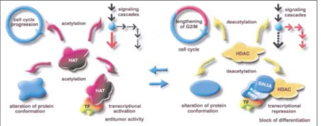
understood [60, 61]. Figure 1.7 demonstrates the interaction occurring with HAT and HDAC.
Figure 1.7. The dynamic equilibrium of HATs and HDACs in the cells results from histone acetylation. The change in histone levels has effect in transcriptional regulation, signal transduction
cascades, cell survival, differentiation, and the activities of target proteins. HATs are connected to the cell-cycle progression, whereas HDAC are connected with extension of cell cell-cycle G1 and G2. Adapted from [61].
1.5. Objectives
Researchers have found that the use of the DR5-specific TRAIL variants D269H/E195R could significantly reduce binding to decoy receptors and improve DR5 specific TRAIL receptor binding, resulting in a lower administrated dose with possibly fewer side effects [35].
The aim of our project is to use the gene therapy for the delivery of rhTRAIL DHER, which can selectively induce apoptosis in activated HSC, and hopefully, cure fibrotic livers. We will investigate the specificity and efficacy of the vector expressed proteins containing the rhTRAIL DHER. To this end, we will use the rhTRAIL mutants to determine their specificity in the context of different hepatic cell types. A specific research line will be the use of rhTRAIL mutants, designed to reduce binding to decoy receptors and to improve DR5 TRAIL receptor binding. Moreover, we will use the
14
rhTRAIL variants (DHER and 4C7) and the wild type rhTRAIL protein, to combine with HDACs (SAHA and Entinostat) and HATs (MG149 and C646) inhibitors, and evaluate the effects in different cancer cell lines (SW948 and H460) and transformed hepatic cells lines (HepG2 and HUH-7).We hope to find a combination that is highly efficient towards cancer cell line, but can protect the normal liver cells.
15
-Chapter 2-
Materials and Methods
2.1.
Cell culture and cell lines
The human embryonic kidney cell line (HEK-293 ATCC® CRL-1573TM), the human colorectal adenocarcinoma cell line (SW948 ATCC® CCL-237TM) and the human hepatic cellular carcinoma cell line (HUH-7 JCRB0403TM) were maintained in complete Dulbecco’s minimum essential medium, with high glucose, GlutaMax™ and pyruvate (DMEM; Gibco®, Life TechnologiesTM, Carlsbad, CA, USA) with 10% of FBS (Gibco®, Life TechnologiesTM, Carlsbad, CA, USA) ) and penicillin (100 IU/mL)/streptomycin (100 μg/mL) (1% P/S). The large lung carcinoma cell line (H460 ATCC® HTB-177TM) and hepatocellular carcinoma cell line (HepG2 ATCC® HB-8065TM) was maintained in complete DMEM with 10% of FBS and penicillin (100 IU/mL)/streptomycin (100 μg/mL). Immortalized human hepatic stellate cell line (LX-2 ), a kind gift provided by Prof. Scott Friedman (Mount Sinai Hospital, New York), was maintained as described previously [20]. All of the cells lines were maintained in a T-75flask at 37ºC with a 5% of CO2 in a humidified atmosphere, and routinely cultured when they were at 90% of confluence. After the experiments with the cells, a T-75 flask was incubated again with the cell line, in order to have the cell line available for other assays.
2.2.
Antibodies and rhTRAIL proteins
The Mouse IgG1 Anti-HA Tag antibody was from InvivoGen (13L18-MM, San Diego, CA, USA). Rabbit polyclonal Anti-TRAIL antibody was from AbCam® (ab2435,
16
Cambridge, England, UK). Polyclonal Swine Anti-Rabbit Immunoglobulins/HRP was from Dako© (P 0217, Agilent Technologies, Santa Clara, CA, USA). Polyclonal Rabbit Anti-Mouse Immunoglobulins/HRP was from Dako© (Z0259, Agilent Technologies, Santa Clara, CA, USA). The proteins rhTRAIL DHER, 4C7 and WT were a kind gift of Dr. R.H. Cool and the proteins were produced in Escherichia coli BL21 (DE3) bacteria using pET15b expression plasmids.
2.3.
Materials
The agar and agarose solution were from InvitrogenTM (Life TechnologiesTM, Carlsbad,CA, USA) and the electrophoresis in agarose gel was performed in a Sub-Cell® GT Cell, a horizontal electrophoresis system from Biorad®(Hercules, California, USA). The solution of Tris-HCl (Tris-HCl, Molecular Biology Grade (Tris-Hydrochloride) from Promega® (#H5123, Fitchburg, Wisconsin, USA). The SDS, and bromophenol blue Loading solutions were from Promega® (Fitchburg, Wisconsin, USA) and the electrophoresis of the 12,5% SDS-polyacrylamide gels and blotting of the gels were performed in Mini-Protean® Electrophoresis System and Mini Trans-Blot® system from Biorad®(Hercules, California, USA), respectively. The T-Flask 75 cm2 and trypsin with 0,5% of EDTA were from Life TechnologiesTM (Carlsbad,CA, USA). The EDTA, Glycine, PBS (Phosphate buffered saline) were from Sigma Aldrich® (St. Louis, MO, USA). The methanol was from VWR® (Radnor, PA, USA). The 30% Acrylamide/Bis solution, Temed and β-mercaptethanol were from Biorad® (Hercules, California, USA). The Tris-acetate-EDTA (TAE) solution was done using Tris Base, Acetic Acid, EDTA and water until one liter. The primers used to the cloning process were created in a Clone Manager® Professional version 9 and purchase in Sigma Aldrich® (St. Louis, MO, USA).
17
2.4.
Construction and production of the vector
pADTRACK-CMV-IGK-HA- rhTRAIL DHER
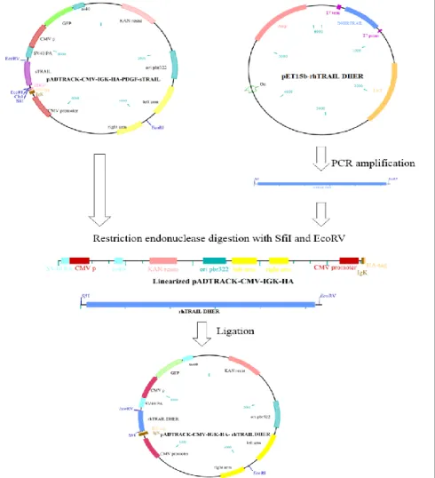
2.4.1. Cloning process to create the vector
One of our goals was to construct a vector containing the rhTRAIL DHER protein. To this end, the gene was amplified from the pET15b-rhTRAIL DHER (kindly provided by Dr. R.H. Cool [35]) and cloned in a shuttle vector, pADTRACK-CMV, for the expression of specific transgenes [62]. The vector pADTRACK-CMV-IGK-HA-PDGF-sTRAIL (kindly provided by M. Arabpour, University of Groningen, The Netherlands) was used to construct the new plasmid pADTRACK-CMV-IGK-HA- rhTRAIL DHER. To construct the new vector, a specific region in the pET15b- rhTRAIL DHER was amplified by PCR with Phusionenzyme(Promega®, Fitchburg, Wisconsin, USA) using a forward primer 5´ GGGCCCAGCCGGCCGTGAGAGAAAGAGGTCCTCAGAG 3´ and a reverse primer 3´GGATATCGGATCCCTATTAGCCAACTAAAAAGGCCCC 5´. The restriction site for SfiI (New England Biolabs®, Ipswich, Massachusetts, USA) was added in the forward primer, and a restriction site for BamHI (New England Biolabs®, Ipswich, Massachusetts, USA) and EcoRV (Promega®, Fitchburg, Wisconsin, USA) was added in the reverse primer. The conditions of the amplification were 98ºC for 30 sec in the initial denaturation, 30 cycles at 98ºC for 10 sec, 60ºC for 15 sec and 72ºC for 1min 45sec and a final extension at 72ºC for 5 min. The PCR product was purified using the Wizard® SV Gel and PCR Clean-Up system (Promega®, Fitchburg, Wisconsin, USA) according with the manufacturer’s instructions.
The amplified product and the vector pADTRACK-CMV-IGK-HA-PDGF-sTRAIL was digested by the restriction endonuclease SfiI and EcoRV, and in the digested vector was added CIP (New England Biolabs®, Ipswich, Massachusetts, USA) to remove the dephosphorylate ends of the DNA, to prevent the reconnection of the plasmid. The products resulting from digestion were cleaned with the Wizard® SV Gel and PCR Clean-Up system (Promega®, Fitchburg, Wisconsin, USA) according with the manufacturer’s
18
instructions. The digested vector and the digested sequence of the rhTRAIL DHER was ligated using the DNA ligase T4 (Promega®, Fitchburg, Wisconsin, USA) kept overnight at 4ºC, and a new plasmid was created (Figure 2.1). The pADTRACK-CMV-IGK-HA- rhTRAIL DHER vector was transformed into Escherichia coli XL1-Blue Bacteria. The transformation was done using 10μL of the ligation with 100μL of bacteria, incubated on ice 10 minutes, 45seconds at 42ºC, and 2 minutes on ice again. After thermal shock LB medium was added without antibiotic and incubated at 37ºC to allow the bacteria to recover from the shock. The bacteria containing the cloned vector were added in LB plates and selected using kanamycin.
Figure 2.1 Schematic diagram of the cloning process to create the pADTRACK-CMV-IgK-HA-rhTRAIL DHER vector. A PCR amplification in the pET15b-pADTRACK-CMV-IgK-HA-rhTRAIL DHER occurs to amplify the
rhTRAIL DHER gene. Then, the restriction endonuclease digestion with SfiI and EcoRV was done to create cohesive ends. Lastly, the ligation of the products was done to create the new vector.
19
2.4.2. Analysis of the cloned vector
In order to analyze whether the ligation of the amplified gene with the shuttle vector occur, two analyzes were performed. The selection in kanamycin produced several colonies, but only fifteen colonies were used to continue with the analysis. After the extraction of the DNA using a PureYieldTM Plasmid Miniprep System (Promega®, Fitchburg, Wisconsin, USA) according with the manufacturer’s instructions, the vector was analyzed through PCR reaction and restriction endonuclease digestions. The gene coding for the rhTRAIL DHER was amplified by PCR with PhusionTM enzyme (Promega®, Fitchburg, Wisconsin, USA) using the same primers used for the cloning process. The restriction endonuclease digestions were prepared in three different reactions: the first with ClaI restriction enzyme (Life TechnologiesTM, Carlsbad,CA, USA); the second with ClaI and EcoRI (New England Biolabs®, Ipswich, Massachusetts, USA) restriction enzymes; and the third with EcoRI restriction enzyme, using as a control non digested DNA. The digestions were held for 1h30min at 37ºC. The bands sizes of the digestions were analysed using the electrophoresis technique in a 1% agarose gel (50 mL of Tris-acetate-EDTA (TAE) 1%, 0.5g agarose) and using the 1Kb Plus DNA Ladder marker from InvitrogenTM (Life TechnologiesTM, Carlsbad, CA, USA). After both analysis two clones were chosen and sent for sequencing in the BaseClear® Company (Leiden, The Netherlands). The sequencing results are in Appendix A.1.
2.4.3. Expression of the protein in HEK-293 cells
After validation and confirmation of the presence of the gene of interest in the vector, it was necessary verified that the vector was expressing the protein of interested, to performed this analysis was used only the clones that was sent to sequence. The vectors pADTRACK-CMV-IGK-HA-rhTRAIL DHER were transfected into HEK-293 cells using the FuGENE® HD Transfection reagent (Promega®, Fitchburg, Wisconsin, USA) according with the instructions of the manufacture. As positive control, the vector
20
pADTRACK-CMV-IGK-HA- rhTRAILWT (kindly provided by M. Arabpour, University of Groningen, The Netherlands) was used, and, as negative, the transfection without DNA.The HEK-293 cells was cultured in a 24-well plate (Nunclon Delta Surface, Thermo Fisher Scientific® Waltham, Massachusetts, USA) with an 80% confluence (around 15x105 cell/well), and allowed to adhere for 24 hours. The cells were cultured in 1mL of complete DMEM, containing 10%FBS and 1% penicillin (100 IU/mL)/streptomycin (100 μg/mL), then incubated at 37ºC in a humidified atmosphere with 5% of CO2.
The efficiency of the transfection was observed in fluorescence microscopy (Axiovert 25 from Zeiss) by the GFP expression in the cells, because the vector contains the gene for GFP.Three days after the transfection, the culture medium (supernatant) was saved and the cells were harvested adding 50 μL of Trypsin containing 0.5% EDTA (Life TechnologiesTM, Carlsbad, CA, USA), then incubated for 20 min at 37ºC in a humidified atmosphere with 5% of CO2. The trypsinized cells were neutralized using 200 μL of the medium collect before. Afterwards, the neutralized cells were centrifuged at 1000g during 5 min (Beckman Coulter®, Brea, California, USA), and the supernatant was collected and saved. The pellet was ressuspended in 25 μL of 100mM Tris-HCl pH 7.8 and underwent 5 cycles of freeze-thawing in liquid nitrogen to promote the disruption of the cells. The final volume for the supernatant was 2mL and, for the pellet, 25 μL. Both the cells and the supernatant were stored at -20ºC.
21
2.5.
Analysis of the expression profile of the
rhTRAIL DHER protein in HEK-293 cells
2.5.1. Analysis of the expression of the protein through western
blotting analysis
After confirming the presence of the gene in the vector, it was necessary to verify that the cloned protein expressed by the vector is working, which was done by western blotting. The supernatant and the lysates cells, obtained in the transfection in HEK-293 cells, were used to search for the specific protein rhTRAIL DHER expressed by the plasmid pAdTRACK-CMV-IGK-HA-rhTRAIL DHER. In 10 μL of the supernatant and 1 μL of the pellet were added 4 μL sodium dodecyl sulfate (SDS) buffer (1M Tris-HCl, pH 6.8, 0.8% SDS, 40% Glycine (w/v), 10% 0.5M EDTA, 0,01% bromophenol blue) and 1 μL of β-mercaptethanol, then warmed at 110ºC during 5 min, and 6μL of marker ( Page Ruler Plus Prestained Protein Ladder, Thermo Fisher Scientific® Waltham, Massachusetts, USA) was added to allow the identification of the size. Allsamples were placed in a 12.5% sodium dodecyl sulphate polyacrylamide gel (SDS Gel) ( 1.5 M Tris-HCl, pH 8.8, 0.5M Tris-Tris-HCl, pH 6.8, 30% acrylamide/bisAA, 20% SDS, 1X Tris-glycine-SDS Buffer (TEMED), 10% Ammonium PerSulfate (APS) ), and resolved by electrophoresis on a SDS running buffer ( 25mM Tris-HCl pH 8.3, 192mM Glycine, 0,1% SDS) during 30min at 70V, then the voltage was increased to 150V during 1hour 20min. After the SDS gel was transferred onto an activated polyvinylidene difluoride (PVDF) membrane (#162-0177 Biorad®, Hercules, California, USA), this membrane was activated for 2 min with 100% methanol, and blotted with electrophoretic transfer for 2 hours at 250mA in 4ºC, with blotting buffer ( 25mM Tris-HCl pH8.3, 192mM Glycine, 10% methanol). The membrane was blocked overnight at 4ºC in PBS (Gibco®, Life TechnologiesTM, Carlsbad, CA, USA) with 0.1% Tween®20 (Promega®, Fitchburg, Wisconsin, USA) and 5% powder milk, after blocking was incubated with the primary antibody. The Mouse IgG1 Anti-HA Tag antibody (1:1000 dilution) and Rabbit
22
polyclonal Anti-TRAIL antibody (1:1000 dilution) were incubated in 10 mL of PBS with 0.1% Tween®20 and 5% of bovine serum albumin (BSA) (A9418 Sigma Aldrich®, St. Louis, MO, USA) during 1hour30min moderately shaking at RT. Then, the membrane was washed with 10 mL of PBS+0.1% Tween®20 (Promega®, Fitchburg, Wisconsin, USA), three times at RT, during 10min shaking smoothly. After washing, the peroxidase conjugated secondary antibodies, as polyclonal Rabbit Anti-Mouse Immunoglobulins/HRP antibody (1:1000 dilution) and polyclonal Swine Anti-Rabbit Immunoglobulins/HRP (1:1000 dilution) antibody were incubated in 10 mL PBS with 0.1% Tween®20 and 5% BSA over 1hour at RT, with moderate shaking. Finally the membrane was washed as mention previously and the signal was revealed with enhanced chemiluminescence substrate (Western Lightning® Plus-ECL, PerkinElmer, Waltham, MA, USA), according to the manufacturer’s instructions. The picture of the membrane was done in a gel imaging for fluorescence and visible applications (GBOX, Syngene, England, UK).
2.5.2. Analysis of the function of protein through survival assay
in SW-948 cells
As described in the previously section to determine whether the express protein is working, first a western blotting analysis was done to detect the protein, and, second, a survival assay was done adding the express proteins to the cells SW948.
The cells SW-948, cultured in flasks T-75cm2, were washed two times with 1xPBS buffer, trypsinized with 2mL during 30min at 37ºC, in a 5% CO2 incubator. Then 2 mL of complete DMEM were added, to neutralize the trypsin. The suspension of cells was counted and seeded in 96-well plate (CostarTM, Thermo Fisher Scientific, Waltham, MA, USA) with 12 x 103 cells/well at a final volume of 100 μL of complete DMEM and allowed to adhere overnight. After 24 hours of plating, different concentrations of the supernatant from transfection (3.1μL, 6.25μL, 10μL, 12.5μL, 25μL, 50μL, and 100μL) were added and 1 μL of pellet from transfection was added to the cells. The positive
23
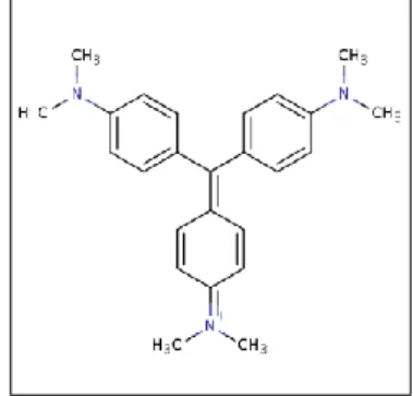
control and the negative control from the transfection in HEK-293 cells were used. To reach the 200 μL as a final volume complete DMEM was added. After treatment for 72hours was removed carefully the culture medium from the wells, and washed slightly with PBS buffer concentrate 1x (warmed at RT). The PBS buffer was removed gently and 50 μL of the crystal violet solution (70% ethanol and 30% crystal violet solution) were added and incubate for 10min at RT. After incubation, the plates were washed twice by immersion in a large beaker with tap water. Then the plates were drained upside down on filter paper, and 100 μL of 1% SDS ( 1%SDS, 100mL sterile water) was added in each well, and put on shaker during 2 hours (until the color was uniform). As a final step the absorbance of each well was measured at 570nm, in a synergy H1 hybrid Multi-Mode Microplate reader (Biotek®,Winooski, VT, USA). The crystal violet (Figure 2.2), a triarylmethane dye, is a simple assay, useful for obtaining information about the adherent cells to the surface of the dishes. This assay stains the DNA. When soluble the amount of dye taken up by the monolayer cells can be quantified in a plate reader [63].
Figure 2.2 . The crystal violet structure. The crystal violet is a triarylmethane dye (methylrosanilide,
tris[4-(dimethylamino)phenyl methanol) useful for cell adhesion studies. Adapted from [64].
The stained cells where an indication that the cells were alive and they adhered to the plate. To calculate the survival of the cells, after the measurement in the plate reader, the equation (1) was used and all the calculations was done in the Microsoft® Excel program.
Survival(%) = Avrg(Abs values of the sample)
Avrg(Abs values of the control only with cells) x100 equation (1)
24
2.6.
Generation
of
adenovirus
plasmid
by
homologous recombination in bacterial cells
2.6.1. Preparation of electrocompetent cells
In order to construct an adenovirus plasmid to produce the protein rhTRAIL DHER and to be able to use in humans, the Escherichia Coli strain BJ5183 bacteria was used, to obtain the homologous recombination of the constructed vector (pAdTRACK-CMV-IGK-HA-rhTRAIL DHER) with an adenoviral backbone vector (pAdEasy-1 adenoviral plasmid). The bacteria BJ5183 was transformed with the backbone vector pAdEasy-1 (kindly provided by M. Arabpour, University of Groningen, The Netherlands). The transformation procedure was the same as described in section 2.4.1. Then the transformed bacteria grew overnight in plates of LB medium (LB medium, 1% agar, 50 μg/mL ampicillin) at 37ºC in an incubator. One colony was selected and grew overnight in 6 mL of LB medium (with 50 μg/mL ampicillin) at 37ºC, in a Thermoshake- incubator shaker (Gerhardt® analytical systems). Subsequently 5mL of the cultured bacteria were added in 250mL of medium LB (with 50 μg/mL ampicillin), a sample was taken and the absorbance at 600 nm (A600) was measured in a Eppendorf photospectrometer (Eppendorf®, Hamburg, Germany).The bacteria were then incubated for 4 hours until the A600=0.75-0.90. The cells were collected in a 250mL flask and incubated on ice for 30 min, then centrifuged (Beckman Coulter®, Brea, California, USA) 4000g at 4ºC during 10 min. The pellet was washed with 250 ml of prechilled washing buffer (10% glycerol in sterile milli Q water), and the step of centrifuge was repeated again but for 30 min. The washing and centrifuge steps were repeated two more times, but decreased the volume of washing buffer, for 125mL and 75mL. The supernatant was discarded leaving only 15mL that were transferred to a 50 mL tube, and washed with 15 mL of washing buffer. Then the result was centrifuged at 2500g at 4ºC during 10 min, and the supernatant was aspirated, leaving only 1 mL. The remaining solution was ressuspended in 2mL of washing buffer, and aliquot 20 μL in cooled down tubes at -80ºC and stored.
25
2.6.2. Generation of recombinant adenoviral plasmid in BJ5183
With the goal of creating an adenovirus containing the gene of interest, the shuttle vector created was recombined with the plasmid pAdEasy-1, would be able to reproduce in the human body. For that reason, the prepared electrocompetent bacteria BJ5183 was taken from the -80ºC and thawed on ice. The plasmid pAdTRACK-CMV-IGK-HA-rhTRAIL DHER contained in XL1-Blue bacteria was grown overnight in medium 2mL of LB with 50 μg/mL of kanamycin and purified by phenol/chloroform extraction and ethanol precipitation using the kit PureYieldTM Plasmid Miniprep System (Promega®, Fitchburg, Wisconsin, USA) according with the manufacturer’s instructions. After purification 0.5-1.0μg of plasmid was linearized by restriction endonuclease with PmeI (New England Biolabs®, Ipswich, Massachusetts, USA) restriction enzyme (reaction of the digestion in Appendix 7.2), and incubated for 2 hours at 37ºC. After the incubation, the digested plasmid was purified using the kit Wizard® SV Gel and PCR Clean-Up system (Promega®, Fitchburg, Wisconsin, USA), following the rules of the manufacturer. Then, 1 μL of purified DNA was added to the electrocompetent bacteria BJ5183 (containing the pAdEasy-1), and incubated on ice for 30 seconds. The bacteria were transferred to a prechilled electroporation cuvette (Micropulser electroporation cuvettes, BioRad®, Hercules, California, USA), and the electroporation was performed at 2500V in the electroporation apparatus (MicroPulser, BioRad®, Hercules, California, USA). After electroporation 500μL of medium LB (without antibiotic) was added to the cuvette. The bacterial suspension was transferred to a micro tube and incubated at 37ºC for 40 min in a Thermoshake-incubator shaker (Gerhardt® analytical systems), to allow bacteria to recover and express the antibiotic resistance. To finish the procedure, 50μL of the bacterial suspension were added in a plate of LB medium (1% agar, 50 μg/mL of kanamycin), and then the bacterial suspension was centrifuged at 5000g during 1 min. The supernatant was removed, and 50 μL were left to ressuspend the pellet and add to other LB plate (with kanamycin) (see Figure Appendix A.3). In other to see the competence of the electrocompetent bacteria and the efficiency of the recombination, 50