Clin Case Rep. 2019;7:865–871. wileyonlinelibrary.com/journal/ccr3
|
8651
|
INTRODUCTION
Systemic lupus erythematosus (SLE) remains a disease of unknown etiology with a mosaic of clinical presentations. Cutaneous lesions are a first sign of SLE in up to one quarter of patients.1 According to histopathologic criteria, cutaneous manifestations include lupus erythematosus (LE)‐specific and LE‐nonspecific lesions. LE‐specific lesions are subdi-vided according to clinical phenotype, histological changes, laboratory abnormalities, and average duration. As the clini-cal and histologiclini-cal features of SLE skin lesions may mimic many other dermatological conditions, a skin biopsy may be required and a correct diagnosis relies on strict clinico‐patho-logical correlation, benefiting from evaluation by a lupus expert or an experienced dermatopathologist.1-3 We hereby
describe diagnostic and management difficulties and a suc-cessful therapeutic outcome in a single SLE patient, applying
current knowledge to discuss a multiplicity of cutaneous lesions.
2
|
CASE REPORT
In April 2012, a previously healthy 12‐year‐old female pre-sented with a malar rash (Figure 1A). Menarche had started at 11 years of age, and the patient had been vaccinated according to the national Portuguese vaccination program including the first dose of the human papilloma virus vaccine, administered 1 month before symptom onset. The clinical characteristics, histological reports, treatments, and outcome are presented in chronological order in Tables 1 and 2. A skin biopsy (Figure 2A) was reported as compatible with a diagnosis of lupus. More specifically, there was a thin epidermis, the basement mem-brane was not thickened, and a mild perivascular lymphocytic
C A S E R E P O R T
Heterogeneous lupus‐specific lesions and treatment outcome, in a
single patient, over a period of time
Melissa Fernandes
1|
Anna V. Taulaigo
1|
Carolina Vidal
1,2|
Patrick Agostini
3|
Nuno Riso
1|
Maria Francisca Moraes‐Fontes
1This is an open access article under the terms of the Creative Commons Attribution License, which permits use, distribution and reproduction in any medium, provided the original work is properly cited.
© 2019 The Authors. Clinical Case Reports published by John Wiley & Sons Ltd.
M. Fernandes and A. V. Taulaigo contributed equally to this paper. 1Unidade de Doenças Auto-imunes/
Serviço de Medicina 7.2, Hospital de Curry Cabral, Centro Hospitalar Universitário de Lisboa Central, Lisboa, Portugal
2Serviço de Medicina Interna, Hospital do Divino Espírito Santo, Ponta Delgada, Açores, Portugal
3Laboratório de Anatomia
Patológica, Centro Hospitalar Universitário do Algarve, Faro‐Portimão, Portugal
Correspondence
Maria Francisca Moraes‐Fontes, Unidade de Doenças Auto-imunes/Serviço de Medicina 7.2, Hospital de Curry Cabral, Centro Hospitalar Universitário de Lisboa Central, Lisboa, Portugal.
Email: mffontes@igc.gulbenkian.pt
Key Clinical Message
The report highlights the importance of strict clinico‐histological correlations when skin biopsies are performed in diagnostic doubt in systemic lupus erythematosus. Furthermore, PUVA is never indicated in autoimmune conditions involving photo-sensitivity, due to high potential for internal and cutaneous aggravation of the dis-ease, as the authors observed in this case.
K E Y W O R D S
discoid lupus erythematosus, heterogeneity, lupus erythematosus tumidus, subacute cutaneous lupus erythematosus, systemic lupus erythematosus, treatment
infiltrate and focal vacuolization were found at the dermoe-pidermal junction. Edema, vessel ectasia, a mild perivascu-lar lymphocytic infiltrate, and mucin deposits were found in the reticular dermis and a lymphocytic infiltrate surrounded hair follicles. At that time anti‐SSA antibodies were present, but there were no other abnormalities in the full blood count, renal function, or urinary sediment. There was improvement with topical hydrocortisone, tacrolimus, and photoprotection. One month later, the patient developed fever and lost 1.5 kg in weight, and 3 months later, the rash on the cheeks returned (Figure 1B). Repeat biopsies in the malar region were per-formed in July 2012 but a tissue orientation error prevented in-terpretation. At that time, a lupus band test from unaffected skin revealed the presence of IgM and IgG granular deposits in the basement membrane. Hydroxychloroquine (HCQ) 400 mg/d was started and the rash improved (Figure 1C). Despite HCQ, in December 2012, symmetrical painful violaceous lesions ap-peared on the tip of the fingers and toes. These resolved with deflazacort 30 mg/d for 1 week, progressively discontinued in the following 3 months. In June 2013, still on HCQ, worsening of the malar rash was documented. In April 2014, the patient reported the onset of pruritic well‐defined hyperkeratotic pap-ules initially in the lower limbs, rapidly spreading to the but-tocks, upper torso, arms, palms of hands and scalp, resulting in severe alopecia (Figure 1D). The complete full blood count, hepatic and renal function tests were within normal ranges. A more extensive profile revealed ANA positivity (1/1280), with an elevated anti‐dsDNA, a low C4 and C3. The patient was then treated with daily deflazacort 30 mg, azathioprine (AZA) 50 mg and anti‐histaminics, with no improvement. At that time, scabies was suspected and topical treatment with ben-zyl benzoate was prescribed on two occasions. Several scalp punch biopsies in September 2014 (Figure 2B) were reported as compatible with lupus, folliculitis being reported in one of
the samples (Figure 2C). No periodic acid‐Schiff (PAS) posi-tive microorganisms were identified, and there was no immu-noglobulin deposition by direct immunofluorescence. The skin condition progressively deteriorated, and both deflazacort and AZA were discontinued. Several discordant histologi-cal diagnosis of perforating dermatosis (Figure 2D) and pso-riasis (Figure 2E) ensued. The patient was then treated with oral isotretinoin, whole body psoralen, and ultraviolet‐A light therapy (PUVA), 3 times a week (oral 8‐Methoxsalen admin-istered before each session with initial, final and total doses of 1.5, 9, and 29.5 J/cm2, respectively). These treatments were
harmful and stopped after eleven sessions due to the develop-ment of generalized, erosive, painful and extremely pruritic disseminated cutaneous lesions with severe alopecia (Figure 1E), after which the patient was admitted to our unit in July 2015. Laboratory tests showed leucopenia (3100/μL), neutro-penia (1680/μL), ANA positivity (1/640), anti‐dsDNA antibod-ies (277 IU/mL; ELISA reference: <25 IU/mL), complement consumption (C3 = 61 mg/dL [normal range: 90‐180 mg/ dL], C4 = 5 mg/dL [normal range: 10‐40 mg/dL]), and sus-tained proteinuria (highest value: 1006 mg/24 h). ELISA tests for anti‐Beta‐2 Glycoprotein1 and anti‐cardiolipin antibodies as well as the lupus anticoagulant assay were negative. The renal biopsy revealed class V membranous glomerulonephritis with granular deposits of immunoglobulins, complement com-ponents, and light chains (Figure S1); tissue and serum anti‐ Phospholipase A2 receptor antibody were negative. In view of her skin condition, off‐label intravenous immunoglobulin (IVIG) was administered (20 g/d × 5 days) together with HCQ 400 mg/d, and mycophenolate mofetil (MMF) was started at the dose of 500 mg bd and increased weekly by 250 mg bd to a maximum dose of 1 g bd, together with enalapril 5 mg/d. On the 20th day of hospitalization due to the ongoing sever-ity of the skin lesions, the patient was treated with rituximab
FIGURE 1 Clinical features. Age 12—cutaneous lesions localized to malar regions (A and B); topical treatment led to improvement without scarring (C); Age 14—generalized rash, started in legs and extending to arms, buttocks, palm of hands and fronto‐temporal regions of the scalp with alopecia (D); Age 15—post PUVA (E); Age 15—post rituximab (F)
(A) (A) (B) (C) (D) (E) (F) (D) (D) (D) (D) (E) (F) (E) (D) (D) (D) (D) (B) (C)
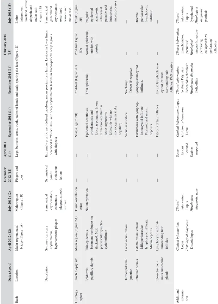
TABLE 1
Clinical characteristics and sequential histological reports Date (A
ge, y) Apr il 2012 (12) Jul y 2012 (12) December 2012 (12) Apr il 2014 (14) Sep tember 2014 (14) No vember 2014 (14) Febr uar y 2015 (15) Jul y 2015 (15) Rash Location Malar r egion, nasal br idg e (F igur e 1A) Malar r egion (F igur e 1B) Fing ers and toes Legs, butt oc ks, ar ms, tr unk
, palms of hands and scalp, spar
ing t he f ace (F igur e 1D) Entir e integument invol ved, se ver e
alopecia and malar r
ash (F igur e 1E) Descr ip tion Symme trical scal y er yt hemat ous, hyper ker at otic plaq ues Symme trical er yt hemat ous, edemat ous plaq ues, smoo th sur face Symme trical
painful violaceous lesions
Extr emel y pr ur itic w ell‐def ined papulosq uamous psor iasif or m lesions. Lesions in lo wer limbs descr ibed as “f olliculitis‐lik e.” Scal y er yt hemat ous lesions in fr ont o‐par ie tal scalp r egions wit h alopecia Symme trical gener alized er yt hemat ous ex cor iated
lesions and malar r
ash His tology repor t Punc h biopsy site Malar r egion (F igur e 2A) Tissue or ient ation er ror — — Scalp (F igur e 2B) Pr e‐tibial (F igur e 2C) Pr e‐tibial F igur e 2D) Tr unk (F igur e 2E) Epider mis, papillar y der mis Thin epider mis, basement membr ane no t thic kened. Mild per iv ascular l ym pho -cytic inf iltr ate No inter pr et ation — — Epider mal acant hosis, or thok er at osis and
folicular plugging. In one of the biopsies t
her e is acute suppur ativ e folliculitis wit h no micr oor ganisms (P AS neg ativ e) Thin epider mis Nor mal epider mis, er osion wit h pus tule Irr egular epider mal acant hosis, subcor neal pus tules and intr acor neal micr oabscesses Der mo epider mal junction Focal v acuolization — — Vacuolar inter face No c hang e Dir ect IF neg ativ e — — Re ticular der mis Edema, v essel ect asia. Mild per iv ascular lym phocytic inf iltr ate. Mucin deposits — — Edemat ous wit h l ym phop -lasmacyt oid inf iltr ate. Fibr
inoid and mucin
deposits Lym phoplasmacyt oid inf iltr ate. — Discr ete per iv ascular lym phocytic inf iltr ate
Pilo‐sebaceous units and eccr
ine glands Lym phocytic inf iltr ate sur rounding hair follicles — — Fibr osis of hair f ollicles Intense l ym phoplasma -cyt oid inf iltr ate sur rounding hair follicles. P AS neg ativ e — — Additional inf or ma -tion Clinical inf or mation :
Lupus Histological diagnosis
: Discoid lupus Clinical inf or mation :
Lupus Histological diagnosis
: none
Some lesions ulcer
ated. Scabies suspected Clinical inf or mation : L upus His tological diagnosis : Lupus Clinical inf or mation : Scabies? Pityr iasis lic henoides c hr onica? His tological diagnosis : Folicullitis Clinical inf or mation : Lym phomat oid
papulosis? Histological diagnosis
: r eactiv e per for ating collag enosis v s per for ating folliculitis Clinical inf or mation : Lupus, lym phoma? His tological diagnosis : psor iasis
TABLE 2
Laboratory results, treatments and outcomes April 2012
July 2012 December 2012 April 2014 September 2014 November 2014 February 2015 July 2015 LABORATORY TESTS Anti-nuclear antibody (ANA) positive,
anti-SSA
Ab positive (double-ID assay). Anti-dsDNA negative. No other changes. ANA positive, anti-SSA Ab positive (method unavailable)
None
ANA 1/1280 (IF – speck
-led), anti-dsDNA 163 [(N <
20
IU/mL - RIA),
anti-Sm, anti-SSA present (double-ID assay)), C4 4 (N 10-40
mg/dL); C3 63
(N 90 – 180
mg/dL)
ANA positive; anti-dsDNA 110 (N
< 20 IU/mL - RIA), C4 6 (N 10-40 mg/dL); C3 63 (N 90 – 180 mg/dL None None
ANA positive (1/640), anti-dsDNA 277 (N
< 100 IU/mL - ELISA, C4 5 (N 10-40 mg/dL); C3 61 (N 90 – 180 mg/dL
Sustained proteinuria (highest value: 1006
mg/24
h)
Class V membranous glomerulone
-phritis with granular deposits of immunoglobulins, complement components and light chains
TREATMENT
Topical hydrocorti
-sone and tacrolimus Started HCQ 400
mg/day
Maintained HCQ DFZ 30
mg/
day for one week – ↓ over 3 months Maintained HCQ Re-started DFZ 30 mg/day Azathioprine 50 mg/day Hydroxyzine 25 mg tds.
Two treatments with topical benzyl benzoate. Maintained HCQ DFZ 30
mg/day
Azathioprine 50
mg/
day.
All treatment was stopped
Oral isotretinoin Whole body psoralen and ultraviolet-A light therapy, 3-times a week (oral 8-Methoxsalen administered before each session with initial, final and total doses of 1,5
J/cm2, 9 J/cm2 and 29,5 J/ cm2, respectively). IVIG 20 g/day x 5 days
Restarted HCQ Micophenolate mofetil Enalapril 20th day hospitalization: Riruximab 1 g preceeded by Methylprednisolone 500
mg,
repeated two weeks later
OUTCOME
Improvement without scarring Improvement without scarring (Fig. 1 c) Resolution without scarring No improvement. New lesions continued to appear. No improvement. New lesions continued to appear. No improve
-ment. New lesions continued to appear.
All treatments were stopped after nine sessions (March 25 to April 20) due to the development of generalized, painful and pruritic crusts with aggravated alopecia, malar rash, fever and generalized lymphadenopathy Complete healing with areas of depigmentation in arms (Fig. 1f) Disease remission (Figure 3)
(RTX) 1 g preceded by methylprednisolone 500 mg, on days 1 and 15, in addition to the above‐mentioned drugs. The skin rash resolved within 2 weeks of the RTX administration, with residual hypopigmentation (Figure 1C); full hair re‐growth was documented at 6 months (Figure 1D) with well‐being and sustained renal remission at 3 years of follow‐up, allowing for successful medication taper (Figure 3), continuing HCQ and MMF as maintenance treatment.
3
|
DISCUSSION AND
CONCLUSIONS
In contrast to lupus nephritis where a renal biopsy has prognostic and therapeutic value with a classification based on well‐recognized features,4 when lupus affects
the skin, lesions cannot be distinguished on the grounds of histology alone.1 Classically, in most cases of SLE,
mucin deposition in the dermis is reportedly prominent. Findings may be subtle, with discrete basal cell liquefac-tive degeneration, papillary dermal edema and perivascu-lar and perifollicuperivascu-lar mild chronic inflammatory infiltrate, indistinguishable from subacute cutaneous lupus ery-thematosus (SCLE) and discoid lupus eryery-thematosus (DLE).5,6 There are, however, histopathological features
that are more frequent in some cutaneous subtypes.7 We
envisage the following scenario based on a retrospective clinico‐pathological correlation: In April 2012, at disease onset, the patient may have presented with acute cutane-ous lupus erythematosus (ACLE), suggested by a scaly localized malar rash. Nevertheless, this was somewhat atypical for ACLE, as the rash was very discrete, there
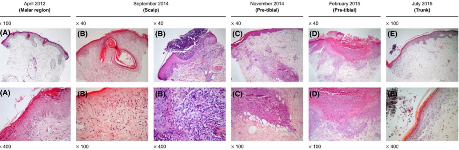
FIGURE 2 Histological features. Temporal correlation with photographs in Figure 1 per lesion: Age 12—localized to malar regions (A); Age 14—scalp (B), pre‐tibial (C); Age 15—pre‐tibial, pre PUVA (D); Age 15—trunk, post PUVA (E)
(A) (B) (B) (C) (D) (E)
(A) (B) (B) (C) (D) (E)
FIGURE 3 Follow‐up disease activity measured by the safety of estrogens in lupus national assessment‐systemic lupus erythematosus disease activity index (SELENA‐SLEDAI) and therapy.
were no systemic features and the lack of scarring after healing was against a diagnosis of DLE. The findings of perivascular and periadnexal lymphocytic infiltration together with mucin deposits, no epidermal change and no thickened basement membrane, were in favor of lupus erythematosus tumidus (LET). The latter corresponded to the morphology of the lesions, characterized by symmetri-cal erythematous and edematous plaques with a smooth surface and no scales, registered 3 months later, in July 2012. Although HCQ was started, the patient presented 8 months later with a rash on the tip of the fingers and toes suggestive of chilblain lupus. Almost 1 year later, in June 2013, the skin lesions worsened on the face, possibly due to ACLE or LET, as there was no residual scarring. From April 2014, we believe the patient presented with SCLE and SLE, on the basis of papulosquamous lesions that spared the central face and laboratory findings. These lesions were highly pruritic, psoriasiform, and not ex-clusive to sun‐exposed areas. By September 2014, histo-logical findings (orthokeratosis and follicular plugs) were suggestive of scalp DLE leading to intensification of im-munosuppression. From then on, a combination of atypi-cal features (the highly pruritic and psoriasiform nature of the lesions), misleading clinical information and re-fractoriness to therapy distanced the diagnostic path away from SLE, the underlying disease. Several misdiagnosis including scabies, folliculitis, a histological diagnosis of reactive perforating collagenosis vs perforating folliculitis and even psoriasis were evoked at the time, leading to an incorrect treatment choice with PUVA, with severe del-eterious consequences. At the time of PUVA treatments, we propose the patient was affected by SCLE, with gen-eralized skin lesions on the entire integument, in addition to SLE. Complete healing with no alopecia and no scar-ring contradict the diagnosis of scalp DLE. Finally, the hypopigmentation that remained after healing was typical for photosensitive SCLE. In summary, the patient seems to have developed several lupus‐specific skin lesions over time, starting at least 2 years before the criteria for the di-agnosis of SLE were fulfilled.8,9 Different manifestations
appeared over time. Initially, ACLE/LET responding fa-vorably to HCQ, immunosuppressants, and sunscreen, and subsequently, SCLE, refractory to therapy. Contrarily to its reportedly favorable prognosis,10 LET seems to have
preceded SLE in this patient. Of note, PUVA treatment is a formal contraindication in patients with photosensitivity.
Metabolic disorders and chronic pruritis may be associ-ated with reactive perforating dermatosis. This is a variant of prurigo nodularis, histologically characterized by epider-mal perforation11 for which ultraviolet (UV) light therapy
is recommended.12 But there was no evidence of epidermal
perforation and not unexpectedly, in this patient, UV light therapy was equivalent to a major form of photoprovocation,
with a deleterious effect, aggravating pre‐existing and pre-cipitating new cutaneous lesions, followed by a renal flare. Furthermore, lesions affecting the palms would not be ex-pected to occur in any type of folliculitis. The use of IVIG was justified by the severity of the presentation. The positive long‐term response to rituximab with a steroid sparing ef-fect has been previously described,13,14 contrasting with the
adverse events associated to the prolonged use of systemic steroids in juvenile SLE patients with skin involvement.15,16
This report emphasizes the divergence of cutaneous lupus manifestations that may present in a single patient over a period of time and the importance of clinico‐patho-logical correlation for a correct diagnostic and therapeutic approach.
CONSENT
The Subject and her Mother have given informed consent for publication.
ACKNOWLEDGEMENTS
Dr. Fernanda Carvalho—Laboratório de Morfologia Renal, Serviço de Nefrologia, Hospital Curry Cabral, CHULC for renal histology;
CONFLICT OF INTEREST
None declared.
AUTHOR CONTRIBUTION
MF and AVT: share co‐first authorship in drafting the manu-script. MF, AVT, CV, NR, and MFMF: responsible for acqui-sition, analysis, and interpretation of data; NR and MFMF: overall responsibility for patient care and critical manuscript review.
ORCID
Maria Francisca Moraes‐Fontes https://orcid.
org/0000-0002-8917-6592
REFERENCES
1. Kuhn A, Landmann A. The classification and diagnosis of cutane-ous lupus erythematosus. J Autoimmun. 2014;48–49:14‐19. 2. Tebbe B, Orfanos CE. Epidemiology and socioeconomic impact of
skin disease in lupus erythematosus. Lupus. 1997;6(2):96‐104. 3. Lipsker D. The need to revisit the nosology of cutaneous lupus
erythematosus: the current terminology and morphologic classifi-cation of cutaneous LE: difficult, incomplete and not always appli-cable. Lupus. 2010;19(9):1047‐1049.
4. Bajema IM, Wilhelmus S, Alpers CE, et al. Revision of the International Society of Nephrology/Renal Pathology Society clas-sification for lupus nephritis: clarification of definitions, and mod-ified National Institutes of Health activity and chronicity indices.
Kidney Int. 2018;93(4):789‐796.
5. Jerdan MS, Hood AF, Moore GW, et al. Histopathologic com-parison of the subsets of lupus erythematosus. Arch Dermatol. 1990;126(1):52‐55.
6. Crowson AN, Magro C. The cutaneous pathology of lupus erythe-matosus: a review. J Cutan Pathol. 2001;28(1):1‐23.
7. Obermoser G, Sontheimer RD, Zelger B. Overview of common, rare and atypical manifestations of cutaneous lupus erythematosus and histopathological correlates. Lupus. 2010;19(9):1050‐1070. 8. Hochberg MC. Updating the American College of Rheumatology
revised criteria for the classification of systemic lupus erythemato-sus. Arthritis Rheum. 1997;40(9):1725.
9. Petri M, Orbai A‐M, Alarcón GS, et al. Derivation and validation of the Systemic Lupus International Collaborating Clinics classifi-cation criteria for systemic lupus erythematosus. Arthritis Rheum. 2012;64(8):2677‐2686.
10. Kuhn A, Bein D, Bonsmann G. The 100th anniversary of lupus erythematosus tumidus. Autoimmun Rev. 2009;8(6):441‐448. 11. Kestner R, Ständer S, Osada N, Ziegler D, Metze D. Acquired
re-active perforating dermatosis is a variant of prurigo nodularis. Acta
Derm Venereol. 2017;97(2):249‐254.
12. Ohe S, Danno K, Sasaki H, Isei T, Okamoto H, Horio T. Treatment of acquired perforating dermatosis with narrowband ultraviolet B.
J Am Acad Dermatol. 2004;50(6):892‐894.
13. Condon MB, Ashby D, Pepper RJ, et al. Prospective observational single‐centre cohort study to evaluate the effectiveness of treating
lupus nephritis with rituximab and mycophenolate mofetil but no oral steroids. Ann Rheum Dis. 2013;72(8):1280‐1286.
14. Aguiar R, Araújo C, Martins‐Coelho G, Isenberg D. Use of rituximab in systemic lupus erythematosus: a single cen-ter experience over 14 years. Arthritis Care Res (Hoboken). 2017;69(2):257‐262.
15. Chiewchengchol D, Murphy R, Morgan T, et al. Mucocutaneous manifestations in a UK national cohort of juvenile‐onset sys-temic lupus erythematosus patients. Rheumatology (Oxford). 2014;53(8):1504‐1512.
16. Fangtham M, Petri M. 2013 update: Hopkins lupus cohort. Curr
Rheumatol Rep. 2013;15(9):360.
SUPPORTING INFORMATION
Additional supporting information may be found online in the Supporting Information section at the end of the article.
How to cite this article: Fernandes M, Taulaigo AV,
Vidal C, Agostini P, Riso N, Moraes‐Fontes MF. Heterogeneous lupus‐specific lesions and treatment outcome, in a single patient, over a period of time. Clin
Case Rep. 2019;7:865–871. https://doi.org/10.1002/ ccr3.2105