Research Article
Chemokine Receptor Expression on Normal Blood CD56
+
NK-Cells Elucidates Cell Partners That Comigrate during the
Innate and Adaptive Immune Responses and Identifies
a Transitional NK-Cell Population
Margarida Lima,
1Magdalena Leander,
1Marlene Santos,
1Ana Helena Santos,
1Catarina Lau,
1Maria Luís Queirós,
1Marta Gonçalves,
1Sónia Fonseca,
1João Moura,
1Maria dos Anjos Teixeira,
1and Alberto Orfao
21Laboratory of Cytometry, Service of Hematology, Hospital de Santo Ant´onio (HSA), Centro Hospitalar do Porto (CHP),
Rua D. Manuel II, 4050-345 Porto, Portugal
2Laboratory of Flow Cytometry, Centro de Investigaci´on del Cancer (CIC), Campus Miguel de Unamuno, 37007 Salamanca, Spain
Correspondence should be addressed to Margarida Lima; [email protected] Received 24 December 2014; Revised 2 March 2015; Accepted 2 March 2015 Academic Editor: Manoj K. Mishra
Copyright © 2015 Margarida Lima et al. This is an open access article distributed under the Creative Commons Attribution License, which permits unrestricted use, distribution, and reproduction in any medium, provided the original work is properly cited. Studies of chemokine receptors (CKR) in natural killer- (NK-) cells have already been published, but only a few gave detailed information on its differential expression on blood NK-cell subsets. We report on the expression of the inflammatory and homeostatic CKR on normal blood CD56+lowCD16+and CD56+highCD16−/+lowNK-cells. Conventional CD56+lowand CD56+high NK-cells present in the normal PB do express CKR for inflammatory cytokines, although with different patterns CD56+lowNK-cells are mainly CXCR1/CXCR2+and CXCR3/CCR5−/+, whereas mostly CD56+highNK-cells are CXCR1/CXCR2−and CXCR3/CCR5+. Both NK-cell subsets have variable CXCR4 expression and are CCR4−and CCR6−. The CKR repertoire of the CD56+lowNK-cells approaches to that of neutrophils, whereas the CKR repertoire of the CD56+highNK-cells mimics that of Th1+T cells, suggesting that these cells are prepared to migrate into inflamed tissues at different phases of the immune response. In addition, we describe a subpopulation of NK-cells with intermediate levels of CD56 expression, which we named CD56+intNK-cells. These NK-cells are CXCR3/CCR5+, they have intermediate levels of expression of CD16, CD62L, CD94, and CD122, and they are CD57−and CD158a−. In view of their phenotypic features, we hypothesize that they correspond to a transitional stage, between the well-known CD56+high and CD56+lowNK-cells populations.
1. Introduction
Natural killer- (NK-) cells were originally identified by their natural ability to kill target cells and are known for a long time as effector cells of the innate immune system, with an important role in controlling several types of tumors and infections [1]. In recent years, NK-cells have also been recognized as regulatory cells, which are able to interact with other cells of the immune system, such as dendritic cells (DC), monocytes/macrophages, and T cells, thereby
influencing the innate and adaptive immune responses [2–
5]. The role of their interaction with neutrophils in shaping the immune response is also being increasingly documented [6,7].
The cytotoxic activity of the NK-cells is controlled by the balance between inhibitory and activating receptors, whose ligands are self-Major Histocompatibility Complex (MHC) class I molecules and molecules expressed on stressed, viral infected, and tumor cells. They comprise, among others, the killer cell immunoglobulin-like receptors (KIR), killer cell Volume 2015, Article ID 839684, 18 pages
lectin type receptors (KLR), and natural cytotoxic receptors (NCR) as well as immunoglobulin Fc receptors (FcR) and complement receptors [8–10].
Meanwhile, the immunoregulatory properties of the NK-cells are mediated, not only by cell-to-cell contact, but also by the soluble factors they produce, which enable them to recruit and to activate other immune cells. These include chemokines (CK), such as MIP-1𝛼 (macrophage inflammatory proteins-1 alpha, CCL3) and MIP-proteins-1𝛽 (CCL4), RANTES (regulated activation, normal T cell expressed and secreted, CCL5), and ATAC (activation-induced, T cell derived, and chemokine-related cytokine, CXCL1). They also comprise cytokines, for example, IFN-𝛾 (interferon-gamma) and TNF-𝛼 (tumor necrosis factor alpha) and growth factors, such as GM-CSF (granulocyte-macrophage colony-stimulating factor) [11,12]. Using adhesion molecules and chemokine receptors (CKR), NK-cells are able to circulate in the blood and to distribute throughout the body, by homing into secondary lymphoid organs (e.g., lymph nodes), localizing in specific nonlymphoid organs (e.g., liver, placenta), and migrating into acute or chronic inflamed tissues, where they participate in the immune responses [13–16]. In some organs, NK-cells exhibit specific phenotypes and functions [17, 18], for example, promoting decidualization of the endometrium, embryo implantation and placenta development [19,20], and influencing the hematopoiesis [21,22].
Two different subsets of mature CD56+ NK-cells have been described in humans, based on the levels of CD56 and CD16 expression: CD56+low CD16+ and CD56+high
CD16−/+low NK-cells from now on designed CD56+high and
CD56+high, respectively [23, 24]. While the former clearly
predominates in the peripheral blood (PB), where they represent around 90% of the circulating CD56+NK-cells, the latter are more represented in secondary lymphoid organs, chronically inflamed tissues and placenta [13–16,19,20].
Apart from the different expression of CD16, the low affinity receptor for IgG (Fc𝛾RIIIA) and CD56, the neural cell adhesion molecule (NCAM), the conventional CD56+ NK-cell subsets also differ in the expression of other adhesion, homing, and costimulatory molecules as well as on the reper-toires of NCR, KIR and KLR, and receptors for cytokines, chemokines, and growth factors [25–29]. In addition, these NK-cell subsets exhibit distinct sialylated forms of CD43 and posttranslational modifications of the P-selectin glycoprotein ligand-1 (PSGL-1) [30,31].
From the functional point of view, CD56+low NK-cells
are essentially cytotoxic, with a greater level of antibody dependent cell mediated cytotoxicity (ADCC) [32], whereas
CD56+highNK-cells have a high proliferative response to low
doses of interleukin- (IL-) 2 (IL-2) and C-kit ligand [33]. In addition, the latter display a more important immunomodu-latory role associated with cytokine production in response to IL-2 and monokines [33]. More recently it became apparent that upon target cell recognition, CD56+low NK-cells are
more prominent cytokine and chemokine producers than
CD56+highNK-cells [34]. These diverse functional properties
would suggest that CD56+lowand CD56+highNK-cells could
be naturally prepared to act in different sites and at different phases of the immune response.
The exact relationship between these NK-cell subsets still remains unclear. Some studies have shown that bone marrow progenitor cells give rise to CD56+high or CD56+low
NK-cells depending on being cultured in the presence of IL-15 alone or in combination with IL-21, respectively [35, 36]. However, more recent data would favor a possible maturation relationship between these NK-cell subsets and suggest that CD56+low NK-cells originate from CD56+high NK-cells [37–
42].
Chemokines are small proteins that control a number of biological activities, including cell development, differ-entiation, tissue distribution, and function [43]. They act by binding chemokine receptors (CKR), a family of seven-transmembrane proteins that are classified by structure according to the number and spacing of conserved cysteines into four major groups given the names CXCR, CCR, CX3CR, and XCR to which four groups of CK correspond: CXCL, CCL, CL, and CX3CL [44]. In addition, CXCL chemokines have been further subclassified into glutamic acid-leucine-arginine tripeptide (ELR) positive or negative, based on the presence or absence of the ELR motif N-terminal to the first cysteine. From a functional point of view, two distinct types of CK have been considered: inflammatory/inducible CK, which are regulated by proinflammatory stimuli and dictate migration to the inflamed tissues and homeo-static/constitutive CK, which are responsible for the homing of the immune cells to the lymphoid organs and tissues. Similarly, two distinct groups of CKR have been described: those that interact mainly with inflammatory/inducible CK and have overlapping specificities and those that are relatively specific for homeostatic/constitutive CK [43,44].
To the best of our knowledge only a few studies analyzed in detail the CKR repertoire on CD56+low and CD56+high NK-cells and the results obtained were somewhat diver-gent [45, 46]. For instance, Campbell et al. have found that CD56+/CD16+(primarily CD56+low) NK-cells uniformly express high levels of CXCR1, CXCR4, and CX3CR1 and low levels of CXCR2 and CXCR3 but no CCR1–6, CCR9, CXCR5, and CXCR6; they also found that CD56+/CD16− (primarily
CD56+high) NK-cells do express CXCR3, CXCR4, CCR5,
and very low levels of CX3CR1, but no CXCR1, CXCR2, CXCR5, CCR1–4, 6, and 9 [45]. In contrast, Berahovich et al. observed that NK-cells are CXCR1+, CXCR3+, and CXCR4+ and contain subsets expressing CCR1, CCR4, CCR5, CCR6, CCR9, CXCR5, and CXCR6 [46]; according to their work, with the exception of CCR4, these CKR are expressed at higher percentages by CD56+highNK-cells [46]. Additionally,
both authors have found CCR7 to be restricted to CD56+high
NK-cells, which has been proved to regulate its selective homing into the lymph nodes (LN) [47, 48], where these cells establish the link between innate and adaptive immunity [47,48].
We have previously characterized the immunopheno-type of blood CD56+low and CD56+high NK-cells [29]. In order to better understand the migration pathways and cell-interactions of these NK-cell subsets and to establish
the normal reference patterns for the study of the NK-cell lymphoproliferative disorders, we decided to investigate the expression of a number of CKR on these NK-cell subsets. At some point in our study, we found that blood CD56+ NK-cells include a minor population of CXCR3/CCR5+NK-cells whose levels of CD56 expression are intermediate between those observed on CD56+lowand CD56+highNK-cells, most of which are CD16+. These cells, from now on referred to as CD56+ int NK-cells, fail to display CD57 and KIR, and they have intermediate levels of CD62L, CD94, and CD122 expression. Based on the results presented herein and on the published data, we discuss the migration routes of the conventional CD56+high and CD56+low NK-cells and
their relevance for the success of the immune response and hypothesize that CD56+ int NK-cells probably represent a
transitional NK-cell state.
2. Material and Methods
2.1. Subjects. We first analyzed by flow cytometry the
expres-sion of a number of CKR on the CD56+NK-cells in the PB of 15 adult healthy individuals (blood donors), 9 males and 6 females, aged from 19 to 54 years (median age of 38 years). After suspecting the existence of a subpopulation of CD56+ int NK-cells, these cells were further characterized using another group of 13 adult healthy individuals (blood donors), 8 males and 5 females, aged from 20 to 64 years (median age of 40 years).
2.2. Ethical Statement. This study was approved by the Ethical
Committee as part of a research project aimed to characterize the CKR on normal and neoplastic T cells and NK-cells in order to better understand the biology of the T cell and NK-cell lymphoproliferative disorders. All individuals gave informed consent to participate in the study.
2.3. Flow Cytometry Studies. Immunophenotyping was
per-formed using a whole blood stain-lyse-and-then-wash direct immunofluorescence technique using FACS lysing solution (Becton Dickinson, San Jos´e, CA) (BD) for erythrocyte lysis and cell fixation and four-color stainings with mono-clonal antibodies (mAbs) conjugated with fluorescein isoth-iocyanate (FITC), phycoerythrin (PE), PE-Cyanine 5 (PC5) or peridinin chlorophyll protein (PerCP), and allophyco-cyanin (APC). These were purchased to BD, Pharmingen (PH; San Diego, CA), Beckman Coulter (BC; Miami, FL), Immunotech (IOT; Marseille, France), and CLB (Amster-dam, Netherlands). Appropriate fluorochrome-conjugated isotype matched mAbs were used as negative controls.
In order to characterize the CKR expression on the conventional CD56+lowand CD56+highNK-cell subsets,
APC-conjugated anti-CD3 (BD; mouse IgG1,𝜅; clone SK7), PC5-conjugated anti-CD56 (IOT; mouse IgG1,𝜅; clone N901/ NKH-1), and FITC-conjugated anti-CD16 (IOT; mouse IgG1,𝜅; clone 3G8) mAbs were used in combination with PE-conjugated mAbs directed against the following CKR (PH): CXCR1 (CD181) (mouse IgG2b,𝜅; clone 5A12), CXCR2 (CD182) (mouse IgG1,𝜅; clone 6C6), CXCR3 (CD183) (mouse
IgG1,𝜅; clone 1C6), CCR4 (CD194) (mouse IgG1,𝜅; clone 1G1), CCR5 (CD195) (mouse IgG2a,𝜅; clone 2D7/CCR5), and CCR6 (CD196) (mouse IgG1,𝜅; clone 11A9).
Subsequently, CD56+ intNK-cells (which, in most of the
normal PB samples, cannot be distinguished from the con-ventional CD56+lowor CD56+highNK-cells using the staining protocol mentioned above) were further characterized using APC-conjugated anti-CD3, PC5-conjugated anti-CD56, PE-conjugated anti-CXCR3 + PE-PE-conjugated anti-CCR5, and one of the following FITC-conjugated mAbs directed against these molecules: anti-CD16 (IOT; mouse IgG1,𝜅; clone 3G8), anti-CD57 (BD; mouse IgM,𝜅; clone HNK-1), anti-CD62L (BD; mouse IgG2a,𝜅; clone SK11), anti-CD94 (PH; mouse IgG1,𝜅; clone HP-3D9), anti-CD122 (CLB; mouse IgG2a,𝜅; clone MIK-b1), and anti-CD158a (BD; mouse IgM,𝜅; clone HP-3E4).
Data acquisition was carried out in a FACSCalibur flow cytometer (BD) equipped with a 15 mW air-cooled 488 nm argon ion laser and a 625 nm neon diode laser, using the CellQUEST software (BD). Information on a minimum of 2× 10⁵events was acquired and stored as FCS 2.0 data files for each staining. For data analysis the Paint-a-Gate PRO (BD) and the Infinicyt (Cytognos, Salamanca, Spain) software programs were used.
Using the first staining protocol, NK-cells were first gated based on their CD3−/CD56+ phenotype; then, the conventional CD56+low and CD56+highNK-cell subsets were
selected based on their levels of CD56 expression and on their differential positivity for CD16 and separately analyzed for the expression of CXCR1, CXCR2, CXCR3, CCR4, CCR5, and CCR6. Using the second staining protocol, in which the anti-CXCR3 and CCR5 mAbs used have the same fluorochrome, we were able to distinguish three populations of CD56+ NK-cells: CD56+lowCXCR3/CCR5−, CD56+ intCXCR3/CR5+, and
CD56+highCXCR3/CR5+. These were separately analyzed for
the expression of CD16, CD56, CD57, CD62L, CD94, CD158a, and CD122.
The percentage of positive cells, the mean fluorescence intensity (MFI, expressed as arbitrary relative linear units scaled from 0 to 10,000), and the coefficient of variation of the MFI (CV, expressed as percentage) were recorded for each molecule tested.
2.4. Statistical Analysis. For all quantitative variables under
study, mean, standard deviation, median, and range values were calculated. The statistical significance of the differences observed between groups was evaluated using the Mann-Whitney𝑈-test (SPSS 10.0, SPSS, Chicago, IL, USA). 𝑃 values less than 0.05 were considered to be associated with statistical significance.
3. Results
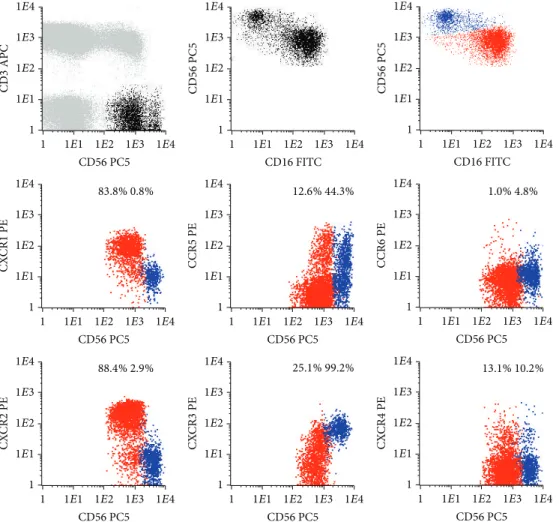
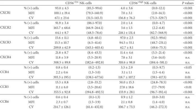
3.1. Chemokine Receptors on Blood CD56+lowand CD56+high NK-Cells. Conventional CD56+low and CD56+high NK-cells present in the normal PB have different CKR repertoires (Figure 1andTable 1).
CD3 APC CD56 PC5 CD56 PC5 CD56 PC5 CD16 FITC CD16 FITC CX CR1 PE C CR5 PE C CR6 PE CD56 PC5 CD56 PC5 CD56 PC5 CX CR2 PE CX CR3 PE CX CR4 PE CD56 PC5 CD56 PC5 CD56 PC5 1 1E1 1E2 1E3 1E4
1 1E1 1E2 1E3 1E4 1
1E1 1E2 1E3 1E4
1 1E1 1E2 1E3 1E4 1
1E1 1E2 1E3 1E4
1 1E1 1E2 1E3 1E4
1
1E1 1E2 1E3 1E4
1 1E1 1E2 1E3 1E4 1
1E1 1E2 1E3 1E4
1 1E1 1E2 1E3 1E4 1
1E1 1E2 1E3 1E4
1 1E1 1E2 1E3 1E4
1
1E1 1E2 1E3 1E4
1 1E1 1E2 1E3 1E4 1
1E1 1E2 1E3 1E4
1 1E1 1E2 1E3 1E4 1
1E1 1E2 1E3 1E4
1 1E1 1E2 1E3 1E4
83.8% 0.8% 12.6% 44.3% 1.0% 4.8%
88.4% 2.9% 25.1% 99.2% 13.1% 10.2%
Figure 1: Representative dot plots illustrating the expression of different chemokine receptors (CKR) on the conventional CD56+low(red
dots) and CD56+high(blue dots) NK-cell subsets present in the normal peripheral blood (PB). In order to obtain the dot plots showed in this
figure, PB cells were stained with APC-conjugated anti-CD3, PC5-conjugated anti-CD56, PE-conjugated anti-CKR, and FITC-conjugated anti-CD16 monoclonal antibodies. Dot plots in the first row illustrate the strategy of gating. Using the CD3/CD56 dot plot, CD56+NK-cells were first identified based on their CD3−/CD56+phenotype (black dots), comparatively to T (CD3+) and B (CD3−CD56−) cells (gray dots). Then, after gating for CD56+NK-cells (first CD56/CD16 dot plot), the CD56+low(red dots) and CD56+high(blue dots) NK-cell populations were identified based on their typical patterns of CD56 and CD16 expression (second CD56/CD16 dot plot). Finally, these NK-cell populations were analyzed for the expression of the CKR (CKR/CD56 dot plots). The numbers above the CD56+lowand CD56+highNK-cells inside the
CKR/CD56 dot plots indicate the percentage of cells staining positively for the correspondent CKR and were obtained after gating separately for each NK-cell population (CKR/CD56 dot plots gated for CD56+lowand CD56+highNK-cells are not shown, for simplicity).
3.1.1. Chemokine Receptors on Conventional CD56+low NK-Cells. Most CD56+lowNK-cells are CXCR1/CXCR2+; that is, the majority expresses high levels of CXCR1 (93.0± 4.5%) and CXCR2 (91.9 ± 3.4%), whose ligands are CXCL8 (IL-8) and other ELR motif containing chemokines involved in inflammation and angiogenesis [49] (Table 1andFigure 1). In addition, these NK-cells are CXCR3/CCR5−/+, which means that a variable proportion of them have low levels of CXCR3 and/or CCR5 (15.6± 11.1% and 13.3 ± 8.8%, resp.) (Table 1
andFigure 1). CXCR3 binds IFN-𝛾 inducible cytokines, such
as CXCL9 (monokine induced by gamma-interferon, MIG), CXCL10 (interferon-induced protein of 10 kD, IP-10), and CXCL11 (interferon-inducible T cell alpha chemoattractant,
I-TAC) and mediates Ca++mobilization and chemotaxis [50–
52]. On the other hand, CCR5 has affinity to CCL3 (MIP-1𝛼), CCL4 (MIP-1𝛽), CCL5 (RANTES), and CCL8 (monocyte chemotactic protein-2, MCP-2) [53,54].
Concerning the expression of constitutive/homeostatic CKR and the fraction of CD56+low NK-cells that expresses CXCR4, a CKR present on most hematopoietic cell types that binds to CXCL12 (stromal cell derived factor type 1, SDF-1) [55, 56] and has been shown to play a pivotal role in hematopoiesis [57] is variable (21.8± 8.7%) (Table 1 and
Figure 1). In contrast, CCR4 is expressed in only a very small percentage of the CD56+low NK-cells (0.8± 0.4%) (Table 1
Table 1: Chemokine receptor expression on the well-known CD56+lowand CD56+highNK-cells observed in blood, as identified based only on the levels of CD56 and CD16 expression.
CD56+lowNK-cells CD56+highNK-cells 𝑃 values
CXCR1 % (+) cells 93.0± 4.5 (85.3–99.6) 4.0± 3.6 (0.0–12.1) <0.001 MFI 100.1± 19.4 (79.3–140.9) 7.0± 3.1 (2.0–14.5) <0.001 CV 67.1± 23.6 (51.5–145.3) 156.8± 76.2 (75.3–329.7) <0.001 CXCR2 % (+) cells 91.9± 3.4 (86.1–97.0) 2.0± 1.4 (0.0–4.7) <0.001 MFI 165.7± 68.6 (66.9–264.1) 3.6± 1.5 (2.2–6.8) <0.001 CV 64.1± 8.7 (48.5–78.6) 218.1± 131.4 (82.7–568.9) <0.001 CXCR3 % (+) cells 15.6± 11.1 (4.8–40.1) 97.0± 2.5 (92.5–99.6) <0.001 MFI 15.5± 10.7 (6.5–41.6) 94.5± 55.1 (48.7–231.1) <0.001 CV 209.8± 62.9 (145.1–403.4) 62.7± 8.1 (49.6–75.3) <0.001 CXCR4 % (+) cells 21.8± 8.7 (8.4–43.5) 11.4± 4.6 (5.5–21.4) <0.001 MFI 11.8± 3.9 (5.3–20.9) 7.8± 3.1 (3.6–14.0) n.s. CV 308.5± 89.8 (182.6–492.8) 318.6± 98.8 (184.4–581.1) n.s. CCR4 % (+) cells 0.8± 0.4 (0.2–1.5) 3.3± 2.9 (0.3–9.7) <0.05 MFI 2.2± 0.6 (1.3–3.0) 3.1± 1.1 (1.5–4.4) n.s. CV 393.3± 195.1 (138.1–677.6) 118.7± 107.2 (59.1–417.3) 0.01 CCR5 % (+) cells 13.3± 8.8 (2.8–33.2) 50.0± 15.3 (24.8–78.5) <0.001 MFI 11.1± 6.0 (3.5–20.6) 27.8± 18.6 (7.7–79.9) <0.01 CV 353.5± 92.8 (194.8–492.5) 133.9± 28.1 (96.7–192.4) <0.001 CCR6 % (+) cells 0.6± 0.4 (0.1–1.3) 0.9± 1.2 (0.0–3.0) n.s. MFI 2.3± 0.7 (1.5–3.9) 2.1± 0.8 (1.4–4.0) n.s. CV 214.7± 126.1 (61.4–432.8) 106.7± 73.5 (46.2–272.3) n.s.
Data were obtained using the gating and analysis strategies described inFigure 1, where representative dot plots of these two conventional NK-cell subsets are
presented.
Results are expressed as mean± standard deviation (minimum–maximum) of the percentage of cells expressing each of the chemokine receptors analyzed
within each CD56+NK-cell population as well as mean± standard deviation (minimum–maximum) of the mean fluorescence intensity (MFI) and coefficient
of variation (CV) of expression. n.s.: not statistically significant.
T helper 2 (Th2) lymphocytes [58] and promotes homing of memory T cells to inflamed skin [59] by means of interaction with CCL17 (thymus and activation-regulated chemokine, TARC) and CCL22 (macrophage-derived chemokine, MDC) [60, 61]. Similar results were obtained for CCR6, which is expressed in only 0.6 ± 0.4% of the CD56+low NK-cells (Table 1). This CKR mediates responsiveness of memory T cells to CCL3 (MIP-1𝛼) [62] and CCL20 (liver- and activation-regulated chemokine, LARC) [63] and has also been implicated in the homing of Langerhans’ cells to the epidermis [64].
3.1.2. Chemokine Receptors on Conventional CD56+high NK-Cells. In contrast to CD56+low NK-cells, the majority of the
CD56+high NK-cells are CXCR1/CXCR2− and CXCR3+; that
is, most of CD56+highNK-cells express high levels of CXCR3
(96.9± 2.5%) whereas only a few are CXCR1+ (4.0 ± 3.6%) or CXCR2+(2.0 ± 1.4%), and a large fraction of them (50.0 ± 15.3%) is CCR5+(Table 1andFigure 1).
Constitutive/homeostatic CKR are also present in
CD56+high NK-cells, with a variable fraction of them
ex-pressing CXCR4 (11.4 ± 4.6%) and only a few being CCR4+
(3.3 ± 2.9%) and CCR6+ (0.9 ± 1.2%, resp.) (Table 1 and
Figure 1).
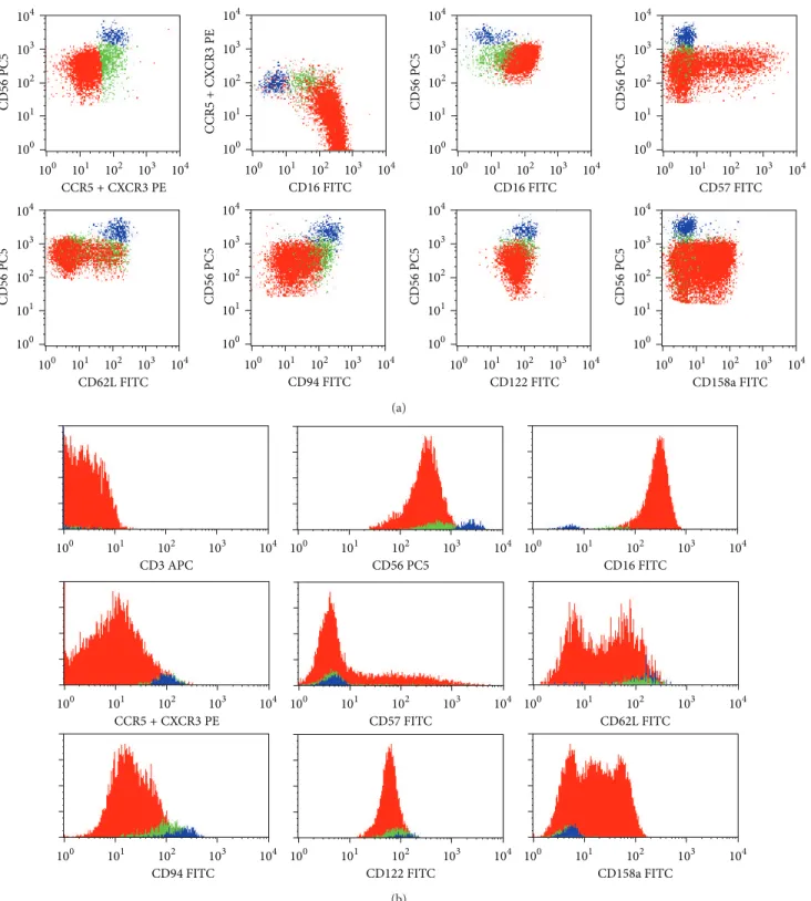
3.2. Identification of a New CD56+𝑖𝑛𝑡 NK-Cell Population in the Peripheral Blood. When analyzing the conventional
CD56+ NK-cell subsets, we observed that the percentage of CD56+low NK-cells staining for CCR5 correlated positively
with the percentage of CD56+lowNK-cells staining for CXCR3
(𝑟 = 0.656; 𝑃 = 0.01). In addition, we found that CCR5+
and CXCR+ CD56+low NK-cells had higher levels of CD56 and lower levels of CD16, as compared to CCR5− (𝑃 = 0.001 and 𝑃 = 0.05, resp.) and CXCR3− (𝑃 = 0.003 and 𝑃 = 0.002, resp.) CD56+lowcounterparts. These observations allow us to investigate if CD56+low cells expressing CCR5
and/or CXCR3 could represent a specific stage in NK-cell differentiation. In accordance, using another staining protocol in which anti-CXCR3 and anti-CCR5 mAbs had the same fluorochrome, we were able to identify three NK-cell populations in the normal PB, based on the expression of CD56, CD16, and the chemokine receptors CXCR3 and/or CCR5 (Figure 2): CD56+low CD16+CCR5/CXCR3− (or sim-ply CD56+low), CD56+ intCD16+/−CCR5/CXCR3+(or simply
CD56+ int), and CD56+high CD16−/+low CCR5/CXCR3+ (or
simply CD56+high) NK-cells.
In the normal PB, CD56+low NK-cells correspond to the
CD56 PC5
CD56 PC5
CD56 PC5 CD56 PC5
CD16 FITC CD16 FITC CD57 FITC
CD62L FITC 100 101 102 103 104 100 101 102 103 104 100 101 102 103 104 100 101 102 103 104 100 101 102 103 104 100 101 102 103 104 100 101 102 103 104 100 101 102 103 104 CCR5 + CXCR3 PE CC R 5+ CX CR 3 PE CD56 PC5 CD56 PC5 CD56 PC5
CD94 FITC CD122 FITC CD158a FITC
100 101 102 103 104 100 101 102 103 104 100 101 102 103 104 100 101 102 103 104 10 0 101 102 103 104 100 101 102 103 104 100 101 102 103 104 100 101 102 103 104 (a) CD3 APC CD56 PC5 CD16 FITC CD57 FITC CD62L FITC
CD94 FITC CD122 FITC CD158a FITC
CCR5 + CXCR3 PE
100 101 102 103 104 100 101 102 103 104 100 101 102 103 104
100 101 102 103 104 100 101 102 103 104 100 101 102 103 104
100 101 102 103 104 100 101 102 103 104 100 101 102 103 104
(b)
Figure 2: Representative dot plots (a) and histograms (b) illustrating the expression of the CD3, CD16, CD56, CD57, CD62L, CD94, CD122, and CD158a molecules on CD56+low CXCR3/CCR5− (red dots), CD56+ int CXCR3/CCR5+ (green dots), and CD56+highCXCR3/CCR5+
(blue dots) NK-cells in normal peripheral blood (PB). In order to obtain the dot plots showed in this figure, PB cells were stained with APC-conjugated anti-CD3, PC5-conjugated anti-CD56, PE-conjugated anti-CXCR3 + PE-conjugated anti-CCR5, and FITC-conjugated monoclonal antibodies against CD16, CD57, CD62L, CD94, CD122, or CD158a molecules. Total CD56+cells were gated using the strategy illustrated inFigure 1. Then, using the CD56/CCR5 + CXCR3 dot plot (first dot plot), three different CD56+ NK-cell populations were identified based on the levels of expression of CD56 and CXCR3/CCR5: CD56+lowCCR5/CXCR3−(red dots), CD56+ intCCR5/CXCR3+(green
dots), and CD56+highCCR5/CXCR3+(blue dots). As it can be seen in the remaining dot plots and histograms, these NK-cell populations differ
on the expression of several cell surface molecules. The percentage of cells staining positively for each molecule analyzed, as well as the mean fluorescence intensity of antigen expression and its coefficient of variation, was calculated after gating separately for each NK-cell population and is shown inTable 1(data is not shown in the figure, for simplicity).
CD56+ intand CD56+highNK-cells are minimally represented
(mean of6 ± 4% and 4 ± 2%, resp.) (Table 2).
Despite representing a minor NK-cell population in most normal PB samples, CD56+ intNK-cells are largely expanded
in some patients with chronic lymphoproliferative disorders of NK-cells (CLPD-NK) (Figure 3).
No differences were observed between these three NK-cell subsets concerning both the NK-cell size and complexity, as evaluated by the forward (FSC) and side light scatter (SSC), respectively, except for a slightly larger size of CD56+high
NK-cells (Table 2). Nonetheless, statistically significant dif-ferences were found concerning the expression of CD56 and CD16 (Figure 2andTable 3) as well as of the other adhesion molecules and homing, cytokine, and killer cell receptors analyzed (Figure 2andTable 4).
3.2.1. Phenotypic Characterization of Blood CD56+𝑖𝑛𝑡 NK-Cells. CD56+ int NK-cells do express CD56 at levels that are intermediate between those observed on CD56+low and
CD56+highNK-cells (MFI of615 ± 149, 466 ± 108, and 2926 ±
578, resp.) (Figure 2andTable 3). They also have intermediate percentages of CD16+ cells (64.6 ± 23.6%), as compared to CD56+low and CD56+highNK-cells (99.9 ± 0.1% and 28.7 ±
9.9%, resp.). In addition, the levels of CD16 expression (MFI of84 ± 55) were in between those observed on CD56+lowand
CD56+highNK-cells (MFI of226 ± 107 and 47 ± 18, resp.).
These three CD56+ NK-cell subsets also differ in the expression of other molecules (Figure 2andTable 4).
Concerning the KLR, only a fraction of CD56+low(47.8 ±
13.7%) expresses dimly CD94, whereas nearly all CD56+ int
(91.4 ± 6.0%) and CD56+high (98.3 ± 1.5%) are CD94+
(Figure 2andTable 4). Curiously, the levels of CD94 expres-sion on CD56+ intNK-cells are in between those observed on CD56+lowand CD56+highNK-cells (MFI of129 ± 34, 71 ± 18, and228 ± 34, resp.).
With respect to the expression of KIR, an opposite pattern is observed. Indeed, a variable fraction of CD56+low NK-cells is CD158a+ (38.9 ± 30.0%), in contrast to that found in CD56+ int and CD56+high NK-cells, which are basically
CD158a− (mean percentage of CD158a− cells of9.9 ± 9.0% and4.1 ± 4.9%, resp.) (Figure 2andTable 4).
Regarding cell adhesion molecules, the percentage of CD62L+cells is significantly lower among CD56+low(35.4 ± 20.4%), as compared to CD56+high NK-cells (97.3 ± 2.4%), intermediate values being observed in the CD56+ intNK-cells (77.3 ± 19.0%). Similar results were obtained for the levels of CD62L expression (MFI of49 ± 9, 119 ± 21, and 139 ± 30, resp.). In addition, a large fraction of CD56+low NK-cells
(66.3 ± 15.6%) expresses variably and heterogeneously the CD57 oligosaccharide, whereas most CD56+ intNK-cells fail
to express this molecule and CD56+highNK-cells are virtually
CD57−(mean % of CD57+cells of15.3±13.7% and 1.3±1.4%, resp.). Once again, the levels of CD57 expression on CD56+ int
NK-cells (MFI of355 ± 166) were in between those observed on CD56+low(MFI of700±436) and CD56+highNK-cells (MFI of165 ± 219).
The low affinity receptor for IL-2 and CD122, which is present in virtually all NK-cells, also exhibit intermediate levels on CD56+ int cells (MFI of77 ± 20), as compared to
CD56+low(MFI of46±11) and to CD56+high(MFI of117±32)
NK-cells (Figure 2andTable 4).
4. Discussion
In the present study we show that CD56+low and CD56+high
NK-cells that circulate in the normal blood have typical and quite different patterns of expression of receptors for inflammatory chemokines. At the same time, we identify and describe a subpopulation of CD56+ int NK-cells that could represent a transitional stage in between the conventional NK-cell subsets referred to above, based on their intermediate levels of CD56 and CD16 expression and on their patterns of chemokine (CXCR3, CCR5), cytokine (CD122), and killer cell (CD94, CD158a) receptors and adhesion molecules (CD62L, CD57).
Differences on the CKR repertoires make the NK-cell subsets naturally able to circulate in the blood, to home into secondary lymphoid organs, or to migrate into inflamed tissues, in different circumstances and with different partners (Figure 4), in response to constitutive and inflammatory chemokines (Table 5).
In accordance, the majority of CD56+high NK-cells are
CXCR3/CCR5+, a pattern of CKR expression that is typically observed in Th1 cells [65], while CD56+low NK-cells do express CXCR1 and CXCR2, the only CKR specific for the ELR+ CXCL chemokines involved in inflammation, thus mimicking neutrophils [66, 67]. In addition, both NK-cell subsets have variable levels of CXCR4 and virtually no CCR4 and CCR6 expression.
CD56+highNK-cells and Th1 cells, the primary cell
pop-ulations responsible for IL-2, IFN-𝛾, and TNF-𝛼 production in response to IL-2 or certain monokines, such as IL-12 and IL-15, are attracted together to chronically inflamed tissues in response to CCR5 (MIP-1𝛼, MIP-1𝛽, RANTES, and MCP-2) and CXCR3 (MIG, IP-10, and I-TAC) chemokine ligands, where they orchestrate the adaptive immune response. Some of these CK, such as RANTES and MIP-1𝛼, also attract proinflammatory CD14+low CD16+monocytes, by acting as
ligands for CCR1 and CCR4, as well as for CCR5 [68]. In agreement, CD56+high NK-cells accumulate within
Th1-type chronic inflammatory lesions in a wide variety of pathological conditions such as rheumatoid arthritis [69], psoriasis [70], sarcoidosis [71], and allograft rejection [72] as well as in sites of intracellular bacterial infections [73], chronic viral infections [74], and tumors [75]. Inside the inflamed tissues and imbibed in the appropriate monokine environment, CD56+high NK-cells are able to engage with
monocytes in a reciprocal fashion [76], thereby amplifying the inflammatory response and having important antitumor and antiviral effects. In the LN, they can induce the matu-ration of DC via IFN-𝛾 and TNF-𝛼 release and/or cell-cell contact-dependent mechanisms [2,3], in that way shaping the subsequent immune response. Moreover, activated NK-cells can kill immature myeloid DC, which have insufficient
T a ble 2: R ela ti ve rep re se n ta tio n and lig h t sca tt er p ro p er ti es o f b lo o d CD5 6 +lo w C C R5/CX C R3 − ,C D 56 +in t C C R5/CX C R3 + ,a n d C D 56 +hig h C C R5/CX C R3 + NK-cel ls ubs ets. AB C 𝑃 val u es CD5 6 +lo w C C R5/CX C R3 − CD5 6 +in t C C R5/CX C R3 + CD5 6 +hig h C C R5/CX C R3 + A ver sus B B ver sus C A ver sus C %C D 56 + NK-cel ls 9 0.3 ± 3.9 (8 3.4–98.0) 6.1 ± 4.0 (1.2–1 4.6) 3.7 ± 2.3 (0.9–8.5) — — — FSC 29 9 ± 9 (2 85–3 15) 30 3 ± 10 (2 84 –3 20) 311 ± 11 (29 1–3 24 ) n .s. 0.0 4 4 0.0 4 1 tSSC 15 1± 7( 14 0 – 16 3) 15 6 ± 7( 14 1– 17 0 ) 15 9 ± 5 (1 47 – 16 8) n.s. n.s. n.s. D at a w er e ob ta ined usin g the ga tin g an d ana ly sis stra tegies des cr ib ed in Figur e 2 ,w her e re p res en ta ti ve do t p lo ts o f th es e thr ee NK-cell subs ets ar e p res en te d. R es u lt s are ex pre ss ed as m ea n ± st an da rd de via tio n (minim um–maxim um) o f th e p er cen tag e o f each NK-cell subs et wi th in to ta lCD5 6 + NK-cells and as me an ± st an da rd de via tio n (minim um–maxim um) o f th e tra n sf o rmed side sca tt er (tSSC) and fo rw ar d sca tt er (FSC) ch an n el o f eac h NK-cell subs et. n.s.: n o t st at istica ll y significa n t.
T a b le 3: C D 56 an d C D 16 ex pre ss ion on bl o o d C D 56 +lo w C C R5/CX C R3 − ,C D 56 +in t C C R5/CX C R3 + ,a n d C D 56 +hig h C C R5/CX C R3 + NK-cel ls ubs ets. AB C 𝑃 val u es CD5 6 +lo w C C R5/CX C R3 − CD5 6 +in t C C R5/CX C R3 + CD5 6 +hig h C C R5/CX C R3 + A ver sus B B ver sus C A ver sus C CD5 6 % (+) cel ls 10 0.0 ± 0.0 (10 0.0-10 0.0) 10 0.0 ± 0.0 (10 0.0-10 0.0) 10 0.0 ± 0.0 (10 0.0-10 0.0) — — — MFI 4 6 6 ± 108 (3 03–6 23) 61 5 ± 14 9 (416–9 9 9) 29 26 ± 57 8 (216 1–3 9 93) 0.0 22 < 0.0 01 < 0.0 01 CV 57 ± 7 (41 –6 6) 72 ± 11 (5 3–9 3) 37 ± 5 (2 7– 4 8) 0.0 01 < 0.0 01 < 0.0 01 CD1 6 %( + ) ce ll s 9 9. 9 ± 0.1 (9 9.9–10 0.0) 6 4.6 ± 23 .6 (26.8–9 6.2) 28.7 ± 9. 9 (9 .9– 43 .5) < 0.0 01 < 0.0 01 < 0.0 01 MFI 226 ± 107 (55–37 2) 84 ± 55 (2 5–201) 47 ± 18 (21 – 47) 0.0 01 0.0 26 < 0.0 01 CV 45 ± 5( 38 – 55 ) 79 ± 21 (45–122) 81 ± 21 (4 4–11 8) < 0.0 01 n .s. < 0.0 01 D at a w er e ob ta ined usin g the ga tin g an d ana ly sis stra tegies des cr ib ed in Figur e 2 ,w her e re p res en ta ti ve do t p lo ts o f th es e thr ee NK-cell subs ets ar e p res en te d. R es u lt s are ex pre ss ed as m ea n ± st an da rd de via tio n (minim um–maxim um) o ft h e p er cen tag e o fp osi ti ve (+) cells w it hin each CD5 6 +NK-cell p o p u la tio n an d o ft he me an fl u o res cence in ten si ty (MFI) and co efficien t o f va ri at io n (CV) o f CD16 an d CD5 6 exp re ssio n. n.s.: n o t st at istica ll y significa n t.
Table 4: CD57, CD62L, CD94, CD122, and CD158a expression on peripheral blood CD56+lowCCR5/CXCR3−, CD56+intCCR5/CXCR3+, and CD56+highCCR5/CXCR3+NK-cell subsets.
A B C 𝑃 values
CD56+lowCCR5/CXCR3− CD56+intCCR5/CXCR3+ CD56+highCCR5/CXCR3+ A versus B B versus C A versus C CD57 % (+) cells 66.3± 15.6 (36.7–87.4) 15.3± 13.7 (1.7–42.7) 1.3± 1.4 (0.0–4.3) <0.001 <0.001 <0.001 MFI 700± 436 (217–1557) 355± 166 (150–703) 165± 219 (9–574) 0.018 0.015 <0.001 CV 114± 34 (77–194) 130± 35 (80–205) 85± 54 (6–155) n.s. n.s. n.s. CD62L % (+) cells 35.4± 20.4 (9.6–73.5) 77.3± 19.0 (41.9–95.7) 97.3± 2.4 (92.8–100.0) <0.001 <0.001 <0.001 MFI 49± 9 (38–65) 119± 21 (77–147) 139± 30 (91–172) <0.001 0.057 <0.001 CV 83± 11 (72–109) 56± 7 (46–71) 47± 9 (35–69) <0.001 0.010 <0.001 CD94 % (+) cells 47.8± 13.7 (34.1–74.1) 91.4± 6.0 (79.2–98.4) 98.3 ± 1.5 (94.5–100.0) <0.001 <0.001 <0.001 MFI 71± 18 (50–106) 129± 34 (77–211) 228± 34 (175–286) <0.001 <0.001 <0.001 CV 56± 7 (44–69) 56± 11 (41–85) 47± 8 (31–56) n.s. 0.039 0.039 CD122 % (+) cells 100.0± 0.0 (100.0-100.0) 100.0 ± 0.0 (100.0-100.0) 100.0 ± 0.0 (100.0-100.0) n.s. n.s. n.s. MFI 46± 11 (31–63) 77± 20 (49–107) 117± 32 (79–182) <0.001 0.001 <0.001 CV 50± 12 (35–72) 56± 9 (43–79) 46± 5 (34–54) n.s. <0.001 <0.001 CD158a % (+) cells 38.9± 30.0 (7.9–92.9) 9.9± 9.0 (0.7–30.0) 4.1± 4.9 (0.1–19.3) 0.001 0.030 <0.001 MFI 30± 8 (18–42) 35± 11 (22–57) 41± 13 (20–68) n.s. n.s. n.s. CV 61± 21 (27–98) 63± 20 (31–96) 69± 29 (35–126) n.s. n.s. n.s.
Data were obtained using the gating and analysis strategies described inFigure 2, where representative dot plots of these three NK-cell subsets are presented.
Results are expressed as mean± standard deviation (minimum–maximum) of the percentage of positive (+) cells within each CD56+NK-cell population,
and as the mean fluorescence intensity (MFI) and coefficient of variation (CV) of CD57, CD62L, CD94, CD122, and CD158a expression in cells that stained positively for these antigens.
n.s.: not statistically significant.
amounts of MHC molecules to activate T cells properly [2,3]. In addition, CD56+highNK-cells also predominate in placenta [77], where they are involved in maternal-fetal tolerance [78,
79].
In contrast, CD56+low NK-cells, which are essentially cytotoxic, and neutrophils, which are phagocytic cells by excellence, predominate in the PB and are equipped with the CXCR1 and CXCR2 chemokine receptors, making them able to comigrate into sites of acute inflammation in response to IL-8 and other ELR motif containing chemokines and to participate in the earliest phase of the innate immune response. As for the neutrophils, migration of CD56+low
NK-cells to inflamed tissues also depends on the interaction of different forms of PSGL-1 expressed on their membrane with the selectin molecules expressed on endothelial cells [31]. Curiously, cytotoxic T lymphocytes (CTL) have also been reported to express CXCR1 [80] and PSGL-1 [81].
Normally, both neutrophils, which are able to neutralize efficiently the extracellular pathogens, after opsonization by antibodies, using Fc receptors for IgG and IgA and CD56+low NK-cells, which mediate antibody dependent cell cytotoxicity via Fc𝛾RIIIa (CD16), circulate in the blood. In that sense, it can be hypothesized that these cells are candidates to establish a bridge between the innate immune response and the antibody mediated adaptive immune response. Evidence is being accumulated in the last years for a cross-talk between neutrophils and NK-cells [6,7]. For instance, NK-cells promote neutrophil recruitment to the inflamed tissues and several NK-cell derived cytokines and growth factors, such as GM-CSF, IFN-𝛾, and TNF-𝛼, act by enhancing
neutrophil survival and by modulating cell surface expression of complement and Fc receptors in neutrophils [82–84]. On the other hand, neutrophils can stimulate the production of IFN-𝛾 by NK-cells [84].
The other anatomical sites in which CD56+low
NK-cells and neutrophils might be concomitantly present to modulate each other’s activity and its contribution to dis-ease are not completely elucidated. Normal liver contains mainly CD56+lowNK-cells, but these cells are different from
the CD56+low NK-cells that circulate in the blood [85]. In addition, CD56+low NK-cells also infiltrate the liver of patients with primary biliary cirrhosis, an antibody mediated autoimmune disease, following the CXCR1/IL-8 axis [85]; curiously, hepatic infiltration by neutrophils is also found in these patients [86]. Moreover, CD56+low NK-cells and neutrophils colocalize in the skin of patients with Sweet’s syndrome, an acute febrile neutrophilic dermatosis that can follow viral infections, autoimmune diseases, and hemato-logic malignancies [84].
Also of note, in this study we confirm previous obser-vations about the lack of expression on both CD56+low and
CD56+high NK-cells of other CKR involved in homing to
nonlymphoid organs and tissues including CCR4 (skin and lung), CCR6 (intestine and liver), CCR9 (small intestine), and CCR10 (skin) [45,87]. This suggests that, unlike mem-ory/effector T cells, CD56+NK-cells may not be divided into cutaneous versus mucosal/intestinal-homing compartments, based on CKR expression [58,88,89].
Of special interest is also the identification of a new popu-lation of NK-cells expressing intermediate levels of CD56 that
T a ble 5: H o m eost at ic an d infla m ma to ry ch emo k ines (CK) an d chemo kine re cep to rs (CKR) in vo lv ed in cell media ted imm une resp o n ses an d their re le va nce fo r co lo caliza ti o n o ft h e N K-cel l subs ets w it h o th er cel ls o f the inna te-dendr it ic cel ls (D C), m o n o cyt es, n eu tr o p hi ls, and ada p ti ve (T cel ls) imm une syst em. C o lo caliza ti o n o f CD5 6 +hig h ,C D 56 +in t ,a n d C D 56 +hig h NK-cel ls wi th o ther imm une cells is signaled in b lue ,g re en, and re d ,r esp ec ti ve ly .B lac k sq ua re s in side th e cells indica te p osi ti ve in terac ti o n s o f the CK an d th e C KR as w el las th e C KR exp ressin g cel ls. CD5 6 +in t NK-cells ar e tra n si tio nal N K-cells, w h os e p ro p er ties ar e in te rm edia te b etw een thos e o fCD5 6 +hig h an d C D 56 +lo w NK-cel ls. C D5 6 +hig h an d C D 56 +lo w N K -c el ls ar eb o tha b let op ro d u ce cy to kines and ch emo k ines, u p o n st im u la ti o n wi th mo no kines and ta rg et cel lr ecog n it io n, re sp ec ti ve ly .Th e C K p ro d u ced by N K-cel ls (e .g., MIP -1 𝛼 ,MIP -1 𝛽 ,a n d R A N T E S)n o t o n ly at tr ac te d p ro infla m ma to ry mo no cy te s (via C CR1 and C C R5), imma tur e m ye lo id D C (via C C R1 an d C CR5), Th 1 cells, and CTL (via C CR5) b u t als o ac ti va ted neu tr o p h ils (vi aC C R 1) to th es it es o f infla m ma ti o n .Th e cyt o k ines p ro d uced by th e N K-cells (e .g., IFN-𝛾,T N F -𝛼 ) ac ti va te m o n o cyt es, D C, neu tr o p h il s, an d Th 1/CTL, th er eb y p o ten ti at in g the cel l media ted imm une resp o n ses. F o r in st ance , ac ti va te d m o n o cyt es/D C ar e ab le to p ro d uce IFN-𝛾 ind u cib le cyt o k ines—CX C L9 (MI G), C X C L1 0 (IP -1 0), and CX CL11 (I-T A C), w hic h recr u it mo re CX CR3 exp re ssin g cells (CD5 6 +hig h NK-cells, Th 1 cells, and CTL). In addi ti o n , C X C L8 (IL -8) p ro d u ced in infla m ed ti ssues by lo ca ll y residen t ac ti va te d cel ls, suc h as m acr o p hag es, ep it he li al cel ls, an d endo theli al cel ls, re cr ui ts neu tr o p h il s, CD5 6 +lo w NK-cells, and CTL, via in terac tio n wi th CX CL1 and CX CL2. Ch emok iner ecepto rs express ed C h emo k ines p ro d uced C h emo k ine recep to r exp re ssin g In fla mma to ry (ind u cib le) Ho m eo st at ic (c ons titut iv e) NK-cells Ot her cells in vo lv ed in cell media ted imm une resp o n ses CC L-3 (MIP-1 𝛼) CC L-4 (MIP-1 𝛽) CCL -5 (RA NT ES) CC L2(M CP-1) CC L8(M CP-2) CC L7(M CP-3) CCL 13 (M CP -4 ) CC L20( LA RC ) CXCL1 0(IP -10) CXCL9 (MIG) CXCL11 (I-TA C) CC L22(MD C) CC L17 (T AR C) CXCL8 (IL-8) CXCL1 (GR O-𝛼) CCL 19 (E LC ) CCL 21 (SL C) CXCL12 (SD F-1) NKCD5 6 +high NKCD5 6 +int NKCD5 6 +low Im matu re mDC Matu rem yel oid DC Co nven tio nal mono cytes (CD16 − ) Proinfl am mato ry mono cytes (CD16 + ) Naiv eT cel ls Th1T cells Cyto toxi cT Lym pho cyte s Neu tro ph ils CC R 1 ◼◼ ◼ ◼ ◼ ◼ ◼◻◻ CC R 2 ◼◼◼◼ ◼◼ ◼ CC R 5 ◼◼◼ ◼ ◼ ◼ ◼ ◼ ◼ CC R 6 ◼ ◻ CX CR1 ◼ ◼ ◼ ◼ ◼ ◼ CX CR2 ◼◼ ◼ ◼ ◼ CX CR3 ◼◼◼ ◼ ◼ ◼ ◼ CC R 4 ◼◼ ◼ CC R 7 ◼◼ ◼ ◼ ◼ CX CR4 ◼◼ ◼ ◼ ◼ ◼ ◼ ◼ ◻
CD3 APC CD56 PC5 CD56 PC5 CD16 FITC CX CR1 PE CX CR2 PE CX CR3 PE C CR5 PE
CD16 FITC CD16 FITC CD16 FITC CD16 FITC
100 101 102 103 104 100 101 102 103 104 100 101 102 103 104 100 101 102 103 104 100 101 102 103 104 100 101 102 103 104 100 101 102 103 104 100 101 102 103 104 100 101 102 103 104 100 101 102 103 104 100 101 102 103 104 100 101 102 103 104 (a) CD3 APC CD56 PC5 CD56 PC5 CD16 FITC CX CR1 PE CX CR2 PE CX CR3 PE C CR5 PE
CD16 FITC CD16 FITC CD16 FITC CD16 FITC
100 101 102 103 104 100 101 102 103 104 100 101 102 103 104 100 101 102 103 104 100 101 102 103 104 100 101 102 103 104 100 101 102 103 104 100 101 102 103 104 100 101 102 103 104 100 101 102 103 104 100 101 102 103 104 100 101 102 103 104 (b)
Figure 3: Illustrative dot plots showing the expression of the CXCR1, CXCR2, CXCR3, and CCR5 chemokine receptors (CKR) in normal peripheral blood (PB) (a), where only CD56+low(red dots) and CD56+high(blue dots) are observed, and in the PB of a patient with a chronic
lymphoproliferative disorder of NK-cells (CLPD-NK) (b), exhibiting a transitional CD56+ intphenotype (green dots); other lymphocytes are
shown in gray. In order to obtain the dot plots showed in this figure, the PB cells were stained with APC-conjugated anti-CD3, PC5-conjugated anti-CD56, PE-conjugated anti-CKR (CXCR1, CXCR2, CXCR3, or CCR5), and FITC-conjugated anti-CD16 monoclonal antibodies. As shown in (a), in the normal PB most CD56+lowNK-cells are CXCR1+and CXCR2+, whereas only a very small fraction of cells stains positively for
CXCR3 and/or CCR5; in contrast, most CD56+highNK-cells are CXCR3+whereas CCR5 is expressed in only a fraction and CXCR1 and
CXCR2 are virtually negative. As it can be seen in (b), the expanded NK-cells from this patient, which have relatively high levels of CD56 expression, are CD16+and they express the CXCR1, CXCR2, CXCR3, and CCR5 molecules in a considerable fraction of cells.
CCR1 CCR1 CCR1 CCR1 CCR2 CCR2 CCR4 CCR7 CCR7 CCR7 CCR5 CCR5 CCR5 CCR5 CCR5 CCR5 CCR5 CXCR1 CXCR1 CXCR1 CXCR1 CXCR1 CXCR1 CXCR2 CXCR2 CXCR2 CXCR3 CXCR3 CXCR2 CXCR3 CXCR3 Immature myeloid dendritic cell myeloid dendritic cell Conventional monocyte monocyte Proinflammatory Macrophage Mature NK-cell NK-cell NK-cell Th1 cell Cytotoxic T lymphocyte Neutrophil Naive T cell
CD56+high CD56+int CD56+low
Figure 4: Diagram illustrating the complex relationship established between NK-cells and the other cells of the innate-dendritic cells (DC), monocytes, macrophages, neutrophils, and adaptive (T cells) immune system, whose homing to lymphoid organs and recruitment to inflamed tissues are mediated by the interaction of homeostatic chemokines constitutively expressed on locally resident cells and inflammatory chemokines, with the correspondent chemokine receptors. CCR7 expression on CD56+highNK-cells, mature DC, and na¨ıve T cells allows these
cells to migrate into the lymph nodes, in response to CCL19 (ELC) and CCL21 (SLC) produced locally. CXCR3/CCR5 expression on CD56+high
NK-cells permits these cells to migrate into inflamed tissues, together with CCR5+proinflammatory monocytes, CCR5/CXCR3+Th1 cells, and CCR5/CXCR3+cytotoxic T lymphocytes (CTL). CXCR1/CXCR2 expression on CD56+lowNK-cells, neutrophils, and CTL permits these cells to migrate into inflamed tissues in response to CXCL8 (IL-8), where they interact together and with activated macrophages. CD56+ int
NK-cells are transitional NK-cells, whose properties are intermediate between those of CD56+highand CD56+lowNK-cells. Dashed arrows
indicate the routes of differentiation. Full arrows indicate the cross-talk between cells mediated by cytokines and chemokines. CCR7 ligands: CCL-19 (ELC) and CCL21 (SLC); CCR5 ligands: CCL3 (MIP-1𝛼), CCL4 (MIP-1𝛽), CCL5 (RANTES), and CCL8 (MCP-2); CXCR3 ligands: CXCL9 (MIG), CXCL10 (IP-10), and CXCL11 (I-TAC); CXCL1 ligands: CXCL-8 (IL-8); CXCL2 ligands: CXCL-8 (IL-8) and other ELR motif containing CXCL chemokine; CXCR4 ligand: CXCL12 (SDF-1); CCR1 ligands: CCL3 (MIP-1𝛼), CCL5 (RANTES), MCP-2, and MCP-3; CCR2 ligands: CCL2 (MCP-1), CCL8 (MCP-2), CCL7 (MCP-3), and CCL13 (MCP-4).
we designed as CD56+ intNK-cells. Similar to CD56+high
NK-cells, most of the CD56+ intNK-cells are KIR− and CD57−;
however, the majority of them display the CD16 molecule, a marker of CD56+lowNK-cells, and they have intermediate levels of CD62L, CD94, and CD122 expression. Due to the fact that the identification of the NK-cell populations by flow cytometry is usually based only on CD56 and CD16 expression, CD56+ intNK-cells are being considered together
with CD56+lowNK-cells on routine blood analysis.
The fact that CD56+ intNK-cells have phenotypic features
intermediate between those of conventional CD56+low and
CD56+highNK-cells would suggest that they could represent
a transitional NK-cell maturation stage.
In line with this hypothesis, evidence for the existence of transitional NK-cell populations with phenotypic fea-tures similar to those of the CD56+ int NK-cells described herein has also been provided in other studies [90, 91]. In accordance, Yu et al. described a CD56+low CD94+high NK-cell subset expressing CD2, CD62L, CD56, KIR, granzymes, and perforin, producing IFN-𝛾 in response to monokines, and exhibiting CD94-mediated redirected killing at levels intermediate between those observed in CD56+low CD94+low
and CD56+high CD94+high NK-cells [90]. In addition, Juelke
el al. reported on a CD56+low CD62L+ NK-cell subset
with the ability to produce IFN-𝛾 and the capacity to kill [91]. Finally, when studying the differentiation of CD56+high
CD94/NKG2A+ into CD56+low CD94/NKG2A− NK-cells, B´eziat et al. found a transitional CD56+low CD94/NKG2A+
NK-cell subset, expressing intermediate levels of CD62L, granzyme-K, CD27, and CD57, among other molecules [92]. Given the immunophenotypic similarities, the CD56+ int
NK-cell population described herein, which comprises less than 10% of the CD56+ NK-cells in the PB from normal healthy individuals, probably corresponds to a subpopulation of the
CD56+low CD94+high NK-cell subset described by Yu et al.,
which accounts for half of the circulating CD56+ NK-cells [90].
Another potential interest of identifying normal NK-cells with intermediate phenotypic features relies on data interpretation in clinical settings. For instance, overrepre-sentation of CD56+ int NK-cells in the PB from patients
with NK-cell lymphocytosis may erroneously be interpreted as phenotypically aberrant (and thus potentially neoplastic) NK-cells. Thus, the knowledge about the immunophenotype of the NK-cell populations that circulate in normal PB, as well as in the PB from patients with inflammatory and infectious conditions [93,94], is essential to a better understanding of the phenotypic heterogeneity of the expanded NK-cell pop-ulations observed in patients with CLPD-NK [95], thereby contributing to distinguishing nonclonal from clonal NK-cell proliferations and reactive from neoplastic conditions [96,97].
5. Conclusions
Differences in the CKR expression on CD56+low and
CD56+high NK-cells may determine their ability to be
rec-ruited into inflamed tissues and colocalize with other cells at sites of inflammation, which is crucial for the success of the immune response. In addition, the phenotypic hetero-geneity of the conventional CD56+low and CD56+high
NK-cells may be largely due to the presence of transitional NK-cell populations, which may be preferentially expanded in some pathological conditions.
Further investigations in this area will help to better understand the terminal differentiation of the NK-cells and the maturation relationship between the NK-cell subsets, their circulation through the body, and their participation in the immune response. In addition, they will give an important contribution to establish phenotypic criteria to differentiate reactive and neoplastic NK-cell proliferations, as well as to better identify the normal cell counterparts from which the neoplastic NK-cells originate.
Abbreviations
ADCC: Antibody dependent cell cytotoxicity APC: Allophycocyanin
BC: Beckman Coulter BD: Becton Dickinson
BDB: Becton Dickinson Biosciences BM: Bone marrow
CK: Chemokines
CKR: Chemokine receptors CTL: Cytotoxic T lymphocytes DC: Dendritic cells
ELR: Glutamic acid-leucine-arginine tripeptide motif
Fc𝛾RIIIa: Low affinity receptor for IgG Fc and alpha chain (CD16)
FCRL: Fc receptor-like
FITC: Fluorescein isothiocyanate FSC: Forward light scatter IFN-𝛾: Interferon-gamma IL: Interleukin
ILT: Immunoglobulin-like transcript IP-10: Interferon-induced protein of 10 kD
(CXCL10)
I-TAC: Interferon-inducible T cell alpha chemoattractant (CXCL11)
KIR: Killer cell immunoglobulin-like receptors KLR: Killer cell lectin type receptors
LARC: Liver- and activation-regulated chemokine (CCL20)
LN: Lymph nodes
MCP-2: Monocyte chemotactic protein-2 (CCL8) MDC: Macrophage-derived chemokine (CCL22) MHC: Major Histocompatibility Complex MIG: Monokine induced by gamma-interferon
(CXCL9)
MIP-1𝛼: Macrophage inflammatory protein 1 alpha (CCL3)
MIP-1𝛽: Macrophage inflammatory protein 1 beta (CCL4)
NCAM: Neural cell adhesion molecule (CD56) NCR: Natural cytotoxicity receptors
NK: Natural killer PB: Peripheral blood PC5: PE-Cyanine 5 PE: Phycoerythrin PH: Pharmingen
RANTES: Regulated upon activation, normal T cell expressed and secreted (CCL5)
SSC: Side light scatter
SDF-1: Stromal cell derived factor type 1 (CXCL12)
TARC: Thymus and activation-regulated chemokine (CCL17)
TGF-𝛽: Transformed growth factor beta Th: T helper
TNF-𝛼: Tumor necrosis factor alpha.
Conflict of Interests
Authors’ Contribution
Margarida Lima carried out concept and research question, literature review, study design, data collection and interpreta-tion, statistical analysis, and report writing. Ana Helena San-tos, Catarina Lau, Jo˜ao Moura, Magdalena Leander, Marlene Santos, Maria dos Anjos Teixeira, Maria Lu´ıs Queir´os, Marta Gonc¸alves, and S´onia Fonseca were responsible for sample processing, data acquisition, and data analysis. Catarina Lau and Maria dos Anjos Teixeira were responsible for data analysis and interpretation. Alberto Orfao was responsible for scientific consulting, paper revision, and discussion. All authors read and approved the final paper.
Acknowledgments
This work has been partially supported by a grant of the “Associac¸˜ao Portuguesa Contra a Leucemia” for the project “caracterizac¸˜ao imunofenot´ıpica de leucemias e linfomas de c´elulas T e NK: padr˜ao de express˜ao de receptores de quimiocinas como determinantes do tropismo tecidular da c´elula neopl´asica e do comportamento cl´ınico-biol´ogico da doenc¸a” and by the “Forum Hematol´ogico do Norte,” Portugal.
References
[1] E. Vivier, E. Tomasello, M. Baratin, T. Walzer, and S. Ugolini, “Functions of natural killer cells,” Nature Immunology, vol. 9, no. 5, pp. 503–510, 2008.
[2] M. A. Degli-Esposti and M. J. Smyth, “Close encounters of different kinds: dendritic cells and NK cells take centre stage,” Nature Reviews Immunology, vol. 5, no. 2, pp. 112–124, 2005. [3] F. Brilot, T. Strowig, and C. Munz, “NK cells interactions with
dendritic cells shape innate and adaptive immunity,” Frontiers in Bioscience, vol. 13, no. 17, pp. 6443–6454, 2008.
[4] A. Moretta, E. Marcenaro, S. Parolini, G. Ferlazzo, and L. Moretta, “NK cells at the interface between innate and adaptive immunity,” Cell Death and Differentiation, vol. 15, no. 2, pp. 226– 233, 2008.
[5] J. Crouse, H. C. Xu, P. A. Lang, and A. Oxenius, “NK cells regulating T cell responses: mechanisms and outcome,” Trends in Immunology, vol. 36, no. 1, pp. 49–58, 2015.
[6] C. Costantini and M. A. Cassatella, “The defensive alliance between neutrophils and NK cells as a novel arm of innate immunity,” Journal of Leukocyte Biology, vol. 89, no. 2, pp. 221– 233, 2011.
[7] P. Scapini and M. A. Cassatella, “Social networking of human neutrophils within the immune system,” Blood, vol. 124, no. 5, pp. 710–719, 2014.
[8] A. Moretta, C. Bottino, M. Vitale et al., “Activating receptors and coreceptors involved in human natural killer cell-mediated cytolysis,” Annual Review of Immunology, vol. 19, pp. 197–223, 2001.
[9] F. Borrego, J. Kabat, D.-K. Kim et al., “Structure and function of major histocompatibility complex (MHC) class I specific recep-tors expressed on human natural killer (NK) cells,” Molecular Immunology, vol. 38, no. 9, pp. 637–660, 2002.
[10] K. Natarajan, N. Dimasi, J. Wang, R. A. Mariuzza, and D. H. Margulies, “Structure and function of natural killer cell
receptors: multiple molecular solutions to self, nonself discrim-ination,” Annual Review of Immunology, vol. 20, pp. 853–885, 2002.
[11] A. A. Maghazachi, “Role of chemokines in the biology of natural killer cells,” Current Topics in Microbiology and Immunology, vol. 341, no. 1, pp. 37–58, 2010.
[12] R. Paolini, G. Bernardini, R. Molfetta, and A. Santoni, “NK cells and interferons,” Cytokine Growth Factor Reviews, 2014. [13] M. J. M¨oller, R. Kammerer, and S. von Kleist, “A distinct
distribution of natural killer cell subgroups in human tissues and blood,” International Journal of Cancer, vol. 78, no. 5, pp. 533–538, 1998.
[14] M. Morris and K. Ley, “Trafficking of natural killer cells,” Current Molecular Medicine, vol. 4, no. 4, pp. 431–438, 2004. [15] P. Carrega and G. Ferlazzo, “Natural killer cell distribution and
trafficking in human tissues,” Frontiers in Immunology, vol. 3, article 347, 2012.
[16] G. Bernardini and A. Santoni, “The pathophysiological role of chemokines in the regulation of NK cell tissue homing,” Critical Reviews in Oncogenesis, vol. 19, no. 1-2, pp. 77–90, 2014. [17] R. Sharma and A. Das, “Organ-specific phenotypic and
func-tional features of NK cells in humans,” Immunology Research, vol. 58, no. 1, pp. 125–131, 2014.
[18] D. K. Sojka, Z. Tian, and W. M. Yokoyama, “Tissue-resident natural killer cells and their potential diversity,” Seminars in Immunology, vol. 26, no. 2, pp. 127–131, 2014.
[19] A. Santoni, C. Carlino, and A. Gismondi, “Uterine NK cell development, migration and function,” Reproductive BioMedicine Online, vol. 16, no. 2, pp. 202–210, 2008.
[20] A. Moffett and F. Colucci, “Uterine NK cells: active regulators at the maternal-fetal interface,” Journal of Clinical Investigation, vol. 124, no. 5, pp. 1872–1879, 2014.
[21] A. M. de Bruin, C. Voermans, and M. A. Nolte, “Impact of interferon-𝛾 on hematopoiesis,” Blood, vol. 124, no. 16, pp. 2479– 2486, 2014.
[22] M. Diederich, F. Morceau, and M. Diederich, “Pro-inflammatory cytokine-mediated anemia: Regarding molecular mechanisms of erythropoiesis,” Mediators of Inflammation, vol. 2009, Article ID 405016, 11 pages, 2009.
[23] M. A. Cooper, T. A. Fehniger, and M. A. Caligiuri, “The biology of human natural killer-cell subsets,” Trends in Immunology, vol. 22, no. 11, pp. 633–640, 2001.
[24] A. Poli, T. Michel, M. Th´er´esine, E. Andr`es, F. Hentges, and J. Zimmer, “CD56brightnatural killer (NK) cells: an important NK
cell subset,” Immunology, vol. 126, no. 4, pp. 458–465, 2009. [25] D. M. Baume, M. J. Robertson, H. Levine, T. J. Manley, P.
W. Schow, and J. Ritz, “Differential responses to interleukin 2 define functionally distinct subsets of human natural killer cells,” European Journal of Immunology, vol. 22, no. 1, pp. 1–6, 1992.
[26] W. E. Carson, T. A. Fehniger, and M. A. Caligiuri, “CD56bright
natural killer cell subsets: characterization of distinct functional responses to interleukin-2 and the c-kit ligand,” European Journal of Immunology, vol. 27, no. 2, pp. 354–360, 1997. [27] M. Frey, N. B. Packianathan, T. A. Fehniger et al.,
“Differen-tial expression and function of L-selectin on CD56bright and
CD56dimnatural killer cell subsets,” The Journal of Immunology,
vol. 161, no. 1, pp. 400–408, 1998.
[28] R. Jacobs, G. Hintzen, A. Kemper et al., “CD5656brightcells differ
cells,” European Journal of Immunology, vol. 31, no. 10, pp. 3121– 3126, 2001.
[29] M. Lima, M. Dos Anjos Teixeira, M. L. Queir´os et al., “Immunophenotypic characterization of normal blood CD56𝑙𝑜+ versus CD56ℎ𝑖+NK-cell subsets and its impact on the under-standing of their tissue distribution and functional properties,” Blood Cells, Molecules, and Diseases, vol. 27, no. 4, pp. 731–743, 2001.
[30] E. Aguado, M. Santamar´ıa, M. D. Gallego, J. Pe˜na, and I. J. Molina, “Functional expression of CD43 on human natural killer cells,” Journal of Leukocyte Biology, vol. 66, no. 6, pp. 923– 929, 1999.
[31] P. Andr´e, O. Spertini, S. Guia et al., “Modification of P–selectin glycoprotein ligand–1 with a natural killer cell–restricted sul-fated lactosamine creates an alternate ligand for L–selectin,” Proceedings of the National Academy of Sciences of the United States of America, vol. 97, no. 7, pp. 3400–3405, 2000.
[32] A. Nagler, L. L. Lanier, S. Cwirla, and J. H. Phillips, “Compara-tive studies of human FcRIII-posi“Compara-tive and nega“Compara-tive natural killer cells,” Journal of Immunology, vol. 143, no. 10, pp. 3183–3191, 1989. [33] M. A. Cooper, T. A. Fehniger, S. C. Turner et al., “Human natural killer cells: a unique innate immunoregulatory role for the CD56brightsubset,” Blood, vol. 97, no. 10, pp. 3146–3151, 2001.
[34] C. Fauriat, E. O. Long, H.-G. Ljunggren, and Y. T. Bryceson, “Regulation of human NK-cell cytokine and chemokine pro-duction by target cell recognition,” Blood, vol. 115, no. 11, pp. 2167–2176, 2010.
[35] J. Parrish-Novak, S. R. Dillon, A. Nelson et al., “Interleukin 21 and its receptor are involved in NK cell expansion and regulation of lymphocyte function,” Nature, vol. 408, no. 6808, pp. 57–63, 2000.
[36] S. Sivori, C. Cantoni, S. Parolini et al., “IL-21 induces both rapid maturation of human CD34+cell precursors towards NK cells and acquisition of surface killer Ig-like receptors,” European Journal of Immunology, vol. 33, no. 12, pp. 3439–3447, 2003. [37] M. J. Loza and B. Perussia, “The IL–12 signature: NK–cell
terminal CD56+high stage and effector functions,” Journal of
Immunology, vol. 172, no. 1, pp. 88–96, 2004.
[38] G. Ferlazzo, D. Thomas, S.-L. Lin et al., “The abundant NK cells in human secondary lymphoid tissues require activation to express killer cell Ig-like receptors and become cytolytic,” Journal of Immunology, vol. 172, no. 3, pp. 1455–1462, 2004. [39] C. Romagnani, K. Juelke, M. Falco et al., “CD56bright
CD16-killer Ig-like receptor-NK cells display longer telomeres and acquire features of CD56dim NK cells upon activation,” Journal of Immunology, vol. 178, no. 8, pp. 4947–4955, 2007.
[40] A. Chan, D.-L. Hong, A. Atzberger et al., “CD56brighthuman
NK cells differentiate into CD56dimcells: role of contact with
peripheral fibroblasts,” The Journal of Immunology, vol. 179, no. 1, pp. 89–94, 2007.
[41] Q. Ouyang, G. Baerlocher, I. Vulto, and P. M. Lansdorp, “Telomere length in human natural killer cell subsets,” Annals of the New York Academy of Sciences, vol. 1106, pp. 240–252, 2007. [42] N. Dulphy, P. Haas, M. Busson et al., “An unusual CD56brightCD16low NK cell subset dominates the early
posttransplant period following HLA-matched hematopoietic stem cell transplantation,” Journal of Immunology, vol. 181, no. 3, pp. 2227–2237, 2008.
[43] D. J. Campbell, C. H. Kim, and E. C. Butcher, “Chemokines in the systemic organization of immunity,” Immunological Reviews, vol. 195, pp. 58–71, 2003.
[44] International Union of Immunological Societies/World Health Organization (IUIS/WHO) Subcommittee on Chemokine Nomenclature, “Chemokine/chemokine receptor nomencla-ture,” Cytokine, vol. 21, no. 1, pp. 48–49, 2003.
[45] J. J. Campbell, S. Qin, D. Unutmaz et al., “Unique subpopula-tions of CD56+NK and NK-T peripheral blood lymphocytes identified by chemokine receptor expression repertoire,” Jour-nal of Immunology, vol. 166, no. 11, pp. 6477–6482, 2001. [46] R. D. Berahovich, N. L. Lai, Z. Wei, L. L. Lanier, and T. J. Schall,
“Evidence for NK–cell subsets based on chemokine receptor expression,” Journal of Immunology, vol. 177, no. 11, pp. 7833– 7840, 2006.
[47] T. A. Fehniger, M. A. Cooper, G. J. Nuovo et al., “CD56bright
natural killer cells are present in human lymph nodes and are activated by T cell-derived IL-2: a potential new link between adaptive and innate immunity,” Blood, vol. 101, no. 8, pp. 3052– 3057, 2003.
[48] M. Vitale, M. Della Chiesa, S. Carlomagno et al., “The small subset of CD56brightCD16− natural killer cells is selectively
responsible for both cell proliferation and interferon-gamma production upon interaction with dendritic cells,” European Journal of Immunology, vol. 34, no. 6, pp. 1715–1722, 2004. [49] P. Romagnani, L. Lasagni, F. Annunziato, M. Serio, and S.
Romagnani, “CXC chemokines: the regulatory link between inflammation and angiogenesis,” Trends in Immunology, vol. 25, no. 4, pp. 201–209, 2004.
[50] J. R. Groom and A. D. Luster, “CXCR3 ligands: redundant, collaborative and antagonistic functions,” Immunology & Cell Biology, vol. 89, no. 2, pp. 207–215, 2011.
[51] K. E. Cole, C. A. Strick, T. J. Paradis et al., “Interferon-inducible T cell alpha chemoattractant (I-TAC): a novel non-ELR CXC chemokine with potent activity on activated T cells through selective high affinity binding to CXCR3,” Journal of Experimental Medicine, vol. 187, no. 12, pp. 2009–2021, 1998. [52] M. Loetscher, P. Loetscher, N. Brass, E. Meese, and B. Moser,
“Lymphocyte-specific chemokine receptor CXCR3: regulation, chemokine binding and gene localization,” European Journal of Immunology, vol. 28, no. 11, pp. 3696–3705, 1998.
[53] C. Combadiere, S. K. Ahuja, H. L. Tiffany, and P. M. Murphy, “Cloning and functional expression of CC CKR5, a human monocytg CC chemokine receptor selective for 1𝛼, MIP-1𝛽, and RANTES,” Journal of Leukocyte Biology, vol. 60, no. 1, pp. 147–152, 1996.
[54] C. J. Raport, J. Gosling, V. L. Schweickart, P. W. Gray, and I. F. Charo, “Molecular cloning and functional characterization of a novel human CC chemokine receptor (CCR5) for RANTES, MIP-1𝛽, and MIP-1𝛼,” The Journal of Biological Chemistry, vol. 271, no. 29, pp. 17161–17166, 1996.
[55] C. C. Bleul, M. Farzan, H. Choe et al., “The lymphocyte chemoattractant SDF-1 is a ligand for LESTR/fusin and blocks HIV-1 entry,” Nature, vol. 382, no. 6594, pp. 829–833, 1996. [56] E. Oberlin, A. Amara, F. Bachelerie et al., “The CXC chemokine
SDF-1 is the ligand for LESTR/fusin and prevents infection by T-cell-line-adapted HIV-1,” Nature, vol. 382, no. 6594, pp. 833– 835, 1996.
[57] J. Juarez and L. Bendall, “SDF-1 and CXCR4 in normal and malignant hematopoiesis,” Histology and Histopathology, vol. 19, no. 1, pp. 299–309, 2004.
[58] R. Bonecchi, G. Bianchi, P. P. Bordignon et al., “Differential expression of chemokine receptors and chemotactic respon-siveness of type 1 T helper cells (Th1s) and Th2s,” Journal of Experimental Medicine, vol. 187, no. 1, pp. 129–134, 1998.