Vol.58, n.4: pp. 577-586, July-August 2015 http://dx.doi.org/10.1590/S1516-8913201500014
ISSN 1516-8913 Printed in Brazil
BRAZILIAN ARCHIVES OF BIOLOGY AND TECHNOLOGY
A N I N T E R N A T I O N A L J O U R N A L
Role of Carnosine and Melatonin in Ameliorating
Cardiotoxicity of Titanium Dioxide Nanoparticles in the
Rats
Nouf Al-Rasheed
1, Laila Faddah
1, Hanan Ibrahim
2,3*, Azza M Mohamed
2,4, Nawal
Al-Rasheed
1and Nayira Abdelbaky
11
Faculty of Pharmacy-
P
harmacology Department; King Saudi University; Riyadh - Saudi Arabia. 2 Faculty of Science- Al Faisalia Campus- Biochemistry Department; King Abdulaziz University; Jeddah - Saudi Arabia.3
Faculty of Medicine-Medical Biochemistry Department; Alexandria University; Alexandria - Egypt. 4Theraputic Chemistry Department; National Research Center; Dokki - Egypt
ABSTRACT
The aim of this work was to study the possible cardiotoxicity of two different doses of 50 nm nano titanium dioxide (n-TiO2) and the possible modulating effects of the use of two natural antioxidants carnosine and melatonin. The
results showed that TiO2- NPsproduced deleterious effects on rat cardiac tissue as confirmed by the increased levels of serum myoglobin, troponin-T and CK-MB. Increased levels of serum Inflammatory markers represented by the tumor necrosis factor alpha (TNF-α) and Interleukin-6 (IL-6) was also noticed. Caspase3 and IGg were elevated compared to the control group in a dose dependant manner. treatment of the rats with Carnosine or melatonin. along with TiO2- NPs administration significantly improved most of the elevated biochemical markers. It
was concluded that the use of Carnosine or melatonin could play a beneficial role against deleterious effects of TiO2- NPs
Key words: Titanium dioxide nanoparticles, cardiotoxicity, carnosine, melatonin
*Author for correspondence: [email protected]
INTRODUCTION
Naturally occurring titanium dioxide, also known as titania, has a wide scale of applications such as in paints, sunscreen or even food colouring (Robertson et al. 2010; Saber et al. 2012). It was thought that TiO2- NPs were non-toxic mineral (Sager et al. 2008). However, many studies suggested that titanium dioxide nanoparticles could be more toxic than their original materials (Long et al. 2007; Zhao Jet al. 2009; Zhao Jet al. 2011; Magaye and Zhao 2012; Liu K et al. 2013). The exposure to nanoparticle can be either accidentally due to occupational exposure, or intentionally through different routs such as nose
by inhalation, mouth by ingestion, skin contact or intravenous injection (Zhu et al. 2012; Khan and Maskat 2014). Owing to their tiny size, NPs can get an access to many biological structures, interacting with molecules such as proteins, lipids and nucleic acids, which may, in turn, interfere with their normal function, damage the subcellular organelles and cause cell death (Buzea et al. 2007; Kang et al. 2008; Park et al. 2008; Zhao and Castranova 2011; Zhu et al. 2012; Tay 2014). There are evidences showing that TiO2 -NPs may induce the cardiotoxicity (Jawad et al. 2011) as well as toxicity of the circulatory system. It was found that red blood cells exposed to TiO2- NPs
hemagglutination and hemolysis. Studies on animal models and in vitro experiments involving various cell lines have further proved the cytotoxicity of TiO2- NPs, including the inflammation of many organs according to their rout of entry, induction of chromatin condensation, nuclear fragmentation, caspase activation, and even apoptosis (Kang et al. 2008; Zhu et al. 2012). The cytotoxicity of TiO2- NPs depend on their physical and chemical properties, especialy their size, as the smaller the particles, the more damage they cause (Sohaebuddin et al. 2010). It was found that TiO2- material catalyzed the water-splitting reaction, producing hydroxyl radicals (Varghese 2009; Mallik et al. 2011). ROS generation in TiO2 -NPs- treated cells was increased according to the increase in its concentration, but ROS was decreased at a higher concentration due to the decrease in cell viability, resulting in fewer viable cells to measure ROS levels (Mallik et al. 2011). L-carnosine, a
dipeptide made up of β alanine and L-histidine was studied first in 1980s by Boldyrev et al. (2012). Later on, antioxidant activity of carnosine was proved, including the antioxidant mechanisms involving its ability to chelate the metal ions, and scavenging reactive oxygen species (ROS) and peroxyl radicals. Many in vitro and in vivo studies have proved the role of carnosine in preventing the formation of advanced lipoxidation products (ALEs) and advanced glycoxidation end-products (AGEs) that are accused in the pathogenesis of aging and in the diseases based on the oxidative sress such as diabetes, atherosclerosis, and Alzheimer's disease (Boldyrev et al. 2013).
Melatonin is a naturally occurring compound that has an antioxidant effect as it is a potant free radical recipient and can inhibit the formation of lipid peroxides, thus, guarding against oxidative stress. Also, it can oppose the oxidative stress in mitochondrial and the cyanide induced-inhibition of ATP by increasing ATP synthesis. It is called as a suicidal antioxidant, as it is consumed during its action (Galano 2011; Athanassiou 2013). It is suggested that melatonin has a role in modulating several physiological processes, including the control of blood pressure, and tumor growth (Abdelkarem and Faddah 2013). Melatonin, a hormone secreted from the pineal body is involved in the light–dark cycle and is also involved in the regulation of the immune system. Melatonin may be involved in cancer prevention
through a role in conservation DNA integrity (Galano 2011; Aversa 2012; Gitto 2012).
The current work aimed to detect the possible toxic effects of low dose (60 mg/kg) as well as high dose (1g/kg) 50 nm n- TiO2 on cardiac tissues of rats. The study was also conducted to determine whether natural antioxidants, such as carnosine and melatonin, could ameliorate the hazardous effects of 50 nm n- TiO2.
MATERIAL AND METHODS
Chemicals
All the chemicals used in the study were products of Sigma and Merck companies of high analytical grade. The n- TiO2 (50 nm ) powders were purchased from Sigma Co.( USA).
Experimental Animals
TiO2-NPs were suspended in 1.0% Tween 80 and treated by ultrasonic vibration for 15 min. The control group was given 1.0% Tween solution. Carnosine and melatonin were given orally for three weeks. After that, the rats of all the groups were kept fasting over-night. Then blood samples were collected from each rat in all the groups for serum separation by centrifugation at 3000 xg for 10 min and used for biochemical tests. Rats of each group were sacrificed under ether anesthesia and the cardiac samples were collected, weighed, and washed using chilled saline solution. The cadiac tissue were minced and homogenized in ice-cold double distilled water to yield 10% homogenates. The homogenates were centrifuged at 4,000 rpm at 4˚C for 15 min, and the supernatants were used for caspase-3 assay.
Biochemical Serum Analysis
Serum were assayed for the cardiac function parameters myoglobin, troponin, and creatine kinase by using standard diagnostic kits. TNF-α was quantified using ELISA kit (Endogen, Woburn, MA). IL-6 was measured by ultra-sensitive ELISA (Quantikine HS Human IL-6 Immunoassay; R&D Systems, Minneapolis, MN) with an analytical CV of 6.3% and a detection level of 0.04 pg/mL (Hang L.1994). IgG level was measured in the serum using a sandwich enzyme linked immunosorbent assay (ELISA) (Sigma Chemical Co., St. Louis, MO).
Biochemical Assay of Cardiac Tissue
Caspase-3 activity assay was done according to Thornberry and Lazebnik (1998). The Comet assay or single cell gel electrophoresis (SCGE) was performed to estimate the DNA damage. The method included the unwinding DNA under alkaline conditions (Singh et al. 1988). The measures used to analyze the electrophoretic patterns were: tail length measured from the middle of the head to the end of the tail and relative DNA content in the tail.
Statistical Analysis
Data were statistically analyzed by comparing the values of different groups with the values of controls. Results are expressed as mean ± Standard Deviation (SD). Significant differences among the values were analyzed using ANOVA test, followed by Bonferroni`s test post-ANOVA. Value of p ≤ 0.05 was considered statistically significant.
RESULTS
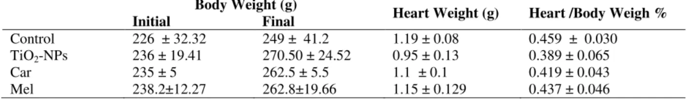
Tables 1 and 2, respectively showed that TiO2-NPs, either in low or in high repeated doses did not significantly affect either the final weight of rats or their heart weight or the heart/body weight ratio.
Table 1 - Effect of Carnosine and Melatonine on body weight, Heart weight, and Heart /body weigh % with low dose of n-TiO2.
Body Weight (g)
Initial Final Heart Weight (g) Heart /Body Weigh %
Control 226 ± 32.32 249 ± 41.2 1.19 ± 0.08 0.459 ± 0.030 TiO2-NPs 236 ± 19.41 270.50 ± 24.52 0.95 ± 0.13 0.389 ± 0.065
Car 235 ± 5 262.5 ± 5.5 1.1 ± 0.1 0.419 ± 0.043
Mel 238.2±12.27 262.8±19.66 1.15 ± 0.129 0.437 ± 0.046
Table 2 - Effect of Carnosine and Melatonine on body weight, Heart weight, and Heart /body weigh % in intoxicated rats with high dose of n-TiO2.
Body Weight (g)
Initial Final Heart Weight (g) Heart /Body Weigh %
Control 212 ± 15.58 260.5 ± 25.05 1.19 ± 0.08 0.459 ± 0.030 TiO2- NPs 226 ± 32.32 249 ± 41.20 0.95 ± 0.13 0.389 ± 0.065
Car 242.67 ± 6.66 271 ± 10.58 1.35 ± 0.06 0.493 ± 0.037
Mel 238.2±12.27 262.8±19.66 ± 0. 0. ± 0.
As regard the cardiac functions represented by myoglobin, troponinT and creatine kinase MB isozyme, both the low and high dose TiO2-NPs
melatonin showed significant reduction in their serum levels compared to TiO2-NPs intoxicated the rats (≤0.01); serum creatine kinase reduction was more prominant in the carnosine treated group
than melatonin treated group, while myoglobin level of both carnosine and melatonin of the high dose of TiO2-NPs did not show any difference than the intoxicated rats.
Table 3 - Effect of Carnosine and Melatonine treatment on cardiac functions markers Myoglobin, Troponin-T and Creatine Kinase MB (CK MB) with low dose of n-TiO2.
Group Myoglobin (pg/ml) Troponin-T (ng/ml) CK MB (ng/ml)
Control 31.16 ±2.38 27.14 ± 3.15 2.46± 0.11
TiO2- NPs 40.8±2.07***a 38.1 ± 3.07***a 3.71 ± 0.11***a
Car 36.26±1.21**ab 31.94 ± 2.09**ab 2.65± 0.25***b
Mel 35.72±2.20**ab 33.12 ± 1.90**ab 2.98± 0.08*abc
Values are expressed as mean ± SD. ***p ≤0.001, ** p ≤0.01), * p ≤0.05, a: Compared to Control group, b: Compared to n-TiO2
group, c: compared to Carnosine group.
Table 4 - Effect of Carnosine and Melatonine treatment on cardiac functions markers Myoglobin,Troponin-T and Creatine Kinase MB (CK MB) with high dose of n-TiO2.
Group Myoglobin (pg/mL) Troponin-T (ng/mL) CK MB (ng/mL)
Control 31.16±2.38 27.14± 3.15 2.64 ± 0.11
TiO2- NPs 46.68±2.79***a 41.18±2.23***a 3.81 ± 0.12***a
Car 46.1±2.07***a 36.24 ± 3**ab 3.18±0.12***ab
Mel 44.14±1.51***a 34.16± 2.25**ab 3.54 ± 0.12***abc
Values are expressed as mean ± SD. *** p ≤0.001, ** p ≤0.01, * p ≤0.05, a: Compared to Control group, b: Compared to n-TiO2 group, c:
compared to Carnosine group.
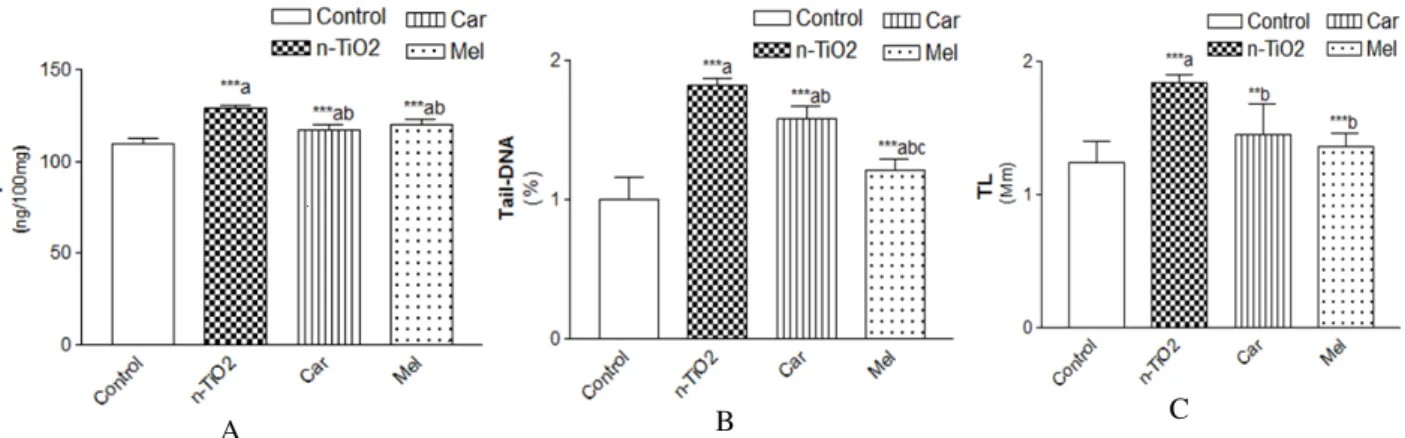
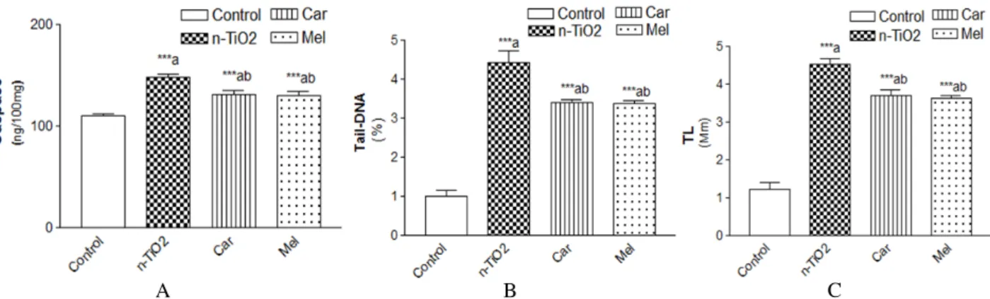
The administration of TiO2-NPs, in either doses, elevated the levels of caspase-3, TDNA and TL in comparison with the normal control (p ≤0.001) as shown in Figures 1 (A, B, C) and 2 (A, B, C). The increase in these biomarkers was pronunced in the rats taken high doses. Oral administration of carnosine or melatonin showed significantly reduced levels of caspase-3,TDNA and TL compared with the control and TiO2-NPs intoxicated counterparts (p≤0.001), with more significant decrease in TDNA of melatonin treated group. There was significant decrease in TL levels than the TiO2-NPs intoxicated group (p≤0.01,
p≤0.001, respectively).
Figure 3 (A, B) showed that TiO2-NPs, in either low or high doses, immunoglobulin G compared with normal control values p≤0.001. The increase in this biomarker was prominant in rats intoxicated with high repeated doses. Treatment with carnosine or melatonin showed significant decrease serum level than TiO2-NPs intoxicated rats p≤0.001, Melatonin treated rats with low as well as high doses of TiO2-NPs showed significantly reduced level Ig G than the carnosine treated rats p≤0.001.
A B C
Figure 1 – A) Effect of carnosine and melatonin on levels of caspase-3 in rats taken low dose n-TiO2.
B) Effect of carnosine and melatonin on Tail – DNA in rats taken low dose n-TiO2.
A B C
Figure 2 – A) Effect of carnosine and melatonin on levels of caspase-3 in rats taken high dose n-TiO2.
B) Effect of carnosine and melatonin on levels of Tail-DNA in rats taken high dose n-TiO2. C) Effect of carnosine and melatonin on levels of Tail length in rats taken
high dose n-TiO2.
A B
Figure3 – A) Effect of carnosine and melatonin on levels of IgG in rats taken low dose n-TiO2. B)
Effect of carnosine and melatonin on levels of IgG in rats taken high dose n-TiO2.
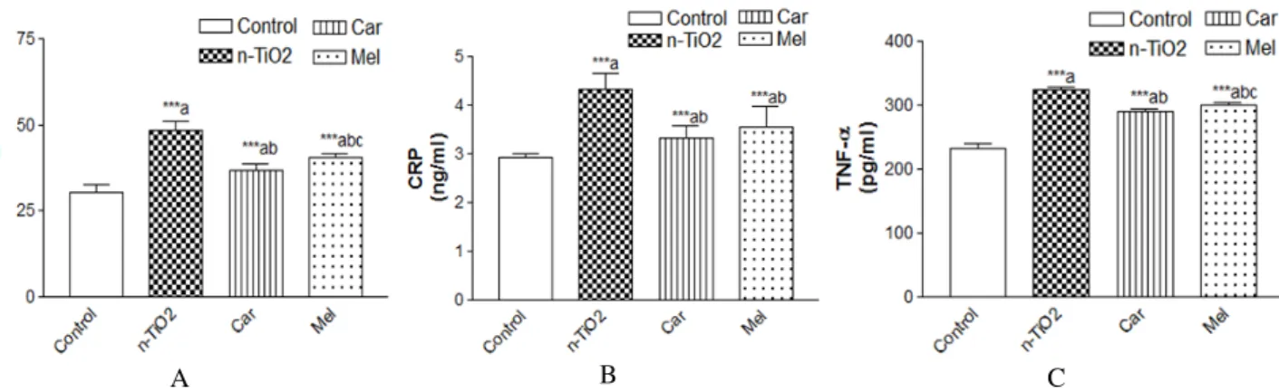
As regards TNF- α, CRP and IL-6, Figures 4 (A, B, C) and 5 (A, B, C) showed that the intake of TiO2-NPs, in either low or high repeated doses increased serum levels of these factors as compared to the control group (p≤0.001). The increase in these biomarkers was pronunced in the
rats with high repeated doses of TiO2. Oral administration of carnosine or melatonin showed significantly reduced levels of the anti-inflammatory markers compared with the control and TiO2-NPs intoxicated counterparts (p≤0.001).
A B C
Figure 4 – A)Effect of carnosine and melatonin on levels of IL-6 in rats taken low dose n-TiO2.
B) Effect of carnosine and melatonin on levels of IgG in rats taken high dose n-TiO2.
A B C
Figure 5 – A) Effect of carnosine and melatonin on levels of IL-6 in rats taken high dose n-TiO2.
B) Effect of carnosine and melatonin on levels of CRP in rats taken high dose n-TiO2.
C) Effect of carnosine and melatonin on levels of TNF-α in rats taken high dose n-TiO2.
DISCUSSION
The present work was conducted to evaluate the effect of ingestion of TiO2 nanoparticles on cardiac tissue of the rats and the possible protective role of oral carnosine as well as oral melatonin. Results revealed no significant changes on the final weight of the ratsor their heart weight or heart/body weight (%). Xu et al. (2013), injected once intravenous saline suspension of TiO2 NPs to the rats. After the rats were sacrificed, different organs were weighed and compared to control, which showed that the spleens coefficients of TiO2 NPs treated rats were significantly increased; while, the coefficients of both liver and kidney were significantly decreased. No significant effects were observed in the coefficients of the heart, lung or brain in TiO2 -NPs treated rats. Duan et al. (2010) used intragastric anatase TiO2 (5 nm) in various doses in mice for continuous 30 days, which resulted decreased body weight. Other studies reported that cultured cardiomyocytes exposed to TiO2 nanoparticles concentrations larger than 0.1 mg/mL showed decrease in their growth rate (Sayes 2006; Pan et al. 2009). In another study on cardiomyocytes cell culture, exposure to 50, 100, and 150 g/mL anatase or rutileTiO2 nanoparticles for two days led to decrease their viability; also, the shape of the cells rendered elongated and seemed to be detached from the surface of cell plate (Song et al. 2013). The present results concerning this aspect could be explained by short duration of exposure to the nanoparticles; also, the animals ingested the nanoparticles and were thus exposed to different metabolism and kinetics than a cell culture.
both OS conditions and NP TiO2. Also, a pathological changes of myocardium was observed by Sha et al. (2013). In a study using single intravenous injection of TiO2 NPs, 14 days after the treatment, no significant differences in the serum levels of TBIL, ALT, AST, ALP, BUN, CREA, or CK in TiO2 NPs treated mice as compared to the control group were found. Histopathological examination revealed that TiO2 NPs could produce different organ lesions, which was dose dependent.There were pathological changes in the brain, lung, spleen, liver and kidneys. No pathological effects were observed in cardiac tissue, which could be due to delayed onset of pathological changes in the heart (Xu et al. 2013). Another study reported that TiO2 NPs caused abnormal pathological changes in the tissues of the heart, lung, testis, ovary, and spleen (Wang et al. 2007). The present results showed elevated levels of caspase-3, TDNA and TL. Serum levels of immunoglobulin G, TNF- α, CRP and IL-6 were also elevated. Some studies have shown that TiO2 nanoparticles could increase the reactive oxygen species, which in turn depleted cellular antioxidants (Long et al. 2007; Lu et al. 2008; Park et al. 2008; Wang et al. 2008; Mallik et al. 2011). Adverse biological effects exerted by NPs is thought to be caused by the oxidative stress (Donaldson et al. 2001; Gurr et al. 2005; Nel et al. 2006) and results in DNA oxidation in the heart, DNA strand breaks, inflammation and even cell necrosis (Sheng et al. 2013). Cardiac muscle is a metabolically active organ, and rich in mitochondria. Any reduction in mitochondrial membrane potential can lead to decreased energy production, which may results cytochrome c release and induce cellular apoptosis via calcium-sensitive proteases, or through the activation of caspases and DNA fragmentation enzymes (Calcineurin et al. 2001). Several studies in different cell lines showed that TiO2 NP could cause genotoxicity represented by DNA damage and chromosomal aberrations (Rahman et al. 2002; Wang et al. 2007; Kang et al. 2008; Xu et al. 2009;
Petković et al. 2011). The exposure of peripheral human lymphocytes to TiO2 NPs can lead to the activation of DNA damage check points and accumulation of tumour suppressor protein p53 (Kang et al. 2008). In vitro and in vivo studies of different experimental models showed that nano-TiO2 could cause genotoxic effects through
indirect mechanisms like oxidative stress and inflammation (Driscoll et al. 1997; Xu et al. 2008; Trouiller et al. 2009; Petković et al. 2011). ROS are important signalling molecules that can affect cell proliferation, inflammation and cell death (Sha et al. 2013). Studies have confirmed the role of oxidative stress in TiO2-induced inflammation where ROS production lead to the activation of pro-inflammatory cascade including tumour necrosis factor TNFα (Peters et al. 2004; Xia et al. 2006; Kang et al. 2008). According to the physical and chemical properties of NPs, they can be recognized and taken up by different types of immune cells, thus trigger an inflammatory response. the expression of TNF-α and IL-6 mRNA increased following the treatment with 100 and 150 μg/mL anatase particles. These cytokines are the molecular messengers that can influence the tissue or cell response to biomaterials (Schutte et al. 2009). Immunomodulation have been also observed in in vivo studies (Skocaj et al. 2011). Larsen et al. (2010) found increased levels of immunoglobulins IgE and IgG1 in the serum and appearance of of eosinophils, neutrophils and lymphocytes in bronchoalveolar lavage fluid after intraperitoneal TiO2 NPs intake. In the present study, oral administration of carnosine or melatonin showed significant reduced levels of all the studied parameters as they were antioxidants, thus opposed the cytotoxic effects of TiO2- NPs due to their physicochemical properties can lead to the generation of reactive oxygen species, resulting their accumulation, which in turn, reduce the activities of antioxidant enzymes and antioxidant contents (Warheit et al. 2007; Sheng et al. 2013).
CONCLUSION
ACKNOWLEDGMENT
This research project was supported by a grant
from the “Research center, Female Scientific and Medical Colleges Center”, King Saud University.
REFERENCES
Abdelkarem HM; Faddah LM. The role of some natural antioxidants in ameliorating the toxic effects of nano titanium dioxide on bone complications in rats. IJCR
2013; 5 (12):4113-4117.
Athanassiou IK. Therapeutic Applications of Melatonin. Adv Endocrinol Metab. 2013;4(1):13-24. Aversa S, Pellegrino S, Barberi I, Reiter R, Gitto E.
Potential utility of melatonin as an antioxidant during pregnancy and in the perinatal period. J Matern Fetal Neonatal Med. 2012; 25: 207-221.
Boldyrev AA. Carnosine: new concept for the function of an old molecule. Biochemistry. 2012; 77: 313-326.
Boldyrev AA, Aldini G, Derave W. Physiology and Pathophysiology of carnosine. Physiol Rev. 2013; 93(4):1803-1845.
Buzea C, Pacheco II, Robbie K. Nanomaterials and nanoparticles: sources and toxicity. Biointerphases. 2007; 2:17-71.
Calcineurin MJ. mitochondrial membrane potential, and cardiomyocyte apoptosis. Circ Res. 2001;88:1220. Donaldson K, Stone V, Clouter A, Renwick L, MacNee
W. Ultrafine particles. Occup Environ Med. 2001; 58: 211-216.
Driscoll KE, Deyo LC, Carter JM, Howard BW, Hassenbein DG, Bertram TA. Effects of particle exposure and particle-elicited inflammatory cells on mutation in rat alveolar epithelial cells.
Carcinogenesis. 1997; 18: 423-430.
Duan Y, Liu J, Ma L, Li N, Liu H, Wang J, et al. Toxicological characteristics of nanoparticulate anatase titanium dioxide in mice. Biomaterials. 2010; 31:894-899.
Galano A, Tan D, Reiter R.Melatonin as a natural ally against oxidative stress: a physicochemical examination. J Pineal Res .2011; 51: 1-16.
Geiser M, Casaulta M, Kupferschmid B, Schulz H, Semmler-Behnke M, Kreyling W. The role of macrophages in the clearance of inhaled ultrafine titanium dioxide particles. Am J Respir Cell Mol Biol. 2008; 38: 371-376.
Gitto E, Aversa S, Salpietro C, Barberi I, Arrigo T, Trimarchi G, et al. Pain in neonatal intensive care: role of melatonin as an analgesic antioxidant. J Pineal Res. 2012; 52: 291-295.
Gurr JR, Wang AAS, Chen CH, Jan KY. Ultrafine titanium dioxide particles in the absence of photoactivation can induce oxidative damage to
human bronchial epithelial cells. Toxicol. 2005; 213(1-2): 66-73.
Hang L, Svanborg C, Andersson G. Determination of Interleukin 6 in Human Urine and Epithelial Cell Supernatants. Int Arch Allergy Immunol. 1994; 105: 397-403 .
Jawad H, Boccaccini AR, Ali NN, Harding SE. Assessment of cellular toxicity of TiO2 nanoparticles for cardiac tissue engineering applications.
Nanotoxicology. 2011;5:372-380.
Jackson M, Robinson G, Ali N, Kousar Y, Mei S, Gracio J, et al. Surface engineering of artificial heart valve disks using nanostructured thin films deposited by chemical vapour deposition and sol-gel methods. J Med Eng Technol. 2006; 30: 323-329.
Kang JL, Moon C, Lee HS, Lee HW, Park EM, Kim HS, et al. Comparison of the biological activity between ultrafine and fine titanium dioxide particles in RAW 264.7 cells associated with oxidative stress.
J Toxicol Environ Health. Part A. 2008; 71: 478-485. Kang SJ, Kim BM, Lee YJ, Chung HW. Titanium dioxide nanoparticles trigger p53-mediated damage response in peripheral blood lymphocytes. Environ Mol Mutagen. 2008; 49: 399-405.
Khan MJ, Maskat MY. Interaction of titanium dioxide nanoparticles with human serum albumin: A spectroscopic approach. Int J Pharm Pharm Sci.
2014; 6(3): 43-46.
Larsen ST, Roursgaard M, Jensen KA, Nielsen GD. Nano titanium dioxide particles promote allergic sensitization and lung inflammation in mice. Basic Clin Pharmacol Toxicol. 2010; 106: 114-117.
Liu H, Ma L, Zhao J, Liu J, Yan J, Ruan J, Fashui Hong. Biochemical toxicity of nano-anatase TiO2 particles in mice. Biol Trace Elem Res. 2009; 129 (1-3): 170-180.
Liu K, Lin X, Zhao j. Toxic effects of the interaction of titanium dioxide nanoparticles with chemicals or physical factors. Intern J Nanomedicine. 2013; 8: 2509-2520.
Long TC, Tajuba J, Sama P, Saleh N, Swartz C, Parker J, et al. Nanosize titanium dioxide stimulates reactive oxygen species in brain microglia and damages neurons in vitro. Environ Health Persp. 2007; 115: 1631-1637.
Lu N, Zhu Z, Zhao X, Tao R, Yang X, Gao Z. Nano titanium dioxide photocatalytic protein tyrosine nitration: a potential hazard of TiO2 on skin. Biochem Biophys Res Commun. 2008; 370: 675-680.
Magaye R, Zhao J. Recent progress in studies of metallic nickel and nickel-based nanoparticles’ genotoxicity and carcinogenicity. Environ Toxicol Pharmacol. 2012; 34: 644-650.
Mallik A, Bryan S,Puukila S, Chen A, Khaper N. Efficacy of Pt-modified TiO2 nanoparticles in cardiac
Nel A, Xia T, Madler L, Li N. Toxic potential of materials at the nanolevel. Science. 2006; 311: 622-7. OECD. OECD Guidelines for Testing of Chemicals. No
420: Acute Oral Toxicity-fixed Dose Method. Organisation for Economic Co-operation and Development, Paris 1992.
Olmedo D, Guglielmotti MB, CabriniRL. An experimental study of the dissemination of titanium and zirconium in the body. J Mater Sci Mater Med.
2002; 13(8): 793-796.
Pan Z, Lee W, Slutsky L, Clark RAF, Pernodet N, Rafailovich MH. Adverse effects of titanium dioxide nanoparticles on human dermal fibroblasts and how to protect cells. Small. 2009; 5(4) : 511-520.
Park EJ, Yi J, Chung KH, Ryu DY, Choi J, Park K et al. Oxidative stress and apoptosis induced by titanium dioxide nanoparticles in cultured BEAS-2B cells.
Toxicol Lett. 2008; 180: 222-229.
Paunesku T, Rajh T, Wiederrecht G, Maser J, Vogt S,
Stojićević N, et al. Biology of TiO2-oligonucleotide
nanocomposites. Nat Mater. 2003; 2: 343-346. Peters K, Unger RE, Kirkpatrick CJ, Gatti AM, Monari
E. Effects of nano-scaled particles on endothelial cell function in vitro: studies on viability, proliferation and inflammation. J Mater Sci Mater Med.
2004;15(4):321–325.
Petković J, Žegura B, Stevanović M, Drnovšek N, Uskoković D, Novak S, et al. DNA damage and alterations in expression of DNA damage responsive genes induced by TiO2 nanoparticles in human hepatoma HepG2 cells. Nanotoxicology. 2011;5:341– 353.
Rahman Q, Lohani M, Dopp E, Pemsel H, Jonas L, Weiss DG, et al. Evidence that ultrafine titanium dioxide induces micronuclei and apoptosis in Syrian hamster embryo fibroblasts. Environ Health Persp. 2002;110:797–800.
Robertson TA, Sanchez WY, Roberts MS. Are commercially available nanoparticles safe when applied to the skin? J Biomed Nanotechnol. 2010; 6:452-468.
Saber AT, Jensen KA, Jacobsen NR, Birkedal R, Mikkelsen L, Moller P, et al. Inflammatory and genotoxic effects of nanoparticles designed for inclusion in paints and lacquers. Nanotoxicology. 2012; 6:453-471.
Sager TM, Kommineni C, Castranova V. Pulmonary response to intratracheal instillation of ultrafine versus fine titanium dioxide: role of particle surface area. Part Fibre Toxicol. 2008; 5(17).
Sayes CM, Wahi R, Kurian PA , Lui I, West JL,
Ausman KD,et al. “Correlating nanoscale titania structure with toxicity: a cytotoxicity and inflammatory response study with human dermal
fibroblasts and human lung epithelial cells,” Toxicol Sci. 2006; 92(1): 174-185.
Schutte RJ, Xie L, Klitzman B, Reichert WM. “In vivo
cytokine-associated responses to biomaterials”.
Biomaterials. 2009; 30 (2): 160-168.
Sha BY, Gao W, Wang SQ, Li W, Liang X, XuF, et al. Nano-titanium dioxide induced cardiac injury in rat under oxidative stress. Food Chem Toxicol. 2013; 58: 280-288.
Sheng L1, Wang X, Sang X, Ze Y, Zhao X, Liu D, Gui S, Sun Q, Cheng J, Cheng Z, Hu R, Wang L, Hong F.
“Cardiac oxidative damage in mice following
exposure to nanoparticulate titanium dioxide”. J Biomed Mater Res. 1(11): 3238-3246.
Singh NP, McCoy MT, Tice RR, Schneider EL. A simple technique foe quantitation of low levels of DNA damage in individaul cells. Exp Cell Res. 1988; 175: 184-191.
Skocaj M, Filipic M, Petkovic J, Novak S.Titanium dioxide in our everyday life; is it safe? Radiol Oncol. 2011; 45(4): 227-247.
Sohaebuddin SK, Thevenot PT, Baker D, Eaton JW, Tang L. Nanomaterial cytotoxicity is composition, size, and cell type dependent. Part Fibre Toxicol.
2010; 7: 22.
Song W, Wang J, Liu M, Li P, Zhou G, Li Z, Fan Y. Titanium Dioxide Nanoparticles Induced Proinflammation of Primary Cultured Cardiac Myocytes of Rat. J Nanomater. 2013; 2013: 1-9. Tay CY, Fang W, Setyawati MI, Chia SL,Tan KS,
Hong CHL et al. hydroxyapatite and Nano-titanium Dioxide Exhibit Different Subcellular Distribution and Apoptotic Profile in Human Oral Epithelium. ACS Appl Mater Interfaces. 2014; 6 (9): 6248-6256.
Thornberry NA and Lazebnik Y. Caspases: enemies within. Science. 1998; 281:1312-1316.
Trouiller B, Reliene R, Westbrook A, Solaimani P, Schiestl RH. Titanium dioxide nanoparticles induce DNA damage and genetic instability in vivo in mice.
Cancer Res. 2009; 69: 8784-8789.
Varghese O, Paulose M, LaTempa T, Grimes C. High-rate solar photocatalytic conversion of CO2 and water
vapor to hydrocarbon fuels. Nano Lett. 2009; 9: 731-737.
Wang J, Zhou G, Chen C, Yu H, Wang T, Ma Y, et al. Acute toxicity and biodistribution of different sized titanium dioxide particles in mice after oral administration. Toxicol Lett. 2007; 168( 2): 176-185. Wang JJ, Sanderson BJS, Wang H. Cyto- and
genotoxicity of ultrafine TiO2 particles in cultured
human lymphoblastoid cells. Mutat Res-Gen Tox En.
2007; 628: 99-106.
Warheit DB, Webb TR, Reed KL, Frerichs S, Sayes CM. Pulmonary toxicity study in rats with three forms of ultrafine-TiO2 particles: Differential responses related to surface properties. Toxicol. 2007; 230: 90-104.
Wu G, Nishikawa T, Ohtani B, Chen A. Synthesis and characterization of carbon-doped TiO2 nanostructures
with enhanced visible light response. Chem Mater. 2007; 19: 4530-4537.
Wu J, Liu W, Xue C, Zhou S, Lan F, Bi L, et al. Toxicity and penetration of TiO2 nanoparticles in
hairless mice and porcine skin after subchronic dermal exposure. Toxicol Lett. 2009; 191(1): 1-8. Zhao J, Bowman L, Zhang X, Vallyathan V, Young
SH,Castranova V et al Titanium dioxide (TiO2)
nanoparticles induce JB6 cell apoptosis through activation of the caspase-8/Bid and mitochondrial pathways. J Toxicol Environ Health A. 2009; 72: 1141-1149.
Zhao J, Castranova V. Toxicology6of nanomaterials used in nanomedicine. Toxicol Environ Health BCrit Rev. 2011; 14: 593-632.
Zhu Y, Eaton JW, Li C. Titanium dioxide (TiO2) nanoparticles preferentially induce cell death in transformed cells in a Bak/Bax-independent fashion. PLoS ONE .2012;7: e5060.
Xia T, Kovochich M, Brant J, Hotze M, Sempf J, Oberley T, et al. Comparison of the abilities of ambient and manufactured nanoparticles to induce cellular toxicity according to an oxidative stress paradigm. Nano Lett. 2006; 6(8): 1794-1807.
Xu A, Chai YF, Nohmi T, Hei TK. Genotoxic responses to titanium dioxide nanoparticles and fullerene in gpt delta transgenic MEF cells. Part Fibre Toxicol. 2009; 6:3.
Xu J, Shi H, Ruth M, Yu H, Lazar Land Yang C. Acute Toxicity of Intravenously Administered Titanium Dioxide Nanoparticles in Mice. PLoS ONE.2013;8(8): e70618.