Vol.59: e16160132, January-December 2016 http://dx.doi.org/10.1590/1678-4324-2016160132
ISSN 1678-4324 Online Edition
BRAZILIAN ARCHIVES OF BIOLOGY AND TECHNOLOGY
A N I N T E R N A T I O N A L J O U R N A L
The Effect of PM
10on Ischemia- Reperfusion Induced
Arrhythmias in Rats
Esmat Radmanesh
1,Mahin Dianat
1*, Mohammad Badavi
1, Gholamreza Goudarzi
2,
Seyyed Ali Mard
1.
1
Physiology Research Center, Department of Physiology, Faculty of Medicine, Ahvaz Jundishapur University of Medical Sciences, Ahvaz, Iran. 2 Department of Environmental Health Engineering, Health Faculty, Ahvaz Jundishapur University of Medical Sciences, Ahvaz, Iran.
ABSTRACT
Epidemiological studies show that particulate matter (PM) is the principal instigator of some adverse clinical symptoms involving cardiovascular diseases. PM exposure can increase experimental infarct size and potentiate myocardial ischemia and arrhythmias in experimental MI models such as ischemia-reperfusion (I/R) injury.The present study was aimed to evaluate the effects of particulate matter (PM10) on ischemia- reperfusion induced arrhythmias with emphasis on the protective role of VA as an antioxidant on them. Male Wistar rats were divided into 8 groups (n=10): Control, VAc, Sham, VA, PM1 (0.5 mg/kg), PM2 (2.5 mg/kg), PM3 group (5 mg/kg), PM3 + VA group. Within 48 hours, PM10 was instilled into trachea in two stages. Then the hearts were isolated, transferred to a Langendorff apparatus, and subjected to global ischemia (30 minutes) followed by reperfusion (60 minutes). The ischemia- reperfusion induced ventricular arrhythmias were assessed according to the Lambeth conventions.In the present study,the number, incidence and duration of arrhythmiasduring30 minutes ischemia were demonstrated to be more than those in the reperfusion stage. PM exposure increased significantly the number, incidence and duration of arrhythmias in the ischemia and reperfusion duration. Vanillic acid reduced significantly the number, incidence and duration of arrhythmias during the ischemia and reperfusion period.In summary, the results of this study demonstrated that the protective and dysrhythmic effects of VA in the PM exposure rats in I/R model are probably related to its antioxidant properties.
Key words: Particullate Matter, Ischemia- reperfusion, arrhythmias,Vanillic Acid, Rat
*
Authors for correspondence: [email protected]
ABBREVIATIONS
PM: particulate matter
PM10: particles with aerodynamic diameter<10 μm VA: vanillic acid
MI: myocardial infarction I/R: ischemia-reperfusion ROS: reactive oxygen species VPB: ventricular premature beats VT: ventricular tachycardia VF: ventricular fibrillation
PVC: premature ventricular contraction CAT: catalase
SOD: superoxid dismutase GPx: glutathion peroxidase CVD: cardiovascular disease MDA: malondialdehyde ECG: electrocardiogram
INTRODUCTION
Particulate matter )PM( is a complex, dynamic mix of liquid and solid particles suspended in the air. Some of the more common PM components include: 1) organic compounds such as aldehydes and polyaromatic hydrocarbons; 2) elemental and organic carbon, nitrates, and sulfates; 3) metals and metal oxides; 4) biological compounds including bacterial products and pollen grains; and 5) Gaseous components (Sumanth, 2006).
Epidemiological studies indicate that particulate matter (PM) is the principal instigator of some
adverse clinical symptoms involving
cardiovascular diseases (Brook et al 2010). One mechanism underlying PM-related CVD is oxidative stress. This occurs when the homeostatic balance of oxidizing agents to antioxidants is upset towards an imbalance of the former. Vanillic acid is oxidized form of vanillin produced during the conversion of vanillin to ferulic acid (Lesage-Meessen et al 1996). Systematical evaluation of the antioxidative properties of vanillic acid (VA) and vanillin by multiple assays has provided the evidence that supports the superiority of antioxidative and radical-scavenging activity of vanillic acid (Tai et al 2012). An American Heart Association’s scientific statement on PM has proposed a role for oxidative stress in altering cardiac function (Brook et al 2010). Antioxidant nutrients and their bioactive compounds common in fruits and vegetables can protect against
environmental toxic agents. Antioxidants, as dietary supplements, can provide protection against reactive species (RS)-induced damage under conditions of oxidative stress elevation to the organism (Poljsak et al 2013).
In our previous study, the effectiveness of vanillic acid on lipid peroxidation, indicated by a reduction inmalondialdehyde (MDA), and the enhancement of endogenous antioxidant enzymes, indicated by
increased glutathione peroxidase (GPx),
superoxide dismutase (SOD), catalase (CAT), and total antioxidant capacity (TAC) in the rat hearts exposed to I/R were demonstrated (Dianat et al 2014b).Some studies identify a significant association between severe cardiac diseasessuch as arrhythmias (Wichmann et al 1989), as well as
other cardiographic abnormalities and air
pollution(Wellenius et al 2002). PM exposure can increase experimental infarct size and potentiate
myocardial ischemia and arrhythmias in
experimental myocardial infarction (MI) models
such as ischemia-reperfusion (I/R)
injury.Nevertheless, it has been suggested that PM exposure may potentially be capable to increase the myocardium sensitivity to ischemia, probably by impairing myocardial blood flow and perfusion (Brook et al 2010).
Heavy metals such as cadmium, chromium, lead, arsenic, anions and cations which are forwarding by particles can cause cardiovascular effects. Heavy metals associated with PM10 play a significant role in air pollution. One study on dusty days in Ahvaz, the capital of Khuzestan province in the southwest of Iran, demonstrated correlations between the heavy metals (Ni,Pb, Cd, Crand Coexcept Zn) and PM10(Shahsavani et al 2012a).Ahvazhas been experiencing desert dust events with 29, 33, 55, 45, and 17 dust storms in 2005, 2006, 2007, 2008, and 2009, respectively (Goudarzi et al 2014).
The present study was aimed to evaluate the effects of particulate matter (PM10) onischemia- reperfusion induced arrhythmiaswith emphasis on the protective role of VA as an antioxidant on them.
MATERIALS AND METHODS
Chemicals
(10%) and Xylazine (2%) were obtained from Alfasan Co. (Netherlands). Krebs salts were purchased from Merck Co. (Germany).
Animals and treatments
Eighty adult male Wistar rats (body weight, 250-300 g) were randomly divided into 8 experimental groups (n=10) as follows: Control (1 ml normal saline, gavage, 10 days), VAc (10 mg/kg of VA, gavage, 10 days) (Dianat et al 2014b), Sham (0.1 ml normal saline, intratrachealinstillation ), VA (10 mg/kg vanillic acid, gavage, 10 days +0.1 ml normal saline, intratracheal Instillation ), PM1 (0.5 mg/kg PM10, intratracheal instillation), PM2 (2.5 mg/kg PM10, intratracheal instillation), PM3 (5 mg/kg PM10, intratracheal instillation), PM3 + VA (5 mg/kg PM10, intratracheal instillation + vanillic acid 10 mg/kg, gavage, 10 days). Given that PM 5mg/kg showed stronger effects on parameters than other dosage, this dose was selected as the effective dose in this experiment. The groups were maintained under the same conditions, relative humidity of 60±5%,temperature 22 ± 2°C and 12 hour dark-light cycle, supplied with food and water ad libitum. Vanillic acid was separately suspended in normal saline and administered to the rats via a gavage needle for ten days. The control group received normal saline orally for the same duration. The animals were maintained in the animal house of Ahvaz Jundishapur University of Medical Sciences, Ahvaz, Iran, and treated in accordance with the guidelines of the Animal Care. The protocol was approved by the Ethical Committee of Laboratory Animals of Ahvaz Jundishapur University of Medical Sciences (No. ajumsAPRC-9316).
Area of study and Sampling procedure
Ahvaz, the well known city in the world regardingits particulate matter being equal to or less than 10 micrometer (Goudie 2014), is located in southwestern of Iran in close vicinity of Iraq, Kuwait and Saudi Arabia. It is located in 31º 20 N, 48º 40 E geographically and has 18 meters elevation above sea level. Ahvaz has been suffering from dust storm during the last decade. Many industries such as National Drilling Company, Ahvaz Steel Company, Carbon Black, Gas, Oil and petroleum refineries gathered together in this city so that ambient air is polluted due to their industrial activities (Heidari-Farsani et al 2014).
On13thof July, 2014, a dust event day, the sample was taken up from Ahvaz by high volume PM10 sampler (Tisch Environmental, INC.145 south Miami AVE). Dust event days as defined based on visibility, wind speed and PM10 concentration by Hoffmann et al. (Hoffmann et al 2008) was categorized as DS2.
The high volume PM10 sampler was equipped with a quartz filter and placed on the roof of the Health Faculty of Ahvaz Jundishapur University of Medical Sciences at the approximate level of 10 m above ground in order to mitigate any barrier on the air flow.The device operated with a flow rate of 1.2-1.8 m3/min for 16h. Filter preparation was conducted based on the procedure presented by Shahsavani et al. (Shahsavani et al 2012b) and Zhang et al. (Zhang et al 2010).
Sample preparation for elemental analysis
One-fourth of the fiber glass filter was cut and sited in a Teflon container.A mixture of Nitric acid, Hydrofluoric acid and Hydrochloric acid was added to it.The filter was digested in a hot oven at 170 degrees Celsius for 4 h, and then the cap of the Teflon container was opened to evaporate all the remaining acids inside it. After cooling, distilled water and concentrated Nitric acid (ratio9: 1V %) were added and shaken for 15 min. The solution was filtered through a Whatman-42 filter paper, diluted to distilled water (25 ml) and then stored in a sterile plastic bottle at 4° C for additional analyses (Shahsavani et al 2012b; Mohd et al 2009).The samples were analyzed to assay target heavy metals by inductively coupled plasma atomic emission spectroscopy (ICP-AES; model: ARCOUS, Germany) (Heidari-Farsani et al 2014).
PM intratracheal instillation
intratracheal instillation.Within 48 hours, PM10 was instilled into trachea in two stages.Forty-eight hours later, the rats were anesthetized with intraperitoneal injection of ketamine-xylazine. 1000 units of Heparin sodiumwas injected intraperitoneal into the rats to avoid blood coagulation and thirty minutes after 0.1 ml normal
saline intratracheal instillation andcertain
concentration of PM, the hearts were isolated.
Preparation of isolated heart
Trachea was cannulated and ventilated with room air by a rodent ventilator. The thoracic cage was opened and a steel cannula was inserted into aorta and tightened with a suture. The heartswere quickly excised and mounted to a Langendorff perfusion apparatus. The heartswere perfusedin retrograde via the aorta at 37 ± 0.1°C and a constant flow rate 10 ml/min .The perfusion Krebs Henseleit buffer consisted of KH2PO4 (1.18 mM), KCl (4.75mM), NaCl (118 mM), CaCl2 (1.75 mM), MgSO4 (1.2 mM), glucose (11.1 mM) and NaHCO3 (25 mM), in double distilled water equilibrated by 95% O2 and 5% CO2 at pH of 7.4. For each experiment, fresh perfusion buffer was filtered through a 1.2-µm microfiber filter (GF/Cglass filters; Whatman). The hearts were perfused for 30 minutes before the induction of ischemia to allow stabilization of coronary perfusion pressure, and then subjected to no flow global ischemia (30 minutes) followed by reperfusion (60 minutes) (Dianat et al 2014a). The left ventricular pressure was measured with a ventricular latex balloon inflated to a diastolic pressure of 5–10 mmHg, connected to a transducer (Gracia-Villalon 2009). The successful induction of ischemia was determined by ST elevation on the electrocardiogram (Dianat et al 2014a).
Evaluation of ischemic- reperfusion induced ventricular arrhythmias
Lead II electrocardiogram (ECG) was recorded by Bio Amp and monitored by a Power Lab system
(ADInstruments, Australia) (Dianat et al
2014c).Ischemia-reperfusion induced ventricular arrhythmias were assessed according to the Lambeth conventions(Curtis et al 2013).Certain types of simple ventricular arrhythmia represent specific elaborations of the ventricular premature beats (VPB). Bigeminy has the minimum sequence VPB. Salvo is defined as a run of 2-3 consecutive VPBs. Sequence of a minimum of 4 consecutive ventricular complexes is defined as Ventricular
tachycardia (VT). Ventricular fibrillation (VF) is defined as a sequence of a minimum of 4
consecutive ventricular complexes without
intervening diastolic pauses, in which the intrinsic shape, the peak–peak interval and the height vary, and the variation between each is non-progressive
(Curtis et al 2013). The criteria for
subcategorization are: regularity of configuration (morphology); the rate (RR interval) and regularity of rate for VT, the duration of the arrhythmia, and the mode of termination for VT and VF. The incidences of the main categories of arrhythmia (VPBs, bigeminy, salvos, VT, and VF) must be analyzed separately. Whether the incidences of subcategories of VTand VF are analyzed is a matter of personal preference (Walker et al 1988).ECG scoring system criteria was in accordance with following formula: Score: (log10 PVCs) + (log10 episodes VT) + 2 [(log10 episodes of VF) + (log10 total duration of VF)] (Miller et al 2012).
Statistical analyses
Results were analyzed using GraphPad Prism6 and expressed as mean ± SD. Two-way ANOVA and one-way ANOVA for multiple comparison tests were used, followed by LSD. p<0.05 was
considered statistically significant.Any
arrhythmias (yes/no categories) were expressed as the total in each group and were analyzed using Fisher’s exact test.
RESULTS
PM10 contents
The present type of dust storm withPM10 500-2000 (μg m-3
h-1), Visibility<1000 m and Wind speed>17 ms-1, according to Hoffmann’s classification, is categorized as DS2 (Hoffmann et al., 2008). PM10 level and its heavy metal content of PM10 include: PM10 575 µg/m3,Ni 2.07 µg/m3, Zn 6.2 µg/m3 ,Al 2.7,Pb 4.75 µg/m3 , Cr 1.7 µg/m3 , Co 1.37 µg/m3 and cd 0.7 µg/m3.
Trajectory analysis
mostly originated from Saudi Arabia and internal
sources such as, Hendijon, Abadan and
Mahshahr(Soleimani et al., 2015).
I/R-induced arrhythmias The number of arrhythmias
The number of arrhythmiasoccurring during 30-minute ischemia was more than that in the reperfusion stage. PM10 increased the number of arrhythmias in the ischemia and reperfusion duration anda significant reduction effect was observed invanillic acid groups (Fig.1).
A:
VP B Big em iny Salvo VT VF
0 5 0 1 0 0 1 5 0 2 0 0 2 5 0
C o n t r o l V A c
S h a m
V A
P M 1 P M 2
P M 3 P M 3 + V A
N u m b e r o f A r r h y th m ia s $ ** * ** *** ** * ** * $ ** * ** * ** * ** * ** $ *** *** ** * * $ ** *** ** * ** * ** $ ***
B:
VP B Big e m inySa l
v o VT VF
0 2 0 4 0 6 0 8 0
1 0 0 C o n t r o l
V A c
S h a m
V A
P M 1
P M 2 P M 3
P M 3 + V A
N u m b e r o f a rr h y th m ia s $ ** * ** * * ** * $ ** * ** * ** * ** * $ ** * ** * ** * *** $ ** * ** * * ** *
Figure1-Effect of PM10 and vanillic acid on the number of Arrhythmias during 30 min ischemia (A) and the first30
VA, gavage, 10 days). Two- way ANOVA was used followed by the LSD test. *p<0.05, **p<0.01, ***p<0.001 vs. Sham group. $p <0.001 vs. Control group.
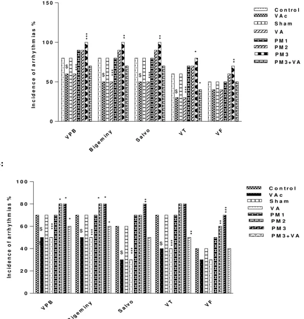
The incidences of arrhythmias
The results of PM10 and vanillic acid on the incidences of I/R induced arrhythmias are shown in Figure 2.
During 30-minute ischemia, the incidences of arrhythmias increased in PM3 group, and vanillic
acid showed a significant reducing effecton them (Fig. 2A).
During the first 30 minutes of reperfusion, PM2 and PM3 demonstrated asignificant increased effecton VPB, bigeminary and VF. Salvo
increased significantly inPM3 group. The
incidence of arrhythmia wasreduced by vanillic acid, significantly (Fig. 2B).
A:
VP B
Big em
iny
Sa
lvo VT VF
0 5 0 1 0 0 1 5 0
C o n t r o l V A c S h a m V A
P M 1 P M 2 P M 3 P M 3 + V A
In
c
id
e
n
c
e
o
f
a
r
r
h
y
th
m
ia
s
%
$ ***
**
*
$ ***
**
$ ***
**
$ *** *
*
**
B:
VP B
Big e m
iny
Sa l
v o VT VF
0 2 0 4 0 6 0 8 0 1 0 0
C o n t r o l V A c S h a m V A P M 1
P M 2 P M 3 P M 3 + V A
In
c
id
e
n
c
e
o
f
a
rr
h
y
th
m
ia
s
%
$ $ **
*
*
* *
$
**
*
*
* *
$ ***
**
$ ***
**
**
**
*
Figure 2-Effect of PM10and vanillic acid on the incidence of Arrhythmias (%) during 30 min ischemia (A) and the
intratrachealinstillation), VA (VA, 10 mg/kg for 10 days, gavage +0.1 ml normal saline, intratracheal instillation), PM1 (0.5 mg/kg PM), PM2 (2.5 mg/kg PM), PM3 group (5 mg/kg PM), PM3 + VA group (5 mg/kg PM + 10 mg/kg
VA, gavage, 10 days). Fisher’s exact test and Two-way ANOVA was used followed by the LSD test.*p<0.05, **p<0.01, ***p<0.001 vs. Sham group. $p <0.001 vs. Control group.
The score of arrhythmias
During the 30-minute ischemia,the score of arrhythmia increased in PM groups but reduced in vanillic acid receiving groups significantly (Fig. 3).
During the first 30 minutes of reperfusion, the score of arrhythmias increased in PM2 and PM3 groups but decreased significantly in vanillic acid receiving groups (Fig. 3).
0 1 2 3 4 5
C o n t r o l V A c S h a m V A P M 1 P M 2 P M 3 P M 3 + V A
I s c h e m i a R e p e r f u s i o n
$
$ ***
** *** ***
*** ***
***
S
c
o
r
e
o
f
a
r
r
h
y
th
m
ia
s
Fig3-Effect of PM10andvanillic acid on the score of arrhythmias during 30 min ischemia and the first30 min of
reperfusion. Results are expressed as mean ± SD of 10 samples per group. Control (normal saline, 1 ml for 10 days, gavage), VAc (VA, 10 mg/kg for 10 days, gavage), Sham (normal saline, 0.1 ml, intratrachealinstillation), VA (VA, 10 mg/kg for 10 days, gavage +0.1 ml normal saline, intratracheal instillation), PM1 (0.5 mg/kg PM), PM2 (2.5 mg/kg PM) , PM3 group (5 mg/kg PM), PM3 + VA group (5 mg/kg PM + 10 mg/kg VA, gavage, 10 days). Two-way ANOVA was used followed by the LSD test. **p<0.01, ***p<0.001 vs. Sham group. $p<0.001 vs. Control group.
The duration of the arrhythmias
Duration of the VT and VF in the ischemia period was more than that in the first 30 minutes of reperfusion.
During 30 minutes of ischemia, the duration of the VT in PM3 group and that of the VF in PM1, PM2
VT VF VT VF
0 1 0 2 0 3 0 4 0
C o n t r o l V A c S h a m V A P M 1 P M 2 P M 3 P M 3 + V A
I s c h e m i a R e p e r f u s i o n
D
u
ra
ti
o
n
(
s
)
$ **
*
**
** $ **
*
**
*
**
*
**
*
*
$ ***
**
**
*
*
$ **
*
**
*
**
*
**
Fig 4-Effect of PM10and vanillic acid on the duration of VT and VF during 30 min ischemia and the first 30 min of
reperfusion.Results are expressed as mean ± SD of 10 samples per group. Control (normal saline, 1 ml for 10 days, gavage), VAc (VA, 10 mg/kg for 10 days, gavage), Sham (normal saline, 0.1 ml, intratrachealinstillation), VA (VA, 10 mg/kg for 10 days, gavage +0.1 ml normal saline, intratracheal instillation), PM1 (0.5 mg/kg PM), PM2 (2.5 mg/kg PM), PM3 group (5 mg/kg PM), PM3 + VA group (5 mg/kg PM + 10 mg/kg VA, gavage, 10 days). Two - way ANOVA was used followed by the LSD test.*p<0.05, **p<0.01, ***p<0.001 vs. Sham group. $p <0.001 vs. Control group.
DISCUSSION
In the present study,the number, incidence and duration of arrhythmias in 30-minute ischemia were demonstrated to be more than those in the reperfusion stage. PM exposure increasedthe number, incidence and duration of arrhythmias in ischemia and reperfusion significantly.
Liao et al. (2009) reported that ambient PM2.5 exposures 1 daybefore the electrocardiography (ECG) measurement were associated with
augmented odds of premature ventricular
contraction (PVC) inwomen(Liao et al 2009). The association between exposure to PM2.5 and increasing PVC frequency was shown in He et al.’s study which indicated an approximate 8% increase in the number of PVCs per 30 minutes for each 10-μg/m3increase in PM2.5 concentration during the same time period (He et al 2011). According to Hoffmann’s classification,the type of dust storm collected from Ahvaz and used in the present study is categorized as DS2 (Hoffmann et al 2008).
2005; Gonick et al 1997). The results of our previous researchshowed that the antioxidant enzymes have significantly decreased in the groups receiving PM, while the activity of antioxidant enzymes in the group receiving vanillic acid increased. Therefore, antioxidant substances by increasing the activity of antioxidant enzymes are able to improve the harmful effects of PM (Dianat et al 2016).
Oxidative stress has been shown to mediate PM-related effects on ECG and cardiovascular function (Brook et al., 2010). Oxidative stress is a consequence of an increased generation of ROS
and/or reduced physiological activity of
antioxidant defense against ROS. Environmental pollutants stimulate a diversity of mechanisms of toxicity on molecular level and oxidative stress leading to the damage to cellular membrane lipids, proteins and DNA (Valavanidis et al 2006). When the antioxidant defense in the human body becomes weak, the inducing inflammatory processes, adaptive, injurious, and reparative processes can often occur by oxidative stress (Cross et al 2002).
There is some evidence that low molecular weight antioxidants are involved in decreasing the
damage caused by certain environmental
pollutants. The current evidence suggests that increased consumption of vegetables and fruits or certain supplements can enhance the protection against many common types of environmentally induced oxidative/ nitrosative stress (O/NS) (Poljsak et al 2013).In the present study, VA reduced the number, incidence and duration of arrhythmia during ischemia and reperfusion, significantly. Positive inotropic properties of VA alone and in combination with exercise were demonstrated in our previous study and also dysrhythmia improvement in young and aged rats was shown in VA group (Dianat et al 2014c ). A decrease in lipid peroxidation by reduction of MDA indicates the efficiency of VA, and improves endogenous antioxidant enzymes such as increased GPx, SOD, and CAT demonstrated in the isolated rat hearts (Dianat et al 2014b).The present study showed that vanillic acid reduced the number, incidence and duration of arrhythmias in ischemia and reperfusion significantly.
CONCLUSION
In summary, the results of this study demonstrated the protective and dysrhythmic effects of VA in
the PM exposure rats in I/R model,which are probably related to its antioxidant properties and also its protective role against lipid peroxidation.
ACKNOWLEDGMENT
The source of data used in this paper was from Ph.D. thesis of Mrs Esmat Radmanesh, a student of Ahvaz Jundishapur University of Medical
Sciences, Ahvaz, Iran. Authors gratefully
acknowledge the help and financial support of Physiology Research Center of Ahvaz Jundishapur University of Medical Sciences (No. ajums APRC-9316).
CONFLICT
OF
INTEREST
DISCLOSURE
The authors declare no conflicts of interest.
REFERENCES
Brook RD, Rajagopalan S, Pope CA3rd, Brook JR, Bhatnagar A, Diez-Roux AV, Holguin F, Hong Y, Luepker RV, Mittleman MA, Peters A, Siscovick D, Smith SC, Whitsel L, Kaufman JD. Particulate matter air pollution and cardiovascular disease: an update to the scientific statement from the American Heart Association. Circulation. 2010; 121: 2331– 2378. doi: 10.1161/CIR.0b013e3181dbece1.
Cross CE, Valacchi G, Schock B, Wilson M, Weber S, Eiserich J,van der VlietA. Environmental oxidant pollutant effects on biologic systems: a focus on micronutrien antioxidant-oxidant interactions. American Journal o Respiratory and Critical Care Medicine. 2002; 166(1): S44– S50.doi:10.1164/rccm.2206015.
Curtis MJ, Hancox JC, Farkas A, Wainwright CL, Stables CL, Saint DA, Clements-Jewery H, Lambiase PD, Billman GE, Janse MJ,Pugsley MK, Ng GA, Roden DM, Camm AJ, Walker MJ. The Lambeth Conventions (II): guidelines for the study of animal and human ventricular and supraventricular arrhythmias. PharmacolTher. 2013; 139: 213–248, doi: 101016/jpharmthera 201304008. Dianat M, Esmaeilizadeh M, Badavi M, Samarbafzadeh A , Naghizadeh B. Protective Effects of Crocin on Hemodynamic Parameters and Infarct Size in Comparison with Vitamin E after Ischemia Reperfusion in Isolated Rat Hearts. Planta Med. 2014a ; 80: 393–98. doi: 10.1055/s-0033-1360383. PMID: 24585091.
Oxidative Stress in Isolated Rat Heart. Iran Red Crescent Med J. 2014b; 16(7): e16664. doi: 10.5812/ircmj.16664.
Dianat M, Radan M, Badavi M, Sarkaki A. The Evaluation of Inotropic Properties and Antidysrhythmic Effect of Vanillic Acid and Exercise on Cacl2 -Induced Arrhythmia in Young and Aged Rats, RJPBCS. 2014c ;5(3):1545-1555. ISSN: 0975-8585.
Dianat M, Radmanesh E , Badavi M , Mard SA , Goudarzi G. Disturbance effects of PM10 on iNOS and eNOS mRNA expression levels and antioxidant activity induced by ischemia–reperfusion injury in isolated rat heart: protective role of vanillic acid. Environ Sci Pollut Re. 2016; 23:5154–5165. doi 10.1007/s11356-015-5759-x.
Garcia-Villalon AL, Monge L, Fernandez N, Salcedo A. Coronary response to diadenosinepentaphosphate after ischaemia–reperfusion in the isolated rat heart. Cardiovasc Res. 2009; 81:336–43. doi:10.1093/cvr/cvn321. PMID: 19029135.
Golomb E, Matza D, Cummings CA, Schwalb H, Kodavanti UP, Schneider A, Houminer E, Korach A, Nyska A, O.M. Shapira OM. Myocardial mitochondrial injury induced by pulmonary exposure to particulate matter in rats. ToxicolPatho. 2012; 40: 779-88. doi: 10.1177/0192623312441409. PMID: 22549975.
Gonick H C, Ding Y, Bondy S C, Ni Z, Vaziri
ND.―Lead-induced hypertension: interplay of nitric
oxide and reactive oxygen species,‖ Hypertension. 1997; 30( 6): 1487–1492.
Goudarzi GH, Hashemi-Shahraki A, Babaei AA, Shirmardi M, Alavi N, Soleimani Z,Bagherian Marzouni M.Particulate matter and bacteria characteristics of the Middle East Dust (MED) storms over Ahvaz, Iran. Aerobiologia. 2014; 30: 345–356. doi: 10.1007/s10453-014-9333-7.
Goudie A S. Desert dust and human health disorders. Environ Int. 2014; 63:101-13.
He F, Shaffer ML, Rodriguez-Colon S, Yanosky JD, Bixler E, Cascio WE, Liao D. Acute Effects of Fine Particulate Air Pollution on Cardiac Arrhythmia: The APACR Study. Environ Health Perspect. 2011;
119: 927-932.
http://dx.doi.org/10.1289/ehp.1002640.
Heidari-Farsani M, Shirmardi M, Goudarzi GH, Alavi-Bakhtiarivand N, Ahmadi-Ankali K, Zallaghi E.(2014). The evaluation of heavy metals concentration related to PM10 in ambient air of Ahvaz city, Iran. J Adv Environ Health Res. 2014;1(2):120-8. http://jaehr.muk.ac.ir.
Hoffmann C, Funk R, Wieland R, Li Y, Sommer M. Effects of grazing and topography on dust flux and deposition in the Xilingele grassland, Inner Mongolia. Journal of AridEnvironments. 2008; 72(5): 792-807. doi:10.1016/j.jaridenv.2007.09.004.
Kodavanti UP, Schladweiler MC, Gilmour PS, Wallenborn JG, Mandavilli BS, Ledbetter AD,Christiani DC, Runge MS, Karoly ED,Costa DL, Peddada S, Jaskot R, Richards JH, Thomas R, Madamanchi NR, Nyska A. The role of particulate matter-associated zinc in cardiac injury in rats." Environ Health. 2008; 116(1):13-20. doi:10.1289/ehp.10379. PMID: 18197293.
Kopp SJ, Barron JT, Tow JP. Cardiovascular actions of lead and relationship to hypertension: a review. Environmental Health Perspectives. 1988; 78: 91– 99.
Lesage-Meessen L, Delattre M, Haon M, Thibault JF, Ceccaldi BC, Brunerie P, Asther M. A two-step bioconversion process for vanillin production from ferulic acid combining Aspergillusniger and Pycnoporuscinnabarinus. J. Biotechnol. 1996; 50:107–113.
Liao D, Whitsel EA, Duan Y, Lin HM, Quibrera PM, Smith R,Peuquet DJ, Prineas RJ, Zhang ZM, Anderson G. Ambient particulate air pollution and ectopy—the environmental epidemiology of
arrhythmogenesis in Women’s Health Initiative
study, 1999–2004. J Toxicol Environ Health A. 2009;72: 30–38.doi: 10.1080/15287390802445483. Manzano-Leon N, Quintana R, Sanchez B, Serrano J,
Vega E, Vazquez-Lopez I, O'Neill MS,Vadillo-Ortega F, De Vizcaya-Ruiz A, Rosas I,Osornio-Vargas AR. Variation in the composition and in vitroproinflammatory effect of urban particulate matter from different sites. J BiochemMolToxicol. 2013;27(1): 87–97. doi: 10.1002/jbt.21471. PMID: 23335408.
Miller LE, Hosick PA, Wrieden J, Hoyt E, Quindry JC. (). Evaluation of arrhythmia scoring systems and exercise-induced cardioprotection. Med Sci Sports
Exerc. 2012;
44:435.doi:10.1249/MSS.0b013e3182323f8b. Mohd TN, Poh SC, Suratman S, Ariffin MM,
ShazaliNA ,Yunus K. Determination of trace metals inairborne particulate matter of Kuala Terengganu, Malaysia. Bull Environ ContamToxicol. 2009; 83(2):199-203. doi: 10.1007/s00128-009-9751-3. PMID: 19436928.
Poljsak B, SuputD ,Milisav I. Achieving the balance between ROS and antioxidants: when to use the synthetic antioxidants.Oxidative Medicine and Cellular Longevity. 2013; Article ID 956792.http://dx.doi.org/10.1155/2013/956792. Quindry J, Hamilton K. Exercise induced
cardioprotection: an overview and critical comparison with ischemic preconditioning. CurrCardiol Rev. 2007; 3:193-201. 5. Sumanth D, Prabhu MD, FAHA, FACC. Environmental pollution: relationship to cardiac dysfunction and heart disease. Sustain. 2006;13: 12-16.
Arahami M, SowlatMH, Yarahmadi M, Saki H, Alimohamadi M, Nazmara S, Motevalian SA, Goudarzi G. The evaluation of PM10, PM2.5, and PM1 concentrationsduring the Middle Eastern Dust (MED) events inAhvaz, Iran, from april through september 2010, Journal of Arid Environments.
2012a; 77: 72-83.
doi:10.1016/j.jaridenv.2011.09.007.
Shahsavani A, Naddafi K, Jaafarzadeh HN, Mesdaghinia A, Yunesian M, Nabizadeh R, Arhami M, Yarahmadi M, Sowlat MH, Ghani M, JonidiJafari A, Alimohamadi M, Motevalian SA, Soleimani Z. Characterization of ionic composition of TSP and PM10 during the Middle Eastern Dust (MED) storms in Ahvaz, Iran. Environ Monit Assess. 2012b; 184 (11): 6683-92. doi: 10.1007/s10661-011-2451-6. PMID: 22146819. Soleimani Z, Parhizgari N, Dehdari Rad H, Akhoond
MR, Kermani M, BagherianMarzouni M, Goudarzi H, Goudarzi G. (). Normal and dusty days comparison of culturable indoor airborne bacteria in Ahvaz, Iran, Aerobiologia. 2015; 31(2): 127-41. doi:10.1007/s10453-014-9352-4.
Tai A, Sawano T, Ito H. Antioxidative properties of vanillic acid esters in multiple antioxidant assays. Biosci Biotechnol Biochem. 2012; 76(2): 314-18. doi:10.1271/bbb.110700.
Valavanidis A, Vlahogianni T, Dassenakis M, Scoullos M. Molecular biomarkers of oxidative stress in aquatic organism in relation to toxic environmental pollutants. Ecotoxicology and Environmental Safety.
2006; 64(2): 178–
189.doi:10.1016/j.ecoenv.2005.03.013.
Valko M, Morris H, Cronin MT.―Metals, toxicity and oxidative stress,‖ CurrentMedicinal Chemistry. 2005; 12( 10):1161–1208.
Walker MJ, Curtis MJ, Hearse DJ, Campbell RW, Janse MJ, Yellon DM, Cobbe SM., Coker SJ, Harness JB, Harron DW, et al. B: The Lambeth Conventions: guidelines for the study of arrhythmias in ischaemia, infarction, and reperfusion. Cardiovasc Res.1988;22:447-455.
Wellenius GA, Saldiva PH, Batalha JR, Krishna Murthy GG, Coull BA, Verrier RL, Godleski JJ. Electrocardiographic changes during exposure to residual oil fly ash (ROFA) particles in a rat model of myocardial infarction. Toxicol. Sci. 2002; 66: 327–335. PMID:11896300.
Wichmann HE, Mueller W, Allhoff P, Beckmann M, Bocter N, Csicsaky MJ, Jung M, Molik BSchoeneberg G. Health effects during a smog episode in West Germany in 1985. Environ. Health Perspect. 1989; 79: 89–99. PMID: 2495934. Zhang W, Zhuang G, Guo J, Xu D, Wang W,
Baumgardner D, Wu Z, Yang W. Sources of aerosol as determined from elemental composition and size distributions in Beijing. Atmospheric Research. 2010; 95(2-3): 197-209. doi:10.1016/j.atmosres.2009.09.017.