Effects of Angiotensin-I and Ischemia on Functional Recovery in
Isolated Hearts
Ubirajara Oliveira de Oliveira
1,2, Álvaro Reischak de Oliveira
1, Luiz Carlos Kucharski
1, Ubiratan Fabres Machado
3,
Maria Claudia Irigoyen
1,3, Beatriz D’Agord Schaan
1,2Universidade Federal do Rio Grande do Sul1; Instituto de Cardiologia – Fundação Universitária de Cardiologia2, Porto Alegre, RS; Universidade de São Paulo3, São Paulo, SP, Brazil
Abstract
Background: Cardiac arrest resuscitation can present myocardial dysfunction determined by ischemic time, and inhibition of the angiotensin-converting enzyme (ACE) can reduce cardiac dysfunction during reperfusion.
Objective: To investigate the effects of angiotensin-I and different periods of ischemia on functional recovery in isolated rat hearts.
Methods: Isolated hearts from Wistar rats (n=45; 250 to 300 g) were submitted to different periods of global ischemia (20, 25 or 30 min) and reperfused (30 min) with Krebs-Henseleit buffer alone or with the addition of 400 nmol/L angiotensin-I, or 400 nmol/L angiotensin-I + 100 µmol/L captopril along the reperfusion period.
Results: The maximal positive derivative of pressure (+dP/dtmax) and rate-pressure product were reduced in hearts exposed to 25 min ischemia (~73%) and 30 min ischemia (~80%) vs. 20 min ischemia. Left ventricular end-diastolic pressure (LVEDP) and perfusion pressure (PP) were increased in hearts exposed to 25 min ischemia (5.5 and 1.08 fold, respectively) and 30 min ischemia (6 and 1.10 fold, respectively) vs. 20 min ischemia. Angiotensin-I caused a decrease in +dP/dtmax and rate-pressure product (~85 to 94%) in all ischemic periods and an increase in LVEDP and PP (6.9 and 1.25 fold, respectively) only at 20 min ischemia. Captopril was able to partially or completely reverse the effects of angiotensin-I on functional recovery in 20 min and 25 min ischemia.
Conclusions: These data suggest that angiotensin-II directly or indirectly participates in the post-ischemic damage, and the ability of an ACE inhibitor to attenuate this damage depends on ischemic time. (Arq Bras Cardiol 2011;97(5):390-396)
Keywords: Angiotensin I/drug effects; ischemia/complications; rats; ventricular function/drug effects.
Mailing Adress: Ubirajara Oliveira de Oliveira •
Av. Princesa Isabel, 370 - Unidade de Pesquisa – Santana - 90620-001 – Porto Alegre, RS, Brazil
E-mail: [email protected], [email protected]
Manuscript received December 22, 2010; revised manuscript received February 18, 2011; accepted February 25, 2011.
Introduction
Cardiac arrest resuscitation has important clinical implications, mainly related to myocardial dysfunction, caused by the duration of the ischemia1,2 or by the stunning heart
when the ischemia does not result in cell death3,4. However,
when the duration of the myocardial ischemia increases, the contractile dysfunction may be due to combinations of reversible and irreversible processes (including apoptosis and/ or necrosis)1,5. Paradoxically, reperfusion of the ischemic area
can result in greater tissue injury, which is mainly mediated by reactive oxygen species that are toxic and can lead to oxidative damage of proteins, lipids, and DNA6,7.
In ischemic myocardium, the local inhibition of angiotensin-converting enzyme (ACE) can improve cardiac function and reduce the biochemical markers of cell necrosis8,9, thus
suggesting the participation of angiotensin-II in tissue damage.
Captopril is the most extensively studied ACE inhibitor, and can effectively reduce the extent of contractile dysfunction after myocardial infarction10,11.
The discovery of the local renin-angiotensin system has led to the search of new experimental approaches to evaluate the activity of this system. Angiotensin-I has been used to estimate the activity of ACE in tissue and in circulation, because it requires their conversion to angiotensin-II to determine a response12-14. In an isolated rat heart model, the release of
angiotensin-II can only be maintained by adding renin and angiotensinogen12, or angiotensin-I12-14 to the perfusion buffer.
Despite the popularity of the use of isolated rat heart preparations in studies of ischemia-reperfusion injury, many different protocols have been employed with variations in the duration of ischemia15,16 and the degree of the metabolic
disturbance17,18, making it difficult to compare the severity of
the protocols. Additionally, most of these studies did not use angiotensin-I in the perfusate, hindering any conclusions about the effects of ACE inhibition. Since the rennin-angiotensin system is closely related to the aggravation of reperfusion injury11, the addition of angiotensin in the perfusate is required
approaches that could really improve cardiac function after ischemia-reperfusion. Thus, this study investigated the effects of angiotensin-I and different periods of ischemia on functional recovery in isolated rat hearts.
Methods
Animals and Isolated Heart Preparation
Male Wistar rats (n=45; 250 to 300 g) were obtained from the Central Animal House of Universidade Federal do Rio Grande do Sul, RS, Brazil. They were maintained on a 12 h/12 h light/dark cycle (lights on from 7 a.m. to 7 p.m.) in an air-conditioned colony room at constant temperature (22 °C), and had free access to commercial chow and water. Animal care followed the government guidelines in compliance with the COBEA (Brazilian College of Animal Experimentation) and was approved (nº 2004313) by the Ethics committee of the Universidade Federal do Rio Grande do Sul, RS, Brazil.
Animals were killed by cervical dislocation, the hearts were excised quickly and perfused via aorta (Langendorff technique) by using an isolated heart apparatus – size 3 (Hugo Sachs Elektronik, Germany)19. The hearts were perfused
with a modified Krebs-Henseleit buffer (composition in mmol/L: NaCl 120, KCl 5.4, MgCl2 1.8, NaHCO3 27, Na2SO4 1.8, NaH2PO4 2, glucose 5.5 and CaCl2 1.4), bubbled with 95% O2-5% CO2 (pH of 7.4), warmed to 37
ºC and maintained a constant flow of 10 mL/min with a peristaltic pump (Miniplus-2, Gilson Medical Electronics, France). The perfusion buffer was previously filtered through a 0.45 mm membrane (Omnipore, Millipore) to remove any contaminant particles. A latex balloon connected to a pressure transducer (TPS-2 Incor, São Paulo, Brazil) through a cannula was inserted into the left ventricle (LV) to measure the contractile function. The balloon volume was adjusted to maintain an end-diastolic pressure of 8 to 10 mmHg at the beginning of the experiment. The hearts were immersed in a glass chamber kept at 37 °C by a water pump with a heater (M3 Lauda, Hugo Sachs Elektronik, Germany). Left ventricular systolic pressure (LVSP), left ventricular end-diastolic pressure (LVEDP), heart rate (HR), maximal positive derivative of LVpressure (+dP/dtmax) and maximal negative derivative of LV pressure (-dP/dtmax) were measured throughout the experiment. Developed pressure (LVDP = systolic - diastolic pressure) and rate-pressure product (heart rate x developed pressure) were calculated. The perfusion pressure (PP) was measured continuously using a pressure transducer (TPS-2 Incor) connected to a side arm of the aortic cannula. The signals from the transducers were transmitted to a data acquisition and analysis system (Isoheart, Hugo Sachs Elektronik, Germany), where the data were continuously recorded for later analysis.
Reperfusion Protocol
After a stabilization period of 25 min, the hearts were submitted to different periods of global ischemia (20, 25 or 30 min) by stopping the perfusion flow. Then, they were reperfused for 30 min with Krebs-Henseleit alone (KH) or with addition of either 400 nmol/L angiotensin-I (Angio) or
400 nmol/L angiotensin-I + 100 μmol/L captopril (AC), by reestablishment of the perfusion flow. In order to ensure the physiologic functioning, the hearts that did not display a HR of at least 210 beats/min and an LVSP or PP of at least 60 mmHg at the end of the stabilization period were excluded.
Statistical Analysis
All parameters measured were expressed as a ratio of the baseline values (comparisons between data obtained before and after ischemia). Data are presented as means ± S.E.M. The groups were compared using two-way ANOVA for repeated measures followed by Tukey’s post hoc test (Sigma Stat version 3.1 for Windows). A value of P < 0.05 was considered statistically significant.
Results
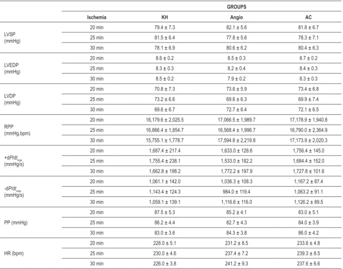
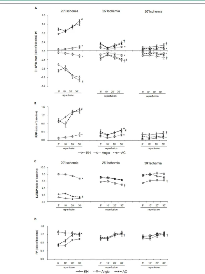
All the isolated perfused hearts presented a similar baseline cardiovascular function (LVSP, LVEDP, LVDP, RPP, HR, PP, +dP/dtmax, and -dP/dtmax) prior to ischemia (Table 1). Hemodynamic data recorded after ischemic times are shown in Figure 1.
Changes in +/- dP/dtmax recovery are shown in Figure 1A: +/- dP/dtmax recovery was significantly reduced in the KH groups submitted to 25 and 30 min of ischemia (74 and 80%, respectively), as compared to the KH group submitted to 20 min of ischemia. Angiotensin-I caused a reduction of 87-94% in +/- dP/dtmax recovery in all ischemic periods as compared to baseline values. These effects of angiotensin-I were completely reverted by captopril in the protocol of 20 min of ischemia. However, after 25 min of ischemia, captopril was only able to reverse the effect of angiotensin-I on the -dP/dtmax recovery. After 30 min of ischemia, captopril did not reverse the effects of angiotensin-I.
Because the LVDP and rate-pressure product recoveries were almost identical, only the rate-pressure product recovery is presented (Figure 1B). The rate-pressure product was significantly reduced in the KH groups submitted to 25 and 30 min of ischemia (73 and 78%, respectively), as compared to the KH group submitted to 20 min of ischemia. Angiotensin-I caused a reduction of 85 to 93% in the rate-pressure product recovery in all ischemic periods as compared to baseline values. This effect of angiotensin-I was reverted by captopril in the groups submitted to 20 min and 25 min of ischemia (85% and 29%, respectively, from baseline values),but not in the protocol of 30 min of ischemia.
Figure 1C represents the LVEDP, which was significantly increased during reperfusion in the KH groups submitted to 25 and 30 min of ischemia (5.5 and 6 fold, respectively), as compared to the KH group submitted to 20 min of ischemia. Angiotensin-I caused an approximate 6.9 fold increase in LVEDP recovery as compared to the KH group submitted to 20 min of ischemia; this effect was reverted by captopril. However, in groups submitted to 25 and 30 min of ischemia, neither angiotensin-I nor captopril had any significant effect on LVEDP.
30 min of ischemia (1.08 and 1.10 fold, respectively) as compared to the KH group submitted to 20 min of ischemia. Angiotensin-I caused an approximate 1.25 fold increase in PP recovery as compared to the KH group submitted to 20 min of ischemia; this effect was partially reverted by captopril. However, in the groups submitted to 25 and 30 min of ischemia, neither angiotensin-I nor captopril had any significant effect on PP.
There were no significant differences in LVSP or HR (data not shown) among the groups, in different protocols of ischemia. There was no interaction between the factors analyzed (treatments and time of reperfusion).
Discussion
This study showed that, in the isolated rat heart model, twenty minutes of global ischemia results in mild/moderate contractile dysfunction, while longer periods of ischemia
can lead to severe contractile dysfunction. Moreover, the effects of angiotensin-I and captopril on functional recovery were better demonstrated when hearts were submitted to 20 min of ischemia. This is the first study evaluating the functional recovery and responsiveness of ACE in rat hearts submitted to three different ischemic periods.
In the isolated heart model, both ischemia and reperfusion induce the release of cardiac enzymes16-18,20.
However, though important, the results shown are inconsistent and illustrate the lack of standardization between different assays and/or sensitivity of the methods used. Furthermore, data are frequently expressed in various different units (unit/mg protein, unit/g dry wt, etc), making it difficult to compare the results. Because of this, we preferred to use multiple functional indices to assess the extent of tissue injury in the ischemic heart, as we considered that this approach would be more reproducible.
The time-course of cardiovascular changes induced by
Table 1 - Baseline values of cardiac function in different groups of treatment and ischemic times
GROUPS
Ischemia KH Angio AC
LVSP (mmHg)
20 min 79.4 ± 7.3 82.1 ± 5.6 81.8 ± 6.7
25 min 81.5 ± 6.4 77.8 ± 5.6 78.3 ± 7.1
30 min 78.1 ± 6.9 80.6 ± 6.2 80.4 ± 6.3
LVEDP (mmHg)
20 min 8.6 ± 0.2 8.5 ± 0.3 8.7 ± 0.2
25 min 8.3 ± 0.3 8.2 ± 0.4 8.4 ± 0.3
30 min 8.5 ± 0.2 7.9 ± 0.2 8.3 ± 0.3
LVDP (mmHg)
20 min 70.8 ± 7.3 73.6 ± 5.9 73.4 ± 6.8
25 min 73.2 ± 6.6 69.6 ± 6.3 69.9 ± 7.4
30 min 69.6 ± 6.7 72.7 ± 6.4 72.1 ± 6.5
RPP (mmHg.bpm)
20 min 16,179.6 ± 2,025.5 17,066.5 ± 1,989.7 17,178.9 ± 1,940.8
25 min 16,866.4 ± 1,854.7 16,568.4 ± 1,996.7 16,790.0 ± 2,364.9
30 min 15,755.1 ± 1,778.7 17,594.8 ± 2,219.8 17,173.9 ± 2,020.3
+dP/dtmax (mmHg/s)
20 min 1,687.4 ± 217.4 1,633.0 ± 128.6 1,756.4 ± 145.0
25 min 1,755.4 ± 238.1 1,533.0 ± 182.2 1,684.4 ± 152.0
30 min 1,662.8 ± 198.2 1,772.2 ± 197.9 1,727.8 ± 101.6
-dP/dtmax (mmHg/s)
20 min 1,061.1 ± 142.0 1,036.3 ± 108.3 1,167.2 ± 87.4
25 min 1,143.4 ± 124.3 984.0 ± 119.4 1,063.2 ± 91.1
30 min 1,059.1 ± 139.1 1,116.6 ± 116.0 1,126.2 ± 89.5
PP (mmHg)
20 min 87.5 ± 5.3 85.2 ± 4.1 83.0 ± 5.1
25 min 86.2 ± 4.4 82.7 ± 4.3 84.0 ± 3.9
30 min 83.0 ± 3.6 84.3 ± 3.8 86.0 ± 4.2
HR (bpm)
20 min 228.0 ± 5.1 231.2 ± 8.5 233.6 ± 4.8
25 min 230.0 ± 4.6 237.4 ± 7.2 239.3 ± 8.5
30 min 226.0 ± 3.8 241.2 ± 9.3 237.6 ± 6.6
KH - Krebs-Henseleit, Angio - angiotensin-I; AC - angiotensin-I+captopril; LVSP - Left ventricular systolic pressure; LVEDP - Left ventricular end-diastolic pressure; LVDP - Left
ventricular developed pressure; RPP - Rate-pressure product; +dP/dtmax - Maximal positive derivative of LV pressure; -dP/dtmax - Maximal negative derivative of LV pressure;
Figure 1 - Recovery of cardiac function after 20, 25 and 30 min of global ischemia. Panel A. Maximal derivatives of LV pressure (+/- dP/dtmax), the negative derivative
global ischemia (20, 25 and 30 min) in the isolated rat heart model showed that when the hearts were exposed to 20 min of ischemia, the functional recovery was reduced in the early phase of reperfusion, but not at the end of reperfusion (30 min), suggesting that the degree of tissue damage was mild to moderate. However, when the ischemic period was increased to 25 or 30 min, a marked decrease in functional recovery was observed, as indicated by the lower +/- dP/dtmax and rate-pressure product and by the higher LVEDP and PP along the reperfusion period. Thus, when the ischemic time exceeds 20 min, the consequence is myocardial contracture, suggesting a high degree of tissue damage. This is probably the cause of the lower responsiveness demonstrated by the cardiac tissue when the period of ischemia was increased from 25 to 30 min.
These findings are in consistency with previous functional and metabolic data from Wang et al21 and Palmer et al16
who reported that mild post-ischemic dysfunction occurred when short periods of global ischemia (15 to 20 min) were employed, but observed a severe myocardial dysfunction with persistent increase of creatine kinase after 25 min of ischemia, suggesting irreversible myocardial injury. These results, together with those of this study, suggest that the transition from mild/moderate to severe tissue damage occurs after 20 min of global ischemia. However, changes in the perfusion buffer [e.g. substrate concentration and/ or composition22, flow23, and temperature24] could modify
this ischemic threshold.
Many reports confirm that ACE activity is present in the cardiac tissue and that the renin-angiotensin system can modulate cardiovascular homeostasis, via its local and systemic systems12-14,25. However, in the isolated rat heart
model, the substrate (angiotensin-I) should be added in the perfusate to study the local ACE12-14.In this study, the
addition of angiotensin-I to the perfusate resulted in a marked decrease in LV functional recovery, as indicated by the lower +/- dP/dtmax and rate-pressure product in all the tested periods of ischemia. This is also supported by the higher LVEDP and PP found in groups submitted to 20 min of ischemia. Earlier reports also showed that angiotensin-I and angiotensin-II reduced cardiac contractility and/ or constrict coronary arteries9,15, but other authors did
not observe any deleterious effect induced by these compounds26. The deleterious effects of angiotensin in
cardiac function may involve increased ROS production, since studies indicate that angiotensin-II could stimulate NADPH oxidase27.
The effects of angiotensin-I on LV functional recovery were totally or partially reversed by captopril, in the groups submitted to 20 and 25 (but not to 30) min of ischemia, suggesting that they depend on the local conversion to angiotensin-II. Multiple factors are likely to have a role in the protective effect of ACE inhibitors in ischemia-reperfusion injury: they decrease leukocyte infiltration28, possess anti-inflammatory effects29 and free
radical scavenging properties30, and effect mediated by
increase the levels of bradykinin and prostaglandin31.
Because ACE inhibition can increase circulating levels of angiotensin-(1-7)32, this may mediate some of these
cardiovascular effects33,34. The inability of captopril to
completely abolish the responses mediated by angiotensin is possibly due to the intrinsic activity of the angiotensin-I, since the formation of angiotensin-II in intact rat hearts is mainly ACE-dependent12,13. Nevertheless, many alternative
pathways to form angiotensin-II (chymase, kallikrein and cathepsin G) could be activated in several pathological conditions such as ischemia, hypercholesterolemia, hypertension and inflammation35.
These results showed that the LV functional recovery was significantly depressed and proportionally less affected by angiotensin-I or captopril, when the heart was exposed to more than 20 min of ischemia. Thus, the low responsiveness of cardiac tissue to drugs is due to ischemic contracture rather than the possible exhaustion of cardiac ACE.
In conclusion, 20 min of global ischemia is probably the best period for the study of mild to moderate myocardial contractile dysfunction, at a level that can be alleviated or exacerbated by the drugs tested. Longer periods of ischemia lead to much more intense myocardial damage, with the development of ischemic contracture, which cannot be reversed by drugs tested during the reperfusion period. Thus, pharmacological approaches tested during these periods may have no effect for methodological reasons. Furthermore, these data suggest that angiotensin-II directly or indirectly participates in the post-ischemic damage, and the ability of an ACE inhibitor in attenuate it depends on the ischemic time.
Acknowledgements
The authors would like to thank Ms. Tania R.G. Fernandes (Instituto de Ciências Básicas da Saúde/Universidade Federal do Rio Grande do Sul (ICBS/UFRGS) for her technical assistance.
Potential Conflict of Interest
No potential conflict of interest relevant to this article was reported.
Sources of Funding
This study was funded by CAPES, CNPq, FAPESP and FAPERGS.
Study Association
References
1. Reimer KA, Heide RSV, Richard VJ. Reperfusion in acute myocardial infarction: effect of timing and modulating factors in experimental models. Am J Cardiol. 1993;72(19):13G-21G.
2. Kern KB, Hilwig RW, Rhee KH, Berg RA. Myocardial dysfunction after resuscitation from cardiac arrest: an example of global myocardial stunning. J Am Coll Cardiol. 1996;28(1):232-40.
3. Bolli R, Marban E. Molecular and cellular mechanisms of myocardial stunning. Physiol Rev. 1999;79(2):609-34.
4. Kloner RA, Jennings RB. Consequences of brief ischemia: stunning, preconditioning, and their clinical implications: part 1. Circulation. 2001;104(24):2981-9.
5. Galinanes M, Hearse DJ. Assessment of ischemic injury and protective interventions: the Langendorff versus working rat heart preparation. Can J Cardiol. 1990;6(2):83-91.
6. Hensley K, Robinson KA, Gabbita SP, Salsman S, Floyd RA. Reactive oxygen species, cell signaling, and cell injury. Free Radic Biol Med. 2000; 28(10):1456-62. 7. Lefer DJ, Granger DN. Oxidative stress and cardiac disease. Am J Med.
2000;109(4):315-23.
8. Arad M, Shotan A, Horowitz LR. Effect of captopril on metabolic and hemodynamic alterations in global ischemia and reperfusion in the isolated working rat heart. J Cardiovasc Pharmacol. 1992;19(3):319-23.
9. Neves LAA, Almeida AP, Klosla MC, Santos RA. Metabolism and angiotensin I in isolated rat hearts: effect of angiotensin converting enzyme inhibitors. Biochem Pharmacol. 1995;50(9):1451-9.
10. Pfeffer MA, Braunwald E, Moyé LA, Basta L, Brown EJ Jr, Cuddy TE, et al. Effect of captopril on mortality and morbidity in patients with left ventricular dysfunction after myocardial infarction: results of the Survival and Ventricular Enlargement Trial. N Engl J Med. 1992;327(10):669-77.
11. Garg R, Yusuf S. Overview of randomized trials of angiotensin-converting enzyme inhibitors on mortality and morbidity in patients with heart failure. Collaborative Group on ACE Inhibitor Trials. JAMA. 1995;273(18):1450-6. 12. de Lannoy LM, Danser AHJ, Bouhuizen AMB, Saxena PR, Schalekamp MADH.
Localization and production of angiotensin II in isolated perfused rat heart. Hypertension. 1998;31(5):1111-7.
13. de Lannoy LM, Schuijt MP, Saxena PR, Schalekamp MA, Danser AH. Angiotensin converting enzyme is the main contributor to angiotensin I-II conversion in the interstitium of the isolated perfused rat heart. J Hypertens. 2001;19(5):959-65. 14. Müller DN, Fischli W, Clozel JP, Hilgers KF, Bohlender J, Ménard J, et al. Local angiotensin II generation in the rat heart: role of renin uptake. Circ Res. 1998;82(1):13-20.
15. Traquandi C, Riva E. Cardiac effects of angiotensin I and angiotensin II: dose-response studies in the isolated perfused rat heart. Pharmacol Res. 1998;37(1):57-65.
16. Palmer BS, Hadziahmetovic M, Veci T, Angelos MG. Global ischemic duration and reperfusion function in the isolated perfused rat heart. Resuscitation. 2004;62(1):97-106.
17. Chocron S, Alwan K, Toubin G, Kantelip B, Clement F, Kantelip JP, et al. Effects of myocardial ischemia on the release of cardiac troponin I in isolated rat hearts. J Thorac Cardiovasc Surg. 1996;112(2):508-13.
18. Bertinchant JP, Polge A, Robert E, Sabbah N, Fabbro-Peray P, Poirey S, et al. Time-course of troponin I release from isolated perfused rat hearts during hypoxia/ reoxygenation and ischemia/reperfusion. Clin Chim Acta. 1999;283(1-2):43-56.
19. Langendorff O. Untersuchungen am uberlebenden saugetierherzen. Pflügers Arch Ges Physiol. 1895;61:291-332.
20. Poston JM, Parenteau GL. Biochemical effects of ischemia on isolated, perfused rat heart tissues. Arch Biochem Biophys. 1992;295(1):35-41. 21. Wang QD, Swardh A, Sjoquist PO. Relationship between ischaemic time
and ischaemia/reperfusion injury in isolated Langendorff-perfused mouse hearts. Acta Physiol Scand. 2001;171(2):123-8.
22. Angelos MG, Murray HN, Gorsline RT, Klawitter PF. Glucose, insulin and potassium (GIK) during reperfusion mediates improved myocardial bioenergetics. Resuscitation. 2002;55(3):329-36.
23. Klawitter PF, Murray HN, Clanton TL, Palmer BS, Angelos MG. Low flow after global ischemia to improve postischemic myocardial function and bioenergetics. Crit Care Med. 2002;30(11):2542-7.
24. Bes S, Roussel P, Laubriet A, Vandroux D, Tissier C, Rochette L, et al. Influence of deep hypothermia on the tolerance of the isolated cardiomyocyte to ischemia-reperfusion. J Mol Cell Cardiol. 2001;33(11):1973-88. 25. Meulemans AL, Andries LJ, Brutsaert DL. Does endocardial endothelium
mediate positive inotropic response to angiotensin I and angiotensin II? Circ Res. 1990;66(6):1591-601.
26. Ford WR, Clanachan AS, Hiley CR, Jugdutt BI. Angiotensin II reduces infarct size and has no effect on post-ischaemic contractile dysfunction in isolated rat hearts. Br J Pharmacol. 2001;134(1):38-45.
27. Li JM, Shah AM. Endothelial cell superoxide generation: regulation and relevance for cardiovascular pathophysiology. Am J Physiol Regul Integr Comp Physiol. 2004;287(5):R1014-30.
28. Ferrari R, Pepi P, Nesta F, Benigno M, Visioli O. Metabolic derangement in ischemic heart disease and its therapeutic control. Am J Cardiol. 1998;82(5A):2k-13k.
29. Peng H, Carretero OA, Liao TD, Peterson EL, Rhaleb NE. Role of N-acetyl-seryl-aspartyl-lysyl-proline in the antifibrotic and anti-inflammatory effects of the angiotensin-converting enzyme inhibitor captopril in hypertension. Hypertension. 2007;49(3):695-703.
30. Anderson B, Khaper N, Dhalla AK. Anti free radical mechanism in captopril protection against reperfusion injury in isolated rat hearts. Can J Cardiol. 1996;12(10):1099-104.
31. Ehring T, Baumgart D, Krajcard M, Hümmelgen M, Kompa S, Heusch G. Attenuation of myocardial stunning by ACE inhibitor ramiprilat through a signal cascade of bradykinin and prostaglandins but not nitric oxide. Circulation. 1994;90(3):1368-85.
32. Iyer SN, Ferrario CM, Chappell MC. Angiotensin-(1–7) contributes to the antihypertensive effects of blockade of the renin–angiotensin system. Hypertension. 1998;31(1 Pt 2):356-61.
33. Almeida AP, Fabregas BC, Madureira MM, Santos RJ, Campagnole-Santos MJ, Santos RA. Angiotensin-(1–7) potentiates the coronary vasodilatatory effect of bradykinin in the isolated rat heart. Braz J Med Biol Res. 2000;33(6):709-13.
34. Oudot A, Vergely C, Ecarnot-Laubriet A, Rochette L. Pharmacological concentration of angiotensin-(1–7) activates NADPH oxidase after ischemia– reperfusion in rat heart through AT1 receptor stimulation. Regulatory Peptides. 2005;127(1-3):101-10.