www.scielo.br/aabc
Crystal habits and magnetic microstructures of magnetosomes
in coccoid magnetotactic bacteria
ULYSSES LINS1, MARTHA R. McCARTNEY2, MARCOS FARINA3, RICHARD B. FRANKEL4 and PETER R. BUSECK5
1Instituto de Microbiologia Professor Paulo de Góes, CCS, Universidade Federal do Rio de Janeiro
Cidade Universitária, 21941-590, Rio de Janeiro, RJ, Brasil
2Center for Solid State Science, Arizona State University, Tempe, Arizona, 85287 USA 3Instituto de Ciências Biomédicas, CCS, Universidade Federal do Rio de Janeiro
Cidade Universitária, 21941-590 Rio de Janeiro, RJ, Brasil
4Department of Physics, California Polytechnic State University
San Luis Obispo, California, 93407, USA
5Departments of Geology and Chemistry/Biochemistry, Arizona State University
Box 871404, Tempe, Arizona, 85287-1404, USA
Manuscript received on September 9, 2005; accepted for publication on February 17, 2006; contributed byRICHARDB. FRANKEL*
ABSTRACT
We report on the application of off-axis electron holography and high-resolution TEM to study the crystal habits of magnetosomes and magnetic microstructure in two coccoid morphotypes of magnetotactic bacte-ria collected from a brackish lagoon at Itaipu, Brazil. Itaipu-1, the larger coccoid organism, contains two separated chains of unusually large magnetosomes; the magnetosome crystals have roughly square projec-tions, lengths up to 250 nm and are slightly elongated along[111](width/length ratio of about 0.9). Itaipu-3
magnetosome crystals have lengths up to 120 nm, greater elongation along[111](width/length∼0.6), and
prominent corner facets. The results show that Itaipu-1 and Itaipu-3 magnetosome crystal habits are related, differing only in the relative sizes of their crystal facets. In both cases, the crystals are aligned with their
[111]elongation axes parallel to the chain direction. In Itaipu-1, but not Itaipu-3, crystallographic positioning
perpendicular to[111]of successive crystals in the magnetosome chain appears to be under biological con-trol. Whereas the large magnetosomes in Itaipu-1 are metastable, single-magnetic domains, magnetosomes in Itaipu-3 are permanent, single-magnetic domains, as in most magnetotactic bacteria.
Key words:magnetite, magnetotaxis, high-resolution transmission electron microscopy.
INTRODUCTION
Magnetotactic bacteria orient and migrate along geomagnetic field lines. Each cell contains mag-netosomes, which are membrane-enclosed, nano-scale, iron-mineral crystals (Bazylinski and Frankel
*Member Academia Brasileira de Ciências Correspondence to: Ulysses Lins E-mail: [email protected]
464 ULYSSES LINS et al.
preferred oxygen concentration in chemically stra-tified sediments or water columns (Frankel et al. 1997).
In micro-aerobic freshwater and marine envi-ronments, magnetotactic bacteria with magnetite (Fe3O4) magnetosomes occur, whereas in sulfidic
environments magnetotactic bacteria that produce greigite (Fe3S4) magnetosomes are present
(Sim-mons et al. 2004). The magnetosome membrane is presumably a structural entity that anchors the min-eral particles at particular locations in the cell, as well as the locus of biological control over the min-eralization process (Matsunaga and Okamura 2003, Schüler 2004).
Magnetosome magnetite crystals in different bacterial species or strains have different, but consis-tent, projected shapes when observed by transmis-sion electron microscopy (Bazylinski and Frankel 2000, Devouard et al. 1998). Idealized crystal habits of magnetosome magnetite crystals in a num-ber of magnetotactic bacteria have been inferred from 2D high-resolution lattice images, assuming low-index faces (Bazylinski et al. 1994, Frankel and Buseck 2000, Mann and Frankel 1989, Mann et al. 1987, Matsuda et al. 1983, Meldrum et al. 1993a, b, Thomas-Keprta et al. 2001). In magnetotactic spir-illa, the idealized crystal habits are equidimensio-nal cuboctahedra comprising{100}and{111}faces
(Mann et al. 1984). In a number of other mag-netotactic bacteria, including cocci and vibrios, the crystals are elongated along the[111]axis parallel to
the magnetosome chain direction and the projected shapes are quasi-rectangular. The idealized habits for the crystals comprise{100}, {111}, and {110}
forms with 6, 8, and 12 faces, respectively. The elon-gation along[111]results in a non-equidimensional
crystal habit with two groups of six{110}faces and
two groups of two and six {111} faces. The six {100} faces remain equidimensional. The result is a prism-like arrangement with a hexagonal cross-section perpendicular to[111]through the center of
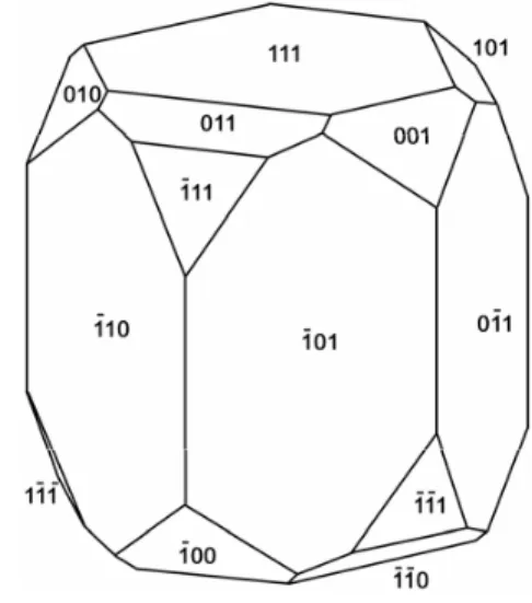
the crystal (Towe and Moench 1981). The remai-ning faces form corner facets at the intersections be-tween the body{110}and end-cap{111}faces (see
Figure 2). The sizes of the crystals, the width/length ratios, and the relative sizes of the corner faces dif-fer between species, resulting in the distinctive pro-jected shapes.
It is possible to determine such complex habits in small crystals with techniques such as electron tomography, which involves carefully controlled TEM tilting experiments (Buseck et al. 2001). However, electron tomography does not give infor-mation about magnetic structure. Off-axis electron holography, on the other hand, can be used to ob-tain both magnetic and structural information about nanometer-sized magnetic crystals. In this paper we report on the application of electron holography and high-resolution transmission electron micros-copy to compare the crystal habits and magnetic structures of magnetosomes between two types of magnetotactic bacteria collected at the same loca-tion in Brazil. The magnetosomes studied here were obtained from two coccoid morphotypes of magnetotactic bacteria in a brackish lagoon at Itai-pu, Brazil, which is located on the coast of Brazil north of Rio de Janeiro (Farina et al. 1994, Lins et al. 1994). At least four coccoid morphotypes with magnetite magnetosomes, Itaipu-1, -2, -3 and -4 (Spring et al. 1998), a rarely observed rod-shaped bacterium (Lins and Farina 1998), and multicel-lular magnetotactic prokaryotes (U. Lins, unpub-lished results), occur in the lagoon. Itaipu-1, the largest coccoid organism, contains two separated chains of magnetosomes; the magnetosome crys-tals have roughly square projections, lengths up to 250 nm and width-to-length ratios of about 0.9 (Fa-rina et al. 1994, Spring et al. 1998). These are the largest-volume magnetosome crystals yet reported. Itaipu-2 and -4 are smaller cocci containing mag-netosome crystals that are smaller, but with simi-lar projected shapes to those in Itaipu-1. Itaipu-3 has magnetosome crystals that are elongated along
[111](width/length∼0.6), with lengths up to 120
re-markable that the crystals in Itaipu-1 are not only all aligned with[111]elongation axes parallel to each
other along the chain, but are also ordered rotation-ally perpendicular to the chain axis, with like corner faces of adjacent crystals facing each other (Lins et al. 2005).
MATERIALS AND METHODS
SAMPLEPREPARATION
Itaipu-1 and Itaipu-3 bacteria predominated in the lagoon at the time the samples for this study were collected. Cells of Itaipu-1 and Itaipu-3 were iso-lated from sediment and water samples by using glass chambers with capillary ends positioned side magnetic coils (Lins et al. 2003). Cells in-side the glass chamber swam in the direction of the north magnetic pole of the coil (i.e., antiparal-lel to the magnetic field generated by the current in the coils), and accumulated at the end of the capillary, from where they were collected and de-posited into Eppendorf plastic tubes. Whole cells, or magnetosomes extracted from disrupted cells, were deposited on holey-carbon TEM grids. A protocol modified from Towe and Moench (1981) was used for disruption of magnetotactic bacteria and isola-tion of crystals. Briefly, cells were concentrated at the bottom of an Eppendorf tube by centrifuga-tion, re-suspended in a solution containing detergent (SDS, about 20% w/v), and magnetically concen-trated with a strong rare-earth magnet positioned at the bottom of the tube. In order to eliminate the detergent and dissolved organic material, the super-natant was discarded, the pellet was re-suspended in distilled water, and the magnetite crystals were reconcentrated at the bottom of the tube. This pro-cess was repeated until no detergent remained in the sample. The isolated magnetite crystals were further treated with a hot NaOH solution (circa 60◦C) and washed in distilled water, as described above. Water drops containing the crystals were spread over the grids. Grids were then air-dried and observed under the transmission electron mi-croscope. In some cases the crystal suspension was
sonicated prior to deposition on the grids. The dis-ruption process resulted in Itaipu-1 and Itaipu-3 magnetosomes mixed together on the TEM grid (Figure 1). High-resolution TEM and selected area electron diffraction (SAED) measurements were made with a Philips CM200 field emission gun (FEG) TEM.
Off-axis electron holography was performed with a Philips CM200 FEG TEM operated at 200 kV. This microscope has a rotatable electrostatic biprism (a 0.6-micron quartz wire coated with gold) located in place of one of the conventional selected-area apertures. It also has a Lorentz mini-lens, which is located in the bore of the objective lens pole-piece. With the conventional microscope objective lens switched off, the Lorentz minilens allows the examination of the magnetosomes in close to field-free conditions with a line resolution of 1.2 nm at 200 kV.
PRINCIPLES OFELECTRONHOLOGRAPHY
In off-axis electron holography, the sample is po-sitioned in the transmission electron microscope so that it covers approximately half the field of view, and a charged electrostatic biprism causes the elec-tron wave that has passed through the specimen to overlap with a reference wave that has only passed through vacuum. The resulting hologram is an in-terference pattern in which amplitude information is contained in the relative amplitude of the cosine-like fringes, and information about the phase shift of the electron wave is contained in their position (Dunin-Borkowski et al. 1998a, b, 2001).
con-466 ULYSSES LINS et al.
Fig. 1 – Transmission electron microscopy image of isolated chains of magnetosomes from Itaipu-1 (large crystals) and Itaipu-3 (small crystals). Scale bar indicates 200 nm.
ventional TEM image, which represents only the intensity of the electron wave and does not contain any phase information.
For a sample with electrostatic potential, V, and magnetic vector field, B, the phase shift,φ, of the electron wave can be expressed as (Reimer 1989)
φ(x, y)= 2π
λE
E+E◦ E + 2E◦
V(r)dz− e
B(r)•dS
whereλ, E, E◦, e,, and V are the electron wave-length, kinetic energy, rest mass energy, charge, Planck’s constant, and electrostatic mean inner po-tential, respectively. In the absence of induced elec-tric fields, the first integral is equal to the mean in-ner potential (Gajdardziska-Josifovska et al. 1993) times the sample thickness. The second integral picks out the components of the magnetic field per-pendicular to the incident-beam direction, i.e., those that normally correspond to the components in the plane of the sample.
The mean inner potential is a quantity that is related to the electron-optical index of refraction and is characteristic for a particular material com-position and density. In the absence of magnetic
fields, it represents a constant that correlates the phase shift with the projected thickness. Normally, this quantity is determined by analyzing samples whose thickness is precisely known. Conversely, once determined, it can be used to measure the local thickness.
Two holograms corresponding to reversed ori-entations of the magnetization are acquired in or-der to analyze both the 3D shape and magnetic flux of small magnetic crystals. Reversals of the mag-netization are obtained in situ by the application of the magnetic field of the objective lens (Dunin-Borkowski et al. 1998a, 2001). The sum of the phases of these two holograms then represents twice the mean inner-potential contribution to the phase if the magnetization has exactly reversed, whereas the difference of the phases gives twice the magnetic contribution.
provide quantitative information about the projec-ted thicknesses of the crystallites, and holograms can be acquired for specific tilts of the crystals to provide 3D morphologies.
RESULTS
It is not possible to tell from the 2D images alone whether any set of lattice fringes is parallel to ter-minating facets or wedge-edges. Whereas the darker contrast in the centers of the crystals may indicate that the crystals are thicker there, the contrast of TEM images is an unreliable indicator of thickness and thus cannot be used to determine morphology (Buseck et al. 2001). Therefore off-axis electron holography was used to measure the cross-sectional thickness of the crystals (Lins et al. 2005) result-ing in the idealized crystal habit shown in Figure 2 for Itaipu-1 crystals. Figure 3a shows the thick-ness contours derived from electron holograms for three Itaipu-1 crystals (labeled 1, 2 and 3) and two Itaipu-3 crystals (labeled 4, and 5). The thickness profiles across crystals 2 and 4 are shown in Figure 3b whereas the thickness profiles across crystals 3 and 5 are shown in Figure 3c. The profiles of crystal 2 and 4 are consistent with a hexagonal cross-section with the electron beam in a[21¯1¯]projection,
corre-sponding to the intersection of two adjacent{110¯ }
planes (Figure 2). This projection has a peak (tent-top) in the center. On the other hand, the profiles of crystals 3 and 5 are consistent with hexagonal cross-sections in a[110¯ ]projection, corresponding to the
electron beam perpendicular to one{110¯ }face and
angled with respect to the adjacent two faces (Fig-ure 2). This projection has a flat region (flat-top) in the center. We previously reported that magnetic contours for Itaipu-3 crystals were always consis-tent with permanent single magnetic domain struc-ture. However, Itaipu-1 crystals presented magnetic contours consistent with permanent single magnetic domain only when crystals were arranged in linear chains (as in Figure 3a). When the chain breaks (be-cause of the use of ultrasound during sample prepa-ration) the magnetic contour lines did not show a
single direction for the magnetization within the crystals. From these results (McCartney et al. 2001) it was concluded that large Itaipu-1 crystals present a metastable single-domain structure.
Fig. 2 – Idealized structure for elongated magnetosomes. Itaipu-1 and Itaipu-3 crystals show similar morphology except for crystal length, width to length ratio, and relative development of the corner faces.
A high-resolution image of part of a crystal from a chain in a [110¯ ] projection (Figure 4) is
consistent with the idealized structure described for Itaipu-1 crystals (Lins et al. 2005). It has a{111}
face at the left end of the crystal and intersections between adjacent[110¯ ]planes at the top and bottom
edges. Well-developed{110}and{100}corner faces
occur, but possible {111}corner faces are
incons-picuous. Thus the idealized structure of the Itaipu-3 crystals is similar to that of the Itaipu-1 crystals (Fig-ure 2), except for length, width to length ratio, and relative development of the corner faces.
468 ULYSSES LINS et al.
Fig. 3 – a) Cluster of three Itaipu-1 magnetosomes (crystals 1, 2 and 3) and two Itaipu-3 magnetosomes (crystals 4 and 5) with thickness contours derived from electron holographic image. (b, c) Thickness profiles for the traverses indicated by the lines. Crystal 1 is in a[110¯ ]projection zone and has a flattop
profile. Crystals 2 and 4 are close to a[21¯1¯]projection zone and have a tent-top profile. Crystals 3 and
Fig. 4 – High-resolution transmission electron microscopy lattice-fringe image of an Itaipu-3 magnetosome. Inset is a diffractogram of the crystal. Scale bar indicates 15 nm.
has a flat-top profile indicative of a[110¯ ]projection,
whereas the crystal on the right has a tent-top profile indicative of a[21¯1¯]projection. These results show
that both the Itaipu-1 and Itaipu-3 crystals have ide-alized habits in which[111]is a three-fold rotational
axis and[110¯ ]and[21¯1¯]are two-fold rotation axes.
However, the surfaces of the Itaipu-3 crystals can be substantially roughened, as shown in Figure 6. This crystal also shows a[111]twin plane through
its center.
DISCUSSION
We used electron holography and high-resolution TEM to determine both the 3D morphology of
bac-terial magnetite crystals and their magnetic micro-structure. The results confirm that both Itaipu-1 and Itaipu-3 bacteria have magnetosome magne-tite crystals with similar non-equidimensional crys-tal habits, although the sizes of the cryscrys-tals and their width to length ratios differ. In addition, the results confirm that the crystals are organized with [111]
axes of elongation parallel to each other along the chain direction.
470 ULYSSES LINS et al.
Fig. 5 – Electrostatic (A) and magnetic (B) contributions to electron holographic phase of four Itaipu-3 magnetosomes in a chain. Scale bar indicates 100 nm in both figures. The thickness profiles along the lines indicated by the arrows crossing the thickness contours are shown in (C) and (D) where half-thickness is plotted with distance along the arrows.
approaching 1000 microns per second (Cox et al. 2002), which requires greater propulsive forces compared to the other magnetotactic bacteria. These greater propulsive forces may result from the bilophotrichous flagellation of many coccoid magnetotactic bacteria (Frankel et al. 1997, Towe and Moench 1981). Many magnetotactic cocci are observed to make rocking motions as they swim. This would occur if the magnetic dipole in each cell were not perfectly aligned along the axis of motility of the cell, and also because flagella in magneto-tactic bacteria are short (similar to or smaller than one helical turn) when compared toEscherichia coli that has very long flagella, and follows straight tra-jectories (Nogueira and Lins de Barros 1995).
The fact that the magnetocrystalline aniso-tropy of magnetite above the Verwey transition is relatively low (Moskowitz 1995) prompts the
ques-tion of whether there is funcques-tional significance to the adoption by the Itaipu cocci and most, if not all, coccoid, magnetotactic bacteria, of non-equidimen-sional crystal habits. Because [111] axes are the
“easy” magnetic axes above the Verwey transition, alignment of the [111] axes of elongation of the
Fig. 6 – High-resolution transmission electron microscopy lattice-fringe image of a twinned Itaipu-3 magnetosome showing a rough surface. Inset is a diffractogram of the crystals. Scale bar indicates 20 nm.
the Verwey transition (Moskowitz 1995) and the slight elongation of the magnetosome crystals in Itaipu-1 suggests that the shape anisotropy of the chain alone is sufficient to keep the magnetization along the chain direction without spontaneous rever-sals. This may explain the presence of large{111}
contact faces between crystals, preventing misalign-ment of the crystals in the chain.
Although increased magnetic anisotropy prob-ably increases the coercive force of the chain, it is not immediately apparent what, if any, effect this has on magnetotaxis. Some bacteria, such as the mag-netotactic spirilla, have equidimensional magnetite crystals and yet form magnetosome chains in which the magnetic flux is well confined to the magnetite crystals in the chain. These bacteria have magnetic dipole moments per unit length that are compara-ble to those in bacteria with magnetosome chains containing elongated crystals (Dunin-Borkowski et
al. 1998b). Also, some bacteria have magnetosome magnetite crystals with an[211]axis of elongation
parallel to the chain direction (Mann et al. 1987). Although the magnetic dipole moment per unit length for chains with these crystals has not been measured, it must be of sufficient magnitude to ac-count for the magnetotactic response of the cells. Thus elongation of magnetosomes along[111]
can-not be explained by simply invoking natural selec-tion (Thomas-Keprta et al. 2001).
nucle-472 ULYSSES LINS et al.
ation and growth of the crystal from a saturated iron solution at the right electrochemical potential and pH such that magnetite is the stable phase (Baeuer-lein 2000, Mann and Frankel 1989, Schüler 1999). Non-equidimensional growth could result from the presence of the nucleating surface or an anisotopic iron ion flux through the membrane that results in crystal habits with lowered symmetry compared to crystals grown in isotropic situations. This might also explain the occurrence of magnetosomes with arrowhead, tooth-shaped or bullet projections, i.e., with even lower symmetry than the magnetosomes in the Itaipu cocci (Blakemore et al. 1981). It would be interesting to have these cocci in pure culture to evaluate the effects of varying concentrations of oxygen, iron, and specific iron chelators on magne-tosome growth.
ACKNOWLEDGMENTS
R.B.F. and P.R.B. acknowledge support from U.S. National Science Foundation grant CHE 9714101. M.F. acknowledges support from Conselho Na-cional de Desenvolvimento Científico e Tecnoló-gico (CNPq) and U.L. acknowledges support from CNPq, Coordenação de Aperfeiçoamento de Pes-soal de Nível Superior (CAPES), Fundação Car-los Chagas Filho de Amparo à Pesquisa do Estado do Rio de Janeiro (FAPERJ), Programa de Apoio a Núcleos de Excelência (PRONEX). Electron mi-croscopy was performed at the Center for High Res-olution Electron Microscopy at Arizona State Uni-versity. U.L. thanks P.R.B. for the opportunity to spend time at Arizona State University to work on the manuscript.
RESUMO
Nós relatamos a aplicação de holografia não-axial e mi-croscopia eletrônica de alta resolução para estudar os hábitos cristalinos de magnetossomos e a microestrutura magnética de dois morfotipos de cocos de bactérias mag-netotáticas coletadas em uma lagoa salobra em Itaipu, Brasil. Itaipu-1, o organismo cocóide maior, contém duas cadeias separadas de magnetossomos atipicamente
grandes; os cristais dos magnetossomos possuem pro-jeções aproximadamente quadradas, comprimentos de até 250 nm e são ligeiramente alongados na direção[111]
(razão largura/comprimento de aproximadamente 0.9). Os cristais dos magnetossomos em Itaipu-3 possuem comprimentos até 120 nm, maior alongamento na dire-ção[111](largura/comprimento∼0.6), e proeminentes
facetas nas extremidades. Os resultados mostram que os hábitos cristalinos dos magnetossomos em Itaipu-1 e Itaipu-3 são relacionados, diferindo apenas nos tama-nhos relativos das suas faces cristalinas. Em ambos os casos, os cristais são alinhados com seus eixos de alon-gamento[111]paralelos à direção da cadeia. Em Itaipu-1, mas não em Itaipu-3, o posicionamento cristalográ-fico, perpendicular à direção[111], de cristais sucessivos
na cadeia de magnetossomos parece estar sobre controle biológico. Enquanto os magnetossomos grandes em Itai-pu-1 são monodomínios magnéticos metaestáveis, em Itaipu-3 eles são monodomínios magnéticos permanentes como na maioria das bactérias.
Palavras-chave: magnetita, magnetotaxia, microscopia eletrônica de transmissão de alta resolução.
REFERENCES
BAEUERLEIN E. 2000. Single magnetic crystals of magnetite (Fe3O4) synthesized in intracytoplasmic vesicles of Magnetospirillum grysphiswaldense. In: BAEUERLEINE (Ed), Biomineralization: From biol-ogy to biotechnolbiol-ogy and medical application, Wein-heim, Germany: Wiley-VCH, p. 61–79.
BAZYLINSKIDAANDFRANKELRB. 2000. Magnetic iron-oxide and iron-sulfide minerals within micro-organisms. In: BAEUERLEINE (Ed), Biomineral-ization: from biology to biotechnology and medi-cal application, Weinheim, Germany: Wiley-VCH, p. 25–46.
BAZYLINSKIDAANDFRANKELRB. 2004. Magneto-some formation in prokaryotes. Nature Rev Micro-biol 2: 217–230.
BAZYLINSKIDA, GARRATT-REEDAJANDFRANKEL RB. 1994. Electron microscopic studies of magneto-somes in magnetotactic bacteria. Microsc Res Tech 27: 389–401.
1981. South-seeking magnetotactic bacteria in the Southern Hemisphere. Nature 286: 384–385. BUSECK PR, DUNIN-BORKOWSKI RE, DEVOUARD
B, FRANKEL RB, MCCARTNEY MC, MIDGLEY PA, POSFAIMANDWEYLANDM. 2001. Magnetite morphology and life on Mars. Proceed Nat Acad Sci USA 98: 13490–13495.
COXBL, POPAR, BAZYLINSKID, LANOILB, DOU -GLAS S, BELZ A, ENGLER DL AND NEALSON KH. 2002. Organization and elemental analysis of P-, S-, and Fe-rich inclusions in a population of freshwater magnetococci. Geomicrobiol J 19: 387– 406.
DERUIJTERWJANDWEISSJK. 1993. Detection limits for quantitative off-axis electron holography. Ultra-microscopy 50: 269–283.
DEVOUARDB, POSFAIM, HUAX, BAZYLINSKIDA, FRANKELRBANDBUSECKPR. 1998. Magnetite from magnetotactic bacteria: Size distributions and twinning. Amer Mineral 83: 1387–1399.
DUNIN-BORKOWSKIRE, MCCARTNEY MR, SMITH DJ AND PARKINSSP. 1998a. Towards quantita-tive electron holography of magnetic thin films using
in situmagnetization reversal. Ultramicroscopy 74:
61–73.
DUNIN-BORKOWSKIRE, MCCARTNEY MR, FRAN -KEL RB, BAZYLINSKIDA, PÓSFAIM ANDBU -SECKPR. 1998b. Magnetic microstructure of mag-netotactic bacteria by electron holography. Science 282: 1868–1870.
DUNIN-BORKOWSKIRE, MCCARTNEYMR, PÓSFAI M, FRANKELRB, BAZYLINSKIDAANDBUSECK PR. 2001. Off-axis electron holography of magne-totactic bacteria: magnetic microstructure of strains MV-1 and MS-1. Eur J Mineral 13: 671–684. FARINAM, KACHARB, LINSU, BRODERICKRAND
LINS DEBARROSH. 1994. The observation of large magnetite (Fe3O4) crystals from magnetotactic bac-teria by electron and atomic force microscopy. J Mi-crosc 173: 1–8.
FRANKELRBANDBUSECKPR. 2000. Magnetite bio-mineralization and ancient life on Mars. Curr Opin Chem Biol 4: 171–176.
FRANKELRB, BAZYLINSKIDA, JOHNSONMSAND TAYLOR BL. 1997. Magneto-aerotaxis in marine coccoid bacteria. Biophys J 73: 994–1000.
GAJDARDZISKA-JOSIFOVSKAM, MCCARTNEYMC, DERUIJTERWJ, SMITHDJ, WEISSJKANDZUO JM. 1993. Accurate measurement of mean inner po-tential of crystal wedges using digital electron holo-grams. Ultramicroscopy 50: 285–299.
LINSUANDFARINAM. 1998. Magnetosome size dis-tribution in uncultured rod-shaped bacteria as deter-mined by electron microscopy and electron spectro-scopic imaging. Microsc Res Tech 42: 459–464. LINSU, SOLÓRZANOGANDFARINAM. 1994. High
volume magnetite (Fe3O4) crystals from magneto-tactic bacteria. Bull Inst Oceanogr Monaco 14: 95– 104.
LINSU, FREITASF, KEIMCN, LINS DEBARROSH, ESQUIVEL DMS AND FARINA M. 2003 Simple homemade apparatus for harvesting uncultured mag-netotactic microorganisms. Braz J Microbiol 34: 111–116.
LINSU, MCCARTNEYMR, FARINAM, FRANKELRB ANDBUSECKPR. 2005. Habits of magnetosome crystals in coccoid magnetotactic bacteria. Appl En-viron Microbiol 71: 4902–4905.
MANNSANDFRANKELRB. 1989. Magnetite biomin-eralization in unicellular organisms. In: MANNS ET AL. (Eds), Biomineralization: Chemical and Bio-chemical perspectives. Weinheim, Germany: Wiley-VCH, p. 389–426.
MANNS, FRANKELRBANDBLAKEMORERP. 1984. Structure, morphology and crystal-growth of bacte-rial magnetite. Nature 310: 405–407.
MANN S, SPARKS N AND BLAKEMORE RP. 1987. Structure, morphology and crystal growth of aniso-tropic magnetite crystals in magnetotactic bacteria. Proc R Soc London Ser B 231: 477–487.
MATSUDA T, ENDO J, OSAKABE N, TONOMURAA AND ARIIT. 1983. Morphology and structure of biogenic magnetite particles. Nature 302: 411–412. MATSUNAGAT ANDOKAMURAY. 2003. Genes and proteins involved in bacterial magnetic particle for-mation. Trends Microbiol 11: 536–541.
MCCARTNEYMR, LINSU, FARINAM, BUSECKPR ANDFRANKELRB. 2001. Magnetic microstructure of bacterial magnetite by electron holography. Eur J Mineral 13: 685–689.
micro-474 ULYSSES LINS et al.
scope study of magnetosomes in a cultured coccoid magnetotactic bacterium. Proc R Soc London Ser B 251: 231–236.
MELDRUM FC, MANNS, HEYWOODBA, FRANKEL RBANDBAZYLINSKIDA. 1993b. Electron micro-scope study of magnetosomes in two cultured vibri-oid magnetotactic bacteria. Proc R Soc London Ser B 251: 237–242.
MOSKOWITZBM. 1995. Biomineralization of magnetic minerals. Rev Geophys 33: 123–128.
NOGUEIRA FS AND LINS DE BARROS HGP. 1995. Study of the motion of magnetotactic bacteria. Eur Biophys J 24: 13–22.
REIMER L. 1989. Transmission electron microscopy. Physics of image formation and microanalysis. Springer Series in Optical Sciences 2nd ed., Berlin: Springer 36: 584.
SCHÜLER D. 1999. Formation of magnetosomes in magnetotactic bacteria. J Mol Microbiol Biotechnol 1: 79–86.
SCHÜLERD. 2004. Molecular analysis of a subcellu-lar compartment: the magnetosome membrane in
Magnetospirillum gryphiswaldense. Arch Microbiol
18: 1–7.
SIMMONS SL, SIEVERT SM, FRANKEL RB, BAZY -LINSKIDAANDEDWARDSKJ. 2004. Spatiotem-poral distribution of marine magnetotactic bacteria in a seasonally stratified coastal salt pond. Appl En-viron Microbiol 70: 6230–6239.
SPRINGS, LINSU, AMANNR, SCHLEIFERK, FER -REIRA LCS, ESQUIVEL DMS AND FARINA M. 1998. Phylogenetic affiliation and ultrastructure of uncultured magnetic bacteria with unusually large magnetosomes. Arch Microbiol 169: 136–147. THOMAS-KEPRTA TL, BAZYLINSKI DA, CLEMETT