Effect of Polyethylene Glycol on the
Formation of Magnetic Nanoparticles
Synthesized by
Magnetospirillum
magnetotacticum
MS-1
Hirokazu Shimoshige1, Hideki Kobayashi2, Toru Mizuki1,3, Yutaka Nagaoka1,3, Akira Inoue1,3, Toru Maekawa1,3*
1Bio-Nano Electronics Research Centre, Toyo University, Kawagoe, Saitama, Japan,2Japan Agency for Marine-Earth Science and Technology, Yokosuka, Kanagawa, Japan,3Graduate School of Interdisciplinary New Science, Toyo University, Kawagoe, Saitama, Japan
*maekawa@toyo.jp
Abstract
Magnetotactic bacteria (MTB) synthesize intracellular magnetic nanocrystals called magne-tosomes, which are composed of either magnetite (Fe3O4) or greigite (Fe3S4) and covered
with lipid membranes. The production of magnetosomes is achieved by the biomineraliza-tion process with strict control over the formabiomineraliza-tion of magnetosome membrane vesicles, up-take and transport of iron ions, and synthesis of mature crystals. These magnetosomes have high potential for both biotechnological and nanotechnological applications, but it is still extremely difficult to grow MTB and produce a large amount of magnetosomes under the conventional cultural conditions. Here, we investigate as a first attempt the effect of poly-ethylene glycol (PEG) added to the culture medium on the increase in the yield of magneto-somes formed inMagnetospirillum magnetotacticumMS-1. We find that the yield of the formation of magnetosomes can be increased up to approximately 130 % by adding PEG200 to the culture medium. We also measure the magnetization of the magnetosomes and find that the magnetosomes possess soft ferromagnetic characteristics and the satura-tion mass magnetizasatura-tion is increased by 7 %.
Introduction
Magnetotactic bacteria (MTB) are Gram-negative prokaryotes that synthesize intracellular magnetic nanoparticles named magnetosomes. Magnetosomes are membrane-bounded crys-tals, which are composed of either magnetite (Fe3O4) or greigite (Fe3S4) and characterized by
the narrow size distribution in each cell ranging from 30 to 120 nm, distinct species-specific crystal morphology and chemical purity, form aligned structures, arranging a single or multiple linear chains within the cells [1–6]. Magnetosomes are formed via some biomineralization pro-cess with strict control over the chemical composition, morphology, size, and intracellular
a11111
OPEN ACCESS
Citation:Shimoshige H, Kobayashi H, Mizuki T, Nagaoka Y, Inoue A, Maekawa T (2015) Effect of Polyethylene Glycol on the Formation of Magnetic Nanoparticles Synthesized byMagnetospirillum magnetotacticumMS-1. PLoS ONE 10(5): e0127481. doi:10.1371/journal.pone.0127481
Academic Editor:John R Battista, Louisiana State University and A & M College, UNITED STATES
Received:December 8, 2014
Accepted:April 14, 2015
Published:May 20, 2015
Copyright:© 2015 Shimoshige et al. This is an open access article distributed under the terms of the
Creative Commons Attribution License, which permits unrestricted use, distribution, and reproduction in any medium, provided the original author and source are credited.
Data Availability Statement:All relevant data are within the paper and its Supporting Information files.
Funding:This work was supported by a grant for the Program for the Strategic Research Foundation at Private Universities S1101017 organized by the Ministry of Education, Culture, Sports, Science and Technology (MEXT), Japan. The funders had no role in study design, data collection and analysis, decision to publish, or preparation of the manuscript.
localization of magnetic minerals. The magnetosome crystals, which are synthesized in the magnetosome membrane vesicles, are covered with lipid bilayer membranes containing various types of proteins. Thanks to the unique characteristics of magnetosomes, MTB are of great in-terest and importance, considering a number of potential applications of them to the biomedi-cal and environmental studies such as drug carriers [7,8], immunoassays [9,10], cell separation [11], enzyme immobilization [12], gene delivery systems [13], and mineral recovery systems [14]. However, the above technologies have not yet been fully developed even at an academic level, let alone on a commercial scale since it is still extremely difficult to grow and produce high yields of magnetosomes under the present growth conditions [15]. Most of the studies on the formation of magnetosomes have focused on three strains of genusMagnetospirillumsuch asM.magnetotacticumMS-1,M.magneticumAMB-1, andM.gryphiswaldenseMSR-1 [15– 17]. It is supposed that the alteration of substances in the culture medium may change the biomineralization process. There have been quite a few studies aiming at the enhancement of the growth rate ofMagnetospirillumand formation of magnetosomes, altering the environ-mental conditions such as the pH and the concentration of oxygen, and adding salt, and some amino acids and proteins to the culture medium [16–21]. Here, we investigate as a first attempt the effect of polyethylene glycol (PEG) added to the culture medium on the increase in the yield of magnetosomes formed inM.magnetotacticumMS-1. We find that the yield of the for-mation of magnetosomes can be increased up to approximately 130% by adding PEG200 to the culture medium. The magnetosomes show soft ferromagnetic characteristics and the saturation mass magnetization of MS-1 is increased by 7%.
Materials and Methods
We obtainedMagnetospirillum magnetotacticumMS-1 (JCM21281T) from the Japan Collec-tion of Microorganisms. MS-1 was grown in 45 ml of the convenCollec-tional Magnetospirillum me-dium (DSMZ 380). Importantly, 10 mM l-1of HEPES was added to the conventional culture medium so that the pH of the culture was kept constant at 7.0. 25μM of ferric quinate, which
had been prepared by mixing 4.5 mg ml-1of FeCl2•6H2O and 1.9 mg ml-1of quinic acid with
1.0 l of distilled water, was added to the culture medium. MS-1 cells were cultured in liquid me-dium under an O2(1%)-N2(99%) atmosphere at 28°C in the dark. The microaerobic
condi-tions were achieved by sparging the culture medium with a mixture of O2(1%)-N2(99%) gases
through a butyl rubber stopper at a volume flow rate of 0.1 l min-1for 10 min. The inoculum used for the initiation of cultures was grown by three sequential transfers at a ratio of 10% (vol/ vol).
First of all, the effect of organic solvent, surfactant, fatty acid, and polysaccharide substances on the growth of MS-1 cells and the formation of magnetosomes was investigated as a prelimi-nary experiment by adding individually 1 ml ofn-dodecane, 1 ml ofn-nonane, 1 ml ofn -oc-tane, 1 ml of cyclooc-oc-tane, 1 ml of diphenylether, 1 ml ofn-hexane, 0.01% (vol/vol)n-octanol, 0.5 and 1.0% (vol/vol) PEG6,000, 0.5 and 1.0% (vol/vol) Triton-X100, 0.5 and 1.0% (vol/vol) oleic acid, 0.5 and 1.0% (vol/vol) olive oil, 0.5 and 1.0% (vol/vol) glycerol, 0.1% (wt/vol) soluble starch, 0.1% (wt/vol) carboxymethylcellulose (CMC), and 0.1% (wt/vol) pectin to the modified Magnetospirillum medium (seeS1 TextandS1 Table). Based on the result obtained by the above preliminary experiment, the effect of the molecular weight of PEGs, in particular, on the growth of MS-1 cells and the formation of magnetosomes was investigated, adding 0.5% (vol/ vol) PEG200, 0.5% (vol/vol) PEG6,000, 0.5% (vol/vol) PEG20,000, and 0.5% (wt/vol)
We evaluated the bacterial cell density in the culture medium, measuring the absorbance of photons of 600 nm wavelength using a spectrometer (DU730, Beckman Coulter). The number of bacterial cells (cells ml-1) was also counted using a Bacteria Counting Chamber (Erma).
The response of MTB grown in the culture medium to an external magnetic field was checked by an optical microscope (DM5000B, LEICA) using a ferrite magnet (150 mm × 100 mm × 25.4 mm) (Niroku Seisakusho).
MS-1 cells were harvested at the stationary phase by centrifugation at 5,000 ×gfor 30 min. The pellets obtained after centrifugation were washed with 1 ml of 10 mM HEPES buffer (pH7.0) and centrifuged again at 5,000 ×gfor 10 min. The supernatant was removed and the pellets were resuspended in 2.5% glutaraldehyde with 10 mM HEPES buffer (pH7.0) overnight at 28°C to fix the cells. After fixation, the cells were washed with 1 ml of 10 mM HEPES buffer (pH7.0) and centrifuged at 5,000 ×gfor 10 min and the supernatant was removed. The above washing procedure was repeated three times. The washed cells were stored at 4°C. The cells were placed on TEM grids (200-mesh Cu Formvar/carbon-coated grid, JEOL) and rinsed three times with sterile distilled water. We observed the bacterial cells with a TEM (JEM-2100, JEOL) and the number of magnetosomes in each cell was counted targeting 130 individual cells, which had been grown under the same growth conditions. The size of magnetosomes was also measured targeting at least 1,000 magnetosomes, which had been formed under the same experimental conditions.
Bacterial cells were washed three times with sterile distilled water and collected by centrifu-gation at 5,000 × g for 10 min at 4°C and stored at -80°C. The pellets were freeze-dried in a vac-uum chamber for the measurement of magnetization. Powder capsules (P125E, Quantum Design), into which the samples had been introduced, were mounted in brass sample holders and the magnetization of magnetosomes was measured by a superconducting quantum inter-ference device (SQUID) magnetometer (MPMS3, Quantum Design) at 300 K. The external magnetic field was changed at the following speeds during the measurement; 10 Oe s-1from 0 to 100 Oe, 100 Oe s-1from 100 to 1000 Oe, and 500 Oe s-1from 1000 to 5000 Oe, where 1 Oe = 103/(4π) A m-1.
Results and Discussion
In order to investigate the effect of organic solvent, surfactant, fatty acid, and polysaccharide on the growth of MS-1 cells and the formation of magnetosomes, strain MS-1 was grown in the presence of various additional substances as preliminary experiments as described above (see S1 Table), assuming that they may affect the structures of the membranes of the cells and/or magnetosomes and as a result, the growth of MS-1 cells and the formation of magnetosomes may be altered. The result of the preliminary experiments is summarized inS1 TextandS1 Table. Among the substances added to the culture medium, 0.5% PEG6,000 was most effective in the growth of MS-1 and the formation of magnetosomes (seeS1 Table,S2 TextandS1 Fig), which suggested that PEG molecules may act positively in the synthetic process of magneto-somes. We therefore added PEG molecules of different molecular weights to the culture medi-um, fixing the concentration of each PEG at 0.5%, and observed the growth of MS-1 and the formation of magnetosomes.
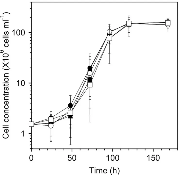
The growth curves of MS-1 cells in the culture medium in the absence of PEG and in the presence of 0.5% PEG200, 0.5% PEG6,000, 0.5% PEG20,000, and 0.5% PEG500,000 are shown inFig 1. The final cell concentration reached (1.55 ± 0.27) × 108, (1.60 ± 0.25) × 108,
of the above cultural conditions according to electron microscopy (seeFig 2). Note however that the size of the cells was smaller in the presence of PEG20,000 and PEG500,000 in the cul-ture medium than that in the presence of PEG200 and PEG6,000, and that the addition of PEG500,000 discouraged the formation of magnetosomes. The number and size distribution of magnetosomes in each cell were determined from TEM images. The average number of magne-tosomes per cell without PEG and with PEG200, PEG6,000, PEG20,000, and PEG500,000 Fig 1. Effect of PEGs added to the culture medium on the growth ofM.magnetotacticumMS-1.The cell concentration was evaluated, counting directly the number of cells in the culture medium using a Bacteria Counting Chamber. The closed circles, squares and triangles, and open circles and squares, respectively, correspond to the growth curves in the absence of PEG and in the presence of PEG200, PEG6,000, PEG20,000, and PEG500,000. The average values were calculated from three independent experiments. The error bars represent the standard deviations.
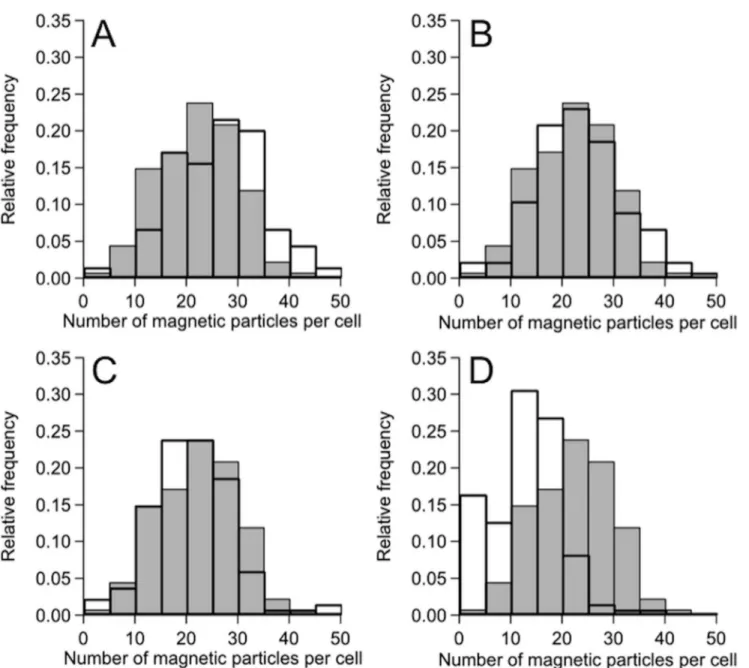
were, respectively, 17.6, 21.8, 17.4, 14.2, and 7.2 (seeTable 1andFig 3). There was approxi-mately 24% increase in the average number of magnetosomes in the case of PEG200 (Fig 3A). Obviously, the culture medium containing PEG200 enhanced the synthesis of magnetosomes. Furthermore, the size of magnetosomes synthesized in the presence of PEG200 was larger than that in the absence of PEG200 by approximately 10%, noting that 34.1 ± 7.9 nm in the former case, while 30.9 ± 7.3 nm in the latter (seeS2 Fig).
The magnetic properties of MS-1 cells grown in the culture medium containing PEG200 were measured using a SQUID at 300 K. The mass magnetization is shown inS3 Fig. The satu-ration mass magnetization, residual mass magnetization and coercivity of strain MS-1 cultivat-ed in the absence of PEG200 were 1.07 A m2kg-1, 0.20 A m2kg-1and 11690 A m-1, whereas those in the presence of PEG200 were 1.15 Am2kg-11, 0.23 A m2kg-1and 8680 A m-1. In other words, the magnetosomes showed soft ferromagnetic characteristics and the saturation mass magnetization increased by approximately 7% by adding PEG200 to the culture medium. It is supposed that the enhancement of the saturation mass magnetization in the presence of PEG200 might have been caused mainly by the increase in the average number of magneto-somes per cell (seeFig 3A), noting that the magnetization can also be changed by the alteration in the crystallinity and the size and number of magnetic domains in the particles, analyses of which were however beyond the scope of the present study. It is supposed that the magneto-somes synthesized in MS-1, which had been cultivated in the presence of PEG200, were well crystallized as in the case of those cultivated in the absence of PEG200, judging by the magnetic characteristics shown inS3 Figand the previous reports on nanoparticles composed of magne-tite (Fe3O4) [22–25].
In summary, the effect of PEG molecules added to the culture medium on the growth of MS-1 and the formation of magnetosomes was investigated for the first time and we found that the yield of magnetosomes was successfully increased by approximately 30% by adding 0.5% PEG200 to the culture medium. It is known that the lipid composition in the cell membranes of Gram-negative bacteria is altered by PEG [26] and that the activation of some membrane transport proteins in the bacterial membranes is affected by the changes in the lipid membrane composition [27]. In the MTB species, the activation of uptake of iron ions into the membrane vesicles is definitely required for the accumulation of iron ions in the magnetosome membrane vesicles and it was suggested that some membrane transport channel proteins may be playing Table 1. Effect of PEGs on the growth ofM.magnetotacticumMS-1 and the formation of magnetosomes.
Added substance
Time (h)
Final cell concentration*(×108 cells ml-1)
Average number of magnetosomes per cell**
Normalized production rate of magnetosome***
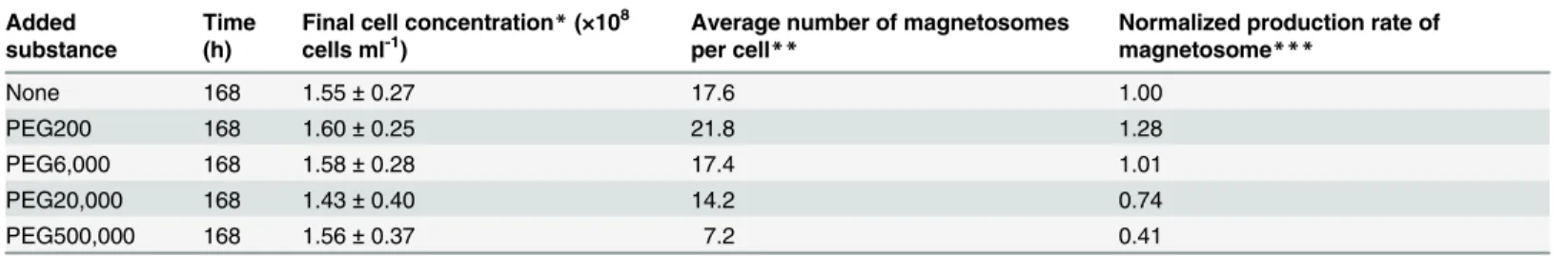
None 168 1.55±0.27 17.6 1.00
PEG200 168 1.60±0.25 21.8 1.28
PEG6,000 168 1.58±0.28 17.4 1.01
PEG20,000 168 1.43±0.40 14.2 0.74
PEG500,000 168 1.56±0.37 7.2 0.41
*The cell concentration was evaluated, counting directly the number of cells in the culture medium using a Bacteria Counting Chamber.
**The average values were obtained from two independent experiments.
***Normalized production rate of magnetosomesPwas defined by
P¼CpNp=ðC0N0Þ;
whereCpandNpare, respectively, thefinal cell concentration and the average number of magnetosomes synthesized in each cell in the presence of
PEG, whereasC0andN0are those in the absence of PEG.
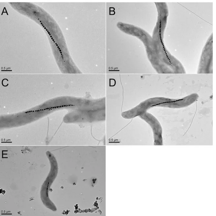
an important role for the formation of magnetosomes in Magnetospirillum species [28–30]. Al-though the actual mechanism of the enhancement of the yield of magnetosomes induced by the addition of PEG200 to the culture medium has not yet been clearly understood, we suppose that PEG200 may promote the uptake of iron ions via the activation of some transport proteins in the magnetosome membranes, knowing that PEG200 permeates the cell walls thanks to its low molecular weight [26]. Since PEG200 is non-toxic and low-cost [31], the present Fig 2. TEM images of magnetosomes synthesized inM.magnetotacticumMS-1 incubated in the culture medium containing PEGs.(A)
Magnetosomes synthesized in the absence of PEG; (B), (C), (D), (E) Magnetosomes synthesized in the presence of 0.5% PEG200, 0.5% PEG6,000, 0.5% PEG20,000, and 0.5% PEG500,000. The scale bars represent 0.5μm.
methodology may well be utilized for the production of magnetosomes in large-scale bioreac-tors. We suppose that mass production of magnetosomes may eventually become possible by discovering appropriate molecules from the point of view of efficient transport of iron ions through the magnetosome membrane vesicles.
Supporting Information
S1 Fig. Number of magnetosomes in each cell grown in the culture medium supplemented with PEG6,000.
(TIFF)
Fig 3. Distribution of the number of magnetosomes in each cell grown in the culture medium containing PEGs.(A), (B), (C), (D) Magnetosomes synthesized in the presence of 0.5% PEG200, 0.5% PEG6,000, 0.5% PEG20,000, and 0.5% PEG500,000. The histograms represent the number distributions of magnetosomes in the absence (gray bars) and presence (solid bars) of PEGs.
S2 Fig. Distribution of the size of magnetosomes in each cell grown in the culture medium supplemented with PEG200.The histograms represent the size distributions of magneto-somes in the absence (gray bars) and presence (solid bars) of PEG200.
(TIFF)
S3 Fig. Mass magnetization of magnetosomes synthesized inM.magnetotacticumMS-1 cultivated in the culture medium in the absence (black circles) and presence (white circles) of PEG200.
(TIFF)
S1 Table. Effect of several substances added to the culture medium on the growth ofM.
magnetotacticumMS-1 and the formation of magnetosomes.
(DOCX)
S1 Text. Effect of substances added to the culture medium on the growth ofM. magnetotac-ticumMS-1 and the formation of magnetosomes.
(DOCX)
S2 Text. Number of magnetosomes in each cell grown in the culture medium.
(DOCX)
Acknowledgments
Part of this study has been supported by a Grant for the Program for the Strategic Research Foundation at Private Universities S1101017 organized by the Ministry of Education, Culture, Sports, Science and Technology (MEXT), Japan, since April 2011. We would like to thank Mr. Keiichi Hirakawa, the Technical Manager of the Bio-Nano Electronics Research Centre, Toyo University, for his assistance with TEM observations.
Author Contributions
Conceived and designed the experiments: HS HK T. Maekawa. Performed the experiments: HS YN. Analyzed the data: HS HK T. Mizuki YN AI T. Maekawa. Contributed reagents/materials/ analysis tools: HS HK T. Mizuki YN AI T. Maekawa. Wrote the paper: HS T. Maekawa.
References
1. Blakemore R (1975) Magnetotactic bacteria. Science 190: 377–379. PMID:170679
2. Bazylinski DA, Frankel RB (2004) Magnetosome formation in prokaryotes. Nat. Rev. Microbiol. 2: 217– 230. PMID:15083157
3. Faivre D, Schüler D (2008) Magnetotactic bacteria and magnetosomes. Chem. Rev. 108: 4875–4898. doi:10.1021/cr078258wPMID:18855486
4. Jogler C, Schüler D (2009) Genomics, genetics, and cell biology of magnetosome formation. Annu. Rev. Microbiol. 63: 501–521. doi:10.1146/annurev.micro.62.081307.162908PMID:19575557 5. Pan Y, Deng C, Liu Q, Petersen N, Zhu R (2004) Biomineralization and magnetism of bacterial
magne-tosomes. Chin. Sci. Bull. 49: 2563–2568.
6. Schüler D (2008) Genetics and cell biology of magnetosome formation in magnetotactic bacteria. FEMS Microbiol. Rev. 32: 654–672. doi:10.1111/j.1574-6976.2008.00116.xPMID:18537832 7. Sun JB, Duran JH, Dai SL, Ren J, Guo L, Jiang W, et al. (2008) Preparation and anti-tumor efficiency
evaluation of doxorubicin-loaded bacterial magnetosomes: magnetic nanoparticles as drug carriers iso-lated fromMagnetospirillum gryphiswaldense. Biotechnol. Bioeng. 101: 1313–1320. doi:10.1002/bit. 22011PMID:18980188
9. Li A, Zhang H, Zhang X, Wang Q, Tian J, Li Y, et al. (2010) Rapid separation and immunoassay for low levels of Salmonella in foods using magnetosome-antibody complex and real-time fluorescence quanti-tative PCR. J. Sep. Sci. 33: 3437–3443. doi:10.1002/jssc.201000441PMID:20886524
10. Wacker R, Ceyhan B, Alhorn P, Schueler D, Lang C, Niemeyer CM (2007) Magneto immuno-PCR: a novel immunoassay based on biogenic magnetosome nanoparticles. Biochem. Biophys. Res. Com-mun. 357: 391–396. PMID:17428442
11. Yoshino T, Hirabe H, Takahashi M, Kuhara M, Takeyama H, Matsunaga T (2008) Magnetic cell separa-tion using nano-sized bacterial magnetic particles with reconstructed magnetosome membrane. Bio-technol. Bioeng. 101: 470–477. doi:10.1002/bit.21912PMID:18421798
12. Matsunaga T, Kamiya S (1987) Use of magnetic particles isolated from magnetotactic bacteria for en-zyme immobilization. Appl. Microbiol. Biotechnol. 26: 328–332.
13. Xiang L, Bin W, Huali J, Wei J, Jiesheng T, Feng G, et al. (2007) Bacterial magnetic particles (BMPs)-PEI as a novel and efficient non-viral gene delivery system. J. Gene Med. 9: 679–690. PMID:17605136 14. Tanaka M, Arakaki A, Staniland SS, Matsunaga T (2010) Simultaneously discrete biomineralization of
magnetite and tellurium nanocrystals in magnetotactic bacteria. Appl. Environ. Microbiol. 76: 5526– 5532. doi:10.1128/AEM.00589-10PMID:20581185
15. Blakemore RP, Maratea D, Wolfe RS (1979) Isolation and pure culture of a freshwater magnetic spiril-lum in chemically defined medium. J. Bacteriol. 140: 720–729. PMID:500569
16. Matsunaga T, Tsujimura N, Kamiya S (1996) Enhancement of magnetic particle-production by nitrate and succinate fed-batch culture ofMagnetospirillumsp. AMB-1. Biotechnol. Tech. 10: 495–500 17. Heyen U, Schüler D (2003) Growth and magnetosome formation by microaerophilicMagnetospirillum
strains in an oxygen-controlled fermentor. Appl. Microbiol. Biotechnol. 61: 536–544. PMID:12764570 18. Sun JB, Zhao F, Tang T, Jiang W, Tian JS, Li Y, et al. (2008) High-yield growth and magnetosome for-mation byMagnetospirillum gryphiswaldenseMSR-1 in an oxygen-controlled fermentor supplied solely with air. Appl. Microbiol. Biotechnol. 79: 389–397. doi:10.1007/s00253-008-1453-yPMID:18425510 19. Zhang Y, Zhang X, Jiang W, Li Y, Li J (2011) Semicontinuous culture ofMagnetospirillum gryphiswal-denseMSR-1 cells in an autofermentor by nutrient-balanced and isosmotic feeding strategies. Appl. Environ. Microbiol. 77 (17): 5851–5856. doi:10.1128/AEM.05962-11PMID:21724877
20. Yang J, Li S, Huang X, Tang T, Jiang W, Zhang T, et al. (2013) A key time point for cell growth and mag-netosome synthesis ofMagnetospirillum gryphiswaldensebased on real-time analysis of physiological factors. Front. Microbiol. 4: 210. doi:10.3389/fmicb.2013.00210PMID:23898327
21. Moisescu C, Ardelean II, Benning L (2014) The effect and role of environmental conditions on magneto-some synthesis. Front. Microbiol. 5: 49. doi:10.3389/fmicb.2014.00049PMID:24575087
22. Mann S, Frankel RB, Blackmore RP (1984) Structure, morphology and ctystal growth of bacterial mag-netite. Nature 310: 405–407.
23. Staniland S, Williams W, Telling N, Laan GVD, Harrison A, Ward B (2008) Controlled cobalt doping of magnetosomesin vivo. Nat. Nanotechnol. 3: 158–162. doi:10.1038/nnano.2008.35PMID:18654488 24. Yang T, Shen C, Li Z, Zhang H, Xiao C, Chen S, Xu Z, et al. (2005) Highly ordered self-assembly with
large area of Fe3O4nanoparticles and the magnetic properties. J. Phys. Chem. B 109: 23233–23236. PMID:16375287
25. Hui C, Shen C, Yang T, Bao L, Tian J, Ding H, et al. (2008) Large-scale Fe3O4nanoparticles soluble in
water synthesized by a facile method. J. Phys. Chem. C 112: 11336–11339.
26. Halverson LJ, Firestone MK (2000) Differential effects of permeating and nonpermeating solutes on the fatty acid composition of Pseudomonas putida. Appl. Environ. Microbiol. 66: 2414–2421. PMID: 10831419
27. Nomura T, Cranfield CG, Deplazes E, Owen DM, Macmillan A, Battle AR, et al. (2012) Differential ef-fects of lipids and lyso-lipids on the mechanosensitivity of the mechanosensitive channels MscL and MscS. Proc. Natl. Acad. Sci. USA. 109: 8770–8775. doi:10.1073/pnas.1200051109PMID:22586095 28. Nakamura C, Burgess JG, Sode K, Matsunaga T (1995) An iron-regulated gene, magA, encoding an
iron transport protein ofMagnetospirillumsp. strain AMB-1.J. Biol. Chem. 270: 28392–28396. PMID: 7499342
29. Grünberg K, Müller EC, Otto A, Reszka R, Linder D, Kube M, et al. (2004) Biochemical and proteomic analysis of the magnetosome membrane inMagnetospirillum gryphiswaldense. Appl. Environ. Micro-biol. 70: 1040–1050. PMID:14766587
30. Nies DH (2003) Efflux-mediated heavy metal resistance in prokaryotes. FEMS Microbiol. Rev. 27: 313–339. PMID:12829273