Angiotensin-(1-7) receptor Mas is an essential modulator of extracellular matrix
protein expression in the heart
Elisandra Gava
a,e, Carlos Henrique de Castro
b,c,e, Anderson J. Ferreira
a,e, Heloísa Colleta
a,e,
Marcos B. Melo
b,e, Natalia Alenina
d, Michael Bader
d, Laser A. Oliveira
a,
Robson A.S. Santos
b,e, Gregory T. Kitten
a,e,⁎
aDepartment of Morphology, Federal University of Minas Gerais, Belo Horizonte, Brazil
bDepartment of Physiology and Biophysics, Federal University of Minas Gerais, Belo Horizonte, Brazil
cDepartment of Physiological Sciences, Federal University of Goiás, Goiânia, Brazil
dMax-Delbrück-Center for Molecular Medicine, Berlin-Buch, Germany
eNational Institute of Science and Technology in Nanobiopharmaceutics, Brazil
a b s t r a c t
a r t i c l e
i n f o
Article history: Received 17 August 2011
Received in revised form 15 December 2011 Accepted 10 January 2012
Available online 26 January 2012
Keywords: Angiotensin
Renin–angiotensin system Fibrosis
Connective tissue Extracellular matrix Mas receptor
In this study we investigated the effects of genetic deletion of the Angiotensin-(1-7) receptor Mas or the Angio-tensin II receptor AT2on the expression of specific extracellular matrix (ECM) proteins in atria, right ventricles
and atrioventricular (AV) valves of neonatal and adult mice. Quantification of collagen types I, III and VI andfi -bronectin was performed using immunofluorescence-labeling and confocal microscopy. Picrosirius red staining was used for the histological assessment of the overall collagen distribution pattern. ECM proteins, metallopro-teinases (MMP), ERK1/2 and p38 levels were quantified by western blot analysis. Gelatin zymography was used to evaluate the activity of MMP-2 and MMP-9. We observed that the relative levels of collagen types I and III and fibronectin are significantly higher in both the right ventricle and AV valves of neonatal Mas−/−mouse hearts
(e.g., collagen type I: 85.28 ±6.66 vs 43.50 ± 4.41 arbitrary units in the right ventricles of Mas+/+mice).
Con-versely, the level of collagen type VI was lower in the right ventricle and AV valves of Mas−/−mice. Adult
Mas−/−mouse hearts presented similar patterns as observed in neonates. No significant differences in ECM
pro-tein level were detected in atria. Likewise, no changes in ECM levels were observed in AT2knockout mouse
hearts. Although deletion of Mas induced a significant reduction in the level of the active form of MMP-2 in ne-onate hearts and a reduction of both MMP-2 and MMP-9 in adult Mas−/−mice, no significant differences were
observed in MMP enzymatic activities when compared to controls. The levels of the active, phosphorylated forms of ERK1/2 and p38 were higher in hearts of both neonatal and adult Mas−/−mice. These observations suggest
that Mas is involved in the selective expression of specific ECM proteins within both the ventricular myocardium and AV valves. The changes in the ECM profile may alter the connective tissue framework and contribute to the decreased cardiac performance observed in Mas−/−mice.
© 2012 Elsevier B.V.Open access under the Elsevier OA license.
1. Introduction
The extracellular matrix (ECM) has been described as a supportive scaffold which is important in both formation and maintenance of tis-sues. In the heart, ECM forms an elaborate, stress-tolerant network, interconnecting myocytes to each other and myocytes to capillaries
within the ventricular wall[1]. Interactions between cells and the
surrounding ECM play critical roles in a number of cellular processes, including migration, proliferation, differentiation and survival. The
interstitial network within the myocardium is composed
predomi-nantly offibrillar collagen types I and III[2]. Cardiacfibrillar collagen
provides structural scaffolding for cardiomyocytes and coronary ves-sels and imparts cardiac tissue with physical properties that include
stiffness and resistance to deformation[2,3]. It is now clear that the
ECM is a dynamic structure whose organization and composition are known to modulate various cellular processes. Events that alter the molecular composition of the ECM, or the structural organization of ECM components, can induce profound changes in cellular
func-tions[4]. Excessive deposition of collagen is thought to contribute to
abnormal stiffness and function of the ventricular myocardium[5].
In many cases these changes are associated with activation of
humor-al systems such as the renin–angiotensin system (RAS)[6].
Components of the circulating and local RAS are closely involved
in the development of myocardial fibrosis in hypertensive heart
⁎ Corresponding author at: Department of Morphology, Av. Antônio Carlos, 6627, ICB, UFMG, 31270-901, Belo Horizonte, MG, Brazil. Tel.: +55 31 3409 2806; fax: +55 31 3409 2810, USA-eFax: +1 815 377 0222.
E-mail addresses:[email protected](A.J. Ferreira),[email protected]
(G.T. Kitten).
0167-0115 © 2012 Elsevier B.V.Open access under the Elsevier OA license. doi:10.1016/j.regpep.2012.01.001
Contents lists available atSciVerse ScienceDirect
Regulatory Peptides
disease and chronic heart failure. The classical effector of this system, the octapeptide Angiotensin (Ang) II, exerts its effects through
specif-ic Ang II receptor isoforms, AT1and AT2. Ang II binds AT1receptor and
stimulates synthesis and deposition of collagen in a dose-dependent manner and suppresses the activity of matrix metalloproteinase (MMP) 1, an enzyme which plays an important role in interstitial
collagen degradation[7]. On the other hand, accumulating lines of
evidence support the broad view that Ang II can bind to the AT2
re-ceptor and induce growth suppression[8]. Conversely, Ang-(1-7), a
biologically active member of the RAS and an endogenous ligand
for the G protein-coupled Mas receptor[9], has been suggested to
act as an antiproliferative[10–14]and anti-fibrotic peptide[13,14].
Ang-(1-7) inhibits growth of cardiomyocytes through Mas-mediated
events, which include ERK1/2 activities [13]. In addition, Ang-(1-7)
and its analog AVE 0991 have been shown to attenuate the
develop-ment of heart failure after myocardial infarction, afinding that suggests
a role for this peptide in cardiac remodeling[15,16]. In keeping with
these data, AVE 0991 also prevented isoproterenol-induced cardiac
remodeling[17]. These effects are apparently independent of changes
in blood pressure since Grobe and colleagues[18,19]have demonstrated
that the anti-fibrotic and anti-hypertrophic actions of Ang-(1-7) are still
observed in Ang II-infused[19]or in DOCA-salt hypertensive rats[18].
Overexpression of the main Ang-(1-7)-forming enzyme, angiotensin-converting enzyme 2 (ACE2), in a rat model of myocardial infarction protected the infarcted myocardium against pathological
remodel-ing and cardiac systolic dysfunction[20]. The anti-fibrotic and
anti-A
B
C
D
E
F
Mas
+/+Mas
-/-Fig. 1.Immunofluorescent localization of Mas in hearts. Mas is present in tricuspid valve (A), right ventricle (C) and atria (E) of adult Mas+/+mouse hearts but absent in the
tricuspid valve (B), right ventricle (D) and atria (F) of adult Mas−/−mouse hearts. No immunostaining was detected in any of the samples when the primary antibody was omitted from the incubation procedure (not shown). Insets are low magnification views of the tissue areas analyzed. Representativefigures of three different animals. Bar = 50μm.
Mas
+/+
Mas
-/-Col I
Col III
FN
Col VI
Mas
+/+
Mas
-/-Right ventricle
Tricuspid valve
0 25 50 75 100 125
Col I Col III FN Col VI
*
*
*
*
Relative Fluorescency
Intensity (AU)
0 25 50 75 100 125
Col I Col III FN Col VI
*
*
*
*
Relative Fluorescency
Intensity (AU)
Right ventricle
0 25 50 75 100 125 150
Col I Col III FN Col VI
*
*
*
*
Relative Fluorescency
Intensity (AU)
0 25 50 75
Col I Col III FN Col VI
Relative Fluorescency
Intensity (AU)
Mas+/+; n=6 Mas-/-; n=6
Tricuspid valve
Mitral valve
Atria
A
B
Fig. 2.Immunofluorescent localization of collagen types I, III and VI andfibronectin in the right ventricle and tricuspid valve of neonatal Mas+/+and Mas−/−mouse hearts (A). The
inset shows the level of immunostaining obtained when the primary antibody was omitted from the incubation procedure. Quantification of ECM proteins in the right ventricle, AV valves and atria of neonatal Mas+/+and Mas−/−mice (B). Values are expressed as arbitrary units (AU). Data are shown as mean ± SEM. *p
hypertrophic actions of ACE2/Ang-(1-7) were also observed in animals (rats and mice) with pulmonary hypertension induced by monocrota-line[21,22]and in rats with pulmonaryfibrosis caused by bleomycin
treatment[23]. Recently, we have shown that deletion of the
Ang-(1-7) receptor Mas markedly decreases cardiac function in adult mice in
part due to changes in collagen levels to a pro-fibrotic profile, i.e.
an increase of collagen types I and III andfibronectin and a decrease
of collagen type VI in left ventricles[24]. However, it is unknown
whether these alterations in ECM extend to other regions of hearts from neonatal and adult mice, such as the right ventricle, atria and atrioventricular (AV) valves. Thus, in the current study we evaluated the presence of Mas in the right ventricle, atria and AV valves of neo-natal and adult mice and checked if its deletion leads to alterations in ECM deposition in these regions. In addition, we investigated
the effects of deletion of the AT2 receptor on the expression of
ECM proteins in adult mouse hearts, since in certain circumstances
AT2receptors appear to be involved in the Ang-(1-7) effects.
2. Material and methods
2.1. Mas knockout mice and AT2knockout mice
Male wild-type (Mas+/+, AT
2
+/y) and Mas and AT
2 knockout
(Mas−/−, AT
2−/y) C57BL/6 mice (adult mice: 8–12 weeks old; neonate
mice: 1 day old) were obtained from the transgenic animal facilities of the Laboratory of Hypertension, Federal University of Minas Gerais, Brazil. All animal procedures were performed in accordance with institutional guidelines (Federal University of Minas Gerais, Brazil).
Genotypes were confirmed by PCR analysis.
2.2. Immunostaining and confocal microscopy
Immunofluorescence-labeling and quantitative confocal microscopy
were used to investigate the distribution and quantity of Mas, collagen
types I, III and VI andfibronectin present in Mas+/+, Mas−/−, AT
2+/yand
AT2−/ymice hearts. Hearts were collected from Mas+/+and Mas−/−
neo-natal (n=6) and adult (n=6) male mice and from AT2+/yand AT2−/yadult
(n=4) male mice, washed in phosphate-buffered saline (PBS) and
cryo-fixed in a−80 °C solution of 80% methanol and 20% dimethyl
sulfox-ide. After 5–7 days of freeze-substitution, samples were embedded in
paraffin following standard methods[24]. Five micrometer thick
sec-tions were mounted on slides, deparaffinized, rehydrated and then
incubated in blocking solution (1% BSA and 0.1% Tween 20 in PBS) at room temperature for 1 h. Sections were incubated overnight at 4 °C with one of the following primary antibodies: rabbit anti-human Mas (1:100, Abcam, Cambridge, MA), rabbit anti-anti-human col-lagen type I (1:400, Rockland Immunochemicals Inc., Gilbertsville, PA), rabbit anti-human collagen type III (1:400, Rockland Immuno-chemicals Inc., Gilbertsville, PA), rabbit anti-human collagen type VI (1:600, Research Diagnostics Inc., Acton, MA) or rabbit anti-human
fibronectin (1:800, Rockland Immunochemicals Inc., Gilbertsville,
PA). After 4–5 rinses in PBS, donkey anti-rabbit IgG conjugated with
Cy3 (1:500, Jackson ImmunoResearch Laboratories, West Grove, PA) was added for 1 h in the dark at room temperature. Following washes with PBS, sections were mounted and viewed with a laser scanning confocal microscope (Zeiss 510Meta). Optimal confocal settings (aperture, gain and laser power) were determined at the beginning of each imaging session and then held constant during the analysis
of all the samples. Nuclei were labeled with 4′
6-diamidino-2-phenylindole dihydrochloride (DAPI) (Molecular Probes, Carlsbad,
Mas+/+ Mas -/-0.00
0.25 0.50 0.75 1.00 1.25 1.50
*
n=4 n=4
Colla
g
en I / GAPDH
Collagen I
GAPDH
A
Mas+/+ Mas -/-0.00
0.25 0.50 0.75 1.00 1.25 1.50
n=2 n=2
Colla
g
en III / GAPDH
Collagen III
GAPDH
B
Mas+/+ Mas -/-0.00
0.25 0.50 0.75 1.00 1.25 1.50 1.75 2.00
*
n=4 n=4
Fibr
onectin / GAPDH
Fibronectin
GAPDH
D
Mas+/+ Mas -/-0.00
0.25 0.50 0.75 1.00 1.25 1.50 1.75 2.00
*
n=8 n=8
Colla
g
en
VI / GAPDH
Collagen VI
GAPDH
C
Fig. 3.Western blotting analysis of the levels of collagen types I (A), III (B) and VI (C) andfibronectin (D) in hearts of neonatal Mas+/+and Mas−/−mice. Densitometry values were
normalized using GAPDH. Data are shown as the SEM. *pb0.05.
A
B
Col I
Col III
FN
Col VI
Mas
+/+
Mas
-/-Mas
+/+
Mas
-/-Right ventricle
Tricuspid valve
Right ventricle
0 25 50 75 100 125
Col I Col III FN Col VI
*
*
*
*
Relative Fluorescency
Intensity (AU)
0 25 50 75 100 125
Col I Col III FN Col VI
*
*
*
*
Relative Fluorescency
Intensity (AU)
0 25 50 75 100 125
Col I Col III FN Col VI
*
*
*
*
Relative Fluorescency
Intensity (AU)
0 25 50 75
Col I Col III FN Col VI
Relative Fluorescency
Intensity (AU)
Mas+/+; n=6 Mas-/-; n=6
Tricuspid valve
Mitral valve
Atria
Fig. 4.Immunofluorescent localization of collagen types I, III and VI andfibronectin in the right ventricle and tricuspid valve of adult Mas+/+and Mas−/−mouse hearts (A).
CA). For quantitative analysis of collagens I, III and VI andfibronectin,
we used the ImageTool 2.0 image analysis program (http://ddsdx.
uthscsa.edu/dig/itdesc.html) to measure thefluorescence intensity in images randomly selected from the right ventricle, AV valves
and atria of Mas+/+and Mas−/−mice and from the right and left
ventricles of AT2+/yand AT2−/ymice. Images were captured at 12 bit
and analyzed in the gray scale range of 0 to 255. Fluorescence inten-sity was measured as an average of the area (i.e. the sum of gray values of all pixels divided by the number of pixels in the area) and
values recorded as arbitrary units (AU). Backgroundfluorescence
was measured and subtracted from the region of interest.
2.3. Histological analysis
Picrosirius red staining was used for histological analysis of total
collagen content in tricuspid valves of neonatal and adult Mas−/−
and Mas+/+mice and right ventricles of adult Mas−/−and Mas+/+
mice (n = 5 per group). Samples were prepared as described above,
sectioned at a thickness of 5μm, stained with picrosirius red (direct
red 80)[25]and then analyzed at 10× magnification using polarizing
filters adapted onto a conventional light microscope.
2.4. Western blotting
Right ventricles from adult mice (n=2–4) and whole hearts from
neonate mice (n=2–8) were collected and homogenized in lysis buffer
containing 1% Nonidet P40, 0.5% sodium deoxycholate, 0.1% SDS, 0.1 mmol/L PMSF, 1 mmol/L pepstatin A, 1 mmol/L leupeptin, and 1 mmol/L aprotinin. Forty micrograms of protein from each sample were separated by electrophoresis on a 10% polyacrylamide gel and
transferred to nitrocellulose membranes. Non-specific binding was
blocked by incubation in 5% milk and 0.1% Tween 20 in Tris-buffered
sa-line. Membranes were probed with specific primary antibodies:
anti-collagen type I (1:500, Rockland Immunochemicals Inc., Gilbertsville, PA), anti-collagen type III (1:3000, Rockland Immunochemicals Inc., Gilbertsville, PA), anti-collagen type VI (1:500, Research Diagnostics
Inc., Acton, MA), anti-fibronectin (1:5000, Rockland
Immunochem-icals Inc., Gilbertsville, PA), anti-p38MAPK (1:1000, Cell Signaling Technology Inc., Danvers, MA), anti-ERK1/2 (1:1000, Cell Signaling Technology Inc., Danvers, MA), anti-MMP-2 (1:500, Chemicon Interna-tional, Billerica, MA), anti-MMP-9 (1:500, Chemicon InternaInterna-tional, Biller-ica, MA) or anti-GAPDH (1:5000, Santa Cruz Biotechnology, Santa Cruz, CA) followed by incubation with enzyme-labeled secondary antibodies. The membrane was incubated in chemiluminescent substrate then
ex-posed tofilm. Immunoreactive bands were quantified by densitometry
using GAPDH, total p38 or total ERK1/2 as a normalization control.
Mas+/+ Mas -/-0.00 0.25 0.50 0.75 1.00 1.25 1.50
**
n=4 n=4 C o lla g e n I / G A P D H Collagen I GAPDHA
Mas+/+ Mas -/-0.00 0.25 0.50 0.75 1.00 1.25 1.50 1.75 2.00
*
n=2 n=2 Fi br on e c ti n / G A PDH Fibronectin GAPDHD
Mas+/+ Mas -/-0.00 0.25 0.50 0.75 1.00 1.25 n=4 n=4 Co ll a g e n V I / G A P D H Collagen VI GAPDH
C
Mas+/+ Mas -/-0.00 0.25 0.50 0.75 1.00 1.25 1.50
**
C o lla g e n III / G
A P D H n=2 n=2 Collagen III GAPDH
B
Fig. 5.Western blotting analysis of the levels of collagen types I (A), III (B) and VI (C) andfibronectin (D) in hearts of adult Mas+/+and Mas−/−mice. Densitometry values were
normalized using GAPDH. Data are shown as the SEM. *pb0.05 and **pb0.001.
Table 1
Echocardiographic analysis of Mas+/+and Mas−/−mice.
Parameter Mas+/+(n = 5) Mas−/−(n = 5)
Cardiac output (mL/min) 18.76 ± 1.30 16.80 ± 1.05 LV internal dimension at diastole (mm) 3.72 ± 0.14 4.20 ± 0.06⁎ LV internal dimension at systole (mm) 2.46 ± 0.14 3.32 ± 0.03⁎ LV ejection fraction (%) 63.48 ± 2.82 42.90 ± 1.26⁎ LV fractional shortening (%) 34.07 ± 2.06 20.93 ± 0.74⁎ LV systolic volume (μL) 37.37 ± 2.70 33.85 ± 2.05 End diastolic LV volume (μL) 59.45 ± 5.04 78.72 ± 2.80⁎ End systolic LV volume (μL) 22.08 ± 2.97 44.87 ± 1.24⁎ Heart rate (bpm) 492.60 ± 25.70 487.80 ± 20.70 RV fractional area change (FAC) (%) 57.58 ± 1.20 38.69 ± 2.30⁎ Data are reported as mean ± SEM. Statistical analyses were performed using Student's ttest. LV: left ventricle; RV: right ventricle. *pb0.05.
2.5. Gelatin zymography
Right ventricles from adult mice (n = 3) and whole hearts from
neonate mice (n = 3–8) were homogenized in 50 mmol/L Tris–HCl
pH 7.4, 0.2 mol/L NaCl, 0.1% Triton, 10 mmol/L CaCl2and 1% Protease
Inhibitor Cocktail (PI860, Sigma Chemical Co., St. Louis, MO) using an ultrasonic cell disruptor. The homogenates were incubated for 2 h at
4 °C and centrifuged at 2080gfor 20 min at 4 °C. The supernatants
were removed and the pellets were suspended once again in the same solution as described above, heated to 60 °C for 5 min and
cen-trifuged at 2080gfor 20 min at 4 °C. Protein was quantified using the
Bradford dye binding assay kit (BioAgency, São Paulo, SP, Brazil). Twenty micrograms of protein extract obtained as above were electrophoresed on a 10% SDS polyacrylamide gel containing 0.1% gelatin (used as protein substrate) at 4 °C under non-reducing con-ditions. After electrophoresis, the gel was washed twice, and then gently shaken under 2.5% Triton X-100 for 30 min at room tempera-ture to remove SDS. The gel was incubated overnight in a 50 mmol/L
Tris–HCl solution pH 7.4, containing 0.01 mol/L CaCl2, 0.1 mol/L NaCl
and 0.03% sodium azide at 37 °C. The gel was then stained with Coo-massie Brilliant Blue (0.5% dye in 20% methanol and 10% acetic acid) for 1 h. Unstained bands indicating gelatinolytic activity were seen after slight destaining with 30% ethanol and 10% acetic acid. Quanti-tative assessment of band intensity was done by densitometry using
the ImageJ image processing program (http://rsb.info.nih.gov/ij/).
All experiments were done in quadruplicate.
2.6. Echocardiographic analysis
Cardiac morphology and function in adult Mas+/+and Mas−/−
mice (9–10 weeks old, n = 5) were assessed noninvasively using a
high-frequency, high-resolution echocardiographic system consisting
of a VEVO 2100 ultrasound machine equipped with a 30–40 MHz
bifrequencial transducer (Visual Sonics, Toronto, Canada). The mice
were anesthetized with 5% isoflurane during 1 min for induction.
Anesthesia was sustained via a nose cone with 1.25% isoflurane.
The anterior chest was shaved and the mice were placed in supine position on an imaging stage equipped with built-in electrocardio-graphic electrodes for continuous heart rate monitoring and a heater to maintain the body temperature at 37 °C. High-resolution images were obtained in the right and left parasternal long and short axes and apical orientations. Standard B-mode images of the heart and
pulsed Doppler images of the mitral and tricuspid inflow were
ac-quired. Left ventricular (LV) dimensions and wall thickness were measured at the level of the papillary muscles in the left and right parasternal short axis during the end-systole and end-diastole. LV ejection fraction (EF), fractional shortening (FS) and mass were measured. All the measurements and calculations were done in accordance with the American Society of Echocardiography. The following M-mode measurements were performed: LV internal di-mensions at diastole and systole (LVIDD and LVIDS, respectively), LV posterior wall dimensions at diastole and systole (LVPWD and LVPWS, respectively) and interventricular septal dimensions at
Mas
+/+Mas
-/-Tricuspid valve
Neonate
Tricuspid valve
Adult
Right ventricle
Adult
Fig. 6.Sections of tricuspid valves from neonatal and adult Mas−/−and Mas+/+mice and right ventricles of adult Mas−/−and Mas+/+mice stained with picrosirius red and
diastole and systole (IVSDD and IVSDS, respectively). Based on these parameters, end diastolic and end systolic LV volumes (EDLVV and ESLVV, respectively), FS, EF, stroke volume (SV) and cardiac output (CO) were calculated. Also, in the right parasternal short axis view, bidimensional images from the right ventricle were ac-quired for fractional area change (FAC) calculation.
2.7. Statistical analysis
Data are reported as mean ± SEM. Statistical analyses were
per-formed using the Mann Whitney test or Student'sttest
(echocardiog-raphy). p values of 0.05 or less were considered significant.
3. Results
First, we evaluated the presence of Mas in the different cardiac
chambers of mice. Immunofluorescence-labeling and confocal
mi-croscopy demonstrated that, in addition to left ventricles[24], Mas
is expressed in the tricuspid valve, right ventricle and atria of adult
Mas+/+mice (Fig. 1A, C and E). As expected, no expression of Mas
was observed in hearts of Mas−/−adult mice (Fig. 1B, D and F).
Fig. 2A shows immunostaining profile of several ECM proteins in
the right ventricles and tricuspid valves of Mas+/+and Mas−/−
neo-natal mice. Quantitative analysis of the level of immunofluorescent
staining revealed that hearts from Mas−/−neonatal mice present
sig-nificantly higher levels of collagen types I and III andfibronectin in
right ventricles and tricuspid valves (Fig. 2B). In contrast, the level
of collagen type VI in Mas−/−neonatal mice was lower. A similar
pat-tern was observed in mitral valves of neonatal Mas−/−mice (Fig. 2B).
However, no significant changes were viewed in the expression of
collagen types I, III and VI andfibronectin in atria of Mas−/−neonatal
mice (Fig. 2B). To confirm the protein expression profile of ECM
pro-teins in hearts of Mas−/−neonatal mice, western blotting analysis
was performed. In agreement with the immunofluorescence-labeling
findings, we found that the levels of collagen types I and III andfi
bro-nectin were higher while the level of collagen type VI was lower in
hearts of Mas−/−neonatal mice (Fig. 3).
A similar pattern of ECM protein expression was observed in
hearts of adult Mas−/−mice. Indeed, Mas de
ficient adult mice
pre-sented significantly higher levels of collagen types I and III andfi
bro-nectin and a lower level of collagen type VI in the right ventricle and
tricuspid valves (Fig. 4A and B). These changes were also detected in
mitral valves of adult Mas−/−mice (Fig. 4B). Nevertheless, no
signif-icant changes in the expression of these proteins were observed in
atria of adult Mas−/−mice (Fig. 4B). Importantly, these alterations
were confirmed by western blotting analysis (Fig. 5). Of note, the
fibrosis observed in Mas−/−mice was accompanied by a significant
reduction in the right ventricular function, i.e. these mice presented a reduced fractional area change (FAC) of the right ventricle. Also,
in keeping with a recent study[24], Mas−/−mice showed an
im-paired left ventricular function (Table 1).
Picrosirius red staining revealed that total collagen content
pre-sent in tricuspid valves of neonatal and adult Mas−/−mice, as well
as in right ventricles of adult Mas−/−mice appears to be significantly
higher when compared with Mas+/+mice (Fig. 6). An increased
bire-fringence pattern observed when polarization microscopy was used
indicates that morefibrous collagen was present. Such an increase
was evident in tricuspid valves of neonatal and adult Mas−/−mice
(Fig. 6). Although sections of the right ventricular wall were stained more intensely with picrosirius red when they were observed with
conventional microscopy (Fig. 6), no significant increases in
polariza-tion were evident (data not shown). Together, these results suggest
Col I
Col III
FN
Col VI
AT2+/y; n=4 AT2-/y; n=4
A
AT
2
+/y
AT
2
-/
y
Right ventricle
0 20 40 60 80
Col I Col III FN Col VI
Relative Fluorescency
Intensity (AU)
B
Right ventricle
C
0 20 40 60 80
Col I Col III FN Col VI
Relative Fluorescency
Intensity (AU)
Left ventricle
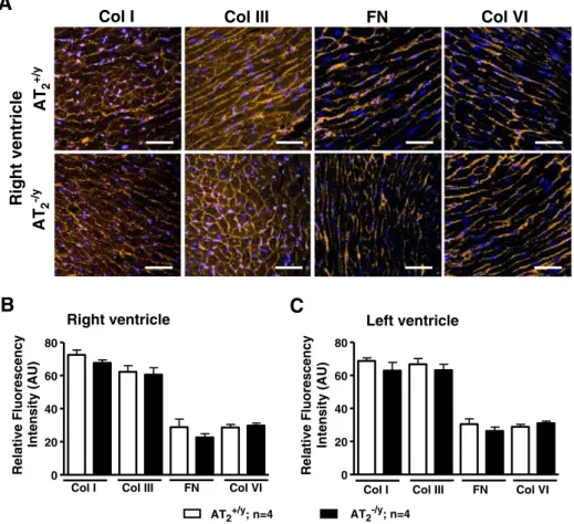
Fig. 7.Immunofluorescent localization of collagen types I, III and VI andfibronectin in the right ventricle of adult AT2+/yand AT2−/ymice hearts (A). Quantification of ECM proteins in
that the increased amount of collagen observed with conventional picrosirius red staining, as well as with immunostaining for collagen
types I and III, is present as microfibrillar material.
Because AT2receptors have been suggested to be involved in some
Ang-(1-7) effects, we evaluated the expression of ECM proteins in
hearts of AT2deficient mice.Fig. 7A shows the immunostaining
pro-file of the levels of collagen types I, III and VI and fibronectin in
right ventricles of AT2+/yand AT2−/yadult mice. Quantitative analysis
of the level of immunofluorescent staining demonstrated that right
(Fig. 7B) and left (Fig. 7C) ventricles from AT2−/ymice present no
sig-nificant alterations in the expression of these proteins as compared
with AT2+/ymice.
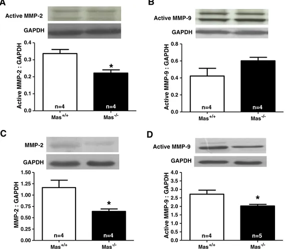
It is well-known that MMPs have a major role in ECM degradation. Thus, our next objective was to investigate if MMPs were involved in
the deposition of ECM proteins in hearts of Mas−/−mice. De
ficiency
of Mas induced a significant reduction in the level of the active form
of MMP-2 in neonate hearts (Fig. 8A). In contrast, no significant
alter-ation was observed in the level of the active form of MMP-9 in hearts
of neonate Mas−/−mice (Fig. 8B). In adult hearts, western blotting
analysis detected the presence of a single MMP-2 band in the gel
whose density was lower in Mas−/− mice (Fig. 8C). Furthermore,
the level of the active form of MMP-9 was significantly lower in
Mas deficient mice (Fig. 8D).
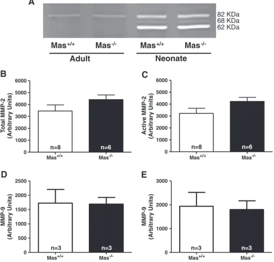
As observed inFig. 9A, gelatinolytic activity of the active form of
MMP-2 (62 kDa) and its precursor form (68 kDa) were detected only in neonate hearts. Although the active form of MMP-9 (82 kDa) was detected on zymographs in both neonate and adult
hearts (Fig. 9A), no significant differences were observed in
metallo-proteinase activity between the groups (Fig. 9B–E).
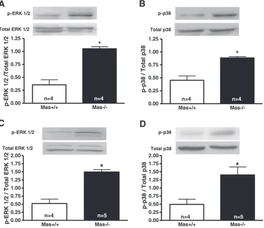
In order to investigate the potential involvement of protein ki-nases in ECM deposition, the levels of phosphorylated ERK1/2 and p38 protein expression were evaluated in hearts of neonatal and
adult Mas−/−mice. We observed that the levels of ERK1/2 and
p-p38 were higher in hearts of both neonatal and adult Mas−/−mice
when compared with control animals (Fig. 10).
4. Discussion
We have previously described that genetic deletion of Mas leads
to a marked impairment of cardiac function[24]. Systolic tension,
+ dT/dt and−dT/dt were significantly lower in isolated hearts of
Mas-deficient mice. Echocardiographic measurements revealed a
lower fractional shortening, posterior wall thickness in systole and a
higher left ventricular end-systolic dimension. In addition, Mas−/−
mice presented a higher coronary perfusion pressure compared
with Mas+/+mice. These alterations appear to be caused, at least
partially, by severe alterations in collagen protein expression in left
ventricles[24]. In the current study, we extended the observation
that Mas is a key modulator of several ECM proteins in mouse hearts.
Neonatal and adult Mas knockout mice showed significantly higher
levels of collagen types I and III andfibronectin in both right ventricle
and AV valves. In contrast, the level of collagen type VI in these
tis-sues was lower in Mas−/−mice. In agreement with our data, many
studies have demonstrated that Ang-(1-7) induces antiproliferative
and anti-fibrotic effects by acting through the Mas receptor[12–14].
Interestingly, the alterations in cardiac expression of ECM proteins
were not observed in the atria of neonatal or adult Mas−/− mice,
Mas+/+ Mas -/-0.0
0.2 0.4 0.6 0.8
n=4 n=4
Active MMP-9 : GAPDH
B
Active MMP-9
GAPDH
Mas+/+ Mas -/-0.0
0.5 1.0 1.5 2.0 2.5 3.0 3.5 4.0
*
n=4 n=5
Active MMP-9 : GAPDH
D
Active MMP-9
GAPDH
Mas+/+ Mas -/-0.00
0.25 0.50 0.75 1.00 1.25 1.50
*
n=4 n=4
MMP-2 : GAPDH
C
MMP-2
GAPDH
Mas+/+ Mas -/-0.0
0.1 0.2 0.3 0.4
*
n=4 n=4
Active MMP-2 : GAPDH
A
Active MMP-2
GAPDH
Fig. 8.Western blotting analysis of the levels of MMP-2 and MMP-9 in neonatal hearts and right ventricles of adult Mas+/+and Mas−/−mice. Active form of MMP-2 in neonate
indicating that the modulation of ECM protein expression by Mas is
re-stricted to specific heart regions.
Previous biochemical and molecular studies have been limited to
the description of the expression of Mas in the whole heart[24,26].
Here, we showed by immunofluorescence-labeling the precise
distri-bution of Mas in different areas of the heart. Mas was presented in the right ventricle, AV valves and atria. Thus, the lack of effects of genetic deletion of Mas in atria could not be attributed to the absence of this receptor in these chambers.
Surprisingly, in contrast to collagen types I and III, the levels of collagen type VI were decreased in ventricles and AV valves of
neo-natal and adult Mas−/−mice, indicating that Mas selectively
regu-lates the expression of specific ECM proteins in mouse hearts.
Collagen type VI forms a microfibrillar extracellular network that is
thought to function as an elastic bridge between cell surfaces or basement membranes and the structural ECM scaffold. It is possible that collagen type VI is important for maintaining an appropriate re-lationship between cells and ECM in cardiac structures exposed to variable states of wall stress. Heart structures must acutely regulate
tensile support in response to alterations in the cardiac cycle[27].
Decreased amounts of collagen type VI may therefore adversely af-fect cardiac function.
Of note, neonatal mice presented a similar pattern of ECM protein expression as observed in adult mice. These results might suggest that the primary cause of the structural and functional disturbances seen in hearts of adult mice is due to changes in cardiac expression of ECM proteins and not due to chronic adaptative alterations. Indeed,
C57BL6 Mas−/−mice presented normal blood pressure as assessed by
intra-arterial catheter measurements [24,28]. Furthermore, it was
found that Mas−/−mice are healthy, grow normally, display no
dif-ference in drinking behavior and show no obvious developmental
abnormalities[29]. However, whether this possibility is true remains
to be elucidated and warrants further investigations. This includes,
but is not limited to, evaluation of shift in elastic versus fibrous
ECM components, changes that may occur in heart function from neonatal to adulthood and associations between heart function and
expression of specific ECM components.
In certain circumstances, and in some tissues, AT2receptors
ap-pear to be involved in the Ang-(1-7) effects [30]. Furthermore,
physical interaction between Mas and AT2in selected tissues such
as the heart has been suggested as a putative mechanism for
Ang-(1-7) actions[31]. In order to investigate the participation of AT2
receptors in the anti-trophic and anti-fibrotic actions of the
Ang-(1-7), we compared the effects of genetic deletion of Mas and AT2
receptors on the ECM protein expression in the heart. In contrast
to the markedfibrotic pattern observed in Mas−/−mice, no
signif-icant alterations in the levels of collagen types I, III and VI andfi
bro-nectin were observed in AT2−/ymice. In agreement with this data,
Ichihara et al.[32]also failed to demonstrate any significant
differ-ence in the collagen deposition between AT2−/yand AT2+/ymice in
normal conditions. Thesefindings suggest that the modulation of ECM
proteins by Ang-(1-7) is AT2-indepentent. Of note, it has been recently
demonstrated that the anti-hypertrophic effect of Ang-(1-7) in
cardio-myocytes is also independent of AT2receptors[33].
0 1000 2000 3000 4000 5000 6000
Active MMP-2
(Arbitrar
y Units)
n=8 n=6
Mas+/+ Mas -/-0
1000 2000 3000 4000 5000 6000
n=8 n=6
T
otal MMP-2
(Arbitrar
y Units)
0 1000 2000 3000
MMP-9
(Arbitrar
y Units)
n=3 n=3
0 500 1000 1500 2000 2500
n=3 n=3
MMP-9
(Arbitrar
y Units)
B
C
D
E
Mas
+/+Mas
-/-Mas
+/+Mas
-/-Adult
Neonate
A
82 KDa
68 KDa
62 KDa
Mas+/+ Mas
-/-Mas+/+ Mas
-/-Mas+/+ Mas
-/-Fig. 9.Representative gelatin zymogram of the active form of MMP-2 (62 kDa), precursor form of MMP-2 (68 kDa) and active form of MMP-9 (82 kDa) (A). Quantification of the
gelatinolytic activity of the total MMP-2 in neonatal mice (B), active form of MMP-2 in neonatal mice (C), active form of MMP-9 in neonatal mice (D) and active form of MMP-9 in right ventricles of adult mice (E). MMP-2 activity was not detected in right ventricles of adult mice. Data are shown as the SEM. (n = 3–8).
The composition of the extracellular matrix, a complex network of structural proteins including collagen types I and III, provides archi-tectural support for the muscle cells and plays an important role in
myocardial function[34]. Collagen type VI is a major microfibrillar
component of extracellular matrices and is predicted to play a key role in the maintenance of tissue integrity by providing a structural link between different components of connective tissues, basement membranes, and cells binding to collagen type I, collagen type III and other matrix components. A number of studies have shown changes in the accumulation, composition, or organization of these interstitial collagens, including types I, III, and VI, during cardiac development and disease. The accumulation of myocardial collagen lead to interstitial
and perivascularfibrosis which has been correlated with left ventricular
early diastolic and systolic dysfunction[6,35,36]. MMP-2 and MMP-9
are expressed by a multitude of cell types including cardiac myocytes
andfibroblasts. It was reported that both enzymes are highly
upregu-lated in hypertrophic and failing hearts and they have been implicated in the progression of ventricular dilatation and the development of heart failure. MMP-2 and MMP-9 degrade various types of collagen,
fibronectin, and others proteins that are accumulating in the
dam-aged myocardium undergoingfibrosis[37]. Thus, we selected these
two metalloproteinases to correlate them to the alterations in the ECM proteins evaluated in our study. In addition, the deposition of ECM proteins in the heart depends on the balance between the syn-thesis of these proteins and their degradation by metalloproteinases
[38]. There is little data available concerning the effects of Ang-(1-7)
and its receptor Mas on the biosynthesis of ECM proteins or on the activity of metalloproteinases. We observed that the level of the
ac-tive form of MMP-2 in hearts of neonatal Mas−/−mice is lower than
in Mas+/+mice while the active form of MMP-9 is not altered. Also,
the level of MMP-2 and of the active form of MMP-9 were significantly
lower in adult Mas−/−hearts. Altogether, thesefindings suggest that
the pro-fibrotic profile observed in Mas−/−mice might be related to
alterations in the expression of metalloproteinases. However, it is important to note that although we were able to detect the presence of MMP-2 and MMP-9 in the heart, the changes observed in the
levels of these MMPs were not accompanied by modifications in
their activities. Indeed, both western blot and gelatin zymography can be used to determine the presence of MMPs in the tissue. Howev-er, the western blot shows the presence of MMPs while zymography determines the activity of MMPs.
The lack of effects observed in the MMP activities in Mas−/−mice
might be explained by a limitation of the technique. Zymography uses gelatin as a substrate which is composed of denatured collagen. In addition to their activities in collagen degradation, 2 and
MMP-9 act in other components of the ECM, such as elastin,fibronectin and
laminin[39]. MMP-2 also acts on decorin which is an important
compo-nent of the ECM[40]. Thus, the divergent results observed between
western blot and zymography may be related to the actions of MMP-2 and MMP-9 on other substrates than collagen. Moreover, low concen-tration of MMPs in our samples might contribute to the lack of effects observed in the MMP activities. Similarly, the discrepancy observed be-tween ECM development and the changes in MMP-9 protein expression when passing from neonatal to adult life may be related to the actions of MMP-9 in other substrates than the proteins evaluated in our study. In fact, MMP-9 may act in other components of the ECM and has high
sub-strate affinity for basement membrane proteins[41–44].
A variety of signal transduction pathways have been shown to be involved in the regulation of ECM deposition/degradation, including
p38 and ERK1/2 MAP kinase pathways[45–47]. In the present study,
we demonstrated that the active, phosphorylated forms of p38 and
ERK1/2 are increased in neonatal and adult Mas−/− mice hearts.
These data are in agreement with previous studies demonstrating that Ang-(1-7) is able to inhibit MAP kinase phosphorylation in different
tissues[48–51]. Thesefindings suggest that the Ang-(1-7)/Mas axis
could be involved in the regulation of the synthesis and/or
Mas+/+ Mas-/-0.00
0.25 0.50 0.75 1.00 1.25
*
n=4 n=4
p-ERK 1/2 /Total ERK 1/2
A
p-ERK 1/2
Total ERK 1/2
Mas+/+ Mas-/-0.00
0.25 0.50 0.75 1.00 1.25
*
n=4 n=4
p-p38 / Total p38
B
p-p38
Total p38
C
Mas+/+ Mas-/-0.00
0.25 0.50 0.75 1.00 1.25 1.50 1.75 2.00
*
n=4 n=5
p-p38 / Total p38
D
p-p38
Total p38
Mas+/+ Mas-/-0.00
0.25 0.50 0.75 1.00 1.25 1.50 1.75 2.00
*
n=4 n=5
p-ERK 1/2 / Total ERK 1/2
p-ERK 1/2
Total ERK 1/2
Fig. 10.Western blotting analysis of the levels of active, phosphorylated p38 p-p38 and ERK1/2 p-ERK1/2 in neonatal hearts (A, B) and in the right ventricles of adult Mas+/+
degradation of collagen and non-collagen proteins by acting in the regulation of the MAP kinase activity. One may argue that, because Ang-(1-7) at pharmacological concentrations can bind and,
eventu-ally, activate AT1receptors[52], this peptide may change the AT1
-mediated stimulation of the ERK/MAPK pathway in the absence of Mas. However, this possibility is unlikely since a recent study has reported that the plasma levels of Ang II and Ang I and the plasma
renin activity are similar in both Mas+/+and Mas−/−mice[53].
Although these data do not exclude this hypothesis, they are an in-dicative that the stimulation of ERK/MAPK observed in our study is
not due to the action of Ang-(1-7) on AT1receptors. Further
exper-iments are obviously needed to confirm this possibility.
5. Conclusions
In summary, our data suggest that Mas is involved in the selective
regulation of the expression of specific ECM proteins within both
ven-tricular myocardium and AV valves. The profile observed may
con-tribute to the decreased cardiac performance viewed in Mas−/−mice.
Acknowledgments
This work was supported in part by CNPq-PRONEX (Conselho
Nacional de Desenvolvimento Científico e Tecnológico—Programa
de Grupos de Excelência), FAPEMIG (Fundação de Amparo à Pesquisa do Estado de Minas Gerais) and CAPES (Coordenação de Aperfeiçoa-mento de Pessoal de Nível Superior). The Zeiss confocal microscope is located in the Centro de Microscopia (CEMEL), Instituto de Ciên-cias Biológicas, Universidade Federal de Minas Gerais.
References
[1] Robinson TF, Cohen-Gould L, Factor SM. Skeletal framework of mammalian heart muscle. Arrangement of inter- and pericellular connective tissue structures. Lab Invest 1983;49:482–98.
[2] Weber KT. Cardiac interstitium in health and disease: thefibrillar collagen net-work. J Am Coll Cardiol 1989;13:1637–52.
[3] Burlew BS, Weber KT. Connective tissue and the heart. Functional significance and regulatory mechanisms. Cardiol Clin 2000;18:435–42.
[4] Gonzalez A, Lopez B, Querejeta R, Diez J. Regulation of myocardialfibrillar collagen by angiotensin II. A role in hypertensive heart disease? J Mol Cell Cardiol 2002;34: 1585–93.
[5] Weber KT, Clark WA, Janicki JS, Shroff SG. Physiologic versus pathologic hypertrophy and the pressure-overloaded myocardium. J Cardiovasc Pharmacol 1987;10(Suppl 6): S37–50.
[6] Villarreal FJ, Dillmann WH. Cardiac hypertrophy-induced changes in mRNA levels for TGF-beta 1,fibronectin, and collagen. Am J Physiol 1992;262:H1861–6. [7] Brilla CG, Zhou G, Matsubara L, Weber KT. Collagen metabolism in cultured adult
rat cardiacfibroblasts: response to angiotensin II and aldosterone. J Mol Cell Car-diol 1994;26:809–20.
[8] Matsubara H. Pathophysiological role of angiotensin II type 2 receptor in cardio-vascular and renal diseases. Circ Res 1998;83:1182–91.
[9] Santos RA, Simoes e Silva AC, Maric C, Silva DM, Machado RP, de Buhr I, et al. Angiotensin-(1-7) is an endogenous ligand for the G protein-coupled receptor Mas. Proc Natl Acad Sci U S A 2003;100:8258–63.
[10] Freeman EJ, Chisolm GM, Ferrario CM, Tallant EA. Angiotensin-(1-7) inhibits vas-cular smooth muscle cell growth. Hypertension 1996;28:104–8.
[11] Strawn WB, Ferrario CM, Tallant EA. Angiotensin-(1-7) reduces smooth muscle growth after vascular injury. Hypertension 1999;33:207–11.
[12] Machado RD, Santos RA, Andrade SP. Opposing actions of angiotensins on angio-genesis. Life Sci 2000;66:67–76.
[13] Tallant EA, Ferrario CM, Gallagher PE. Angiotensin-(1-7) inhibits growth of cardiac myocytes through activation of the Mas receptor. Am J Physiol Heart Circ Physiol 2005;289:H1560–6.
[14] Iwata M, Cowling RT, Gurantz D, Moore C, Zhang S, Yuan JX, et al. Angiotensin-(1-7) binds to specific receptors on cardiacfibroblasts to initiate antifibrotic and anti-trophic effects. Am J Physiol Heart Circ Physiol 2005;289:H2356–63.
[15] Loot AE, Roks AJ, Henning RH, Tio RA, Suurmeijer AJ, van Boomsma F, et al. Angio-tensin-(1-7) attenuates the development of heart failure after myocardial infarc-tion in rats. Circulainfarc-tion 2002;105:1548–50.
[16] Ferreira AJ, Jacoby BA, Araujo CA, Macedo FA, Silva GA, Almeida AP, et al. The nonpeptide angiotensin-(1-7) receptor Mas agonist AVE-0991 attenuates heart failure induced by myocardial infarction. Am J Physiol Heart Circ Physiol 2007;292:H1113–9.
[17] Ferreira AJ, Oliveira TL, Castro MC, Almeida AP, Castro CH, Caliari MV, et al. Isopro-terenol-induced impairment of heart function and remodeling are attenuated by the nonpeptide angiotensin-(1-7) analogue AVE 0991. Life Sci 2007;81:916–23. [18] Grobe JL, Mecca AP, Mao H, Katovich MJ. Chronic angiotensin-(1-7) prevents
car-diacfibrosis in DOCA-salt model of hypertension. Am J Physiol Heart Circ Physiol 2006;290:H2417–23.
[19] Grobe JL, Mecca AP, Lingis M, Shenoy V, Bolton TA, Machado JM, et al. Prevention of angiotensin II-induced cardiac remodeling by angiotensin-(1-7). Am J Physiol Heart Circ Physiol 2007;292:H736–42.
[20] Der Sarkissian S, Grobe JL, Yuan L, Narielwala DR, Walter GA, Katovich MJ, et al. Cardiac overexpression of angiotensin converting enzyme 2 protects the heart from ischemia-induced pathophysiology. Hypertension 2008;51:712–8. [21] Ferreira AJ, Shenoy V, Yamazato Y, Sriramula S, Francis J, Yuan L, et al. Evidence for
angiotensin-converting enzyme 2 as a therapeutic target for the prevention of pulmonary hypertension. Am J Respir Crit Care Med 2009;179:1048–54. [22] Yamazato Y, Ferreira AJ, Hong KH, Sriramula S, Francis J, Yamazato M, et al.
Pre-vention of pulmonary hypertension by Angiotensin-converting enzyme 2 gene transfer. Hypertension 2009;54:365–71.
[23] Shenoy V, Ferreira AJ, Qi Y, Fraga-Silva RA, Diez-Freire C, Dooies A, et al. The angiotensin-converting enzyme 2/angiogenesis-(1-7)/Mas axis confers cardio-pulmonary protection against lungfibrosis and pulmonary hypertension. Am J Respir Crit Care Med 2010;182:1065–72.
[24] Santos RA, Castro CH, Gava E, Pinheiro SV, Almeida AP, Paula RD, et al. Impairment of in vitro and in vivo heart function in angiotensin-(1-7) receptor MAS knockout mice. Hypertension 2006;47:996–1002.
[25] Junqueira LC, Bignolas G, Brentani RR. Picrosirius staining plus polarization mi-croscopy, a specific method for collagen detection in tissue sections. Histochem J 1979;11:447–55.
[26] Metzger R, Bader M, Ludwig T, Berberich C, Bunnemann B, Ganten D. Expression of the mouse and rat mas proto-oncogene in the brain and peripheral tissues. FEBS Lett 1995;357:27–32.
[27] Klewer SE, Krob SL, Kolker SJ, Kitten GT. Expression of type VI collagen in the developing mouse heart. Dev Dyn 1998;211:248–55.
[28] Walther T, Wessel N, Kang N, Sander A, Tschope C, Malberg H, et al. Altered heart rate and blood pressure variability in mice lacking the Mas protooncogene. Braz J Med Biol Res 2000;33:1–9.
[29] Walther T, Balschun D, Voigt JP, Fink H, Zuschratter W, Birchmeier C, et al. Sus-tained long term potentiation and anxiety in mice lacking the Mas protoonco-gene. J Biol Chem 1998;273:11867–73.
[30] Walters PE, Gaspari TA, Widdop RE. Angiotensin-(1-7) acts as a vasodepressor agent via angiotensin II type 2 receptors in conscious rats. Hypertension 2005;45: 960–6.
[31] Castro CH, Santos RA, Ferreira AJ, Bader M, Alenina N, Almeida AP. Evidence for a functional interaction of the angiotensin-(1-7) receptor Mas with AT1 and AT2 receptors in the mouse heart. Hypertension 2005;46:937–42.
[32] Ichihara S, Senbonmatsu T, Price Jr E, Ichiki T, Gaffney FA, Inagami T. Angiotensin II type 2 receptor is essential for left ventricular hypertrophy and cardiacfibrosis in chronic angiotensin II-induced hypertension. Circulation 2001;104:346–51. [33] Flores-Munoz M, Smith NJ, Haggerty C, Milligan G, Nicklin SA. Angiotensin1-9
antagonises pro-hypertrophic signalling in cardiomyocytes via the angiotensin type 2 receptor. J Physiol 2011;589:939–51.
[34] Weber KT, Sun Y, Katwa LC, Cleutjens JP, Zhou G. Connective tissue and repair in the heart. Potential regulatory mechanisms. Ann N Y Acad Sci 1995;752:286–99. [35] Reddi AS. Collagen metabolism in the myocardium of normal and diabetic rats.
Exp Mol Pathol 1988;48:236–43.
[36] Tschope C, Walther T, Koniger J, Spillmann F, Westermann D, Escher F, et al. Pre-vention of cardiacfibrosis and left ventricular dysfunction in diabetic cardiomy-opathy in rats by transgenic expression of the human tissue kallikrein gene. FASEB J 2004;18:828–35.
[37] Panek AN, Bader M. Matrix reloaded: the matrix metalloproteinase paradox. Hypertension 2006;47:640–1.
[38] McAnulty RJ, Laurent GJ. Collagen synthesis and degradation in vivo. Evidence for rapid rates of collagen turnover with extensive degradation of newly synthesized collagen in tissues of the adult rat. Coll Relat Res 1987;7:93–104.
[39] Chow AK, Cena J, Schulz R. Acute actions and novel targets of matrix metallopro-teinases in the heart and vasculature. Br J Pharmacol 2007;152:189–205. [40] Imai K, Hiramatsu A, Fukushima D, Pierschbacher MD, Okada Y. Degradation
of decorin by matrix metalloproteinases: identification of the cleavage sites, kinetic analyses and transforming growth factor-beta1 release. Biochem J 1997;322(Pt 3):809–14.
[41] Parsons SL, Watson SA, Brown PD, Collins HM, Steele RJ. Matrix metalloprotei-nases. Br J Surg 1997;84:160–6.
[42] Nagase H, Woessner Jr JF. Matrix metalloproteinases. J Biol Chem 1999;274: 21491–4.
[43] Woessner Jr JF. Matrix metalloproteinases and their inhibitors in connective tis-sue remodeling. FASEB J 1991;5:2145–54.
[44] Vu TH, Werb Z. Matrix metalloproteinases: effectors of development and normal physiology. Genes Dev 2000;14:2123–33.
[45] Booz GW, Baker KM. Molecular signalling mechanisms controlling growth and function of cardiacfibroblasts. Cardiovasc Res 1995;30:537–43.
[46] Gao X, He X, Luo B, Peng L, Lin J, Zuo Z. Angiotensin II increases collagen I expres-sion via transforming growth factor-beta1 and extracellular signal-regulated ki-nase in cardiacfibroblasts. Eur J Pharmacol 2009;606:115–20.
[47] Touyz RM, He G, El Mabrouk M, Schiffrin EL. p38 Map kinase regulates vascular smooth muscle cell collagen synthesis by angiotensin II in SHR but not in WKY. Hypertension 2001;37:574–80.
[48] Su Z, Zimpelmann J, Burns KD. Angiotensin-(1-7) inhibits angiotensin II-stimulated phosphorylation of MAP kinases in proximal tubular cells. Kidney Int 2006;69: 2212–8.
[49] Giani JF, Gironacci MM, Munoz MC, Turyn D, Dominici FP. Angiotensin-(1-7) has a dual role on growth-promoting signalling pathways in rat heart in vivo by stimulating STAT3 and STAT5a/b phosphorylation and inhibiting angiotensin II-stimulated ERK1/2 and Rho kinase activity. Exp Physiol 2008;93:570–8. [50] Sampaio WO, Henrique de Castro C, Santos RA, Schiffrin EL, Touyz RM.
Angiotensin-(1-7) counterregulates angiotensin II signaling in human endothelial cells. Hypertension 2007;50:1093–8.
[51] Gava E, Samad-Zadeh A, Zimpelmann J, Bahramifarid N, Kitten GT, Santos RA, et al. Angiotensin-(1-7) activates a tyrosine phosphatase and inhibits glucose-induced signalling in proximal tubular cells. Nephrol Dial Transplant 2009;24:1766–73. [52] Clark MA, Tallant EA, Diz DI. Downregulation of the AT1A receptor by
pharmaco-logic concentrations of Angiotensin-(1-7). J Cardiovasc Pharmacol 2001;37: 437–48.