http://www.uem.br/acta ISSN printed: 1679-9275
ISSN on-line: 1807-8621
Doi: 10.4025/actasciagron.v37i3.19745
Ultrastructural and cytochemical analysis of physic nut callus tissue
in response to different combinations of growth regulators
Dalilhia Nazaré dos Santos, Claudinéia Ferreira Nunes, Joyce Dória Rodrigues Soares*, Eduardo Alves, Cláudia Regina Gontijo Labory, Moacir Pasqualand Leila Aparecida Salles Pio
Departamento de Agricultura, Universidade Federal de Lavras, Cx. Postal: 3037, 37200‑000 Lavras, Minas Gerais, Brazil. *Author for correspondence. E-mail: [email protected]
ABSTRACT. This study aimed to induce callus formation in Jatropha curcas L. and to evaluate the ultrastructure and cytochemical behavior of the calli. Calluses were induced with 2,4-D, picloram-PIC, kinetin (Kin) and BAP: (1) control; (2) 4.52 μM 2,4-D; (3) 9.04 μM 2,4-D; (4) 4.14 μM PIC; (5) 8.28 μM PIC; (6) 4.52 μM 2,4-D + 2.32 μM KIN; (7) 9.04 μM 2,4-D + 4.64 μM KIN; (8) 4.14 μM PIC + 2.32
μM KIN; (9) 8.28 μM PIC + 4.64 μM KIN; (10) 4.52 μM 2,4-D + 2.22 μM BAP; (11) 9.04 μM 2,4-D + 4.44 μM BAP; (12) 4.14 μM PIC + 2.22 μM BAP and (13) 8.28 μM PIC + 4.44 μM BAP. It was evaluated the percent coverage of the explants by callus (% CEC) and performed scanning electron microscopy (SEM) and acetocarmine/Evans blue double staining to analyze the embryogenic potential of the calli. As shown by scanning electron microscopy and acetocarmine/Evans blue staining, we found that J. curcas callus formation was optimal with 4.52 μM of 2,4-D.
Keywords:Jatropha curcas L., embryogenesis, microscopy, cytochemistry.
Ultraestrutura e citoquímica de calos de pinhão-manso em resposta a balanços entre
fitorreguladores
RESUMO. Objetivou-se induzir a formação de calos em Jatropha curcas L., avaliar a ultraestrutura e o comportamento citoquímico dos calos. Os calos foram induzidos com 2,4-D, picloran, cinetina e BAP: (1) testemunha; (2) 4,52 μM 2,4-D; (3) 9,04 μM 2,4-D; (4) 4,14 μM PIC; (5) 8,28 μM PIC; (6) 4,52 μM 2,4-D + 2,32 μM KIN; (7) 9,04 μM 2,4-D + 4,64 μM KIN; (8) 4,14 μM PIC + 2,32 μM KIN; (9) 8,28 μM PIC + 4,64 μM KIN; (10) 4,52 μM 2,4-D + 2,22 μM BAP; (11) 9,04 μM 2,4-D + 4,44 μM BAP; (12) 4,14 μM PIC + 2,22 μM BAP and (13) 8,28 μM PIC + 4,44 μM BAP. Foi avaliado a porcentagem de cobertura dos explantes por calos (%CEC), microscopia eletrônica de varredura (MEV) e dupla coloração com carmim acético/azul de evans. A calogênese é otimizada em meio MS acrescido de 4,52 μM de 2,4-D, condição em que pela análise de microscopia eletrônica de varredura e dupla coloração carmim acético/azul de evans há expressão de potencial embriogênico nos calos de J. curcas.
Palavras-chave:Jatropha curcas L., embriogênese, microscopia, citoquímica.
Introduction
Fossil fuels, which are the dominant biomass, are required in large quantities in the transport, agriculture, industry, trade and domestic sectors. However, these fuels are finite sources of energy, and, thus, there is a need for research on alternative sources of energy (SAHOO et al., 2012). Indeed, the use of biomass as a renewable energy source is considered essential for the development and maintenance of human civilization (DIVAKARA et al., 2010).
To meet the demand for bioenergetics, current research has focused on the cultivation of oilseed plants. Jatropha (Jatropha curcas L.) is a viable and promising alternative for the production of
biodiesel; however, the available jatropha culture and breeding methods are insufficient to support detailed analyses (SILVA et al., 2012). In this sense, the development of plant tissue culture, which is used as a tool in plant genetic transformation, organ and tissue regeneration in transgenic plants and the physiological studies of plant development, can make a significant contribution to this field.
Callus formation is a technique used to study the growth and development of the callus, and it is commonly used to facilitate plant propagation by either organogenic or embryogenic routes.
2007). This is possible because these plant hormones are directly involved in the control of gene activity at the level of transcription and translation (GUERRA et al., 1999).
However, the appropriate balance between growth regulators is extremely important. The most widely used hormone in callus induction is the auxin 2,4-D (2,4-dichlorophenoxyacetic acid). According to George et al. (2008), 2,4-D is the most commonly used hormone in the regulation of embryogenic callus induction, although picloram (4-amino-3,5,6-trichloro-2-pyridinecarboxylic acid) is also widely used (PERÁN-QUESADA et al., 2004).
The identification and confirmation of the regenerative potential of the callus tissue may be achieved using several techniques. Ultrastructural analysis has been used in several studies of somatic embryogenesis to provide details of the external and internal morphology of embryogenic tissues (NOGUEIRA et al., 2007); this tool was used to identify the morphological differences between darkened and undarkened calli in J. curcas (HE et al., 2009). According to Varshney et al. (2011), approaches of this nature contribute significantly to the understanding of the culture system in vitro.
Acetocarmine and Evans blue staining, in turn, enable the differentiation of embryogenic cultures because callus cells may adopt two different morphological conformations (STEINER et al., 2005).
Some callus cells are small and isodiametric with a dense cytoplasm, these cells are more reactive to acetocarmine, staining a red color (GEORGE et al., 2008), and are considered embryogenic (STEINER et al., 2005). Other callus cells are elongated due to high vacuolation and are considered non-embryogenic: these cells are permeable to Evans blue and, therefore, stain blue (SUAREZ et al., 2004).
Herrera et al. (2011) used a colorimetric assay to evaluate small murici (Byrsonima intermedia) in vitro and claimed that such methods can be used to elucidate regeneration processes.
The objective of the present study was to obtain calli from cotyledon explants of Jatropha curcas L. and evaluate the ultrastructure and cytochemical behavior of the callus tissue in the presence of different combinations of growth factors.
Material and methods
There were used seeds harvested in 2008 from the matrix Oracília, access selected from the germplasm bank of the city of Janaúba, Minas Gerais State, provided by company NNE Minas Agroforestry.
The seed coats were removed by mechanical shock, and thereaftersubjected to the second sterilization procedure described by Nunes et al. (2008). In a laminar flow hood, the endosperms were excised to expose the embryos, which were placed individually in 25×150 mm test tubes (sealing plastic lid) containing 15 mL of MS medium (MURASHIGE; SKOOG, 1962). The embryos were incubated until seedlings with fully expanded cotyledons were obtained.
The cotyledons (Figure 1A) were cut into 1 cm2
samples using a scalpel in a sterile environment in a laminar flow hood. These explants were inoculated on MS medium supplemented with growth regulators: two auxins, 2,4 dichlorophenoxyacetic acid (2,4-D) and 4-amino 3,5,6 trichloro-2-pyridinecarboxylic (picloram), and two cytokinins, kinetin (KIN) and 6-benzyl amino purine (BAP). The following treatment combinations were used: (1) control; (2) 4.52 μM 2,4-D; (3) 9.04 μM 2,4-D; (4) 4.14 μM PIC; (5) 8.28 μM PIC; (6) 4.52 μM 2,4-D + 2.32 μM KIN; (7) 9.04 μM 2,4-D + 4.64 μM KIN; (8) 4.14 μM PIC + 2.32 μM KIN; (9) 8.28
μM PIC + 4.64 μM KIN; (10) 4.52 μM 2,4-D + 2.22 μM BAP; (11) 9.04 μM 2,4-D + 4.44 μM BAP; (12) 4.14 μM PIC + 2.22 μM BAP and (13) 8.28
μM PIC + 4.44 μM BAP.
The MS medium was prepared by adding 87.6
μL of sucrose and was solidified with 5.5 g L-1 agar
(Isofar®); the medium was autoclaved at 121°C for 20 min. at 1 atm. The cultures were maintained in the dark in a growth room at 25 ± 2°C in a completely randomized design with 13 treatments and four replicates of three tubes each. The percent coverage of the explants by callus (% CEC) was evaluated at 30 and 60 days, and a grade of 0 (zero), 1 (a) or 2 (two) was assigned to each explant, representing 0, 50 and 100%, respectively, callus coverage of the explant. The Scott-Knott test was used to assess the between-group differences (FERREIRA, 2011).
To prepare the samples for SEM (Scanning Electron Microscopy) observation, sections of approximately 1 cm2 of the callus mass were cut
from each treatment and immersed in modified Karnovsky's fixative (2.5% glutaraldehyde and 2.5% formaldehyde in 0.05 M sodium cacodylate buffer [pH 7.2] and 0.001 M CaCl2) for 24h.
Subsequently, the samples were pre-fixed in aldehyde and washed three times in 0.05 M cacodylate buffer for approximately 10 min. and then immersed in 1% osmium tetroxide (OsO4) in
0.05 M cacodylate buffer (pH 7.2) for 1h at room temperature in a fume hood. The OsO4-fixed
dehydrated using solutions with increasing concentrations of acetone (25, 50, 75, 90 and 100%). The samples remained in each solution for approximately 10 min. and were immersed in 100% acetone solution three times.
A cytochemical analysis was performed using a fraction of the reclaimed material. For this purpose, fractions were collected from 100 mg of callus tissue from each treatment, and the material was subjected to double acetocarmine / Evans blue staining according to the methodology described by Steiner et al. (2005). The stained cells were mounted on slides and imaged using a digital camera coupled to an Olympus BX 60 light microscope equipped with a 10× objective. An RGB (red, green, blue) image analysis was performed using the histogram tool in Adobe® Photoshop® CS3 version 10.0.
Results and discussion
The percent coverage of the explants (% CEC) progressed over the time depending on the mixture of growth regulators applied (Table 1). By 60 days, the explants in all of the treatments had achieved 100% coverage.
Table 1. Percent coverage by callus (% CEC) at 30 and 60 days of culture of Jatropha curcas cotyledon explants inoculated on MS medium containing different combinations of growth regulators (1).
Growth regulators (μM) % CEC at 30 days % CEC at 60 days
T1 - control 0 c 0 b
T2 - 4,52μM 2,4-D 50 b 100 a
T3 - 9,04μM 2,4-D 50 b 100 a
T4 - 4,14 μM PIC 50 b 100 a
T5 - 8,28 μM PIC 100 a 100 a
T6 - 4,52μM 2,4-D + 2,32 μM KIN 0 c 100 a T7 - 9,04μM 2,4-D + 4,64 μM KIN 0 c 100 a T8 - 4,14 μM PIC + 2,32 μM KIN 50 b 100 a T9 - 8,28 μM PIC + 4,64 μM KIN 50 b 100 a T10 - 4,52μM 2,4-D + 2,22 μM BAP 0 c 100 a T11 - 9,04μM 2,4-D + 4,44 μM BAP 0 c 100 a T12 - 4,14 μM PIC + 2,22 μM BAP 0 c 100 a T13 - 8,28 μM PIC + 4,44 μM BAP 0 c 100 a (1)Values followed by same letter in a column do not differ statistically from each other, as determined by the Scott-Knott test with a 5% probability cut-off.
At 30 days, the best response (100% CEC) was observed in the 8.28 μM of PIC treatment. Many of the other treatments provided equivalent results; notably, both 4.52 and 9,04 μM of 2,4-D produced the same response. Although 8.28 μM of PIC produced the best response statistically, it was not morphogenetically ideal, as the calli were extremely aqueous in texture. According to Grando et al. (1993), aqueous calli are formed by tissue that is spongy, white/translucent and with little structure; similarly, the calli obtained in this study disintegrated at the touch, making subculture challenging (Figure 1B, C, D and E).
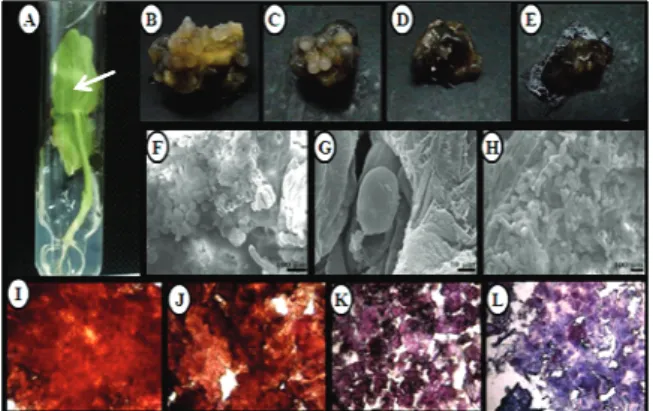
Figure 1. (A) In vitro-grown Jatropha curcas seedlings from which the explants were excised. The textural appearance of the Jatropha curcas calli grown on (B) 4.52μM of 2,4-D (C) 9.04 μM of 2,4-D, (D) 4.14 μM of PIC and (E) 8.28 μM of PIC. Photomicrographs of Jatropha curcas calli grown on (F) 4.52 μM of 2,4-D (round cells), (G) 4.52 μM of 2,4-D (embryoid structure in the globular stage) and (H) 4.14 μM of PIC (elongated cells). Cell masses of
Jatropha curcas grown on (I) 4.52 μM of 2,4-D, (J) 9.04 μM of 2,4-D (acetocarmine staining), (K) 4.14 μM of PIC and (L) 8.28 μM of PIC (Evans blue staining).
The texture is considered one criterion for the acceptance or rejection of a callus. Within this context, Titon et al. (2007) ruled out the use of PIC for callus regeneration in eucalyptus (Eucalyptus spp.) because the resulting callus had a compact texture.
The callus tissue grown on 4.52 and 9.04 μM of 2,4-D demonstrated a similar performance and consistent texture. Upon treatment with this plant regulator, the calli showed prominent structures that were similar to a globular embryo. These structures are characteristic of the first phase of the histological differentiation of this organ and are, therefore, the first indication of success in a somatic embryogenesis protocol.
This result is important because callus induction is dependent on the hormonal balance of auxins and cytokinins. In developing J. curcas, the supply of auxin in the culture medium is likely sufficient to balance the endogenous cytokinin contents of the explants. Thus, these results indicate that the addition of cytokinins (KIN and BAP) to the nutrient medium may provided a concentration of this regulator that is too high relative to the auxin due to an imbalance in the auxin / cytokinin ratio, thus decreasing the rate of callus formation.
Based on the superiority of 2,4-D and PIC, the SEM and cytochemistry analyses focused on these regulators. The SEM analysis showed that the cell masses treated with 4.52 μM of 2,4-D were rounded and that structures similar to embryoid calogênica appeared between the surfaces (Figure 1 F and G). These structures were also observed by Ferreira et al. (2005) to induce somatic embryogenesis in cupuaçu (Theobroma grandiflorum) under the effect of TDZ (Thidiazuron). The developmental stages of somatic embryos include globular, heart, torpedo and cotyledon stages. Our SEM images show the somatic embryos of J. curcas in the initial phase, i.e., the globular stage (Figure 1G).
The cell masses under the influence of 4.14 μM de PIC were more elongated compared to the round cells observed under the 2,4-D treatment. This morphogenetic difference demonstrates that both chemicals are growth regulators, as an isodiametric shape is characteristic of embryogenic cells (STEINER et al., 2005).
Ultrastructural analysis by SEM is a tool that helps to clarify the morphogenetic capacity of explants cultured in vitro, and the SEM data herein confirmed the embryogenic potential of the cotyledon explants of J. curcas. The images of the double acetocarmine / Evans Blue-stained cell masses are shown in Figure 1 (Photomicrographs stages I, J, K and L).
The RGB analysis of the double acetocarmine / Evans Blue-stained cell masses showed that the level of red that developed did not differ statistically among the different growth regulators and concentrations applied (Figure 2). That is, the masses of callusfrom the cultures with both 2,4-D as PIC reacted similarly to the acetocarmine, demonstrating that both growth regulators caused a morphogenetic response in the explants.
The differences in the blue bars indicate that the calli subjected to the different treatments reacted differently to the Evans blue dye (Figure 2). The
masses calogênicas that were more reactive to this dye were those that developed in the presence of PIC, and both doses produced equivalent blue staining. The masses calogênicas developed under 2,4-D did not respond as strongly to the Evans blue dye: comparing the two dosages of 2,4-D, the 4.52
μM concentration resulted in less reaction with Evans blue (Figure 2).
0 20 40 60 80 100 120 140
4,52µM 2,4-D 9,04µM 2,4-D 4,14µM Pic 8,28µM Pic
a
a
a a
c
b
a
a
le
v
e
l o
f c
o
lo
c
r
(
b
y
te
)
Growth regulator
Figure 2. Levels of red and blue in the images of the Jatropha curcas calli masses grown on different auxins and stained with acetocarmine and Evans blue. *The bars marked with the same letter are not significantly different, as determined by the Scott-Knott test at 5% probability.
Silva et al. (2012) also used this double-staining technique to identify callus masses with embryogenic potential in small murici (Byrsonima intermedia Juss A) and found that the three subcultures had a better embryogenic potential than the original. Similarly, Herrera et al. (2011) observed a higher percentage of viable cells with characteristics of embryogenic callus from small murici using FDA dye (fluorescein diacetate).
The data presented here demonstrate that the reaction to acetocarmine was identical for both growth regulators; however, the 2,4-D-treated cells were less reactive to Evans blue, particularly at the concentration of 4.5 μM. According to Steiner et al. (2005) and George et al. (2008), a positive reaction to acetocarmine versus Evans blue identifies embryogenic cells. Thus, the J. curcas callus grown on 4.52 μM of 2,4-D exhibited a higher embryogenic potential.
Conclusion
Acknowledgements
The authors wish to thank the National Council for Scientific and Technological Development (CNPq) and the Foundation for Research Support of Minas Gerais (FAPEMIG) for financial support.
References
DIVAKARA, B. N.; UPADHYAYA, H. D.; WANI, S. P.; LAXMIPATHI GOWDA, C. L. Biology and genetic improvement of Jatropha curcas L.: A review. Applied Energy, v. 87, n. 3, p. 732-742, 2010.
FERREIRA, D. F. Sisvar: a computer statistical analysis system. Ciência and Agrotecnologia, v. 35, n. 6, p. 1039-1042, 2011.
FERREIRA, M. G. R.; CARVALHO, C. H. S.; CARNEIRO, A. A.; DAMIÃO FILHO, C. F. Indução de embriogênese somática em cupuaçu (Theobroma grandiflorum Schum.). Revista Brasileira de Fruticultura, v. 27, n. 3, p. 500-503, 2005.
GEORGE, E. F.; HALL, M. A.; DE KLERK, G. J. Plant propagation by tissue culture. 3rd ed. Dordrecht: The Background, 2008.
GRANDO, M. F.; EICHLER, L.; TANABE, C. R.; SANTOS, J. F.; SANTOS, C. M. Indução de calos e regeneração de plantas em três genótipos de aveia. Revista Brasileira de Fisiologia Vegetal, v. 5, n. 2, p. 139-144, 1993. GUERRA, M. P.; TORRES, A. C.; TEIXEIRA, J. B. Embriogênese somática e sementes sintéticas. In: TORRES, A.C.; CALDAS, L. S.; BUSO, J. A. (Ed.).
Culturas de tecidos e transformação genética de plantas, v. 2. n. 1, p. 533-568, 1999.
HE, Y.; GUO, X.; LU, R.; NIU, B.; PASAPULA, A. V.; HOU, P.; CAI, F.; XU, Y.; CHEN, F. Changes in morphology and biochemical indices in browning callus derived from Jatropha curcas hypocotyls. Plant Cell Tissue and Organ Culture, v. 98, n. 1, p. 11-17, 2009. HERRERA, R. C.; PAIVA, R.; STEIN, V. C.; SALGADO, C. C.; MAGALHÃES, M. M.; SOARES, F. P. Índice mitótico e viabilidade celular de calos embriogênicos de murici-pequeno. Revista de Ciências Agrarárias, v. 54, n. 1, p. 30-34, 2011.
KIELSE, P. V. N.; FRANCO, E. T. H.; FRASSETTO, E. G. Indução de calogênese em explantes de Parapiptadenia rígida.
Revista Brasileira de Biociências, v. 5, n. 2, p. 84-86, 2007. MURASHIGE, T.; SKOOG, F. A revised medium for rapid growth and bioassays with tobacco tissue cultures.
Physiologia Plantarum, v. 15, n. 3, p. 473-497, 1962. NOGUEIRA, R. C.; PAIVA, R.; OLIVEIRA, L. M.; SOARES, G. A.; SOARES, F. P.; CASTRO, A. H. F.; PAIVA, P. D. O. Indução de calos em explantes foliares de murici-pequeno (Byrsonima intermedia A. Juss.). Ciência e Agrotecnologia, v. 31, n. 2, p. 366-370, 2007.
NUNES, C. F.; PASQUAL, M.; SANTOS, D. N.; CUSTÓDIO, T. N.; ARAUJO, A. G. Diferentes suplementos no cultivo in vitro de embriões de pinhão-manso. Pesquisa Agropecuária Brasileira, v. 43, n. 1, p. 9-14, 2008.
OLIVEIRA, A. L.; KIDO, E. A.; BENKO-ISEPPON, A. M.; KIDO, L. M. H. Efeito dos Fitorreguladores BAP e 2,4-D Sobre a indução de calos em Vigna unguiculata. Revista Brasileira de Biociências, v. 5, n. 2, p. 69-71, 2007.
PERÁN-QUESADA, R.; SÁNCHEZ-ROMERO, C.; BARCELÓ-MUNÓZ, A.; PLIEGO-ALFARO, F. Factors effecting maturation of avocado somatic embryos.
Scientia Horticulturae, v. 1, n. 102, p. 61-73, 2004. SAHOO, N.; VOUKKARASU, M. T.; BEHERA, P. R.; SATPATHY, G.; PANDA, P. K. Direct shoot organogenesis from hypocotyl explants of Jatropha curcas L. an important bioenergy feedstock. Global Change BiologyBioenergy, v. 4, n. 1, p. 234-238, 2012.
SILVA, L. C.; PAIVA, R.; VARGAS, D. P.; SILVA, D. P. C.; HERRERA, R. C.; BARBOSA, S.; COSTA-NETTO, A. P. Cell viabiliy of Byrsonima intermedia A Juss calli.
Journal of Agricultural Science and Technology, v. 1, n. 6B, p. 713-720, 2012.
SILVA, R. C.; CAMILLO, J.; SCHERWINSKI-PEREIRA, J. E. A method for seedling recovery in Jatropha curcas after cryogenic exposure of the seeds.
International Journal of Tropical Biology, v. 60, n. 1, p. 473-482, 2012.
STEINER, N.; VIEIRA, F. N.; MALDONADO, S.; GUERRA, M. P. Effect of carbon source on morphology and histodifferenciation of Araucaria angustifolia
embriogenic cultures. Brazilian Archives of Biology and Technology, v. 48, n. 6, p. 895-903, 2005.
SUAREZ, M.; FILONOVA, L.; SMERTENKO, A.; SAVENKOV, E.; CLAPHAM, D.; ARNOLD, S. V.; ZHIVOTOVSKY, B.; BOZHKOV, P. Metacaspase-dependent programmed cell desth is essential for plant embryogenesis. Current Biology, v. 14, n. 9, p. 339-340, 2004. TITON, M.; XAVIER, A.; OTONI, W. C.; MOTOIKE S. Y. Efeito dos reguladores de crescimento dicamba e picloram na embriogênese somática em Eucalyptus grandis.
Revista Árvore, v. 31, n. 3, p. 417-426, 2007.
VARSHNEY, A.; SANGAPILLAI, R.; PATIL, S. M.; JOHNSON, S. T. Histological evidence of morphogenesis from various explants of Jatropha curcas L.
Trees, v. 25, n. 1, p. 689-694, 2011.
Received on February 4, 2013. Accepted on April 17, 2013.