Vol.57, n.6: pp. 860-866, November-December 2014 http://dx.doi.org/10.1590/S1516-8913201402611
ISSN 1516-8913 Printed in Brazil
BRAZILIAN ARCHIVES OF BIOLOGY AND TECHNOLOGY
A N I N T E R N A T I O N A L J O U R N A L
In vitro Callus Induction and Plant Regeneration of Celosia
argentea- An Important Medicinal Plant
Dalilah Abu Bakar, Bakrudeen Ali Ahmed
*and Rosna Mat Taha
Institute of Biological Sciences; Faculty of Science; University of Malaya; Kuala Lumpur - Malaysia
ABSTRACT
Celosia argentea (Var.) cristata (Amaranthaceae) is a widely cultivated ornamental plant, which has antibacterial, astringent, haemostatic, hypertensive, ophthalmic, and parasitic significance. This study describes a protocol for in vitro callus induction and plant regeneration from leaf and stem explants of C. argentea using Murashige and Skoog (MS) medium. Callus culture was initiated and established from seedling, leaf, and stem explants. Explants were cultured on MS medium supplemented with auxin alone (0.5 mg/L Naphthaleneacetic acid (NAA), 2, 4-Dicholorophenoxyacetic acid (2, 4-D). Green and red compact callus (98%) were induced using MS medium supplemented with 0.5 mg/L NAA and 1.0 mg/L Benzyladenine (BA). Two different concentrations (1.0 mg/L BA + 0.5 mg/L NAA and 1.0 mg/L BA + 1.0 mg/L NAA) successfully induced plant regeneration with multiple shoots (1.5 and 0.9 shoots per explants, respectively). Successful shoots were transferred to rooting medium supplemented with 1.0 mg/L Indole-3-acetic acid (IAA) (80%) at 35th day. Acclimatization was done, which resulted in 90% of the plantlets surviving in garden soil. This protocol could be used to micropropagate C. argentea for conservation, commercial natural product production. The high frequency of callus indicated potential of C. argentea for secondary metabolite production (celosin) in pharmaceutical industry.
Key words:Celosia argentea, amaranthaceae, in vitro, micropropagation, callus, plant regeneration
*Author for correspondence:[email protected]
INTRODUCTION
Celosia argentea (Amaranthaceae), also known as
cock’s comb, has wide diversity and is widespread around tropical Africa, tropical and subtropical Asia and America. It is a traditional vegetable in West and Central Africa (Grubben and Denton 2004). These plants are widely grown for ornamental purpose in the tropics and subtropics
such as in Malaysia. C. argentea variety can be
distinguished based on their different forms of
cockscomb (Grant 1954). Seedlings,
inflorescences, and young leaves of C. argentea
are used as vegetables in China and some other countries (Palada and Crossman 1999). The size of
C. argentea is up to two meters. It has an
herbaceous stem; leaves are simple, opposite, entire, and often covered in woolly hair. It bears pinkish or white flowers and the arrangement is simple with branched spikes or racemes and inflorescences (Koh et al. 2009; Peter et al. 2011; Verma 2011).
Traditionally, dried leaves, inflorescences, and
seeds of C. argentea are used in Chinese medicine
(Wong 1994; Xu et al. 1996). It is used internally
compounds found in C. argentea contribute to pharmacological properties such as antibacterial,
antimitotic, antineoplastic, diuretic,
hypoglycaemic, hepatoprotective,
immunomodulatory, cytoprotective, and wound healing (Hayakawa et al. 1998; Wiart 2000; Vetrichelvan et al. 2002; Gnanamani et al. 2003). The compounds found in the plant consist of celosin, nicotinic acid, celogenamide A, celogentin A- D, H, J, and K, moroidin and some others. The leaves have high concentrations of calcium, phosphorus, potassium, sodium, magnesium, iron, zinc, and copper; also, they possess trace levels of chromium, manganese, nickel and lead. The low concentrations of lead and other heavy metals make it suitable for consumption as vegetables (Ayodele and Olajide 2011).
Tissue culture has been implemented to regenerate
the plants and induce callus formation.
Micropropagation has been employed to enhance the mass production, quality, and sterilized condition of the targeted plant. This is the reason tissue culture works needed to be developed before carrying out any molecular work (Ghorpade et al. 2012). This study focused on standardizing efficient techniques for callus induction and micropropagation with specific objectives of (i) Callus development from leaf and stem explants of
C. argentea; (ii) In vitro regeneration of C.
argentea using different concentrations of plant
growth regulators; and (iii) Mass propagation of C.
argentea into complete plants.
MATERIAL AND METHODS
Plant Material
C. argentea seeds were purchased from Jusco
nursery area in Kuala Lumpur, Malaysia. They were washed thoroughly in running tap water for 30 min, including 2% (v/v) Teepol (Reckitt Benckiser, Malaysia) for 5 min, then washed with 70% ethanol for 3 min, followed by another wash with 5% sodium hypochlorite for 1 min, then by
0.1% mercury chloride (HgCl2) for 1 min. Prior to
culture, seeds were rinsed three times with sterile distilled water. The explants were cultured in MS medium (Murashige and Skoog 1962).
Media and Culture Condition
Approximately 1.0 cm of leaf and stem explants of
C. argentea were cultured in MS medium
supplemented with single PGRs ranging from
0.5-2.5 mg/L 2, Dicholorophenoxyacetic acid (2, 4-D), Naphthaleneacetic acid (NAA), 1.0-3.0 mg/L Benzyladenine (BA), and Kinetin (KN), and combinations of different concentrations. The medium was supplemented with 30 g/L sucrose (Duchefa, Germany), gelled with 0.8% agar (Oxoid, England) and the pH was adjusted to 5.7-5.8. Around 2.0 mL of medium was used for each culture tubes (25x150 mm). Cultures were
maintained at 25±2°C under 16/8 h photoperiod
with a light intensity of 35µ Em-2s-1 from Phillips
cool- white fluorescent tubes with 55-65% relative humidity.
Callus Induction, Shoot Regeneration and Multiple Shoots
Leaf and stem explants of C. argentea were
cultured on MS medium with PGRs that consisted of different concentrations and combination of PGRs 0.5:1.0; 1.0:1.0; and 1.0:0.5 mg/L of BA: NAA and 0.1:1.0; 1.0:1.0; 1.0:0.1 mg/L of KN: 2, 4-D. The leaf and stem explants were surface injured using scalpel to give a few slight cut and faced the media abaxially. The role of media nature and biomass of callus was studied in leaf
and stem explants of C. argentea. The regenerated
shoots were transferred to the MS medium with supplemented PGRs to provide mass plant production.
Root Induction
The minimal length of shoots were developed up to 5.0 cm and above, then shoots were cut and transferred to rooting MS medium supplemented with 0.5-2.5 mg/L Indole-3-acetic acid (IAA) and Indole-3-butyric acid (IBA). After 35 days, the successful root induction was observed, the numbers of roots as per shoot was recorded and tabulated.
Acclimatization
Plantlets were successfully developed from rooted plantlets. The roots were washed in running tap water to remove the medium. Rooted shoots were
planted in Plantaflor® soil in plastic pots covered
with a plastic bag that had holes for about four weeks duration. The plantlets were removed from the pots and transferred into the field. The soil
used was Plantaflor® Young Plant Substrate,
Callus Biomass
Callus was collected at week 3 and weighed using fresh and dry weight. Combination of PGRs was used to determine to find the maximum biomass. The callus was transferred into Petri dish to weigh the fresh weight, then stored in oven 35°C for 2 to 3 days and dry weight was recorded. Callus
biomass was calculated as follows; (Fresh weight
– Dry weight)/Dry weight
Statistical Analysis
All the experiments were repeated three times with 10 explants per treatment. The data pertaining to
callus induction, shoot initiation, shoot
regeneration, and rooting were subjected to mean separation and standard deviation, carried out using SPSS Version 14. The significance of differences among means was carried out using
Duncan’s multiple range test (DMRT) at P ≤ 0.05
(Gomez and Gomez 2003). The results were expressed as mean ± SE of the total experiments.
RESULTS AND DISCUSSION
Surface Sterilization
C. argentea sterilization protocol was optimized
with minor modification from Taha and Wafa (2012). Five different sets were used by adjusting
the time and concentrations of sodium
hypochlorite and mercury chloride. The first set consisting of high to low concentrations of sodium hypochlorite had no germination and after that gradually the concentration was increased, which showed, 24.7% success rate. When the mercury chloride was added in the protocol, the germination rate significantly increased, which under optimized condition was as 83.3% survival germination rate and less than 10% contamination rate in other methods. Other methods showed the absence of germination (50-80%). All germination
data were recorded on the starting of 2nd week.
Callus induction
Callus was induced on MS medium with different concentrations and combination of PGRs using leaf and stem explants. The callus could be differentiated by biomass; and the color and structure of the callus. Suitable callus induction
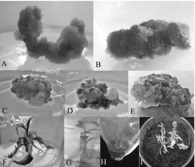
was on MS medium supplemented with 0.5 mg/L NAA and 1.0 mg/L BA for both green compact callus (Fig. 1A) and red compact callus (Fig. 1B). Greenish-reddish-white compact and friable callus (Fig. 1C), reddish-white compact (Fig. 1D) and friable callus and reddish; white compact callus (Fig. 1E) was also observed in both leaf and stem explants. Most of the friable callus were obtained from leaf explants. Previous study (Taha and Wafa 2012) had shown to have white, yellow, orange,
green and black callus in C. cristata. All the
explants were able to produce reddish purple callus on MS medium containing 0.5-2.0 mg/L BAP and 0.5 mg/L NAA.
Fresh weight of callus biomass for leaf and stem
explants of C. argentea are shown in Figure 2. The
highest fresh weight was achieved on 0.5 mg/L NAA and 1.0 mg/L BA (stem explants) with 2.03 g/L, followed by 0.5 mg/L BA and 1.0 mg/L NAA with 1.62 g/L, and 1.0 mg/L KN and 0.1 mg/L 2, 4-D with 1.44 g/L. The concentration of 0.5 mg/L NAA and 1.0 mg/L BA produced the highest yield for stem explants (0.16 g/L), while 0.5 mg/L BA and 1.0 mg/L NAA, and both leaf and stem explants of 0.1 mg/L 2, 4-D and 1.0 mg/L KN showed the highest biomass, 0.11 g/L, 0.10 g/L and 0.10 g/L, respectively (Fig. 3).
The most desired type of callus are red compact callus for the betalain content (Schliemann et al. 2001), and green compact callus could have other
compounds found in C. argentea such as celosin
E, celosin F, and celosin G, together with cristatain (Wu et al. 2011). Based on the biomass, the concentration that induced the highest yield was stem explants on 0.5 mg/L NAA and 1.0 mg/L BA, 0.16 g/L, which produced numerous green and red compact callus. The optimum time for
callus growth was from the 5th day of culture until
week 6 (lag phase). After that, the callus biomass was significally reduced for the next 2 to 3 weeks.
For Alternanthera tenella, an amaranthaceae
family, MS basal medium was supplemented with 1.0 mg/L 2, 4-D and 1.0 mg/L KN and, 2.5 mg/L NAA and 1.0 mg/L BA for callus induction
(Salvador et al. 2009). In A. philoxeroides (Mart.),
the callus induction took place using MS medium
with 2.2 µm BA and 2.2 µm 2, 4-D in 20 days of
Figure 1 - A: green compact callus; B: red compact callus; C: greenish reddish compact and friable callus; D: reddish white compact and friable callus; E: reddish white compact callus; F: shoot regeneration; G: root induction at 35th day; H: plantlets hardening inside culture room; I: acclimatization.
Figure 2 - The fresh weight (g/L) callus biomass induced from leaf and stem explants of Celosia argentea.
Shoot Induction
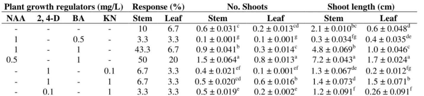
The best results for shoot regeneration were obtained in 0.5 mg/L NAA and 1.0 mg/L BA. The shoot started to regenerate at day 5 and continued to develop new shoots. After one week, the shoots were initiated only on the stem explants with MS medium with 0.5 mg/L NAA and 1.0 mg/L BA (50%) (Fig. 1F), with 1.5 shoots as the highest yield, followed by 1.0 mg/L NAA and 1.0 mg/L BA (40%), with 0.9 shoots, and MS medium without PGR (10%), with 0.5 shoots (control). With 0.5 mg/L NAA and 1.0 mg/L BA, the length of initiated shoots was about 0.2-1.0 cm. By the
end of 8th week, five shoots were formed (50%
shoot growth). The length of the shoots was increased and around 5-7 cm. With 1.0 mg/L NAA and 1.0 mg/L BA, developed shoots were stunted as the callus formed and managed to be regenerated again after two weeks and the lengths
were about 2-3cm (20%). Even though C.
argentea induced callus, direct shoot regeneration
was observed from stem explants and not from callus. Both leaf and stem explants responded under the same conditions; however, stem explants gave a higher yield for shoot induction.
Table 1 - Shoots regenerated from Celosia argentea leaf and stem explants after 24 days.
Plant growth regulators (mg/L) Response (%) No. Shoots Shoot length (cm)
NAA 2, 4-D BA KN Stem Leaf Stem Leaf Stem Leaf
- - - - 10 6.7 0.6 ± 0.031c 0.2 ± 0.013cd 2.1 ± 0.010bc 0.6 ± 0.048d 1 - 0.5 - 3.3 3.3 0.1 ± 0.001g 0.1 ± 0.001g 0.3 ± 0.034fg 0.4 ± 0.035de 1 - 1 - 43.3 6.7 0.9 ± 0.041b 0.3 ± 0.014c 4.8 ± 0.069b 1.0 ± 0.046c 0.5 - 1 - 50 20 1.5 ± 0.064a 0.8 ± 0.013a 7.2 ± 0.043a 1.7 ± 0.024a - 1 - 0.1 6.7 3.3 0.4 ± 0.021ef 0.1 ± 0.001ef 1.3 ± 0.067de 0.2 ± 0.012fg - 1 - 1 6.7 3.3 0.5 ± 0.020cd 0.6 ± 0.016b 1.4 ± 0.073d 1.5 ± 0.071b - 0.1 - 1 3.3 3.3 0.5 ± 0.019e 0.2 ± 0.002e 1.2 ± 0.091f 0.26 ± 0.091f
Values are mean of 10 replicates per treatment and repeated trice. Values with the same superscript are not significantly different at the 5 % probability level according to DMRT.
Rooting and Acclimatization
Plantlets around 5 to 7 cm were cut and transferred to MS medium supplemented with 2, 4-D, NAA, IAA and IBA. However, 2, 4-D and NAA did not
induce the roots on C. argentea, but the basal
callus induction instead. IBA concentrations (1.0 and 1.5 mg/L) gave the highest rooting response, 3.4 roots per shoot. The length and numbers of roots were observed with 1.0 mg/L IAA (Fig. 1G) and was the most suitable concentration of auxin. It showed the highest roots per shoots with
dispersed arrangement. At 35th day, the root
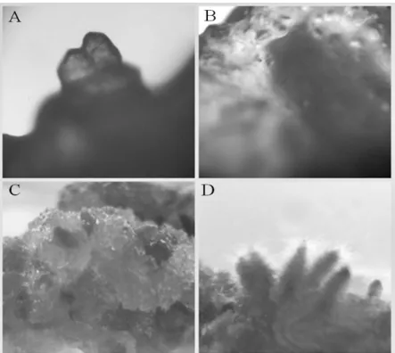
formation from organogenesis was observed in both the leaf and stem explants that had formed callus from explants on 1.0 mg/L BA + 0.5 mg/L NAA and 1.0 mg/L KN + 0.1 mg/L 2, 4-D,
respectively. The callus organogenesis
development is shown in Figure 4A-D. After two months, the plant regeneration and root induced plantlets were hardened (Fig. 1H) inside the
culture room with 25±2°C under 16/8 h
photoperiod. The hardened plantlets (Fig. 1I) were
successfully acclimatized to the natural
environment. About 90% of the plantlets survived in the garden soil. Those plantlets were compared with other plantlets.
Table 2 - The effect of auxin on root induction from
Celosia argentea after 35 days culture.
Plant Growth Regulators Number of Roots,
(mg/L) Per Shoot
IAA
0.5 3.4 ± 0.85d
1 4.8 ± 1.03a
1.5 3.4 ± 0.85bc
2 3.9 ± 0.88b
2.5 1.5 ± 0.41e
IBA
0.5 0.8 ± 0.32e
1 3.4 ± 0.85a
1.5 3.4 ± 0.86ab
2 1.5 ± 0.41c
2.5 1.4 ± 0.34cd
Figure 4 - Observation of callus cultures derived from explants of in vitro propagated C. argentea A: heart stage in organogenesis B: callus generating roots C: formation of roots on callus surface D: organogenesis roots.
CONCLUSION
The present study has shown successful callus induction and plant regeneration from leaf and stem explants derived from direct regeneration.
Plant regeneration was achieved using
combination of BA and NAA. The most high volume red compact callus obtained was through the combination of 1.0 mg/L BA + 0.5 mg/L NAA, while greenish compact callus was obtained through 1.0 mg/L KN and 0.1 mg/L of 2, 4-D and 1.0 mg/L BA + 0.5 mg/L NAA. The highest root development was achieved by using MS medium, supplemented with 1.0 mg/L IAA with 80%
response and 4.8 roots. The medicinal value of C.
argentea offers potential health development as
rapid multiplication increase rate of achievement.
ACKNOWLEDGEMENT
The authors would like to thank University of Malaya for the financial support (RG078- 12BIO) and facilities provided to successfully carry out this research.
REFERENCES
Ayodele JT, Olajide OS. Proximate and Amino Acid Composition of Celosia argentea Leaves. Niger. J Basic Appl Sci. 2011; 19(1):162-165.
Duke JA, Ayensu ES. Medicinal Plants of China, Vol. 1 & 2. United States of America: Reference Publication, Inc.; 1985.
Gao J, Li J, Luo C, Yin L, Li S, Yang G, et al. Callus induction and plant regeneration in Alternanthera philoxeroides. Mol Biol Rep. 2011; 38(2):1413-7. Ghorpade P, Siddiqui A, Patil MJ, Rub RA.
Pharmacognostic and phytochemical evaluation of
Celosia argentea. Pharmacogn J. 2012; 4(33):07-15. Gnanamani A, Shanmuga PK, Radhakrishnan N, Babu
M. Antibacterial activity of two plant extracts on eight burn pathogens. J Ethnopharmacol. 2003; 86: 59-61.
Gomez KA, Gomez AA. Statistical procedures for agricultural research with emphasis on rice. International Rice Research Institute, Los Bans, Philippines: IRRI Publ., 264; 1976.
Grant WF. A Cytological Study of Celosia argentea, C. argentea var. Cristata, and Their Hybrids. Bot Gaz. 1954; 323-336.
Hayakawa Y, Fujii H, Hase K, Ohnishi Y, Sakukawa R, Kadota S, et al. Anti-metastatic and immunomodulating properties of the water extract from Celosia argentea seeds. Biol Pharm Bull.
1998;21:1154-1159.
Koh HL, Chua TK, Tan CH. A Guide to Medicinal Plants: An Illustrated Scientific and Medicinal Approach. Ed. 42 & 43. Singapore: World Scientific Publishing Co. Pte. Ltd.; 2009.
Jain SK, Defilipps RA. Medicinal Plants of India, Vol. 1 and 2. United States of America: Reference Publication, Inc; 1991.
Murashige T, Skoog FA. Revised media for rapid growth and bioassay with tobacco tissue cultures.
Physiol Plant. 1962; 15:473-497.
Palada MC, Crossman SMA. Evaluation of tropical leaf vegetables in the Virgin Islands. In: Janick J, editor. Perspectives on New Crops and New Uses. Alexandria: ASHS Press; 1999. p. 388-393.
Peter KLN, Richard TC, Hugh TWT. Singapore Biodiversity: An Encyclopedia of the Natural Environment. Singapore: Tien Wah Press Pte. Ltd.; 2011.
Salvador MJ, Pereira PS, França SC, Candido RC, Ito IY, Dias DA. Bioactive Chemical Constituents and Comparative Antimicrobial Activity of Callus Culture and Adult Plant Extracts from Alternanthera tenella. Z Naturforsch. 2009; 64:373-381.
Santosh SB, Sohan SC, Anupamaa S, Devanand BS, Manohar JP. Anti- inflammatory activity of an isolated flavonoid fraction from Celosia argentea
Linn. J Med Plants Res. 2008; 2(3): 52-54.
Schliemann W, Cai Y, Degenkolb T, Schmidt J, Corke H. Betalains of Celosia argentea. Phytochemistry.
2001; 58:159-175.
Taha RM, Wafa SN. Plant Regeneration and Cellular Behaviour Studies in Celosia cristata Grown In Vivo
and In Vitro. Sci World J. 2012.
Verma BK. Introduction to Taxonomy Of Angiosperms. New Delhi:Phi Learning Pvt. Ltd.; 2011. p. 309-311.
Vetrichelvan T, Jeegadeesan M, Devi BA. Antidiabetic Activity of Alcoholic Extract of Celosia argentea
Linn. Seeds in Rats. Biol Pharm Bull. 2002;25:526-528.
Wiart C. Medicinal Plants of Southeast Asia. Malaysia: Pelanduk Publications (M) Sdn. Bhd; 2000.
Wong KY. Chinese Herbal Medicine. Hong Kong: Wokman Press; 1994.
Wu Q, Wang Y, Guo M. Triterpenoid Saponins from the Seeds of Celosia argentea and Their Anti-inflammatory and Antitumor Activities. Chem Pharm Bull. 2011; 59(5):666-671.
Xu GJ, Ji GZ, Ji GH, Ji GM. Encyclopedia of Chinese Medicine. Beijing: China Medicine Sci. Technol; 1996.