Usefulness of Preoperative Venography in Patients with Cardiac
Implantable Electronic Devices Submitted to Lead Replacement or
Device Upgrade Procedures
Caio Marcos de Moraes Albertini,
1Katia Regina da Silva,
1Joaquim Maurício da Motta Leal Filho,
1Elizabeth Sartori
Crevelari,
1Martino Martinelli Filho,
1Francisco Cesar Carnevale,
2Roberto Costa
1Instituto do Coração (InCor) - Faculdade de Medicina da Universidade de São Paulo,1 São Paulo, SP – Brazil Hospital das Clinicas da Faculdade de Medicina da Universidade de São Paulo,2 São Paulo, SP – Brazil
Mailing Adress: Roberto Costa •
Av. Dr. Enéas de Carvalho Aguiar, 44, Postal Code 05403-900, Cerqueira César, São Paulo, SP - Brazil
E-mail: [email protected]
Manuscript received October 02, 2017, revised manuscript May 07, 2018, accepted June 12, 2018
DOI: 10.5935/abc.20180164
Abstract
Background: Venous obstructions are common in patients with transvenous cardiac implantable electronic devices, but they rarely cause immediate clinical problems. The main consequence of these lesions is the difficulty in obtaining venous access for additional leads implantation.
Objectives:We aimed to assess the prevalence and predictor factors of venous lesions in patients referred to lead reoperations, and to define the role of preoperative venography in the planning of these procedures.
Methods:From April 2013 to July 2016, contrast venography was performed in 100 patients referred to device upgrade, revision and lead extraction. Venous lesions were classified as non-significant (< 50%), moderate stenosis (51-70%), severe stenosis (71-99%) or occlusion (100%). Collateral circulation was classified as absent, discrete, moderate or accentuated. The surgical strategy was defined according to the result of the preoperative venography. Univariate analysis was used to investigate predictor factors related to the occurrence of these lesions, with 5% of significance level.
Results: Moderate venous stenosis was observed in 23%, severe in 13% and occlusions in 11%. There were no significant differences in relation to the device side or the venous segment. The usefulness of the preoperative venography to define the operative tactic was proven, and in 99% of the cases, the established surgical strategy could be performed according to plan.
Conclusions: The prevalence of venous obstruction is high in CIED recipients referred to reoperations. Venography is highly indicated as a preoperative examination for allowing the adequate surgical planning of procedures involving previous transvenous leads. (Arq Bras Cardiol. 2018; 111(5):686-696)
Keywords:Pacemaker, implantable defibrillators, phlebography, venous stenosis, extraction of leads, risk factors.
Introduction
Venous obstructions frequently occur in patients with transvenous cardiac implantable electronic devices (CIED), with an estimated 14 to 64% prevalence.1-11 Those lesions are
mostly asymptomatic, although visible collateral circulation in the thoracic region is usually found. Although deep venous thrombosis, pulmonary thromboembolism, or superior vena cava syndrome were found in 1.6 to 12% of the cases, the difficulty in gaining access to implant new additional leads or other types of transvenous devices has been the main consequence of those lesions.12-16
Recent studies have shown an increase in the number of reoperations in which it is necessary to handle the
intravascular territory with leads previously implanted.17-23
The increase in this type of procedure is due to three main factors: (1) patients’ increasing longevity, which is directly related to the longer period of time leads remain in the territory and, consequently, to a greater chance of dysfunction of the stimulation system’s components; (2) an increase in comorbidities leading to an increase in the occurrence of infectious complications, whose treatment necessarily requires the complete CIED removal17-23 and (3) an increasing
prevalence heart failure and, consequently, of the need to
upgrade from the conventional pacemaker to more advanced modes, such as implantable cardioverter‑defibrillator (ICD), or cardiac resynchronization therapy (CRT), which require the implantation of additional leads.24-27
Digital subtraction venography provides excellent characterization of the venous anatomy and has been deemed the gold standard for studying venous lesions in CIED patients.11,28-30 Although other imaging techniques are used for
recirculation in thoracic computed tomography images, these methods are not as accurate as digital venography to quantify and define where obstructions are located and any collateral circulation developed.31-34
This study is part of a prospective registry, with data derived from medical practice, and its goals are: (1) to identify the prevalence, degree and location of venous lesions in CIED patients with an indication of reoperation; (2) to identify predisposing factors of these venographic changes; and (3) to define the role of digital subtraction venography when intravascular reinterventions are planned in individuals with leads previously implanted.
Methods
Study Design and Population
This is a cross-section analysis derived from a cohort where thromboembolic complications are studied in patients submitted to lead revision or upgraded procedures. This study was conducted in a high-complexity cardiology hospital and it was approved by that hospital’s Committee of Ethics in Research. All subjects signed a free and informed consent form.
From April 2013 to July 2016, patients who met the following criteria were consecutively included: (1) having CIED implanted at the territory of the superior vena cava for more than six months; (2) being between 18 and 90 years of age; (3) having an indication for lead revision or upgrade procedures. The following candidates were not included: (1) individuals with creatinine > 1.5 mg/dL due to the risk of renal damage from iodinated contrast; (2) candidates that had known allergy to iodinated contrast media; and (3) those who declined to participate in the study.
Considering the high rates of venous lesions in these patients, a convenience sample of 100 patients was defined to detect the outcomes studied.
Study Outcomes
The outcomes of the study included: (1) venographic findings of significant venous obstructions and collateral circulation, and (2) usefulness of the preoperative venographic findings when planning and performing the surgical procedure.
Study Workflow
Patients with an indication of reoperation for implantation of additional leads, replacement or removal of previously-implanted transvenous leads, and who met the eligibility to the study were submitted to preoperative evaluation comprising patient background assessment, clinical evaluation and evaluation of imaging exams.
Thorax radiography was conducted to help determining the position of the leads in use or abandoned.
The venous system was evaluated using digital subtraction venography through images acquired with an Allura DSA unit or Allura Xper FD20 (Philips, The Netherlands) to bilaterally assess the axillary, cephalic, subclavian, innominate (or brachiocephalic trunk) veins, and superior vena cava.
Continuous infusion of low-osmolality nonionic iodinated contrast media (Visipaque-Iodixanol, 320 [652 mg/mL Iodixanol], GE, Healthcare, Europe) was performed using a MEDRAD injection pump with controlled volume (100 mL to 120 mL) and infusion speed (10 mL/s at 600 psi pressure). All exams were simultaneously evaluated by two specialists: a Vascular Interventional Radiologist and a Cardiac Pacing Specialist.
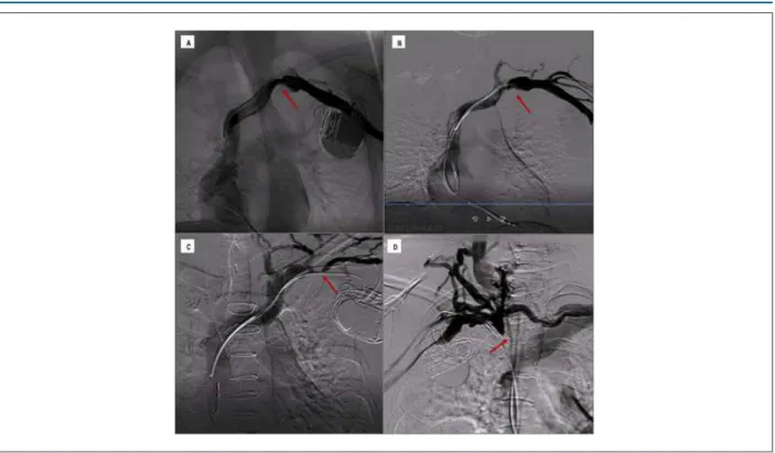
The images obtained were classified according to the presence or absence of venous lesions and of collateral circulation. Venous lesions were classified according to their stenosis level: without significant alteration (< 50%), moderate stenosis (51-70%), severe stenosis (71-99%), and occlusion (100%).
Surgical Procedures
Surgical procedures were performed according to the hospital’s usual routines, always under the supervision of an anesthesiologist. Operations were grouped in three main types: (1) Implanting new leads without further removal (due to dysfunction of a previously implanted lead, or upgrade procedures); (2) Replacing leads with the removal of previously implanted leads; or (3) Isolated lead extraction.
Operations were planned according to the radiological function of the venous territory obtained through venography: (1) In cases where the venous pattern was deemed without significant lesions or with moderate lesions, no special care was taken to implant new leads and, similarly, the decision of removing a deactivated lead was made at the surgical team’s discretion. (2) In cases with stenosis deemed severe or occlusions, surgical planning considered: a) careful evaluation of the venography to check the possibility of using the ipsilateral internal jugular vein; b) preparing the patient for transvenous lead extraction to provide access for the new lead when using the ipsilateral internal jugular was not possible; c) reserving material for attempts to go beyond a lesion and perform venous dilation.
The decision whether to remove or abandon in situ the previously abandoned leads or the ones that would be deactivated in the current surgical procedure was made considering the following criteria: (1) patient’s age and life expectancy; (2) number of leads remaining in the superior vena cava at the end of the surgical procedure performed in this study; (3) risk of worsening the lesions observed in the venography.
Although the criteria for defining an access to deactivated leads and whether to remove or abandon them were previously discussed with the surgical team involved in the study, the final decision on both topics was to be made by the team itself during the procedure due to the intraoperative findings and technical resources available.
Agreement between Planned and Actually Performed Procedure
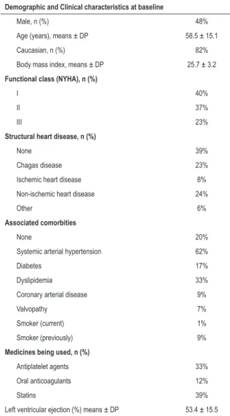
Table 1 – Demographic and clinical characteristics of the study subjects
Demographic and Clinical characteristics at baseline
Male, n (%) 48%
Age (years), means ± DP 58.5 ± 15.1
Caucasian, n (%) 82%
Body mass index, means ± DP 25.7 ± 3.2
Functional class (NYHA), n (%)
I 40%
II 37%
III 23%
Structural heart disease, n (%)
None 39%
Chagas disease 23%
Ischemic heart disease 8%
Non-ischemic heart disease 24%
Other 6%
Associated comorbities
None 20%
Systemic arterial hypertension 62%
Diabetes 17%
Dyslipidemia 33%
Coronary arterial disease 9%
Valvopathy 7%
Smoker (current) 1%
Smoker (previously) 9%
Medicines being used, n (%)
Antiplatelet agents 33%
Oral anticoagulants 12%
Statins 39%
Left ventricular ejection (%) means ± DP 53.4 ± 15.5
SD: Standard deviation; NYHA: New York Heart Association. lesion or subclavian vein occlusion; (3) whether lead extraction
or other unconventional technique was required to gain access in cases of critical lesion affecting the subclavian vein, internal jugular vein and venous brachiocephalic trunk.
Care Provided for Study Subjects
The risks associated with the present study were related to the use of iodinated contrast media. Special care was taken to reduce the risk of renal damage following digital subtraction venography, although adverse reactions related to the use of non-ionic iodinated contrast agents are rare. Diabetic patients receiving oral hypoglycemic metformin hydrochloride were instructed to discontinue the use of that drug for 48 hours before the test and resume use 48 hours after the test. The cases of allergic reactions to iodinated contrast during or after the exams were treated according to the institution’s protocol for allergic reactions to contrast.
Electronic Data Collection and Management
The demographic, clinical and surgical data obtained were stored at the database developed in the REDCap system (Research Electronic Data Capture)35 hosted at the hospital’s server.
Variables Studied and Statistical Analysis
The following data were analyzed as independent variables for the risk of occurrence of the outcomes studied: demographic data, preoperative clinical data at baseline, type of CIED, and type of procedure performed.
The data recorded in the database (REDCap) were exported in the format of Excel worksheets (Microsoft Excel) and analyzed using SAS software (Statistical Analysis System). Initially all variables were analyzed descriptively. The quantitative variables were analyzed by considering the minimal and maximum values, means, standard deviation and median. The qualitative variables were analyzed by calculating the absolute and relative frequencies. We compared means using Student t-test, and tested homogeneity among the variable proportions using chi-square test. The significance level chosen for statistical tests was 5%.
The outcomes of the study were described according to absolute and relative frequencies. The calculation of Odds Ratio (OR) and its confidence intervals at 95% were used as an effect measure between exposure variables and outcome development.
Results
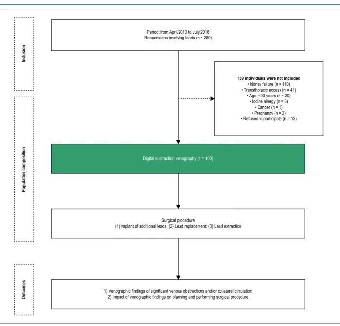
Of 289 patients with an indication of reoperation involving the handling of leads, 100 were included in this study. (Figure 1)
The population was balanced with regard to gender, had a predominance of Caucasian individuals (82%) and a mean age of 58.5 ± 15.1 years, with median 60. Most individuals studied were oligosymptomatic for heart failure (77%), with a left ventricular ejection fraction of 53.4 ± 15.5, 39% of which had no structural cardiac disease identified. Only 20% of cases did not have any comorbidity. One third of this population was using antiplatelet agents, while anticoagulants were used by 12% of the patients (Table 1).
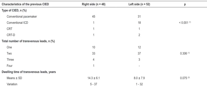
There was a balance in the number of cases with devices implanted on the right side (48%) and those on the left side (52%). Marking differences were observed, however, concerning time since implantation, with an average 14.3 ± 6.1 years for the right side, and 8.0 ± 7.9 years for the left side; as to the type of device, there were more conventional pacemakers on the right, while the four device types were more evenly distributed for the left side. (Table 2)
Results of Digital Subtraction Venography
blood vessel lumen), only 4 had collateral circulation. On the other hand, out of the 24 individuals with venous lesion deemed severe or with venous occlusion, just 2 did not present collateral circulation in their venography. Therefore, finding collateral circulation in venography was observed to be a strong marker of the presence of venous lesion, increasing 4.9 times the prevalence rate (CI 95% 3.05 – 8.10; p < 0.0001) of those lesions (Figures 2 and 3).
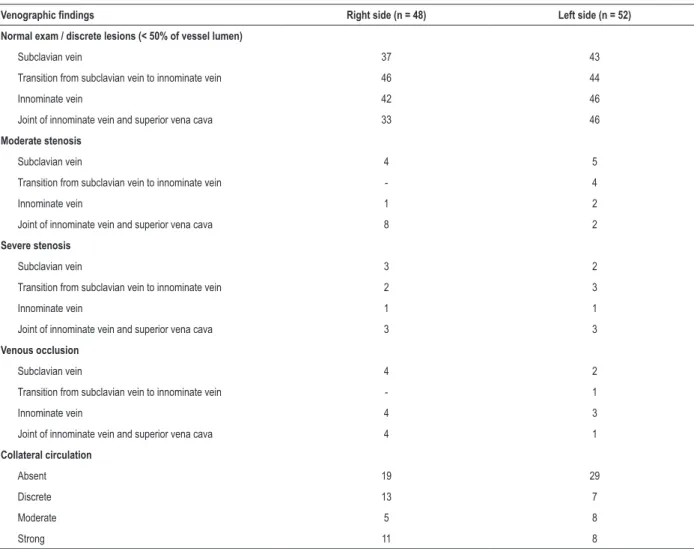
Despite the differences of time since implantation and types of devices implanted, there was balance between the findings of venous lesions (p = 0.865) and of collateral circulation (p = 0.715) in patients with devices implanted on the right and left sides. Regardless of the side the CIED had been implanted, subclavian veins and the transition from subclavian veins to the brachiocephalic trunk were the regions that presented the
highest number of significant lesions (Table 3). No significant lesions were identified in the superior vena cava.
Indication of surgical procedure
The main reason to perform a surgical procedure was lead dysfunction, in 71 patients. Upgrade procedures was the cause of reoperation in 25 cases. Only for 4 patients the operation was caused solely by a need of lead removal (Table 4).
Leads were removed from 52 patients. Transvenous extraction with mechanical or laser sheaths was performed in 36 patients, while leads were removed through simple traction in just 16 cases. At the end of the operation, only 4 patients remained without any transvenous lead implanted, and in most cases (90%), two or three leads remained in the venous territory.
Figure 1 – Composition of the population studied and Study phases.
Period: from April/2013 to July/2016 Reoperations involving leads (n = 289)
Digital subtraction venography (n = 100)
189 individuals were not included
• kidney failure (n = 110) • Transthoracic access (n = 41)
• Age > 90 years (n = 20) • Iodine allergy (n = 3)
• Cancer (n = 1) • Pregnancy (n = 2) • Refused to participate (n = 12)
Surgical procedure
(1) implant of additional leads; (2) Lead replacement; (3) Lead extraction
1) Venographic findings of significant venous obstructions and/or collateral circulation 2) Impact of venographic findings on planning and performing surgical procedure
Inclusion
Population composition
Figure 2 – Distribution of the four types of venous lesions and their associations with the presence of collateral circulation. Collateral Circulation
Absent
Present 60
50
40
30
20
10
0
V
enous lesions (%)
Lesion < 50% Moderate Severe Occlusion
49
4
13
10 11
2
11
Table 2 – Characteristics of the cardiac device being used at the time of inclusion in the study according to the side of the implant
Characteristics of the previous CIED Right side (n = 48) Left side (n = 52) p
Type of CIED, n (%)
Conventional pacemaker 45 31
Conventional ICD 1 18 < 0.001 (1)
CRT 1 1
CRT-D 1 2
Total number of transvenous leads, n (%)
One 10 12
Two 33 37 0.306 (1)
Three 4 3
Four 1
-Dwelling time of transvenous leads, years
Means ± SD 14.3 ± 6.1 8.0 ± 7.9 0.075 (2)
Variation 5 - 37 1 - 32
CIED: cardiac implantable electronic device; ICD: implantable cardioverter-defibrillator; CRT: cardiac resynchronization therapy; CRT-D: cardiac resynchronization therapy associated with implantable cardioverter-defibrillator. (1) Chi-square test; (2) Student t-test
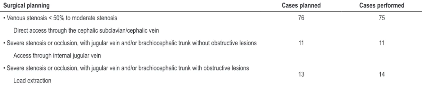
Usefulness of Venography to Define Surgical Planning Agreement between the surgical strategy based on the analysis of digital subtraction venography and the surgical procedure actually performed occurred in 99 out of the 100 patients operated. Lack of agreement, which occurred
In all the cases studied, surgical planning was based on the findings of preoperative venography. Of the 53 patients without significant lesions, there were 28 cases in which we decided to implant new leads without removing the old ones, while in 22 cases the implantation of new leads was combined with removal of old ones in order to avoid overpopulation. There was complete removal of the system in other 3 cases.
On the other hand, of the 23 cases where moderate stenosis had been diagnosed, there were 14 in which there was the implantation of new leads combined with the removal of old ones; only in 9 cases our decision was to implant new leads and maintain the old ones.
In the 24 cases where new leads did not require any removal and severe stenosis or venous occlusion had been diagnosed, the findings in the venography showed that in 13 cases the internal jugular vein and the ipsilateral brachiocephalic trunk of the implant were free from any obstructions. Of those, only in 2, because the patients were young, a transvenous extraction procedure was planned to avoid overpopulation of leads. Of the 11 cases where no extraction was performed, there were 5 in which the internal jugular vein was used as access. In the other 5 cases, it was possible to go beyond the lesion in the subclavian vein with the aid of 0,14” hydrophilic wire guides. Of the 8 cases where the internal jugular veins could not be used as access because there was obstruction in the ipsilateral venous brachiocephalic trunk, in only one case the medical team chose to conduct a new contralateral implantation. In the remainder (7), transvenous extraction was the chosen access.
Leads were removed without implanting new ones in only 4 cases: in 3, to treat an infection related to the device, and in 1 to remove a dysfunctional lead which was causing noise in an ICD. In this last case the venography showed venous occlusion.
Prognostic Factors of Venographic Alterations
Despite the high rate of venographic outcomes in the patients studied, it was not possible to identify prognostic factors for the occurrence of venographic alterations. The following variables were tested as probable prognostic factors: gender, age at the time of the venographic study, cardiopathy at baseline, functional class for heart failure, use of oral anticoagulants and antiplatelet agents, having an ICD lead, CIED implantation side, time since CIED implantation, number of leads implanted, left ventricular ejection, and previous procedures of reoperation (Figure 4).
Discussion
Venous obstructions seldom cause immediate clinical problems. However, when new leads have to be implanted, the presence of those lesions can make the procedure impossible with conventional techniques. Thus, digital subtraction venography has been mostly used because it allows identifying precisely how serious venous lesions are, as well as their location, thus allowing the planning of proper surgical strategy.11,28-30
Figure 3 – Classification of venous lesions and collateral circulation. Examples of the four types of lesion according to the classification adopted in the study.
Table 3 – Distribution of venographic findings according to the CIED side and the anatomical location of the lesion
Venographic findings Right side (n = 48) Left side (n = 52)
Normal exam / discrete lesions (< 50% of vessel lumen)
Subclavian vein 37 43
Transition from subclavian vein to innominate vein 46 44
Innominate vein 42 46
Joint of innominate vein and superior vena cava 33 46
Moderate stenosis
Subclavian vein 4 5
Transition from subclavian vein to innominate vein - 4
Innominate vein 1 2
Joint of innominate vein and superior vena cava 8 2
Severe stenosis
Subclavian vein 3 2
Transition from subclavian vein to innominate vein 2 3
Innominate vein 1 1
Joint of innominate vein and superior vena cava 3 3
Venous occlusion
Subclavian vein 4 2
Transition from subclavian vein to innominate vein - 1
Innominate vein 4 3
Joint of innominate vein and superior vena cava 4 1
Collateral circulation
Absent 19 29
Discrete 13 7
Moderate 5 8
Strong 11 8
CIED: cardiac implantable electronic device.
Table 4 – Characteristics of surgical procedures performed in the study
Characteristics of Surgical Procedures n = 100
Procedure performed, (%)
Implant of additional lead without removing previously implanted lead 48
Implant of additional lead with removal of previously implanted lead 48
Only lead removal 4
Total number of transvenous leads at the end of the procedure, (%)
None 4
One 6
Two 41
Three 42
Four 7
CIED side at the end of the procedure, n (%)
Right 45
Left 54
Subxiphoid 1
Figure 4 – Risk factors for the occurrence of significant venous lesions (> 50% of obstruction of blood vessel lumen) and/or presence of collateral circulation.
Variables OR (IC 95%) p
Male
Age ≥ 60 years
Chagas disease
Ischemic cardiopathy
Non-ischemic cardiopathy
FC NYHA III-IV
Use of anticoagulants
Use of antiplatelet agents
ICD lead
CIED side (right)
Time of implant ≥ 14 years
Transvenous lead ≥ 3
LVEF < 55
Previous reoperations
0 1 2 3 4 5 6 7 8 9 10 11
0.76 (0.35 – 1.67)
1.12 (0.51 – 2.45)
1.75 (0.60 – 5.09)
2.33 (0.53 – 10.34)
0.77 (0.27 – 2.16)
1.24 (0.47 – 3.26)
1.31 (0.39 – 4.44)
1.23 (0.54 – 2.82)
1.24 (0.48 – 3.15)
1.57 (0.71 – 3.45)
0.82 (0.26 – 2.59)
0.73 (0.15 – 3.49)
1.80 (0.75 – 4.34)
0.89 (0.41 – 1.94) 0.495
0.776
0.304
0.265
0.062
0.669
0.666
0.625
0.659
0.263
0.733
0.691
0.189
0.761
Odds Ratio
Table 5 – Agreement between the surgical strategy defined using preoperative venography and the surgical procedure performed
Surgical planning Cases planned Cases performed
• Venous stenosis < 50% to moderate stenosis 76 75
Direct access through the cephalic subclavian/cephalic vein
• Severe stenosis or occlusion, with jugular vein and/or brachiocephalic trunk without obstructive lesions 11 11
Access through internal jugular vein
• Severe stenosis or occlusion, with jugular vein and/or brachiocephalic trunk with obstructive lesions
13 14
Lead extraction
The high prevalence of individuals with lesions deemed significant in this study was compatible with other experiences reported in the literature.1-11 Regardless of lesion seriousness,
their distribution was balanced among the subclavian veins, the venous brachiocephalic trunk or the transitional areas of those veins.
Despite the particularities existing among the anatomy of the veins draining the left side and the right side of the thorax, the venographic study did not identify significant differences in the frequency of those findings, in how serious the stenosis was, or in the location of the lesions between the two sides. However, there were differences in the average time leads had
remained implanted, i.e., longer for patients who had the device implanted on the right side, which may have increased the rate of occurrences of lesions in the right territory. On the other hand, despite the balance between the numbers of leads implanted, the number of defibrillator leads, which is deemed a risk factor for venous lesions, was significantly higher in the cases where the CIED had been implanted on the left side.1-4-8
1. Mond HG, Crozier I. The Australian and New Zealand cardiac pacemaker and implantable cardioverter-defibrillator survey: calendar year 2013. Heart Lung Circ. 2015;24(3):291-7.
2. Oginosawa Y, Abe H, Nakashima Y. The incidence and risk factors for venous obstruction after implantation of transvenous pacing leads. Pacing Clin Electrophysiol. 2002;25(11):1605-11.
3. Costa SS, Scalabrini Neto A, Costa R, Caldas JG, Martinelli Filho M Incidence and risk factors of upper extremity deep vein lesions after permanent transvenous pacemaker implant: a 6-month follow-up prospective study. Pacing Clin Electrophysiol. 2002;25(1):1301-6.
4. Lickfett L, Bitzen A, Arepally A, Nasir K, Wolpert C, Jeong KM et al. Incidence of venous obstruction following insertion of an implantable cardioverter defibrillator. A study of systematic contrast venography in patient presenting for their first elective ICD generator replacement. Europace. 2004;6(1):25-31.
5. Van Rooden CJ, Molhoek SG, Rosendaal FR, Schalij MJ, Meinders AE, Huisman MV. Incidence and risk factors of early venous thrombosis associated with permanent pacemaker leads. J Cardiovasc Electrophysiol. 2004;15(11):1258-62.
6. Rozmus G, Daubert JP, Huang DT, Rosero S, Hall B, Francis C. Venous thrombosis and stenosis after implantation of pacemakers and defibrillators. J Interv Card Electrophysiol. 2005;13(1):9-19.
References
looked for. In this respect, we suggest maintaining dynamic venography images, which allow following the iodinated contrast path. Often enough, when the contrast passes exclusively through the collateral circulation, it fully fills up the blood vessel lumen soon after the critical lesion, which prevents it from being detected in still images.
The high rate of patients with severe or occlusive lesions observed in this study, which agrees with the data in the literature, evidenced the importance of venography for surgical planning. In cases where significant venous lesions could not be identified, the surgical team were able to plan a procedure in which deactivated leads should (or should not) be extracted by considering solely factors such as patient age or the number of leads that would remain in the venous territory. On the other hand, in patients where moderate lesions were observed, the medical team could plan which leads should be extracted in order to avoid an overpopulation of leads that could worsen obstructions. And, finally, in the cases where severe or occlusive venous lesions were observed, the knowledge of the venous anatomy was of essence to plan the surgery, since it raises the possibility of using the ipsilateral jugular vein or the need of extracting leads to gain proper access.
Since causes are multifactorial, the literature is controversial as to defining predictive factors of thromboembolic complications in CIED patients.2-11-36-37 In this respect, the absence of risk
factors for venous lesions found in this study sample confirms the importance of preoperative venography in patients requiring lead reoperations, since it was not possible to identify any subgroup of individuals less subject to venous obstructions.
Study Limitations
Although this study is part of a prospective registry derived from medical practice, due to the non-inclusion criteria used, our conclusions cannot be extended to children, to individuals over 90 years of age and to those with renal dysfunction with serum creatinine over 1,5 mg/dL.
As to the rate of venous alterations found and their predisposing factors, this analysis has the same limitations as other cross-sectional studies, as they were assessed at a particular time.
Conclusions
The high prevalence of severe obstructions or venous occlusions in CIED patients makes a transvenous implant
of new leads difficult in a considerable number of patients. Sometimes, using non-conventional techniques, such as the extraction of leads to achieve access, can be mandatory. The lack of predisposing factors and the absence of clinical signs of venous obstruction, which occurs in most patients with severe or occlusive lesions, can hinder the planning of a surgery. Thus, digital subtraction venography is quite useful to define a surgical strategy in operations for lead revision or upgrade procedures. The finding of collateral veins in this exam has a high predictive value for diagnosing severe and occlusive lesions.
Author contributions
Conception and design of the research and Writing of the manuscript: Albertini CMM, Silva KR, Costa R; Acquisition of data: Albertini CMM, Leal Filho JMM, Crevelari ES; Analysis and interpretation of the data: Albertini CMM, Silva KR, Leal Filho JMM, Costa R; Statistical analysis: Silva KR; Critical revision of the manuscript for intellectual content: Albertini CMM, Silva KR, Martinelli Filho M, Carnevale FC, Costa R.
Potential Conflict of Interest
No potential conflict of interest relevant to this article was reported.
Sources of Funding
This study was funded by FAPESP and CAPES.
Study Association
This article is part of the thesis of Doctoral submitted by Caio Marcos de Moraes Albertini, from Instituto do Coração – Faculdade de Medicina da Universidade de São Paulo.
Ethics approval and consent to participate
7. Korkeila P, Nyman K, Ylitalo A, Koistinen J, Karjalainen P, Lund J, et al. Venous obstruction after pacemaker implantation. Pacing Clin Electrophysiol. 2007;30(2):199-206.
8. Haghjoo M, Nikoo MH, Fazelifar AF, Alizadeh A, Emkanjoo Z, Sadr-Ameli MA. Predictors of venous obstruction following pacemaker or implantable cardioverter-defibrillator implantation: a contrast venographic study on 100 patients admitted for generator change, lead revision, or device upgrade. Europace. 2007;9(5):328-32.
9. Costa R, Silva KR, Rached RA, Martinelli Filho M, Carnevale FC, Moreira LFP, Stolf NAG. Prevention of venous thrombosis by warfarin after permanent transvenous leads implantation in high-risk patients. Pacing Clin Electrophysiol. 2009;32(Suppl 1):S247-51.
10. Pieper CC, Weis V, Fimmers R, Rajab I, Linhart M, Schild HH, et al. Venous obstruction in asymptomatic patients undergoing first implantation or revision of a cardiac pacemaker or implantable cardioverter-defibrillator: A Retrospective Single Center Analysis. RoFo. 2015;187(11):1029-35.
11. Boczar K, Zabek A, Haberka K, Hardzina M, Debski M, Rydlewska A, et al. Venous stenosis and occlusion in the presence of endocardial leads. Adv Clin Exp Med. 2016;25(1):83-91.
12. Lin CT, Kuo CT, Lin KH, Hsu TS. Superior vena cava syndrome as a complication of transvenous permanent pacemaker implantation. Jpn Heart J. 1999;40(4):477-80.
13. Sbragia P, Nait-Saïdi L, Trigano JA, Saadjian A, Barnay P, Lévy S. Intra-atrial thrombosis and pulmonary embolism complicating pacemaker leads for cardiac resynchronization therapy. J Interv Card Electrophysiol .2003;9(1):25-7.
14. Aryana A, Sobota KD, Esterbrooks DJ, Gelbman AI. Superior vena cava syndrome induced by endocardial defibrillator and pacemaker leads. Am J Cardiol .2007;99(12):1765-7.
15. Noheria A, Ponamgi SP, Desimone C V, Vaidya VR, Aakre CA, Ebrille E, et al. Pulmonary embolism in patients with transvenous cardiac implantable electronic device leads. Europace. 2015;18(2):246-52.
16. Korkeila P, Mustonen P, Koistinen J, Nyman K, Ylitalo A, Karjalainen P, et al. Clinical and laboratory risk factors of thrombotic complications
after pacemaker implantation: a prospective study. Europace.
2010;12(6):817-24.
17. Brasil. Ministério da Saúde. DATASUS - Secretaria Executiva. [Citado em 2017 dez 12]. Disponível em: http://w3.datasus.gov.br/datasus/datasus.php
18. Li X, Ze F, Wang L, Li D, Duan J, Guo F, et al. Prevalence of venous occlusion in patients referred for lead extraction: implications for tool selection. Europace. 2014;16(12):1795-9.
19. Harrison JL, Prendergast BD, Sandoe JA. Guidelines for the diagnosis, management and prevention of implantable cardiac electronic device infection. Heart. 2015;101(4):250-2.
20. Uslan DZ, Sohail MR, St Sauver JL, Friedman PA, Hayes DL, Stoner SM,et al. Permanent pacemaker and implantable cardioverter defibrillator infection: a population-based study. Arch Intern Med. 2007;167(7):669-75.
21. Klug D, Balde M, Pavin D, Hidden-Lucet F, Clementy J, Sadoul N, et al. PEOPLE Study Group. Risk factors related to infections of implanted pacemakers and cardioverter-defibrillators: results of a large prospective study. Circulation .2007;116(12):1349-55.
22. de Oliveira JC, Martinelli M, Nishioka SA, Varejão T, Uipe D, Pedrosa AA, et al. Efi cacy of antibiotic prophylaxis before the implantation of pacemakers and cardioverter-defibrillators: results of a large, prospective, randomized, double-blinded, placebo-controlled trial. Circ Arrhythm Electrophysiol. 2009;(1):29-34.
23. Greenspon AJ, Patel JD, Lau E, Ochoa JA, Frisch DR, Ho RT, et al. 16-year trends in the infection burden for pacemakers and implantable cardioverter- defibrillators in the United States 1993 to 2008. J Am Coll Cardiol. 2011;58(10):1001-6.
24. Martinelli M, Lorga A, Fagundes AA, Barros ARC, De Paola AAV, Pedrosa A, et al; Sociedade Brasileira de Cardiologia. Diretrizes Brasileiras de Dispositivos Cardíacos Eletrônicos Implantáveis (DCEI). Arq Bras Cardiol. 2007;89(6):e210-37.
25. Epstein AE, DiMarco JP, Ellenbogen KA, Estes NA 3rd, Freedman RA, Gettes LS, et al. American College of Cardiology Foundation; American Heart Association Task Force on Practice Guidelines; Heart Rhythm Society. 2012 ACCF/AHA/HRS focused update incorporated into the ACCF/AHA/HRS 2008 guidelines for device-based therapy of cardiac rhythm abnormalities: a report of the American College of Cardiology Foundation/ American Heart Association Task Force on Practice Guidelines and the Heart Rhythm Society. Circulation. 2013;127(3):e283-352.
26. Brignole M, Auricchio A, Baron-Esquivias G, Bordachar P, Boriani G, Breithardt OA, et al. 2013 ESC guidelines on cardiac pacing and cardiac resynchronization therapy: the task force on cardiac pacing and resynchronization therapy of the European Society of Cardiology (ESC). Developed in collaboration with the European Heart Rhythm Association (EHRA). Europace. 2013;15(8):1070-118.
27. Yancy CW, Jessup M, Bozkurt B, Butler J, Casey DE Jr, et al.; American College of Cardiology Foundation/American Heart Association Task Force on Practice Guidelines. 2013 ACCF/AHA guideline for the management of heart failure: a report of the American College of Cardiology Foundation/ American Heart Association Task Force on practice guidelines. Circulation. 2013;128(16):e240-327.
28. Marx E, Schulte HD, Balau J, Buyusch KH. Phlebographic and clinical early and late findings in transvenously implanted pacemaker electrodes. Z Kreislaufforsch .1972;61(2):115-23.
29. Stoney WS, Addlestone RB, Alford WC Jr, Burrus GR, Frist RA, Thomas CS Jr. The incidence of venous thrombosis following long term transvenous pacing. Ann Thorac Surg. 1976;22(2):166-70.
30. Fritz T, Richeson JF, Fitzpatrick P, Wilson G. Venous obstruction: a potential complication of transvenous pacemaker electrodes. Chest. 1983;83(3):534-9.
31. Mustafa BO, Rathbun SW, Whitsett TL, Raskob GE. Sensitivity and specificity of ultrasonography in the diagnosis of upper extremity deep vein thrombosis: a systematic review. Arch Intern Med. 2002; 25:162(4):401-4.
32. Baarslag HJ, van Beek EJ, Koopman MM, Reekers JA. Prospective study of color duplex ultrasonography compared with contrast venography in patients suspected of having deep venous thrombosis of the upper extremities. Ann Intern Med .2002;136(12):865-72.
33. Bettmann MA. Noninvasive and venographic diagnosis of deep vein thrombosis. Cardiovasc Intervent Radiol. 1988;11(Suppl)S15-20.
34. Baldt MM, Zontsich T, Kainberger F, Fleischmann G, Mostbeck G. Spiral CT evaluation of deep venous thrombosis.
35. Harris PA, Taylor R, Thielke R, Payne J, Gonzalez N, Conde JG. Research electronic data capture (REDCAP) – A metadata-driven methodology and workflow process for providing translational research informatics support. J Biomed Inform 2009;42(2):377-81.
36. Hosoda J, Toshiyuki I, Matsumoto K, Sugano T, Ishigami T, Kimura K , et al. Clinical significance of collateral superficial vein across clavicle in patients with cardiovascular implantable electronic device. Circ J. 2014;78(8):1846-50.