Changes in arterial partial pressure and end-expired pressure of carbon dioxide
in prepubescent and adult bitches undergoing ovariohysterectomy by
videolaparoscopy or conventional laparotomy
Variações nas pressões parciais arteriais e expiradas de dióxido de carbono em cadelas pré-púberes e adultas submetidas à ovário histerectomia vídeo laparoscópica ou convencional
Luciana Branquinho Queiroga1* Gabriela Marques Sessegolo1 Fabiane Reginatto dos Santos1 Letícia Mendes Fratini1 Verônica Santos Mombach1 Manuel Eduardo Tadeo Robayo Trujillo1 Gabriela Friedrich Lobo D Ávila1 Débora Carneiro da Cruz1 Stella de Faria Valle1
Eduardo Raposo Monteiro1 Carlos Afonso de Castro Beck1
ISSNe 1678-4596
INTRODUCTION
Laparoscopy allows broad and minimally invasive visualization of the abdominal cavity; additional advantages include low postoperative pain scores, shorter surgical recovery times, and lower surgical wound complication rates (MAITI et al., 2013). Carbon dioxide (CO2) is the gas used to induce pneumoperitoneum due to its low cost and high blood solubility; it also allows electrocoagulation and is easily eliminated through pulmonary ventilation (FUKUSHIMA
et al., 2011).However, capnopneumoperitoneum favors cranial displacement of the diaphragm and formation of pulmonary atelectasis. These changes lead to reductions of pulmonary compliance and respiratory volumes, elevation of PIP, and a worsening in pulmonary ventilation-perfusion relationship (V/Q) (IRWIN& WONg, 2014).
Significant amounts of CO2 can be absorbed through
the peritoneal membrane, resulting in hypercapnia and respiratory acidosis if the patient’s minute volume (MV) is not increased (IRWIN & WONg, 2014). Pediatric and neonatal human patients that
1Faculdade de Veterinária, Universidade Federal do Rio grande do Sul (UFRgS), 91530-020, Porto Alegre, RS, Brasil. E-mail:[email protected]. *Corresponding author.
ABSTRACT: This paper aimed to determine arterial partial pressure of carbon dioxide (PaCO2), end-expired CO2 pressure (ETCO2), and
the difference between arterial and end-expired CO2 pressure (Pa - ETCO2) in prepubescent and adult bitches undergoing videolaparoscopic or conventional ovariohyterectomy (OH). Forty bitches were randomly assigned to four groups: Conventional Adult (CA), Conventional Pediatric (CP), Videolaparoscopic Adult (VA) and Videolaparoscopic Pediatric (VP). Pulse rate (PR), respiratory rate (RR), systolic, mean, and diastolic arterial pressures (SAP, MAP, DAP), ETCO2, peak inspiratory pressure (PIP), pH, arterial partial pressure of oxygen (PaO2),
PaCO2, base excess (BE) and HCO3
- were measured. Based on the PaCO
2 and ETCO2 values, Pa-ETCO2 was determined. There was no
significant difference in PaCO2 between the VA (42.5±5.2 to 53.7±5.2) and VP (48.4±5.4 to55.4±5.7) groups. During the postoperative period,
all groups presented with hypertension. However, mild hypertension (SAP 150 to 159mmHg) was observed in the VP group as compared to severe hypertension (SAP>180mmHg) in the CA group, suggesting that both the age range and videolaparoscopic OH are associated with lower levels of hypertension during the postoperative period in dogs.
Key words: pneumoperitony, videolaparoscopy, hypercapnia, dogs, pediatric.
RESUMO: O objetivo deste estudo foi determinar a pressão parcial de dióxido de carbono (PaCO2), pressão ao final da expiração de CO2 (ETCO2) e diferença artério-alveolar de CO2 (Pa–ETCO2) em cadelas pré-púberes e adultas submetidas à ovário-histerectomia
(OH) videolaparoscópica ou convencional. Foram distribuídas 40 cadelas em quatro grupos: Convencional Adulto (CA), Convencional
Pediátrico (CP), Videolaparoscópico Adulto (VA) e Videolaparoscópico Pediátrico (VP). Foram mensurados frequência de pulso (FP),
frequência respiratória (FR), pressões arteriais sistólica (PAS), média (PAM) e diastólica (PAD), ETCO2, pressão de pico inspiratória
(PIP), pH, pressão parcial arterial de oxigênio (PaO2), PaCO2, excesso de bases (EB) e HCO3
-. Com base nos valores de PaCO 2 e ETCO2
encontrados, foi determinada a Pa–ETCO2. Não foram encontradas diferenças significativas nos valores de PaCO2 entre os grupos VA
(42.5±5.2-53,7±5,2) e VP (48.4±5.4 - 55,4±5,7). Todos os grupos apresentaram hipertensão arterial no período pós-operatório. Entretanto, o grupo VP apresentou hipertensão moderada (PAS 150-159mmHg) em comparação ao grupo CA, que apresentou hipertensão severa
(PAS>180 mmHg), sugerindo que tanto a faixa etária, quanto a execução de OH por videolaparoscopia, estão associadas a menores taxas
de hipertensão pós-operatória em cadelas.
Palavras-chave: pneumoperitônio, videolaparoscopia, hipercapnia, cães, pediátrico.
experience capnopneumoperitoneum are especially sensitive to hypercapnia by absorption of CO2 due to their extensive peritoneal surface (McHONEY et al., 2003). To the authors’ knowledge, there are no studies about the relationship between the extent of hypercapnia and the age range of dogs undergoing videolaparoscopy.
The present study aimed to determine the variation of PaCO2, ETCO2, and Pa-ETCO2 in prepubescent and adult bitches that underwent videolaparoscopic or conventional OH. The hypothesis was that in bitches that underwent videolaparoscopic OH, the PaCO2 would be higher in prepubescent animals than in adult animals. A secondary objective of the study was to compare the PR and blood pressure between groups during the intra-operative and immediate postoperative periods.
MATERIALS AND METHODS
Forty bitches with body weights between 4 and 12kg, considered healthy based on physical examination (mucous membrane color evaluation, rectal temperature, hydration score, abdominal and lymph node palpation, and cardiorespiratory auscultation), complete blood count and biochemical exams (ALT, BUN, creatinine, and albumin), abdominal ultrasonography, and chest radiography, were used in this study. Dogs were randomly and equally assigned to four groups (n=10): Conventional Pediatric (CP group), pediatric (3-7 months) bitches undergoing conventional OH; Conventional Adult (CA group), adult (1-6 years) bitches undergoing conventional OH; Videolaparoscopic Pediatric (VP group), pediatric bitches undergoing videolaparoscopic OH; and Videolaparoscopic Adult (VA group), adult bitches undergoing videolaparoscopic OH. Bitches included in pediatric groups (CP and VP) were those aged
3-7 months without a history of first estrus cycle
(swelling or engorgement of the external vulva and bloody vaginal discharge). Bitches in the adult groups (CA and VA) were those aged 1 to 6 years
with a previous history of first estrus cycle.
Food but not water was withheld for 12 hours. No surgery was performed in dogs presenting clinical signs of estrus in the day of surgery. Dogs received intramuscular (IM) morphine (0.3mg.kg-1)
as premedication. Fifteen minutes after intramuscular (IM) morphine administration (0.3mg.kg-1), anesthesia
was induced with intravenous (IV) propofol (5mg.kg -1) until tracheal intubation was possible. Anesthesia was maintained with isoflurane in 100% oxygen
(1L.min-1), delivered through a circle rebreathing
system. Animals were submitted to mechanical ventilation (ORIgAMI 668 ventilator, TAKAOKA®,
Sao Bernardo do Campo, Brazil) limited by pressure, with PIP ranging from 10 to 15cmH2O. The RR and PIP were adjusted to maintain the ETCO2 between 35
and 45mmHg. Concentration of isoflurane delivered
by the non-precision vaporizer was adjusted to maintain moderate depth of anesthesia: ventromedial
rotation of the eyeball, loss of palpebral reflex, loss
of jaw tone, MAP between 60 and 90mmHg. For
intraoperative analgesic rescue, fentanyl (2μg.kg-¹,
IV) was administered in cases of PR>160bpm, and/or MAP>90mmHg. In cases of persistent hypotension (MAP<60mmHg) for more than 10 minutes,
dopamine (7μg.kg-1.min-1, IV) was administered.
Atropine (0.02mg.kg-1, IV) was administered in cases
of PR<60bpm associated with MAP<60mmHg. During the surgical procedure, the bitches were maintained on a thermal mattress and received lactated Ringer’s solution (10mL.kg-1.h-1, IV).
A lead II ECg and esophageal temperature were continuously monitored. A 22-g over-the-needle catheter was placed into the dorsal pedal artery for blood sampling, continuous monitoring of PR, and SAP, MAP, and DAP with a rigid tubular system
filled with heparinized solution (5IU.mL-1). Pressure
transducer was zeroed at the level of the manubrium of the sternum. A mainstream capnography sensor was positioned between the orotracheal tube and the “Y” piece of the breathing system for monitoring the ETCO2. Before each procedure, the capnography sensor was reset and calibrated according to the manufacturer’s recommendations. All variables were monitored using a multi-parameter monitor (Dash, gE Healthcare®, Chicago, IL, USA). Bladder
catheterization was performed and sodium ampicillin (22mg.kg-1, IV) was administered 30 minutes before
beginning the surgical procedure.
Arterial blood samples were collected in heparinized syringes for blood gas evaluation, and cardio circulatory and respiratory variables were recorded at the following moments: M0, during
isoflurane anesthesia, 15 minutes after initiation of
mechanical ventilation and immediately before the onset of surgery (CA and CP) or before the onset of pneumoperitoneum (VA and VP); M1 and M2 of the VP and VA groups were recorded after cauterization and section of the cervix (M1) and after cauterization and section of the second ovarian arteriovenous complex (M2). In the CP and CA groups, M1 and M2 were recorded after ligation and section of
after ligation and section of the cervix (M2). Time points M3, M4, M5, and M6 were recorded every 15 minutes after extubation until 60 minutes. Arterial blood samples were analyzed immediately using a portable analyzer (i-STAT, Abbot Laboratories, Abbott Park, IL, USA) and Eg7+ model cartridges. Based on the values of PaCO2 and ETCO2, Pa-ETCO2 was calculated.
In dogs that underwent laparotomy, removal of the uterus and ovaries was performed
using the modified three tweezers technique. In the
groups that underwent videolaparoscopic OH, the technique was performed through three access portals with 5-mm diameters (Ø). Pneumoperitoneum was
maintained with a flow of 3.5 L.min-1 and 6 mmHg
pressure. All dogs were administered tramadol (3mg. kg-1, IM), meloxicam (0.2mg.kg-1, IV), and dipyrone
(25mg.kg-1, IV) immediately prior to closure of
the skin. After the end of the surgical procedure,
isoflurane was discontinued and the bitches were allowed to recover. 100% oxygen was supplied until extubation (return of cough reflex). Analgesic rescue
with morphine (0.3mg.kg-1, IM) was provided if
animals presented clinical signs of pain (vocalization, reluctance to move, reaction to palpation of the surgical wound, or aggression) during immediate anesthesia recovery.
Unless otherwise expressed, data are presented as mean ± standard deviation. Weight and surgery time were compared between groups using analysis of variance (ANOVA) and a Tukey’s test (symmetrical distribution). For comparisons between
groups in age and volume of CO2 (asymmetric distribution), a Kruskal-Wallis and Dunn test were performed. For simultaneous intra and intergroup comparisons, the generalized estimating equations (gEE) model was applied. The least significant difference (LSD) test was applied when there was a significant effect of group, time, or interaction (group×time) for ETCO2, PaCO2, Pa-ETCO2, RR, PIP, pH, HCO3-, PaO
2, PR, SAP, MAP, or DAP
data. A p-value <0.05 was considered significant. All analyses were performed using SPSS Statistics for Windows, Version 21.0 (IBM Corp., Armonk, NY, USA).
RESULTS
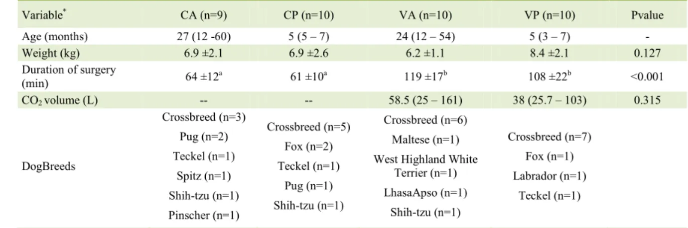
Thirty-nine bitches completed the study. One animal in the CA group experienced cardiac
arrest during the manipulation of the first ovary.
The patient responded adequately to resuscitation maneuvers and was discharged after 24 hours. Data from this dog were excluded from statistical
analysis. There was no significant difference in
weight between the groups. There was no difference in age between the VA and VP groups, or between
the CA and CP groups. There was also no significant
difference between the CO2 volume used in the VA and VP groups (Table 1).
Intraoperative analgesic rescue with
fentanyl was administered to 8 animals in the VP group and 7 in the VA group immediately after pneumoperitoneum (before M1). Other 6 animals
Table 1 - Demographic data of the 39 bitches included in the study distributed in the CA (Conventional Adult), CP (Conventional Pediatric), VA (Videolaparoscopic Adult),and VP (Videolaparoscopic Pediatric) groups.
Variable* CA (n=9) CP (n=10) VA (n=10) VP (n=10) Pvalue
Age (months) 27 (12 -60) 5 (5 – 7) 24 (12 – 54) 5 (3 – 7) -
Weight (kg) 6.9 ±2.1 6.9 ±2.6 6.2 ±1.1 8.4 ±2.1 0.127
Duration of surgery
(min) 64 ±12
a 61 ±10a 119 ±17b 108 ±22b <0.001
CO2 volume (L) -- -- 58.5 (25 – 161) 38 (25.7 – 103) 0.315
DogBreeds
Crossbreed (n=3) Pug (n=2)
Teckel (n=1)
Spitz (n=1)
Shih-tzu (n=1) Pinscher (n=1)
Crossbreed (n=5) Fox (n=2)
Teckel (n=1)
Pug (n=1)
Shih-tzu (n=1)
Crossbreed (n=6) Maltese (n=1)
West Highland White Terrier (n=1)
LhasaApso (n=1) Shih-tzu (n=1)
Crossbreed (n=7)
Fox (n=1)
Labrador (n=1)
Teckel (n=1)
*
in the CA group and 7 in the CP group were given
analgesic rescue during manipulation of the first
ovarian arteriovenous complex (before recording variables at M1). Atropine was administered to 3 animals in the CA group and dopamine was administered to 1 animal each in the CA and VP groups. Four dogs in the CA group and 2 dogs in the VP group received analgesic rescue once in the immediate postoperative period. The number of dogs administered rescue analgesia intraoperatively and postoperatively did not differ between groups.
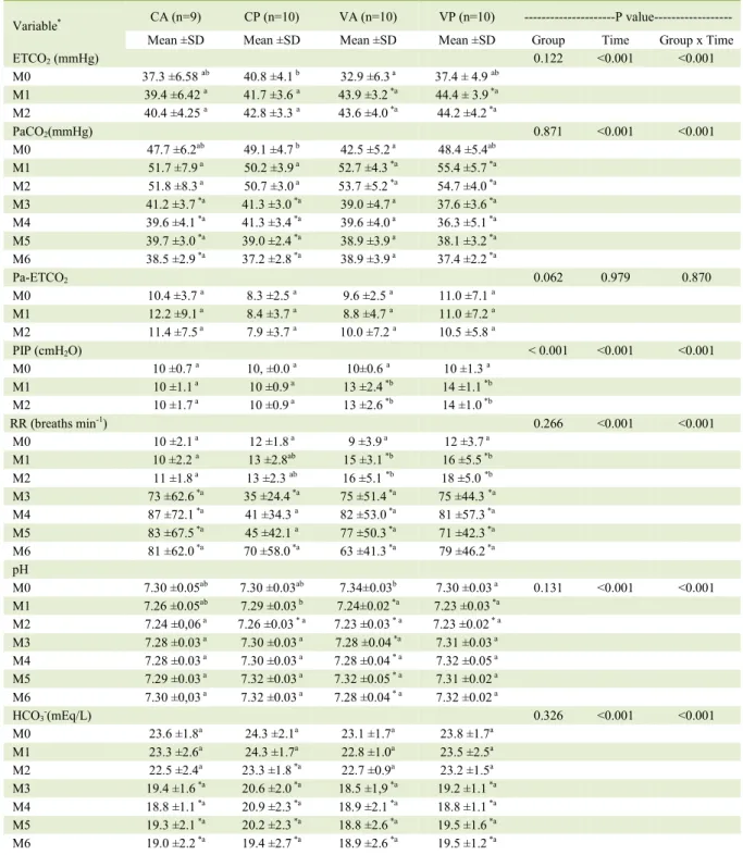
Duration of surgery was significantly
higher in the VA and VP groups than in the CA and CP
groups (P<0.001). There was a significant increase
of ETCO2 at M1 and M2 compared to M0 in the VA group, and at M2 compared to M0 in the VP group (P<0.001). Values for PaCO2 increased significantly at M1 and M2 in the VA and VP groups compared to
M0 (P<0.001). Significantly higher PIP and RR were
required intraoperatively in the VA and VP groups compared to the CA and CP groups (P<0.001).
From M3 to M6, all groups had significantly higher
values of RR than at M0 (P<0.001). From M3 to M6, PaCO2 values were significantly lower than at M0 in the CA, CP, and VP groups (P<0.001). There
was no significant difference in Pa-ETCO2 among the groups or over time (Table 2).
Mean values for PaO2 were within the expected range for dogs according to the inspired
oxygen fraction (>90% at M1 and M2; 21% from
M3 to M6; data not shown). Arterial pH values remained below the reference values for dogs throughout the evaluation period. All animals developed respiratory acidosis intraoperatively due to PaCO2 increase, and metabolic acidosis due to BE and HCO3-decreases during the postoperative
period (Table 2).
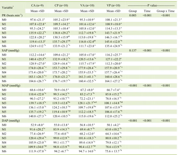
There was no significant difference in
PR between groups or compared to M0 during the intraoperative period (M1 and M2). At selected time
points from M3 to M6, PR increased significantly
in all groups compared to M0, with a trend towards higher values in the prepubescent (CP and VP) groups. Arterial blood pressures were higher in the CA and CP groups than in the VA and VP groups during the
intraoperative period; significant differences in MAP
and DAP were observed at M1. Higher SAP, MAP and DAP values were observed in all groups during the postoperative period as compared to M0. The highest values were observed in the CA group and the lowest values were observed in the VP group, with
significant differences in MAP and DAP between groups at specific moments (Table 3).
DISCUSSION
The main findings of the present study were: a) age (CP and VP groups versus CA and VA groups) did not influence PaCO2or Pa-ETCO2 values in bitches undergoing conventional or videolaparoscopic OH; and b) videolaparoscopic OH (VA and VP groups versus CA and CP groups), despite resulting in longer surgical times, seemed to cause a lower pressure response to surgical stimulation and a lower frequency of postoperative hypertension, notably in prepubescent bitches (VP group).
The increases in ETCO2 and PaCO2 values at M1 and M2 in the VA and VP groups are likely to have resulted from peritoneal CO2 absorption from capnopneumoperitoneum (IRWIN& WONg, 2014). In the same time points, it is suggested that higher intraoperative values of PIP and RR were required in VA and VP due to cranial displacement of the diaphragm resulting from pneumoperitoneum, which in turn decreases pulmonary compliance and total lung volume (IRWIN& WONg, 2014).
The hypothesis of the study was rejected
as there was no significant difference in PaCO2
between the VA and VP groups. It is possible that
the absence of a significant difference of CO2
absorption between the VP and VA groups was due to the age range of patients in the VP group (3 to 7 months). In this age range, the peritoneal surface-to-body mass ratio is closer to that reported in adult animals (TRANQUILLI et al., 2007). Our results might have been different if the bitches were younger than 3 months, but it is not common in clinical practice to perform OH on animals at such an early age.
At time point M0, mean Pa-ETCO2 values in all groups (8.3-11.0mmHg) were above the physiological range for dogs (2-5mmHg) anesthetized under spontaneous ventilation or positive pressure ventilation (HIgHTOWER et al., 1980). The high values of Pa-ETCO2 possibly resulted from atelectasis. High O2-inspired fractions may have resulted in rapid reabsorption of gases from alveoli and alveolar collapse (DANTZKER et al., 1975). Despite mean values of Pa-ETCO2 were high at M0,
there was no significant change in any of the groups
intraoperatively. Absence of an increase in Pa-ETCO2 at M1 and M2 in the VA and VP groups suggested that
the low inflation pressures used (6mmHg) to maintain
Table 2 - Comparison between CA (Conventional Adult), CP (Conventional Pediatric), VA (Videolaparoscopic Adult), and VP (Videolaparoscopic Pediatric) groups and time points of variables end-expired pressure of carbon dioxide (ETCO2), arterial
partial pressure of CO2 (PaCO2), difference between arterial and end-expired pressure of CO2 (Pa-ETCO2), respiratory rate
(RR), peak inspiratory pressure (PIP), pH, and HCO3
-Variable* CA (n=9) CP (n=10) VA (n=10) VP (n=10) ---P value---
Mean ±SD Mean ±SD Mean ±SD Mean ±SD group Time group x Time
ETCO2 (mmHg) 0.122 <0.001 <0.001
M0 37.3 ±6.58 ab 40.8 ±4.1 b 32.9 ±6.3 a 37.4 ± 4.9 ab
M1 39.4 ±6.42 a 41.7 ±3.6 a 43.9 ±3.2 *a 44.4 ± 3.9 *a
M2 40.4 ±4.25 a 42.8 ±3.3 a 43.6 ±4.0 *a 44.2 ±4.2 *a
PaCO2(mmHg) 0.871 <0.001 <0.001
M0 47.7 ±6.2ab 49.1 ±4.7 b 42.5 ±5.2 a 48.4 ±5.4ab
M1 51.7 ±7.9 a 50.2 ±3.9 a 52.7 ±4.3 *a 55.4 ±5.7 *a
M2 51.8 ±8.3 a 50.7 ±3.0 a 53.7 ±5.2 *a 54.7 ±4.0 *a
M3 41.2 ±3.7 *a 41.3 ±3.0 *a 39.0 ±4.7 a 37.6 ±3.6 *a M4 39.6 ±4.1 *a 41.3 ±3.4 *a 39.6 ±4.0 a 36.3 ±5.1 *a
M5 39.7 ±3.0 *a 39.0 ±2.4 *a 38.9 ±3.9 a 38.1 ±3.2 *a
M6 38.5 ±2.9 *a 37.2 ±2.8 *a 38.9 ±3.9 a 37.4 ±2.2 *a
Pa-ETCO2 0.062 0.979 0.870
M0 10.4 ±3.7 a 8.3 ±2.5 a 9.6 ±2.5 a 11.0 ±7.1 a M1 12.2 ±9.1 a 8.4 ±3.7 a 8.8 ±4.7 a 11.0 ±7.2 a
M2 11.4 ±7.5 a 7.9 ±3.7 a 10.0 ±7.2 a 10.5 ±5.8 a
PIP (cmH2O) < 0.001 <0.001 <0.001
M0 10 ±0.7 a 10, ±0.0 a 10±0.6 a 10 ±1.3 a
M1 10 ±1.1 a 10 ±0.9 a 13 ±2.4 *b 14 ±1.1 *b
M2 10 ±1.7 a 10 ±0.9 a 13 ±2.6 *b 14 ±1.0 *b
RR (breaths min-1) 0.266 <0.001 <0.001
M0 10 ±2.1 a 12 ±1.8 a 9 ±3.9 a 12 ±3.7 a
M1 10 ±2.2 a 13 ±2.8ab 15 ±3.1 *b 16 ±5.5 *b M2 11 ±1.8 a 13 ±2.3 ab 16 ±5.1 *b 18 ±5.0 *b M3 73 ±62.6 *a 35 ±24.4 *a 75 ±51.4 *a 75 ±44.3 *a
M4 87 ±72.1 *a 41 ±34.3 a 82 ±53.0 *a 81 ±57.3 *a
M5 83 ±67.5 *a 45 ±42.1 a 77 ±50.3 *a 71 ±42.3 *a
M6 81 ±62.0 *a 70 ±58.0 *a 63 ±41.3 *a 79 ±46.2 *a
pH
M0 7.30 ±0.05ab 7.30 ±0.03ab 7.34±0.03b 7.30 ±0.03 a 0.131 <0.001 <0.001
M1 7.26 ±0.05ab 7.29 ±0.03 b 7.24±0.02 *a 7.23 ±0.03 *a
M2 7.24 ±0,06 a 7.26 ±0.03 * a 7.23 ±0.03 * a 7.23 ±0.02 * a
M3 7.28 ±0.03 a 7.30 ±0.03 a 7.28 ±0.04 *a 7.31 ±0.03 a
M4 7.28 ±0.03 a 7.30 ±0.03 a 7.28 ±0.04 * a 7.32 ±0.05 a
M5 7.29 ±0.03 a 7.32 ±0.03 a 7.32 ±0.05 * a 7.31 ±0.02 a
M6 7.30 ±0,03 a 7.32 ±0.03 a 7.28 ±0.04 * a 7.32 ±0.02 a
HCO3-(mEq/L) 0.326 <0.001 <0.001
M0 23.6 ±1.8a 24.3 ±2.1a 23.1 ±1.7a 23.8 ±1.7a M1 23.3 ±2.6a 24.3 ±1.7a 22.8 ±1.0a 23.5 ±2.5a M2 22.5 ±2.4a 23.3 ±1.8 *a 22.7 ±0.9a 23.2 ±1.5a
M3 19.4 ±1.6 *a 20.6 ±2.0 *a 18.5 ±1,9 *a 19.2 ±1.1 *a
M4 18.8 ±1.1 *a 20.9 ±2.3 *a 18.9 ±2.1 *a 18.8 ±1.1 *a
M5 19.3 ±2.1 *a 20.2 ±2.3 *a 18.8 ±2.6 *a 19.5 ±1.6 *a
M6 19.0 ±2.2 *a 19.4 ±2.7 *a 18.9 ±2.6 *a 19.5 ±1.2 *a
*Described by mean ± standard deviation or median (minimum-maximum); a, bEqual letters do not differ by the Tukey test at 5%
It has been reported in anesthetized dogs that increasing the traction force of the ovary and ovarian ligament resulted in progressively increases in the intensity of the noxious stimulus (BOSCAN et al., 2011). In the present study, the lower values of MAP and DAP observed in the VA and VP groups at M1 and M2 suggested the occurrence of lower nociceptive stimulation during OH in these groups (DEVITT et al.,
2005). These findings may have resulted from minimal
traction forces of the ovarian ligament required during videolaparoscopic surgery. Due to particularities of
each surgical technique, time points M1 and M2 were associated to different noxious stimuli in the
CA and CP groups (ligation and section of the first
ovarian arteriovenous complex followed by ligation and section of the cervix) as compared to the VA and VP groups (manipulation of the uterine cervix followed by the ovarian arteriovenous complex). Although, this design might have biased the results of this study, the authors considered unlikely that this was the determining factor for the higher MAP and DAP values recorded in the CA and CP groups
Table 3 – Comparison between CA (Conventional Adult), CP (Conventional Pediatric), VA (Videolaparoscopic Adult), and VP (Videolaparoscopic Pediatric) groups and time points of variables pulse rate (PR), systolic arterial pressure (SAP), mean arterial pressure (MAP), and diastolic (DAP).
Variable* CA (n=9) CP (n=10) VA (n=10) VP (n=10) ---P value---
Mean ±SD Mean ±SD Mean ±SD Mean ±SD group Time group x Time
PR (beats.min-1) 0.003 <0.001 <0.001
M0 97.6 ±21.1a 105.2 ±23.9 a 95.3 ±10.9 a 108.1 ±21.3 a M1 107.8 ±22.8 a 109.5 ±14.2 a 101.6 ±12.6 a 108.9 ±18.0 a M2 95.3 ±24.2 a 105.3 ±10.4 a 105.8 ±12.0 a 114.3 ±13.3 a
M3 135.9 ±22.2*a 138.8 ±29.2*a 112.7 ±19.8 *a 143.7 ±21.9 *a
M4 122.8 ±20.2 a 130.3 ±15.9ab 113.0 ±19.8 * a 146.3 ±16.7 *b
M5 119.4 ±13.3a 135.7 ±29.3*ab 116.0 ±32.4ab 145.4 ±12.0 *b
M6 124.9 ±112 *a 133.9 ±21.2 *a 111.7 ±23.8 a 135.4 ±24.9 *a
SAP (mmHg) 0.137 <0.001 <0.001
M0 112.2 ±14.6 a 109.6 ±21.2 a 105.0 ±17.0 a 116.2 ±25.7 a
M1 140.4 ±25.5*a 132.9 ±18.2 *a 120.5 ±13.6 *a 127.1 ±22.2a
M2 128.9 ±27.0 a 128.9 ±16.4 * a 115.7 ±17.9 a 112.3 ±20.0 a
M3 174.0 ±20.4 * a 153.7 ±29.8 * a 177.9 ±28.4 * a 157.9 ±20.9 * a
M4 173.4 ±20.0 * a 173.7 ±20.2 * a 155.9 ±33.5 * a 157.7 ±26.4 * a
M5 183.1 ±26.3 * a 176.0 ±21.2 *a 161.3 ±41.1 *a 160.4 ±28.6 *a
M6 184.6 ±26.7 *a 176.7 ±19.7 *a 160.4 ±32.5 *a 164.1 ±27.2 *a
MAP (mmHg) <0.001 <0.001 <0.001
M0 68.6 ±10.6 a 70.9 ±16.5 a 67.2 ±8.0 a 66.7 ±7.4 a M1 110.4 ±22.0 *a 99.3 ±14.2 *a 83.2 ±7.3 *b 85.8 ±15.2 *b
M2 90.3 ±27.2 a 93.2 ±10.7 *a 72.2 ±23.1 a 78.8 ±16.5 *a
M3 139.7 ±18.5*a 119.3 ±12.8*bc 128.1 ±21.7*acd 108.1 ±14.8 *bd
M4 136.1 ±15.0 *a 124.2 ±10.5 *ac 109.7 ±19.8*bc 107.6 ±13.9 *b
M5 135.7 ±19.2 *a 119.4 ±10.9 *ab 112.2 ±18.5 *b 106.4 ±17.6 *b
M6 140.8 ±27.3 *a 120.4 ±10.5 *a 115.0 ±19.6 *a 112.0 ±25.2 *a
DAP (mmHg) <0.001 <0.001 <0.001
M0 52.9 ±8.0 a 55.0 ±13.8 a 56.8 ±10.5 a 50.1 ±4.2 a
M1 91.6 ±20.2*a 83.9 ±14.3 *a 69.4 ±8.7 *b 63.0 ±10.2 *b
M2 77.4 ±26.9 a 77.6 ±8.0 *a 68.2 ±12.0 a 64.3 ±14.6 *a M3 120.4 ±29.4 *a 99.0 ±12.9 *a 101.6 ±18.3 *a 80.8 ±10.2 *b
M4 105.9 ±23.0 *a 99.1 ±11.7 *a 89.4 ±14.9 *a 79.8 ±12.1 *a
M5 109.9 ±34.0 *ab 98.8 ±13.9 *b 90.4 ±12.7 *ab 78.6 ±13.9 *a
M6 111.9 ±37.8 *a 94.2 ±6.7 *a 94.7 ± 14.0 *a 75.6 ± 13.7 *b
*
because the highest MAP values in all groups were observed at M1, regardless of the structure being manipulated.
During the postoperative period (M3 to M6),
all groups presented hypertension (SAP≥150mmHg).
However, mild hypertension (SAP150-159mmHg) was observed in the VP group as compared to severe hypertension (SAP>180mmHg) in the CA group
(BROWN et al., 2007). These findings indicated that
both the age range (KUSTRITZ, 2014) and the use of videolaparoscopicsurgery (FERANTI et al., 2016) are associated with lower levels of hypertension during the postoperative period in dogs undergoing OH. High blood pressure may result from acute pain (MURREL, 2008). Nevertheless, all animals in this study received analgesics considered adequate for the management of pain associated to OH in dogs (FERANTI et al., 2016). Only 6 out of 40 animals presented with clinical signs of postoperative pain and were administered rescue analgesia.
CONCLUSION
Pediatric bitches submitted to laparoscopic
OH with 6mmHg inflation pressure of CO2 do not
develop higher ETCO2, PaCO2, or Pa-ETCO2 values than adult bitches undergoing the same surgical procedure. Higher PIP values are required to maintain normocapnia in bitches undergoing laparoscopic OH compared to conventional OH. Laparoscopic OH in pediatric bitches appeared to result in lower blood pressure increases in response to surgical stimulation and lower levels of postoperative arterial hypertension as compared to conventional OH in adult bitches.
BIOETHICS AND BIOSSECURITY COMMITTEE APPROVAL
This study was approved by the by the Ethics Committee for Animal Use (CEUA) of Universidade Federal do Rio grande do Sul (UFRgS), (protocol number 26609).
DECLARATION OF CONFLICTING OF INTERESTS
The authors declared no potential conflicts of interest with respect to the research, authorship, and/or publication of this article.
AUTHORS’ CONTRIBUTIONS
LBQ, ERM and CACB conceived and designed experiments. LBQ, gMS, FRS, LMF, VSM, METRT, gFLD e DCC performed the experiments, SFV carried out the lab analyses. LBQ, CACB and ERM prepared the draft of the manuscript. All authors critically revised the manuscript and approved of the final version.
REFERENCES
BOSCAN, P. et al. A dog model to study ovary, ovarian ligament and visceral pain. Vet. Anaesth Analg. 2011 May; 38(3):260-6. Available from: <https://onlinelibrary.wiley.com/doi/abs/10.11 11/j.1467-2995.2011.00611.x>. Accessed: Mar. 10, 2017. doi: 10.1111/j.1467-2995.2011.00611.x.
BROWN, S.; et al. Guidelines for Identification, evaluation, and management of systemic hypertension in dogs and cats.
J VetInternMed, v. 21, p. 542 – 558, 2007. Available from: <http://onlinelibrary.wiley.com/doi/10.1111/j.1939-1676.2007. tb03005.x/full>. Accessed: Mar. 15, 2017. doi: 10.1111/j.1939-1676.2007.tb03005.x.
DANTZKER, D. R., et al. Instability of lung units with low VA/Q ratios during O2 breathings. J Appl Physiol, v. 38, p. 886 -895, 1975. Available from: <http://jap.physiology.org/content/38/5/886. short>. Accessed: Mar. 5, 2017.
DEVITT, C. M.; et al. Duration, complications, stress, and pain of opens ovariohysterectomy versus a simple method of laparoscopic-assisted ovariohysterectomy in dogs. JAVMA, v. 227, n. 6, p. 921–927, 2005. Available from: <http://avmajournals.avma.org/ doi/abs/10.2460/javma.2005.227.921>. Accessed: Feb. 26, 2017. doi: 10.2460/javma.2005.227.921.
FERANTI, J. P. S.; ET AL. Ovariectomia laparoscópica ou convencional em cadelas: análise hemodinâmica e álgica. Rev. Bras. Med. Vet. v. 38, n: 1, p. 73-78, 2016. Available from: <http:// rbmv.org/index.php/BJVM/article/view/257>. Accessed: Mar. 4, 2017. doi: 10.2430/00000000000000.
FUKUSHIMA, F. B.; et al. Cardiorespiratoryandbloodgasaltera-tionsduringlaparoscopicsurgery for intra-uterineartificialinsemi -nation in dogs. The Canadian Veterinary Journal, v. 52, n. 1, p. 77–79, 2011. Available from: <http://pubmedcentralcanada.ca/ pmcc/articles/PMC3003583/>. Accessed: Jan. 13, 2017.
HIgHTOWER, C. E.; et al. End-tidal partial pressure of CO2 as an estimate of arterial partial pressure of CO2 during various ventilatory regimens in halothane-anesthetized dogs. American Journal of Veterinary Research, v. 41, n. 4, p. 610–612, 1980. Available from: <http://europepmc.org/abstract/med/6773449>. Accessed: Jan. 21, 2017.
IRWIN, M. g.; WONg, S. S. C. Anaesthesia and minimally inva-sive surgery. Anaesthesia and Intensive Care Medicine, v. 16, n. 1, p. 17–20, 2014. Available from: <http://www.sciencedirect. com/science/article/pii/S1472029911002670>. Accessed: Apr. 11, 2015. doi: 10.1016/j.mpaic.2011.11.004.
KUSTRITZ, M. V. R. Pros, Cons, and Techniques of Pediatric Neutering. Veterinary Clinics of North America: Small Animal Practice, v. 44, n. 2, p. 221–233, 2014. Available from: <http:// www.sciencedirect.com/science/article/pii/S019556161300199X>. Accessed: Mar. 15, 2017.
IN=_eJournals/images/JPLOgO.gif&IID=397&isPDF=YES>. Accessed: Feb. 25, 2017. doi: 10.5005/jp-journals-10033-1182.
McHONEY, M; et al. Carbon Dioxide Elimination During Laparoscopy in Children Is Age Dependent. Journal of Pediatric Surgery, v. 38, n. 1, p. 105–110, 2003. Available from: <http:// www.sciencedirect.com/science/article/pii/S0022346802630295>. Accessed: Jan. 29, 2017. doi: 10.1053/jpsu.2003.50021.
MURREL, J. C.; et al. Application of a modified form of the Glasgow pain scale in a veterinary teaching center in the Netherlands. The Veterinary Record, v. 162, p. 403–408, 2008. Available from: <https:// www.ncbi.nlm.nih.gov/pubmed/18375984>. Accessed: Jan. 20, 2015.