CLINICAL SCIENCE
The effects of volatile induction and maintenance of
anesthesia and selective spinal anesthesia on QT
interval, QT dispersion, and arrhythmia incidence
Ender Ornek,1Dilsen Ornek,2Z. Peren Alkent,3Abdu¨lselam Ekin,3Meleksah Basaran,3Bayazit Dikmen3 1Etlik Ihtisas Education and Training Hospital, Department of Cardiology - Ankara/Turkey.2Etlik Ihtisas Education and Training Hospital, Department of Anesthesia - Ankara/Turkey.3Ankara Numune Education and Training Hospital, Department of Anesthesia - Ankara/Turkey.
OBJECTIVE: The effects of sevoflurane general anesthesia and bupivacaine selective spinal anesthesia on QT dispersion (QTd) and corrected QT (QTc) interval were investigated.
METHODS AND MATERIALS:This prospective, randomized, double-blind study was conducted between July and September 2009 in the Urology and General Surgery operating rooms. Forty ASA I–II patients undergoing noncardiac surgery were randomized into two groups: Group R (n = 20) and Group V (n = 20). In Group R, 5 mg bupivacaine was administered into the spinal space. Anesthesia induction in Group V was established with sevoflurane+0.1 mg/kg vecuronium using the maximum vital capacity technique. Anesthesia was maintained with
2–3% sevoflurane+50% N2O/O2inhalation. All patients were tested with a 24-hour Holter ECG device. QT, QTc, and QTd intervals were measured using 12-lead ECG records at 1 and 3 minutes during preinduction, postinduction, postincision and postextubation periods. Mean arterial pressure (MAP), heart rate and ECG records were measured simultaneously.
RESULTS:None of the patients displayed arrhythmia. There was no significant difference between the groups with regard to QTd values (p.0.05). However, QTc was longer in Group V than in Group R after the induction of anesthesia at 3 minutes, after the intubation at 1 and 3 minutes, and after the incision at 1 and 3 minutes. MAP and heart rate were generally higher in Group V (p,0.05).
CONCLUSION:Although Volatile Induction and Maintenance of Anesthesia (VIMA) with sevoflurane might prolong the QTc interval and did not result in arrhythmia, selective spinal anesthesia with bupivacaine was not associated with alterations in the QT interval or arrhythmia.
KEYWORDS: Anesthesia; Bupivacaine; Sevoflurane; Complication; Arrhythmia.
Ornek E, Ornek D, Alkent ZP, Ekin A, Basaran M, Dikmen B. The effects of volatile induction and maintenance of anesthesia and selective spinal anesthesia on QT interval, QT dispersion, and arrhythmia incidence. Clinics. 2010;65(8):763-767.
Received for publication onFebruary 28, 2010;First review completed onApril 23, 2010;Accepted for publication onMay 8, 2010
E-mail: [email protected]
Tel.: 65 65 7373 823
INTRODUCTION
The effects of anesthetics on the cardiovascular system have a complicated character, and almost all of the effects lead to dose-related myocardial depression and decreases in heart rate and arterial pressure. In anesthesia practice, individual responses of patients against procedures such as induction, intubation, and surgical stimulation are influ-enced by many factors, including preoperatively used drugs, anesthesia type, preferred anesthetic agents, and the autonomic nervous system.1,2
Many of the anesthetics used in anesthesia practice interact with the QT interval. Depolarization and
repolar-ization of the myocardium take place within the QT interval. Varying QT intervals have been associated with hetero-geneous repolarization and ventricular arrhythmias.3,4 Therefore, prolonged QT intervals may be harmful, at least in cases with myocardial pathologies, necessitating anesthe-sia methods that do not influence the QT interval.5
QT dispersion (QTd) is defined as the difference between the longest and shortest QT intervals in 12-lead ECG. QTd is particularly recognized as the indirect measurement of the repolarization and has been associated with complex ventricular arrhythmias.3
Several studies investigating the effects of sevoflurane on QT, QTd, and QTc have revealed differing results.6,7 We found no study investigating the effects of selective spinal anesthesia on QT interval and arrhythmia. In the present study, we aimed to investigate the effects of selective spinal anesthesia with bupivacaine and sevoflurane inhalation anesthesia (VIMA) on QT interval, QTd, QTc, QTcd, and arrhythmia.
METHODS AND MATERIALS
Our prospective, randomized, double-blind study was conducted between July and September 2009 in the Urology and General Surgery clinics. Following the approval of the ethics committee and collection of informed consent, 40 ASA I–II patients 18–65 years of age who were scheduled to undergo urologic intervention or inguinal hernia surgery were included in the study. Patients with a history of a chronic obstructive lung disease, cardiovascular disease, electrolyte imbalance, diabetes mellitus, chronic alcoholism, idiopathic, congenital, or acquired prolonged QT syndrome, chronic drug use, or a Mallampati score$2 were excluded from the study.
Hemodynamic monitoring included mean arterial pres-sure (MAP), heart rate (HR), and peripheral oxygen saturation (SpO2). In addition, 24-hour Holter ECG was performed on all patients (Delmar Impresario, USA) to maintain a continuous ECG record.
The cases were randomly split into two groups: Group V (n = 20) and Group R (n = 20). The allocation sequence was generated by a table of random numbers. None of the cases received premedication. Anesthesia induction in Group V was established using the maximum vital capacity techni-que with 8% sevoflurane + 50% N2O/O2. After the achievement of neuromuscular block with 0.1 mg kg21 vecuronium, endotracheal intubation was performed. Anesthesia was maintained with 2–3% sevoflurane + 50% N2O/O2inhalation.
In Group R, selective spinal anesthesia was performed by delivering 5 mg 0.5% hyperbaric bupivacaine to the L4–L5 space via the midline approach to the patients in the sitting position. A local anesthetic agent was administered within 20 seconds without aspiration of the CSF, and the patients were kept sitting at a 45
˚
angle until the achievement of T10 anesthesia (maximum 10 minutes). Sensory block was evaluated with the pinprick test, while motor block was assessed with the Bromage scale. Surgery was allowed when the sensory block reached the T10level.The study was designed to examine the following: to elevate the crystalloid infusion speed to 15 mg/kg/h when MAP dropped below 30% and persisted at that level for more than 30 seconds; to carry out 5 mg/kg/h colloid infusion upon persistence of the hypotension for 15 minutes; to apply 5 mg ephedrine when the cases failed to normalize after another 15 minutes; to increase the anesthesia depth when MAP increased above 30%; and to administer 0.1 mg nitroglycerine relative to the MAP. We also planned to deliver 0.5 mg atropine when the HR dropped below 50.
We did not use anticholinesterase or opioid therapy in case they would influence the QT and QTc intervals.
Data analyses of both of the groups were performed at the times noted in Table I. MAP, HR, and SpO2 values were recorded. ECG analyses were carried out.
ECG records were examined with a Holter monitor by the same cardiologist. The QT interval was analyzed through-out the whole operation. The QT interval was recognized as the duration between the beginning of the QRS complex and the end of the T wave in the ECG. The mean QT interval was calculated manually, and the QT interval measurement was corrected for heart rate using the Bazett formula (12).
QTc(sn) = QT/ ffiffiffiffiffiffiffiffi RR
p
QTd(sn) = QTmax2Qtmin
QTcd(sn) = QTcmax2Qtcmin
QTc(sn): corrected QT interval, QTd(sn): QT dispersion time, QTcd(sn): corrected QT dispersion time, QT max: maximum QT interval, QT min: minimum QT interval.
The study was conducted with the significance level set at alpha = 0.05 and beta = 0.8 power. The study included 40 subjects (n1 = n2 = 20). No patient was excluded from the study. The power, based on the QTc values of the groups, was found to be 0.83.
The data obtained in the present study were evaluated with SPSS version 12 (SPSS, Chicago, IL). We used the chi-square test for dependency between the variables, the Mann-Whitney U test for comparisons between groups, and the Wilcoxon signed-rank test for comparisons within the groups. The Mann-Whitney U test was used for all comparisons of the demographic characteristics with the exception of gender. Gender was compared using the chi-square test. A p value ,0.05 was considered significantly significant.
RESULTS
No significant differences in demographic characteristics were found between the groups (Table 2).
The HR values did not significantly differ within either group. However, the Group V HR values were high after the induction (p: 0.02) (Figure 1).
The MAP values were not significantly different within groups, but the Group R MAP values were lower than the Group V values at all measurement time points. However, none of the cases demonstrated a need for therapeutic intervention for blood pressure changes (Figure 2).
When compared within groups, the QTd, QTc, and QTcd values displayed no significant differences at any of the
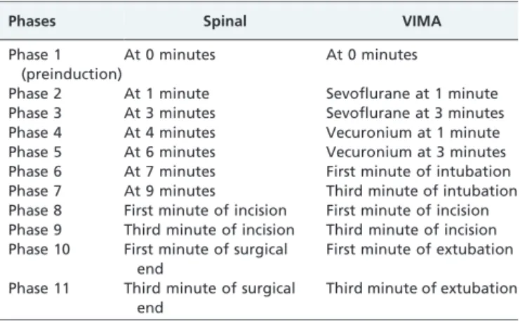
Table 1- Measurement phases in VIMA and spinal anesthesia.
Phases Spinal VIMA
Phase 1 (preinduction)
At 0 minutes At 0 minutes
Phase 2 At 1 minute Sevoflurane at 1 minute
Phase 3 At 3 minutes Sevoflurane at 3 minutes
Phase 4 At 4 minutes Vecuronium at 1 minute
Phase 5 At 6 minutes Vecuronium at 3 minutes
Phase 6 At 7 minutes First minute of intubation
Phase 7 At 9 minutes Third minute of intubation
Phase 8 First minute of incision First minute of incision Phase 9 Third minute of incision Third minute of incision Phase 10 First minute of surgical
end
First minute of extubation
Phase 11 Third minute of surgical end
Third minute of extubation
Table 2- Demographic data (mean¡SD).
Group R (n = 20) Group V (n = 20) p
Age (years) 42.9¡11.88 (18–65) 44.5¡10.47 (26–64) 0.567
Weight (kg) 80.36¡10.35 78.40¡9.93 0.456
Height (cm) 168.46¡8.42 167.36¡9.23 0.632
Gender (F/M) 12/18 14/16 0.602
ASA (I/II) 12/18 13/17 0.793
Operation length (min.)
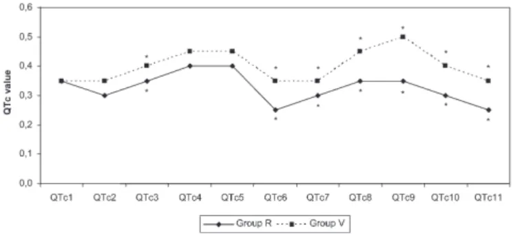
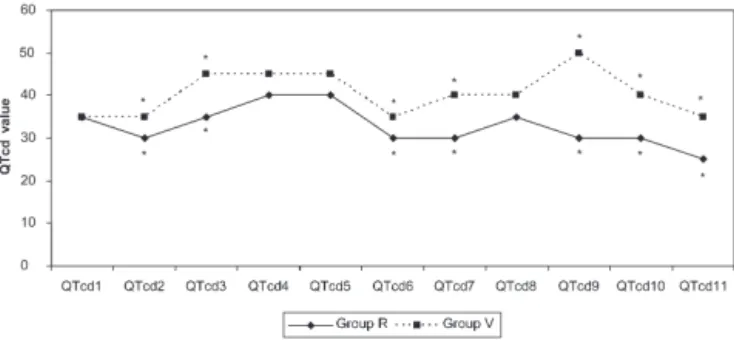
measured time points. Intergroup comparisons exhibited a high QTd at 3 minutes after extubation in Group V. QTc was significantly higher in Group V than in Group R at 3 minutes after the induction, 1 and 3 minutes after the intubation, and 1 and 3 minutes after the incision (Figures 3 and 4) (p,0.05). QTcd values were significantly higher in Group V at 1 and 3 minutes after the induction, 1 and 3 minutes after the extubation, and 1 and 3 minutes after the incision (Figure 5).
DISCUSSION
Several conditions, such as diabetes mellitus, arrhythmia, ischemic cardiac diseases, pulmonary disease, uremia, electrolyte and acid/base disorders, and prolonged QT syndrome, and the drugs used in their treatment, such as antihypertensives, beta-blockers, antidiabetics, and opioids, are known to influence the QT interval.8
A prolonged QT interval should be considered an important symptom because it can lead to serious ven-tricular arrhythmias.8 It is not easy to differentiate the underlying cause of changes in QT interval among factors such as delivered pharmaceutical agents, drug interactions, and sympathetic activation occurring upon laryngoscopy and intubation.1
Michaloudis et al. applied VIMA with sevoflurane and isoflurane and found that isoflurane prolonged the QTc interval but did not change QTd or QTcd, whereas sevoflurane did not affect those three parameters; overall, they determined that sevoflurane was a good agent.9Guler et al. showed that sevoflurane, isoflurane, and halothane prolonged QTd and QTcd in patients who had no congenital heart disease. However, they found no statistically signifi-cant difference between the agents and concluded that those anesthetic agents caused myocardial repolarization abnorm-alities by increasing QTcd. In the same study, prolonged QTcd was found to have the potential to play a role in arrhythmias observed among people receiving anesthesia
who had no cardiovascular disease. In light of the fact that those agents can prolong QTcd further, Guler et al. noted that this parameter could be used during the development of new agents.10
Saarnivaara et al.5 reported that changes in ventricular repolarization might cause a prolonged QTc interval in cases where anesthetic agents have induced sympathoa-drenal hyperactivity. Prolonged QTc interval has been reported to show autonomic nervous system imbalance in the heart, thereby lowering the ventricular fibrillation and leading to ventricular arrhythmia.11 Abe et al.6 reported ventricular tachycardia development under sevoflurane and N2O anesthesia. A study using the single-breath induction technique with sevoflurane found marked prolongation of and increased development of arrhythmia in the QT interval.12 A study comparing 1 MAC sevoflurane and desflurane reported a prolonged QTc interval and increased QTd upon delivery of each of those two agents.13Karagoz et al.14conducted a study to analyze the effects of halothane, isoflurane, and sevoflurane, in which they reported no changes in the QTc interval as a result of 2% sevoflurane and 50% O2 + 50% N2O. In the current study, QTc was higher in Group V than in Group R at every measured time point, and significant differences were identified during times of intense perioperative sympathetic activation, particularly at 3 minutes after the induction and 1 and 3 minutes after the incision (p,0.05). Given the previous finding claiming the presence of a direct relationship between plasma catecholamine levels and QT interval, it is probably no coincidence that QTc was markedly prolonged during times of intense sympathetic activation.15
In the present study, a QTc of 506 ms, recognized as the upper limit of the normal QTc interval, was not reached in any of the cases. Again, no prolonged QTc exceeded the basal value more than 75 ms.
Figure 1- Heart rates in the two study groups (beats per minute).
Figure 2- MAP values in the two study groups (mmHg).
Figure 3- QTc values in the two study groups (seconds).
Although some studies have noted that prolonged QTc contributes to the development of cardiac arrhythmias,5,20 similar to the results of the study of Saarnivaara et al.,5we did not identify a relationship between the prolonged QTc duration and the development of arrhythmia in the present study. As in our study, this study found that heart rate was generally high during the periods of prolonged QTc, and the authors attributed this to perioperative anxiety and elevated sympathetic activity. In view of the fact that amiodarone, a type III antiarrhythmic agent, prolongs the QT interval while having no influence over QTd or ventricular extrasystole formation, we believe that the electrophysiologic mechanism playing a part in arrhythmia development should have been clearly identified.17
The normal range of QTd is 300–600 ms.18 QTd demonstrates the regional homogeneity of ventricular myocardial cells during repolarization and reflects the physiologic desynchronization of myocardial excitability. A prolonged QTd interval is an important determinant of ventricular arrhythmias and sudden cardiac deaths. Moreover, while some studies note that QTd values longer than 65 ms are important in ventricular tachycardia etiology, there are also studies that describe 70 ms as a normal value. In a detailed study on this subject, QTd was found to provide reliable data on prognosis estimation and foreknowledge of cardiac problems stemming from myo-cardial repolarization abnormalities. However, the same study recommended that a marked repolarization abnorm-ality be considered in cases with a QTd value above 100 ms.8 In the current study, although QTd was longer in Group V at 3 minutes after the extubation (phase 10), it was not higher than 57 ms in any of the cases. In view of the variation from the initial phases in the VIMA and spinal anesthesia groups, there was no statistically significant difference with regard to the QTc, QTd, and QTcd values.
A multicenter study conducted on 17,000 patients in 2000 investigated the effects of spinal anesthesia on arrhythmia and found that 70.2% of the cases displayed tachycardia, bradycardia or arrhythmia. Most of those arrhythmias were of spontaneous and remitting minor types. Additionally, 30.3% showed sinus arrhythmia, whereas 27.2% and 13.8% exhibited premature beat and bradycardia, respectively.19 Moreover, another prospective spinal anesthesia study on 40,460 patients reported the rate of bradycardia incidence before cardiac arrest as 6.4/10.000. All of the cases that experienced arrest responded to resuscitation. In that study, the formation of varying degrees of cardiac blocks were noted.20 In contrast to that study, our study preferred selective spinal anesthesia, and its influence over arrhyth-mia was not determined. We believe that the difference
between the studies may be due to the different methods applied.
Bupivacaine is an anesthetic agent that is frequently used in current anesthesia practice. Although most of the toxic reactions are associated with high plasma concentrations of bupivacaine, cases of mortality and morbidity have been reported, even at low doses. Systemic complications exhibiting reduced cardiac output and changes in the central nervous system have been observed, even at a 1mg/ml plasma concentration.21Hotverd et al. conducted a study showing that bupivacaine at a 2 mg/ml plasma concentration had a negative inotropic effect and prolonged cardiac conduction. That study also implicated bupivacaine in re-entry arrhythmias. Bupivacaine is 4 times more effective than lidocaine and causes cardiovascular and other systemic toxicities 4–17-times more frequently than lido-caine.22Studies with bupivacaine have reported supraven-tricular tachycardia, atriovensupraven-tricular block, early vensupraven-tricular beats in various forms, QRS changes, and fatal ventricular fibrillation. Moreover, independent of dose, the most common rhythm abnormality in hypoxia and acidosis is bradycardia with a wide QRS complex.
In animal studies, bupivacaine, etidocaine, mepivacaine, and lidocaine delivered as an i.v. bolus and infusion have been reported to cause an increase in the area below the T wave, prolong the QT interval, and induce the formation of a U wave following the T wave; these effects were more significant in the etidocaine and bupivacaine groups. The effects of those agents over the temporal dispersion of the effective refractory period were reported as 37.4 ms in lidocaine, 48.3 ms in mepivacaine, 92.5 ms in etidocaine, and 98.1 ms in bupivacaine. Moreover, a relationship between prolongation of the effective refractory period and ventri-cular arrhythmia was also noted.22
Decreases in systemic blood pressure can lead to changes in heart rate and cardiac rhythm alterations.2 However, hypotension during anesthesia is described as a reduction of the basal value of arterial pressure below 20%. In the current study, while none of the groups demonstrated changes exceeding that limit in blood pressure or heart rate values, MAP and HR were significantly higher in Group V than in Group R in measurements performed after intubation, incision, and extubation. Lowrie et al. found a considerable increase in HR, SAP, and DAP parallel to the elevations in plasma norepinephrine and epinephrine concentrations. Moreover, they reported a significant rise in epinephrine level and heart rate within the first five minutes of extubation. In the present study, elevation in the heart rate during intubation and extubation in the VIMA group was consistent with the results of the study of Lowrie et al.15
In our study, MAP values showed a significant increase in the VIMA group compared with the spinal group. This result is consistent with the sympathetic discharge results obtained during the intubation and extubation phases in the studies of Tanaka et al. and Lowrie et al.15,24
Nevertheless, selective spinal anesthesia has not been compared with VIMA with regard to QTd, QTc, QTcd, and arrhythmia incidence.
In conclusion, although VIMA with sevoflurane might prolong the QTc interval and did not result in arrhythmia, selective spinal anesthesia with bupivacaine was not associated with alterations in the QT interval or arrhythmia.
REFERENCES
1. Fee JPH, Thompson GH. Comparative tolerability profiles of the inhaled a n a e s t h e t i c s . D r u g S a f e ty . 1 9 9 7 ; 1 6 : 1 5 7 – 7 0 , d o i : 1 0 .2 1 6 5 / 00002018199716030-00002.
2. Pagel PS, Kampine JP, Schmeling WT, Warltier DC. Influence of volatile anesthetics on myocardial contractility in vivo: Desflurane versus i s o f l u r a n e . A n e s t h e s i o l o g y . 1 9 9 1 ; 7 4 : 9 0 0 – 7 , d o i : 1 0 . 1 0 9 7 / 00000542199105000-00016.
3. Hingham PD, Campell RWF. QT dispertion: A review. Br J Anaesth. 1994;71:508–10.
4. Statters DJ, Malik M, Ward DE, Camm AJ. QT dispersion: problems of methodology and clinical significance. J Cardiovasc Electrophysiol. 1994;5:672–85, doi: 10.1111/j.1540-8167.1994.tb01190.x.
5. Saarnivaara L, Klemola UM, Lindgren L, Rautiainen P, Suvanto A. QT interval of the ECG, heart rate and arterial pressure using propofol, methohexital or midazolam for induction of anaesthesia. Acta Anaesth Scand. 1990;34:276–81, doi: 10.1111/j.1399-6576.1990.tb03085.x. 6. Abe K, Takad K, Yoshiya I. Intraoperative torsade de pointes ventricular
tachycardia and ventricular fibrillation during sevoflurane anesthesia. Anesth Analg. 1998;86:701–2, doi: 10.1097/00000539-199804000-00004. 7. Gallagher JD, Weindling SN, Anderson G, Fillinger MP. Effects of
sevoflurane on QT interval in a patient with congenital long QT syndrome. Anesthesiology. 1998;89:1569–73, doi: 10.1097/ 00000542199812000-00038.
8. Malik M, Batchvarov VN. Measurement, interpretation and clinical potential of QT dispersion: J Am Coll Cardiol. 2000. 15;36:1749–66, doi: 10.1016/S0735-1097(00)00962-1.
9. Michaloudis D, Fraidakis O, Petrou A, Farmakalidou H, Neonaki M, Christodoulakis M, et al. Effects of isoflurane or sevoflurane on QT and Qtc dispersion during induction of anaesthesia. BJA June 1999;82, supp 1. 10. Gu¨ler N, Bilge M, Eryonucu B ve ark. Anestezi indu¨ksiyonu esnasy´nda volatil anestezik ajanlary´n miyokardiyal repolarizasyon u¨zerine etkileri. Tu¨rk Kardiyol Dern Ara. 1999;27:31–6.
11. Schouten EG, Dekker JM, Meppelink P et al. QT interval prolongation predicts cardiovascular mortality in an apperently healty population Circulation. 1991;84:1516–23.
12. S¸en S, O¨ zmert G, Boran N, Turan H, C¸alıs¸kan E. Comparison of single-breath vital capacity rapid inhalation with sevoflurane % 5 and propofol induction on QT interval and haemodynamics for laparoscopic surgery. Eur J Anaesth. 2004;21:543–6.
13. Yıldırım H, Adanır T, Atay A, Katırcıog˘lu K, Savacı S. The effects of sevoflurane, isoflurane and desflurane on QT interval of the ECG. Eur J Anaesth. 2004;21:566–70.
14. Karago¨z AH, Bas¸gu¨l E, C¸ eliker V, Aypar U¨ . The effect of inhalational anaesthetics on QTc interval. Eur J Anaesth. 2005;22:171–4.
15. Lowrie A, Johnston PL, Fell D, and Robinson SL. Cardiovascular and plasma catecholamine responses at tracheal extubation. Br J Anaesth. 1992;68:261–3, doi: 10.1093/bja/68.3.261.
16. Lindgren L. ECG changes during halothane and enflurane anaesthesia for E.N.T. surgery in children. Br J Anaesth. 1981;53:653–62, doi: 10.1093/ bja/53.6.653.
17. Tanskanen PE, Kytta JV, Randell TT. QT interval and QT dispersion during the induction of anaesthesia in patients with subarachnoid haemorrhage: a comparison of thiopental and propofol. European Journal of Anaesthesiology. 2002;19:749–54.
18. Higham PD, Campbell RWF. QT dispersion. Br Heart J. 1994;71:508–10, doi: 10.1136/hrt.71.6.508.
19. Youngs PJ, Littleford J. Arrhythmias during spinal anesthesia: Can J Anaesth. 2000;47:385–90, doi: 10.1007/BF03018964.
20. Auroy Y, Narchi P, Messiah A, Litt L, Rouvier B, Samii K. Serious Complications Related to Regional Anesthesia: Results of a Prospective Survey in France: Anesthesiology. 1997;87:479–86, doi: 10.1097/ 00000542-199709000-00005.
21. Zabel M, Klingenheben T, Franz MR, Hohnloser SH. Assesment of QT dispersion for prediction of mortality of arrhytmic events after myocardial infarction. Results of a prospective, long term follow-up study. Circulation. 1998;97:2543–50.
22. Kasten GW. Anıide local anesthetic alterations of effective refractory period temporal dispersion: relationship to ventricular arrhythmias. Anesthesiology. 1986;65:61–6, doi: 10.1097/00000542-198607000-00010. 23. Hotvedt R, Refsum H, Helgesen KG. Cardiac electrophysiologic and
hemodynamic effects related to plasma levels of bupivacaine in the dog. Anesth Analg. 1985;64:388–94, doi: 10.1213/00000539-198504000-00003. 24. Tanaka S, Tsuchida H, Nakabayashi K, Seki S, Namiki A. The effects of