DOI: 10.5935/2359-4802.20180013
ORIGINAL ARTICLE
Mailing Address: Maria das Neves Dantas da Silveira Barros•
Rua Cardeal Arcoverde, 85/1501 Bloco A. Postal Code: 52011-240, Graças, Recife, PE – Brazil. E-mail: [email protected]; [email protected]
Predictors of Coronary Artery Obstructive Disease in Acute Pulmonary Edema of
Unclear Origin
Maria das Neves Dantas da Silveira Barros,1,2,3 Vander Weyden Batista de Sousa,2 Isabelle Adjanine Borges de Lima,2 Cecília Raquel Bezerra Marinho Nóbrega,2 Isabelle Conceição Albuquerque Machado Moreira,2 Suzana Marine Martins Dourado,2 Bruna Maria Simões Andrade,2 Virgínia da Silva Batista,2 Maria Cleide Freire Clementino da Silva,2 Luís Cláudio Correia1
Escola Bahiana de Medicina e Saúde Pública - EBMSP,1 Salvador, BA; Pronto-Socorro Cardiológico de Pernambuco/Universidade de Pernambuco - Procape/UPE;2 Coordenação de Aperfeiçoamento de Pessoal de Nível Superior/ Programa de Doutorado Sanduíche no Exterior CAPES/PDSE,3 – Brazil
Manuscript received November 27, 2016, revised manuscript August 08, 2017, accepted August 21, 2017
Abstract
Background: Cardiogenic Acute Pulmonary Edema (APE) is considered one of the main medical emergencies, and it
is the extreme manifestation of acute heart failure. The main etiology of heart failure is ischemic heart disease. To date, the definition of ischemic etiology in acute pulmonary edema was based on criteria such as: clinical history of ischemic heart disease, noninvasive examinations and, in other patients, coronary angiography. Classified as such, ischemic heart disease has been shown to be its main etiology. The high prevalence between these two diseases was evaluated, but not by the exclusive angiographic criterion, the gold standard of this pathology and the reason of this study.
Objective: To evaluate the predictors of obstructive coronary artery disease in patients with acute pulmonary
edema of unclear origin.
Method: Patients admitted to a cardiovascular disease referral emergency unit were recruited to undergo coronary
angiography if the acute pulmonary edema etiology was not adequately elucidated. Obstructive coronary disease was considered if at least one epicardial vessel had 70% of occlusion.
Results: Obstructive coronary disease was classified by coronary angiography in 149 consecutively evaluated
patients, and coronary artery obstruction was the outcome variable of the predictor model. Among the variables related to coronary disease, the predictor variables were the history of coronary artery disease (p < 0.001) and myocardium segmental deficit at the echocardiogram (p < 0.02).
Conclusion: The antecedent of coronary disease and the myocardium segmental deficit at the echocardiogram
were able to discriminate patients with acute pulmonary edema associated with obstructive coronary disease. Troponin values classified by two cardiologists as secondary to an acute non-ST-segment elevation myocardial infarction, and chest pain preceding the clinical picture were not able to discriminate patients with or without coronary obstruction and thus, the diagnosis of obstructive coronary disease should not be pursued based on the troponin value and/or chest pain preceding the clinical picture. (Int J Cardiovasc Sci. 2018;31(2)133-142)
Keywords: Heart Failure; Pulmonary Edema; Coronary Artery Disease; Risk Factors; Myocardial Ischemia.
Introduction
Acute Pulmonary Edema (APE) is the extreme expression of acute heart failure (HF) in the emergency room, being classified as cardiogenic (APE) or non-cardiogenic (Acute Respiratory Distress Syndrome – ARDS). The etiology and pathophysiological mechanisms differ between them. The first occurs due to the increase
cost among the modalities of acute HF, with the first one being cardiogenic shock.1 APE 30-day mortality is still considered high. In the study by Figueras et al.,2 it was observed that the mortality rate in 216 consecutive patients admitted with APE in a single center was 14.1%.
Severe Coronary Artery Disease (CAD) is often the anatomical substrate in patients with APE, as shown by Graham et al.3 in a study of 119 patients with APE, of which 71 (60%) had a significant coronary lesion; 35 (49%), lesion in three epicardial vessels; and 7 (10%), left coronary trunk lesion.
Ischemic heart disease is the most frequent etiology of chronic HF worldwide4 and in Brazil.5 APE is a severe clinical expression of acute HF and obstructive CAD (arterial lumen mostly occluded) is often related to this clinical syndrome. The present study aimed to determine the predictors of obstructive CAD in this extreme HF clinical condition. We believe that we can contribute to the decision-making concerning patients who have this syndrome and with a suspected ischemic substrate for its occurrence.
Methods
An observational, cross-sectional, diagnostic study with prospective data collection was performed, analyzing 149 patients between 40 and 80 years of age, consecutively admitted to a public emergency unit, specialized in cardiology, after registration in the hospital admission authorization form and patient history was reviewed by the research team. If the APE etiology was not clearly defined, such as uremic syndrome, previously known valvulopathy, well-defined cardiomyopathy at the echocardiogram and ST-segment elevation myocardial infarction, the patient was invited to participate in the study. Kidney dysfunction with creatinine >2 mg% was an exclusion criterion.
Creatinine values above 2mg% were included in the study if a coronary angiography (CA) had been requested by the attending physician prior to the study protocol evaluation. After signing the Free and Informed Consent form (CAAE 05503912.6.0000.5544), the CA was requested, aiming at defining the degree of coronary obstruction. Patients who had undergone the examination 1 year prior to the study and had normal results or had CAD definition without new therapeutic possibilities were not submitted to a new examination. The CA was analyzed by a single physician who was only aware of the reason for the hospitalization, APE. Obstructive CAD was considered severe if more than two-thirds (70%) of
the arterial lumen, including total vessel occlusion, were occupied and if an epicardial vessel was affected: right coronary artery and its branches, circumflex artery and its branches, anterior descending artery, diagonal artery and 50% if the left main coronary artery was affected. The CA was part of the research protocol because, as part of the primary objective, it was necessary to determine the predictors of severe obstructive coronary disease, and this is the gold standard examination for the investigation of this pathology.
The electrocardiogram was performed on the first and second days, and was analyzed by an arrhythmologist, a specialist on the subject. All patients underwent Doppler echocardiography. Ultrasensitive troponin (Roche laboratory equipment) was performed on the first and second days and two cardiologists were asked to interpret the results if the obtained values were compatible with APE precipitated by or associated with an acute coronary event. The diseases considered to be present, such as systemic arterial hypertension, diabetes mellitus, prior stroke, valvulopathy, asthma and chronic obstructive pulmonary disease, were taken into account based on the information provided by the patient. Smoking was considered when the patient was a current smoker, or when the patient had been an ex-smoker for less than 2 years.
Statistical analysis
To create the explanatory model for the occurrence of obstructive coronary disease, the following variables were collected: age, gender, presence of chest pain on admission, suggestive of myocardial ischemia prior to dyspnea, presence of previous dyspnea, history of coronary disease (classic angina pectoris, history of acute myocardial infarction, surgical or percutaneous myocardial revascularization); electrocardiographic alteration
suggestive of myocardial ischemia (ST depression ≥ 0.05 mV − or negative T wave ≥ 2 mm in leads correlated to a coronary
To characterize the study population and perform the bivariate analysis, the continuous variables were shown as means and standard deviation, when they had a normal distribution, or by medians and interquartile ranges. The normality hypothesis was verified by the Kolmogorov-Smirnov test. When searching for an association between the obstructive coronary artery disease predictive variables, Pearson's chi square test was used for the comparison of proportions, whereas the Mann-Whitney test was used for the comparison of medians and the Student's t test for the comparison of the means. The variables that showed statistical significance of up to 10% (p < 0.10) were eligible for the multivariate model. The logistic regression model was applied. The Hosmer-Lemeshow test was used for the model assessment, and model performance was assessed through the area under the curve (AUC) of the Receiver Operating Characteristic (ROC) curve. The statistical software Stata for Windows, version 12, was used for the statistical analyses.
Results
Of the 149 patients, 89 (59%) had severe obstructive CAD. Most patients were elderly and female (Table 1).
A moderate lesion alone was observed in 9 patients (6%) of the 149 coronary angiograms evaluated. In most cases, the affected coronary artery was restricted to an epicardial vessel, with the right coronary artery being the one most frequently involved. An affected left main coronary artery and the involvement of three vessels were observed in 11 (12%) and 15 (17%) of the 89 patients, respectively, as described in Table 2.
Table 3 shows the detailed involvement of the epicardial vessels. It was observed that severe coronary artery disease more often affected the population with diabetes, peripheral obstructive arterial disease, those with a history of coronary artery disease and stroke.
As for the ejection fraction at the echocardiogram, the mean value was above 45% and in most cases, it was evaluated using the Teichholz method. The median troponin I on the first day was 0.086 ng/mL (normal value 0.014 ng/mL), and the interquartile range was 0.036 and 0.201 ng/mL in patients with APE. Troponin was considered positive when two independent cardiologists classified it as suggestive of an APE-related acute coronary event, a fact observed in 77 patients (51.3%) (Table 1).
Mean systolic and diastolic blood pressures were high. The most often found alteration in the electrocardiogram was ST depression, verified in most assessed patients (53.5%).
An intracoronary thrombus was visualized in 4 (3%) of the 122 patients in which CA was performed during hospitalization. One had elevation of CK-MB mass and troponin, with very high values – respectively, 86 mg/mL (reference value of 4.54 mg/mL) and 1.29 ng/mL (reference value of 0.014 ng/mL) –, suggesting values compatible with coronary artery occlusion.
The electrocardiogram showed ST depression of 1 mV in the inferior wall, V4 to V6 and ST elevation of 1 mV in aVR and V1. The CA showed a thrombus in the anterior
descending artery and severe lesions in the diagonal and right coronary arteries.
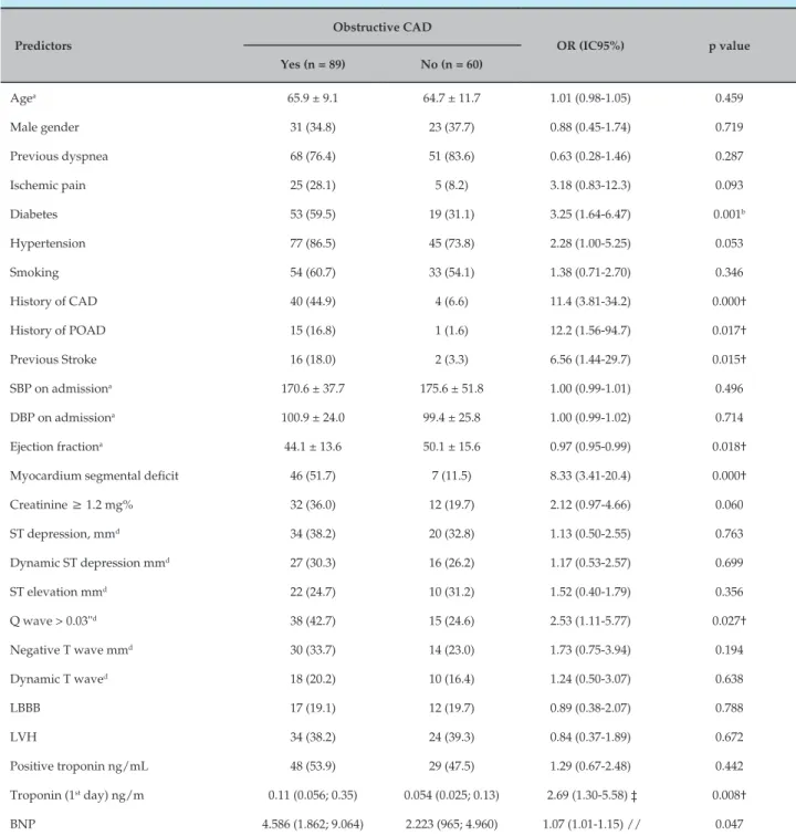
The univariate analysis of the possible predictors of obstructive CAD is described in Table 4. Ischemic chest pain, diabetes mellitus, systemic arterial hypertension, history of CAD, history of obstructive peripheral arterial disease, stroke, ejection fraction, myocardium segmental deficit at the echocardiography, presence of Q wave and evaluation of the absolute measurement of troponin on the first day were associated with obstructive CAD (p < 0.1).
The variables that were included in the multivariate model and the independent variables of obstructive CAD were history of CAD (p < 0.000) and myocardium segmental deficit (p < 0.02), as described in Table 5.
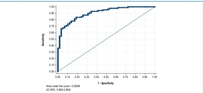
The model performance was evaluated by the area under the curve (AUC) of receiver operating characteristic (ROC) curve, as shown in Figure 1. The AUC was shown to have a good discriminatory power with a C-statistics of 0.905 (95% confidence interval - 95%CI: 0.862-0.954).
Discussion
Table 1 – Baseline characteristics of patients with acute pulmonary edema of unclear origin
Characteristic Total (149)
Age 65.4 ± 10.2
Male gender 54 (36.2)
Previous dyspnea 118 (79.2)
Ischemic pain 30 (20.1)
Diabetes 71 (47.6)
Hypertension 121 (81.2)
Smoking 86 (57.7)
History of CAD 44 (29.5)
History of POAD 16 (10.7)
Previous Stroke 18 (12.1)
SBP on admission 171.8 ± 42.7
DBP at admission 99.6 ± 23.3
Ejection fraction 46.4 ± 14.6
Ejection fraction <40% 56 (37.6)
Segmental myocardium deficit 53 (35.6)
Creatinine> 1.2 mg% 44 (29.5)
Creatinine mg% (131 patients) 1.0 (0.8; 1.31)
BNP pg / mL (123 patients) 3.856 (1.441; 6.634)
ST depression *, in mm 54 (53.5)
ST elevation *, in mm 32 (31.7)
Negative T wave * ≥ 2 mm 44 (43.6)
Q Wave ≥ 0.03" 53 (52.5)
Positive troponin ng/mL 76 (51.0)
Value of Troponin ng/mL (1st measurement) 0.087 (0.036; 0.201)
ECG
Atrial fibrillation 9 (6.0)
RBBB 3 (2.0)
LBBB 28 (18.8)
LVH 72 (48.3)
Results shown as n (%), mean ± standard deviation or median (P25, P75). * Among patients without left bundle branch block (101 patients). This elevation was
not considered as acute myocardial infarction with ST-elevation, but elevation of other etiologies, such as left ventricular hypertrophy and early repolarization. CAD: coronary artery disease; POAD: peripheral obstructive artery disease; SBP: systemic arterial pressure; DBP: diastolic blood pressure; BNP: B-type natriuretic peptide; ECG: electrocardiogram; RBBB: right bundle branch block; LBBB: left bundle branch block; LVH: left ventricular hypertrophy.
The mean systolic and diastolic blood pressures were high in the APE population, and the majority had an ejection fraction above 45% (Teichholz method). As for
Table 2 – Coronary angiography in patients with acute pulmonary edema of unclear origin
Coronary disease %
Without coronary disease 61 (41%)
Exclusive moderate CAD 9 (6%)
Moderate and severe CAD* 30 (20%)
Severe CAD 89 (59%)
ADA Lesion 46 (52%)
LMCA 11 (12%)
CX Lesion 47 (53%)
RCA Lesion 55 (62%)
* Severe CAD ≥ 70% or sub-occluded artery. CAD: coronary artery disease; ADA: anterior descending artery; LMCA: left main coronary artery;
CX: circumflex artery; RC: right coronary artery.
Table 3 – Detailed topography of the 89 patients with severe obstructive lesions in patients with acute pulmonary edema of unclear origin
Coronary artery vessels n (%)
With LMCA 11 (12)
1 vessel with obstructive CAD 37 (42)
2 vessels with obstructive CAD 26 (29)
3 vessels with obstructive CAD 15 (17)
Topography
RCA (55 patients with severe lesion) 55 (62)
Proximal 20 (36)
Medium 27 (49)
Distal 13 (24)
ADA (46 patients with severe lesion) 46 (52)
Proximal 22 (48)
Ostial 9 (20)
Distal 11 (24)
Cx Artery (47 patients with severe lesion) 47 (53)
Proximal 13 (28)
Distal 23 (48)
Marginal of Cx 34 (72)
Table 4 – Predictors of obstructive coronary artery disease (CAD) in acute pulmonary edema of unclear origin
Predictors
Obstructive CAD
OR (IC95%) p value Yes (n = 89) No (n = 60)
Agea 65.9 ± 9.1 64.7 ± 11.7 1.01 (0.98-1.05) 0.459
Male gender 31 (34.8) 23 (37.7) 0.88 (0.45-1.74) 0.719
Previous dyspnea 68 (76.4) 51 (83.6) 0.63 (0.28-1.46) 0.287
Ischemic pain 25 (28.1) 5 (8.2) 3.18 (0.83-12.3) 0.093
Diabetes 53 (59.5) 19 (31.1) 3.25 (1.64-6.47) 0.001b
Hypertension 77 (86.5) 45 (73.8) 2.28 (1.00-5.25) 0.053
Smoking 54 (60.7) 33 (54.1) 1.38 (0.71-2.70) 0.346
History of CAD 40 (44.9) 4 (6.6) 11.4 (3.81-34.2) 0.000†
History of POAD 15 (16.8) 1 (1.6) 12.2 (1.56-94.7) 0.017†
Previous Stroke 16 (18.0) 2 (3.3) 6.56 (1.44-29.7) 0.015†
SBP on admissiona 170.6 ± 37.7 175.6 ± 51.8 1.00 (0.99-1.01) 0.496
DBP on admissiona 100.9 ± 24.0 99.4 ± 25.8 1.00 (0.99-1.02) 0.714
Ejection fractiona 44.1 ± 13.6 50.1 ± 15.6 0.97 (0.95-0.99) 0.018†
Myocardium segmental deficit 46 (51.7) 7 (11.5) 8.33 (3.41-20.4) 0.000†
Creatinine ≥ 1.2 mg% 32 (36.0) 12 (19.7) 2.12 (0.97-4.66) 0.060
ST depression, mmd 34 (38.2) 20 (32.8) 1.13 (0.50-2.55) 0.763
Dynamic ST depression mmd 27 (30.3) 16 (26.2) 1.17 (0.53-2.57) 0.699
ST elevation mmd 22 (24.7) 10 (31.2) 1.52 (0.40-1.79) 0.356
Q wave > 0.03"d 38 (42.7) 15 (24.6) 2.53 (1.11-5.77) 0.027†
Negative T wave mmd 30 (33.7) 14 (23.0) 1.73 (0.75-3.94) 0.194
Dynamic T waved 18 (20.2) 10 (16.4) 1.24 (0.50-3.07) 0.638
LBBB 17 (19.1) 12 (19.7) 0.89 (0.38-2.07) 0.788
LVH 34 (38.2) 24 (39.3) 0.84 (0.37-1.89) 0.672
Positive troponin ng/mL 48 (53.9) 29 (47.5) 1.29 (0.67-2.48) 0.442
Troponin (1st day) ng/m 0.11 (0.056; 0.35) 0.054 (0.025; 0.13) 2.69 (1.30-5.58) ‡ 0.008†
BNP 4.586 (1.862; 9.064) 2.223 (965; 4.960) 1.07 (1.01-1.15) // 0.047
a Mean ± standard deviation; b statistically significant association (p < 0.05); c median as reference; d in patients without left bundle branch block (101 patients);
this elevation was not considered as acute myocardial infarction with ST elevation, but elevation of other etiologies, such as left ventricular hypertrophy and early repolarization; f increased chance of obstructive coronary disease at each increase of 1,000 BNP units. Results expressed as n (%), mean ± standard deviation
and median - P25; P75. OR: odds ratio; 95% CI: 95% confidence interval; POAD: peripheral obstructive arterial disease; SBP: systemic blood pressure; DBP: diastolic blood pressure; LBBB: left bundle branch block; LVH: left ventricular hypertrophy; BNP: B-type natriuretic peptide.
values were not able to differentiate between obstructive and non-obstructive CAD. The troponin elevation occurred, respectively, in 63% and 48% of the cases. This points to the following question: were more
0.00 0.10 0.20 0.30 0.40 0.50 0.60 0.70 0.80 0.90 1.00
Sensitivity
0.00 0.10 0.20 0.30 0.40 0.50 0.60 0.70 0.80 0.90 1.00
1 - Specificity
Area under the curve = 0.9048 (CI 95%: 0.862-0.954)
Figure 1 – Receiver operating characteristic (ROC) curve of the multivariate predictive model of obstructive coronary disease in patients with acute pulmonary edema of unclear origin. 95%CI: 95% confidence interval.
Table 5 – Predictors of obstructive coronary disease in patients with acute pulmonary edema of unclear origin
Predictors ORajustada (IC 95%) p-value
Isquemic chest pain 2.48 (0.38 – 15.9) 0.339
Diabetes 2.37 (0.85 – 6.58) 0.098
Hypertension 1.70 (0.50 – 5.75) 0.392
History of CAD 13.4 (2.81 – .63.6) 0.001
History of POAD 6.76 (0.63 - 72.5) 0.114
History of Stroke 5.62 (0.86 - 36.7) 0.071
Ejection fraction 0.97 (0.93 - 1.01) 0.100
Segmental myocardial contractility deficit 6.21 (1.92 - 20.1) 0.002
Creatinina ≥ 1.2 mg% 2.08 (0.65 – 6.62) 0.214
Q wave 1.53 (0.45 - 5.21) 0.497
Ultrasensitive troponin (1st measure ng/ml) 2.76 (0.41 – 18.8) 0.299
BNP 1.01 (0.99 - 1.03) 0.417
* Goodness-of-fit of the model: Hosmer-Lemeshow test - p = 0.378. * Area under the curve “0.905 (95%CI: 0.862-0.954)”. CAD: coronary artery disease; POAD: peripheral obstructive arterial disease; BNP: B-type natriuretic peptide"
myocardial infarction with ST elevation, due to the greater extent of myocardial involvement. In that study, in a target population of 256 consecutively admitted patients with APE, 3 (1%) had acute
In the study by Figueras et al.,2 acute myocardial infarction with ST elevation was present in 30% of patients admitted with this pathology.
Classifying serial troponin measurements, increased in APE, as secondary to an acute coronary event is not reasonable in this model and brings costly consequences for the health system,8 since the presence of CAD in acute HF increases the time and costs of hospitalization, according Purek et al.9
It is noteworthy the fact that alterations in the electrocardiogram, which may suggest ischemia, such as changes in ST (depression) and T wave, were not part of the predictor model, suggesting that such changes can be found with or without obstructive CAD.10 This was verified in the series of nine cases by Littmann,11 in which negative and deep T waves and QT increase were not associated with coronary disease at the electrocardiogram after 24 hours of APE. One must always consider that other causes of ST depression may occur, such as left ventricular hypertrophy and drug use (such as digoxin) and, perhaps, the relative ischemia itself, which may occur during an episode of APE, where hypoxia is invariably present and, when associated with an increase in troponin, can be considered a type-2 acute myocardial infarction.10
The exact mechanism of the found alterations is poorly defined, attributing the electrophysiological responses of myocardial cells to adrenergic stimulation and myocardial hypoxia, which usually accompany the clinical condition of APE. In this series, only four patients underwent invasive investigation.
That study was not intended to evaluate mortality, but it was 15% over a one-year period – emphasizing that contact was established with only 48% of the study population. In the study by Figueras et al.,2 mortality at 30 days was 14%. That demonstrates disease severity, even in the present day.
Severe valvular disease occurred in 15 cases (10%). It is worth remembering that no prior valvulopathy was known and that 11 cases (73%) had severe aortic stenosis, which is characteristic of the study population’s profile, whose mean age was 65.4 ± 10.2.
This study shows that severe obstructive coronary disease was present in most patients with acute pulmonary edema, but variables commonly used in the emergency room to identify its presence failed, such as the interpretation of troponin levels and chest pain
preceding the clinical picture. Of course, we expect to find the predictors related to this condition – such as history of coronary disease and the segmental deficit at the echocardiogram – in this pathology.
Study limitations and prospects
The present study showed that obstructive coronary disease is prevalent in patients with APE: 59% of patients had some artery with severe obstructive lesion as demonstrated by the invasive hemodynamic study. Percutaneous intervention was not performed in most individuals with this condition, that is, increased troponin and obstructive CAD; as previously mentioned, the research was not designed to answer this question.
Moreover, considering the high frequency of troponin positivity in all the acute conditions affecting the heart, and the intersection of symptoms in the different etiologies, one expects a misdiagnosis when there is no specific marker for a given disease, such as the electrocardiogram in acute myocardial infarction with ST elevation and total atrioventricular block. This has occurred in clinical practice, in which symptoms associated with myocardial ischemia and positive troponin have elicited the request for a CA, interpreting the whole set as an acute ischemic syndrome.
Another limitation would be the CAD progression in patients who were submitted to CA in less than 1 year and the disease occurred during this period. It is assumed, however, that any research process restricted to a single research center results in limitations, considering the differences of conduct and clinical interpretations found in health units.
Conclusion
1. Harjola VP, Costa S, Sund R, Ylikangas S, Siirilä-Waris K, Melin J, et al; FINN-AKVA Study Group. The type of acute heart failure and the costs of hospitalization. Int J Cardiol. 2010;145(1):103-5. doi: 10.1016/j. ijcard.2009.05.058.
2. Figueras J, Peña C, Soler-Soler J. Thirty day prognosis of patients with acute pulmonary oedema complicating acute coronary syndromes. Heart. 2005;91(7):889-93. doi: 10.1136/hrt.2004.043703.
3. Graham SP, Vetrovec GW. Comparison of angiographic findings and demographic variables in patients with coronary artery disease presenting with acute pulmonary edema versus those presenting with chest pain. Am J Cardiol. 1991;68(17):1614-8. PMID: 1746462.
4. Ponikowski P, Voors AA, Anker SD, Bueno H, Cleland, JG, Coats AJ, et al. 2016 ESC Guidelines for the diagnosis and treatment of acute and chronic heart failure: the Task Force for the diagnosis and treatment of acute and chronic heart failure of the European Society of Cardiology (ESC). Eur Heart J. 2016;37(27):2129-200. doi: 10.1093/eurheartj/ehw128.
5. Albuquerque DC, Souza Neto JD, Bacal F, Rohde LE, Bernardez-Pereira S,
Berwanger O, et al; Investigadores Estudo BREATHE. I Brazilian registry of heart failure – clinical aspects, care quality and hospitalization outcomes. Arq Bras Cardiol. 2015;104(6):433-42. doi: 10.5935/abc.20150031. Erratum in: Arq Bras Cardiol. 2015;105(2):208.
6. Pena-Gil C, Figueras J, Soler-Soler J. Acute cardiogenic pulmonary edema: relevance of multivessel disease, conduction abnormalities and silent ischemia. Int J Cardiol. 2005;103(1):59-66. doi: 10.1016/j.ijcard.2004.08.029.
7. Haaf P, Drexler B, Reichlin T, Twerenbold R, Reiter M, Meissner J, et al. High-sensitivity cardiac troponin in the distinction of acute myocardial
infarction from acute cardiac noncoronary artery disease.Circulation.
2012;126(1):31-40. doi: 10.1161/CIRCULATIONAHA.112.100867.
8. Felker GM, Teerlink JR. In Mann DL; Douglas PZ; Libby P; Bonow RO. Acute Heart Failure. Braunwald’s Heart Disease. 10th ed. Elsevier. 2015. p. 484-511.
9. Purek L, Christ A, Klima T, Pfisterer ME, Perruchoud AP, Mueller C. Coronary artery disease and outcome in acute congestive heart failure. Heart. 2006;92(5):598-602. doi: 10.1136/hrt.2005.066464.
10. Thygesen K, Alpert JS, Jaffe AS, Simoons ML, Alpert JS, White HD, et al; Writing Group on the Joint ESC/ACCF/AHA/WHF Task Force for the Universal Definition of Myocardial Infarction; ESC Committee for Practice Guidelines (CPG) Third universal definition of myocardial infarction. Eur Heart J. 2012;33(20):2551-67. doi: 10.1093/eurheartj/ehs184.
11. Littmann L. Large T wave inversion and QT prolongation associated with pulmonary edema: a report of nine cases. J Am Coll Cardiol. 1999;34(4):1106-10. PMID: 10520798.
References
deficit. The value of troponin did not independently predict the presence of obstructive coronary disease in a scenario of acute heart failure, and the identification of the presence of obstructive coronary disease involved few revascularization procedures. This raises questions about the usefulness of pursuing the diagnosis of coronary disease in this clinical scenario.
Author contributions
Conception and design of the research: Barros MNDS, Correia LC. Acquisition of data: Barros MNDS,
Sousa VWB, Lima IAB, Nóbrega CRBM, Moreira ICAM, Dourado SMM, Andrade BMS, Batista VS, Silva MCFC,
Correia LC. Analysis and interpretation of the data: Barros MNDS, Correia LC. Statistical analysis: Barros MNDS, Correia LC. Obtaining financing: Barros MNDS, Correia LC. Writing of the manuscript: Barros MNDS, Correia LC. Critical revision of the manuscript for intellectual content: Barros MNDS, Correia LC.
Potential Conflict of Interest
No potential conflict of interest relevant to this article was reported.
Sources of Funding
There were no external funding sources for this study.
Study Association
This article is part of the thesis of Doctoral submitted by Maria das Neves Dantas da Silveira Barros from Escola Bahiana de Medicina e Saúde Pública – EBMSP.
Ethics approval and consent to participate