Use of non-invasive ventilation in acute pulmonary
edema and chronic obstructive pulmonary disease
exacerbation in emergency medicine: predictors of
failure
Utilização da ventilação não invasiva em edema agudo de
pulmão e exacerbação da doença pulmonar obstrutiva crônica na
emergência: preditores de insucesso
Juliana Nalin de Souza Passarini1, Lair Zambon1, André Moreno Morcillo2, Carolina Kosour3, Ivete Alonso Bredda Saad4
1. Hospital Estadual Sumaré - HES - Sumaré (SP), Brazil.
2. Department of Pediatrics, College of Medical Sciences, Universidade Estadual de Campinas - UNICAMP - Campinas (SP), Brazil.
3. Intensive Care Unit, Clinical Hospital, Universidade Estadual de Campinas - UNICAMP - Campinas (SP), Brazil.
4. Department of Surgery, Clinical Hospital, Universidade Estadual de Campinas - UNICAMP - Campinas (SP), Brazil.
ABSTRACT
Objective: his study analyzed acute respiratory failure caused by acute pulmonary edema, as well as chronic obstructive pulmonary disease exacerbation, that was treated with non-invasive mechanical ventilation to identify the factors that are associated with the success or failure non-invasive mechanical ventilation in urgent and emergency service.
Methods: his study was a
prospective, descriptive and analytical study. We included patients of both genders aged ≥18 years who used non-invasive mechanical ventilation due to acute respiratory failure that was secondary to acute pulmonary edema or chronic obstructive pulmonary disease exacerbation. Patients with acute respiratory failure that was secondary to pathologies other than acute pulmonary edema and chronic obstructive pulmonary disease or who presented with contraindications for the technique were excluded. Expiratory pressures between 5 and 8 cmH2O and inspiratory pressures between 10 and 12 cmH2O were used. Supplemental oxygen maintained peripheral oxygen saturation at >90%. he primary outcome was endotracheal intubation.
Results: A total of 152 patients were included. he median non-invasive mechanical ventilation time was 6 hours (range 1 - 32 hours) for chronic obstructive
pulmonary disease patients (n=60) and 5 hours (range 2 - 32 hours) for acute pulmonary edema patients (n=92). Most (75.7%) patients progressed successfully. However, reduced APACHE II scores and lower peripheral oxygen saturation were observed. hese results were statistically signiicant in patients who progressed to intubation (p<0.001). BiPAP (Bi-level Positive Airway Pressure portable ventilator), as continuous positive airway pressure use increased the probability of endotracheal intubation 2.3 times (p=0.032). Patients with acute pulmonary edema and elevated GCS scores also increased the probability of success.
Conclusion: Respiratory frequency >25 rpm, higher APACHE II scores, BiPAP use and chronic obstructive pulmonary disease diagnosis were associated with endotracheal intubation. Higher GCS and SpO2 values were associated with NIV success. Non-invasive mechanical ventilation can be used in emergency services in acute respiratory failure cases caused by acute pulmonary edema and chronic obstructive pulmonary disease exacerbation, but patients with variables related to a higher percentage of endotracheal intubation should be specially monitored.
Keywords: Continuous positive airway pressure; Pulmonary edema; Pulmonary disease, chronic obstructive; Respiratory insuiciency; Emergency
This study was conducted at the Hospital Estadual Sumaré - HES - Sumaré (SP), Brazil, and at the College of Medical Sciences, Universidade Estadual de Campinas - UNICAMP - Campinas (SP), Brazil.
Conflicts of interest: None.
Submitted on February 10, 2012 Accepted on May 18, 2012
Corresponding author: Juliana Nalin de Souza Passarini
INTRODUCTION
Acute respiratory failure (ARF) is a clinical condition in which the respiratory system cannot maintain adequate blood pressure values of oxygen and carbon dioxide. The deterioration of pulmonary gas exchange is a common occurrence in urgent and emergency services.
The management of this clinical condition generates a dilemma between prompt endotracheal intubation (EI) or the implementation of non-invasive mechanical ventilation (NIV), which is an effective alternative because it reduces the need for EI and its related risks. NIV is an increasingly frequent and safe procedure.(1-6)
NIV is defined as the ventilatory support of positive pressure without the use of a tracheal prosthesis through the upper airway using interfaces.(1-9) The
primary advantage of this method is the prevention of complications from invasive ventilation, such as the aspiration of gastric contents, oropharynx trauma, ventilator-associated pneumonia (VAP), tracheal stenosis and pneumothorax.(10)
The main indications for NIV are associated with chronic obstructive pulmonary disease (COPD) exacerbation and acute cardiogenic pulmonary edema (APE).(4,11-14) The main contraindications include
a decreased level of consciousness, psychomotor agitation, hemodynamic instability, obstruction and trauma of the upper airway and undrained pneumothorax.(1,8)
The most discussed NIV methods include continuous positive airway pressure (CPAP), which uses a single pressure level during both phases of the respiratory cycle, and ventilation with two levels of pressure (BiPAP - bi-level positive airway pressure), which uses an inspiratory positive airway pressure (IPAP) and an expiratory positive airway pressure (EPAP).(1,8,9)
The nasal mask, oronasal mask and the total facial mask are the most commonly used interfaces. The chance of NIV success increases with the appropriate choice of modality and interface and a well-trained team.(1,8,10) A portion of urgent care (UC) services
and intensive care units (ICU) do not rely on the NIV technique, due to a lack of equipment and professionals who are experienced with the method.(15)
Patients should not be totally dependent on the NIV for survival, and they should be capable of assisting with the technique.(1,2,8,10)
The best predictors of NIV success relate to pH variation during the first hour of ventilation and the clinical improvement of the patient.(6,7,16,17)
Another predictor is the analysis of disease severity, which is scored using the Acute Physiology and Chronic Health Evaluation II (APACHE II). Other factors, such as the level of understanding, advanced age, extensive pneumonia and adaptation and/or interface deficiency, must be considered in the success of NIV.(2,3,6,10,16,18-21)
Patients who do not respond to therapy and exhibit a decline in clinical and laboratory standards may require EI due to the failure of NIV technique or disease severity. EI is indicated in patients with a sudden drop in peripheral blood oxygen saturation (SpO2) after mask removal and who do not improve after 2 h of NIV.(2,10)
This study examined patients with ARF due to APE and COPD exacerbation who received NIV to identify the factors that are associated with the success or failure of this technique and its efficacy in an urgent and emergency reference service (UER) in a secondary/tertiary-level hospital.
METHODS
This study was a prospective, descriptive and analytical study that was approved by the College of Medical Sciences Ethics Committee - UNICAMP (#726/2010). The Ethics Committee authorized the study without patient consent requirements. We conducted the survey through the collection of database variables from a continuous databank and records in the UER unit at Hospital Estadual Sumaré from October 2007 to June 2010.
We included patients of both genders aged ≥ 18 years who used NIV due to ARF secondary to APE or COPD exacerbation. Patients with insufficient data for analysis, ARF secondary to different pathologies and APE and COPD patients with NIV contraindications were excluded.
The motives for NIV indication, clinical diagnosis, age and the clinical signs of respiratory distress, including increased respiratory rate (RR), accessory muscle use, expiratory effort, intercostal retracting, nasal flaring and paradoxical breathing, were recorded. We also noted the SpO2 and calculated the APACHE II prognostic index.
10 and 12 cmH2O, depending on the use of equipment with one or two pressure levels and according to the patient’s tolerance. The RR was maintained at <30 rpm (ideal for the patient’s tidal volume), and supplemental oxygen maintained the SpO2 >90. The choice of equipment sometimes depended on availability at the time of admission and the severity of the case. The duration and success or failure of NIV was recorded after equipment installation. Patients who exhibited clinical and gasometric improvement were continued on the same therapy. However, patients who exhibited a worsening of clinical symptoms or any contraindications for the technique received EI, which was the outcome variable.
Statistical analysis
We processed the data using the Statistical Package for Social Science software (SPSS) 16.0 (SPSS Inc., Chicago, IL, USA).
We used the Mann-Whitney U test to compare the distribution of quantitative variables of two independent groups that were not normally distributed. These data are expressed as medians (min-max).
We used the χ2 or Fisher’s exact test to evaluate the association between dependent and independent qualitative variables when indicated. he crude odds ratio and its 95% conidence interval (95% CI) was determined using Epi-Info software, version 6.04d (CDC, USA). We used non-conditional logistic regression, Forward Stepwise Wald method, with a 0.05 probability of inclusion and a 0.10 probability of exclusion in the model. Variables that exhibited a p<0.2 in the prior bivariate analysis were included in the model. We adopted a 5% signiicance level in all cases.
RESULTS
The sample consisted of 152 patients, including 76 females. A total of 92 (60.5%) patients presented with a diagnostic hypothesis of APE, and 60 (39.5%) patients presented with COPD. The median age of the APE group was 63.5 (30 - 93) years and 67.5 (29-87) years in the COPD patients. The initial analysis of the APACHE II score revealed no statistically significance difference between the COPD and APE groups (p=0.399).
The median duration of NIV use was 6 (1 - 32) hours for COPD patients (n=60) and 5 (2 - 32) hours for APE patients. No statistically significant difference between NIV duration or the adopted mode was
observed between groups (p=0.184 and p=0.817, respectively) (Table 1). The SpO2 of the COPD and APE groups at the time of hospitalization in the UER was similar (p=0.112). However, progression to EI was different between groups (p=0.001).
Table 1 - Patient characteristics and outcomes according to diagnosis
Variable DPOC
(N=60)
APE (N=92)
p value
APACHE II 16 (7-27) 15 (5-29) 0.399
Age (years) 67.5 (29-87) 63.5 (30-93) 0.811
SpO2(%) 82 (67-93) 87 (55-98) 0.112
NIV duration (hours) 6 (1-32) 5 (2-32) 0.184
NIV modality 0.817
CPAP 6 (2-32) 6 (1-32)
BiPAP 6 (1-30) 5 (1-31)
RR>25 44 (73.4) 71 (77.2) 0.590
Accessory muscle use 40 (66.6) 42 (45.6) 0.011
Expiratory effort 45 (75) 50 (54.3) 0.010
Intercostal retraction 18 (30) 37 (40.2) 0.200
Nasal flaring 1 (1.6) 6 (6.52) 0.246
Outcome 0.528
Death in the UER 1 (1.6) 5 (5.5)
Transfer 56 (93.4) 79 (85.9)
Hospital discharge 3 (5) 8 (8.6)
Intubation 19 (51.4) 18 (48.6) 0.089
COPD - chronic obstructive pulmonary disease; APE - acute pulmonary edema; NIV - non-invasive ventilation; RR - respiratory rate, UER - urgent and emergency reference unit. The results are expressed as a number (percentage) or median (min-max). Chi-square, Mann-Whitney U or Fischer’s exact tests.
Signs of respiratory distress were analyzed at the time of hospitalization in the UER, including RR >25 rpm, accessory muscle use, expiratory effort, intercostal retraction and nasal flaring. The use of accessory muscles was present in 66.6% of patients in the COPD group, and expiratory effort was observed in 75% of COPD patients. The use of accessory muscles was observed in 45.6% of patients in the APE group, and expiratory effort was observed in 54.3% of APE patients (p=0.011 and p=0.010, respectively) (Table 1). The other variables were not statistically significant between groups.
Eleven (7.2%) of the 152 patients that were treated in the UER were discharged, 44 (28.9%) patients were sent to the ICU and 91 (59.9%) patients were admitted to the ward. A third of the patients died: 6 patients in the UER and 43 patients in other sectors (ward or ICU). The relationships among diagnosis and death, progression to discharge or transfer were examined, and no statistical significance was observed (p=0.528).
exhibited lower SpO2 values - 79 (55 - 90) % - compared to patients who improved with NIV use. The median SpO2 was 86 (66 - 98) % in these patients. A statistically significant difference (p<0.001) in the APACHE II score was observed. Patients who progressed to EI exhibited higher scores than did patients who did not require intubation (19.4 (8 - 29) and 15 (5 - 28), respectively) (Table 2).
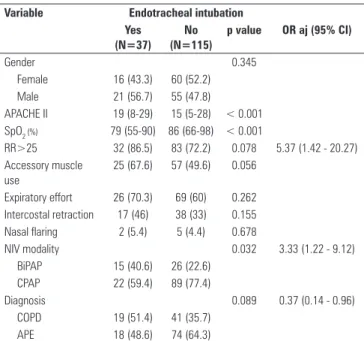
Table 2 - Risk factors for endotracheal intubation
Variable Endotracheal intubation Yes
(N=37) No (N=115)
p value OR aj (95% CI)
Gender 0.345
Female 16 (43.3) 60 (52.2)
Male 21 (56.7) 55 (47.8)
APACHE II 19 (8-29) 15 (5-28) < 0.001 SpO2 (%) 79 (55-90) 86 (66-98) < 0.001
RR>25 32 (86.5) 83 (72.2) 0.078 5.37 (1.42 - 20.27) Accessory muscle
use
25 (67.6) 57 (49.6) 0.056
Expiratory effort 26 (70.3) 69 (60) 0.262 Intercostal retraction 17 (46) 38 (33) 0.155 Nasal flaring 2 (5.4) 5 (4.4) 0.678
NIV modality 0.032 3.33 (1.22 - 9.12)
BiPAP 15 (40.6) 26 (22.6)
CPAP 22 (59.4) 89 (77.4)
Diagnosis 0.089 0.37 (0.14 - 0.96)
COPD 19 (51.4) 41 (35.7)
APE 18 (48.6) 74 (64.3)
RR - respiratory rate; NIV - non-invasive ventilation; BiPAP - bi-level positive airway pressure; CPAP -continuous positive airway pressure; COPD - chronic obstructive pulmonary disease; APE - acute pulmonary edema. The results are expressed as a number (percentage) or median (min-max). Chi-square, Mann-Whitney U or Fischer’s exact tests. OR aj - adjusted Odds Ratio, 95 CI - 95% confidence interval (OR aj and CI relate to results after multivariate analysis).
No diference between COPD and APE was observed in the 37 patients who progressed to EI. he impact of gender, clinical signs and NIV type on EI outcome was investigated. Only NIV type exhibited a statistically signiicant diference (p=0.032). Fifteen (40.6%) of the 37 EI patients received BiPAP versus 22 patients (59.4%) who received NIV as CPAP (Table 2). BiPAP use increased the probability of EI occurrence 2.3 times compared to patients who used CPAP (p=0.032).
Table 2 describes the results of the multivariate logistic regression analysis. RR >25, NIV, clinical diagnosis, Glasgow coma scale (GCS), SpO2 and APACHE II scale remained in the model. Progression to EI was higher in patients with RR >25 (adjusted OR= 5.37, 95% CI=1.42-20.27), patients who received BiPAP (adjusted OR 3.33, 95% CI=1.22-9.12) and patients with higher Apache II scores (adjusted OR= 1.16, 95% CI=1.04-1.28).
Progression to EI was 63% lower in APE patients (adjusted OR=0.37, 95% CI=0.14-0.96). Higher values of SpO2 GCS increased the probability of favorable NIV.
DISCUSSION
This study evaluated the predictive rates of success or failure of NIV in an urgent and emergency service to delineate the clinical and laboratory situations that favor the use of this procedure in specific patient populations.
his study evaluated EI-related factors in patients with APE and COPD exacerbation who received NIV in a UER and demonstrated that an RR >25, a higher APACHE II score and the use of BiPAP predicted EI. he predictors of successful NIV were ARF caused by APE and higher Glasgow coma scores (GCS) and SpO2.
NIV use can prevent EI and its complications, such as airway trauma and VAP, during ARF episode in patients with COPD and APE. NIV intervention reduces hospital costs because hospitalization time is reduced.(7,9,10,17-19,22)
A systematic review and meta-analysis by Lightowler et al.(20) demonstrated that NIV should
be the first treatment choice in COPD patients, especially patients with serious disease exacerbation who exhibit respiratory acidosis (pH<7.35). These authors advocate the installation of NIV prior to the worsening of acidosis to avoid EI and reduce mortality. The use of positive pressure in APE patients promotes alveolar fluid redistribution and the recruitment of collapsed alveoli, which reduces dyspnea, normalizes metabolism and favors oxygenation.(1,8,13) CPAP and
BiPAP are safe, and both of these techniques decrease the need for intubation. CPAP and BiPAP should be associated with conventional drug therapy in APE patients.(1,8,12,13,18)
Pladeck et al.(7) demonstrated that an average age
that was identical to our study. In addition, 30 patients in this study with the same diagnosis also received NIV. However, NIV was used an average 19.9 hours in COPD patients and 7.8 hours in APE patients.
No statistical significances between diagnosis and death, hospital discharge or ward transfer were observed in this study. Tomii et al.(14) analyzed the
use in patients with COPD exacerbation prevented 3 to 9 ICU admissions/year.
This study demonstrated a rate of NIV success that was consistent with previous reports.(2-6) Schettino
et al. demonstrated an NIV failure in 18% of APE patients and 24% of COPD patients.(23)
NIV failure in this study was associated with an RR >25 rpm, disease severity as demonstrated by a APACHE II high score, the BiPAP type of NIV and a diagnosis of COPD. However, higher GCS and SpO2 values reduced the probability of EI. Confalonieri et al. demonstrated that the patients who presented an RR ≥30 rpm, GCS <11, APACHE II ≥29 and pH <7.25 at admission exhibited a >70% risk of NIV failure.(24) Shirakabe et al.(12) analyzed 343 patients
with heart failure who received NIV and demonstrated that the lowest level of arterial blood pH predicted NIV failures. This research analyzed pH indirectly to calculate APACHE II. Hess(3) and Kaya et al.(2) also
related high APACHE II scores to NIV failure. A prospective and multicenter study by Antonelli et al.(5)
demonstrated that the risk of NIV failure was greater in patients with high disease severity scores, old age, the presence of acute respiratory distress syndrome (ARDS) or pneumonia and the absence of clinical improvement 1 hour after NIV treatment. Spada et al.(25) analyzed the SpO
2 and demonstrated that greater
SpO2 was associated with NIV success. These authors emphasized the utility of this predictor because it can be quickly and non-invasively acquired.
The use of accessory muscles and expiratory effort were more noticeable at admission in patients with respiratory failure due to APE or COPD exacerbation. Pladeck et al.(7) concluded that these
signs or dyspnea were eligible criteria for NIV use in COPD and APE patients.
We also observed that NIV modality but not gender or clinical signs was related to EI. Patients who received NIV through BiPAP presented unfavorable progress, and these patients were 2.3 times more likely to require EI compared to patients who received CPAP. This result is probably due to the greater disease severity in the patients who received BiPAP because these patients exhibited median APACHE II scores of 16.
However, the largest and most recent multicenter controlled and randomized clinical study, which examined 1,069 patients who were admitted for respiratory failure due to APE in 26 diferent emergency services, demonstrated no relationship between NIV
modality and EI outcome.(13) Nouira et al.(11) observed
similar efects of BiPAP use compared to CPAP on EI outcomes. Nonetheless, BiPAP is associated with faster improvements in respiratory failure in APE patients.
This clinical study has several methodological limitations. This observational research was not random. A convenient and non-consecutive sample was analyzed because the respiratory failure in patients who were admitted in the emergency department arose from a variety of health problems, which may underlie the absence of criteria for the selection of EI and limits the comparisons between NIV modalities (BiPAP and CPAP).
CONCLUSION
he analysis of APE and COPD patients who received NIV in an urgent and emergency unit revealed that most of these patients progressed successfully. A multivariate analysis using logistic regression demonstrated that an FR >25, higher APACHE II scores, BiPAP use and a COPD diagnosis increased the probability of EI. Similarly, higher GCS and SpO2 values reduced the probability of unfavorable developments.
RESUMO
Objetivo: Analisar os casos de insuficiência respiratória aguda decorrente de edema agudo de pulmão e de agudiza-ção da doença pulmonar obstrutiva crônica, submetidos à ventilação mecânica não invasiva, a fim de identificar fato-res associados ao sucesso ou ao insucesso do método em um serviço de urgência e emergência.
Métodos: Estudo descritivo e analítico prospectivo. Fo-ram incluídos pacientes de ambos os gêneros, com idade ≥18 anos, que utilizaram ventilação mecânica não invasiva devido ao quadro de insuficiência respiratória secundária a edema agudo de pulmão ou agudização da doença pulmo-nar obstrutiva crônica. Foram excluídos os pacientes com insuficiência respiratória aguda secundária a patologias diferentes de edema agudo de pulmão e doença pulmonar obstrutiva crônica, ou que apresentavam contraindicação para a técnica. A rotina da instituição é utilizar a pressão expiratória entre 5 e 8 cmH2O, e a inspiratória entre 10 a 12 cmH2O, além de suplementação de oxigênio para man-ter a saturação periférica de oxigênio >90%. A variável “des-fecho” considerada foi a intubação endotraqueal.
pacien-REFERENCES
1. Schettino GP, Reis MA, Galas F, Park M, Franca SA, Okamoto VN, et al. Ventilação mecânica não-invasiva com pressão positiva. Rev Bras Ter Intensiva. 2007;19(2):245-57.
2. Kaya A, Çileda A, Cayli I, Onen ZP, Sen E, Gülbay B. Associated factors with non-invasive mechanical ventilation failure in acute hypercapnic respiratory failure. Tuberk Toraks. 2010;58(2):128-34.
3. Hess DR. The evidence for noninvasive positive-pressure ventilation in the care of patients in acute respiratory failure: a systematic review of the literature. Respir Care. 2004;49(7):810-29. Review.
4. Collins SP, Mielniczuk LM, Whittingham HA, Boseley ME, Schramm DR, Storrow AB. The use of noninvasive ventilation in emergency department patients with acute cardiogenic pulmonary edema: a systematic review. Ann Emerg Med. 2006;48(3):260-9; 269.e1-4. Review.
5. Antonelli M, Conti G, Moro ML, Esquinas A, Gonzalez-Diaz G, Confalonieri M, et al. Predictors of failure of noninvasive positive pressure ventilation in patients with acute hypoxemic respiratory failure: a multi-center study. Intensive Care Med. 2001;27(11):1718-28.
6. Antón A, Güell R, Gómez J, Serrano J, Castellano A, Carrasco JL, et al. Predicting the result of noninvasive ventilation in severe acute exacerbations of patients with chronic airflow limitation. Chest. 2000;117(3):828-33.
7. Pladeck T, Hader C, Von Orde A, Rasche K, Wiechmann HW. Non-invasive ventilation: comparison of effectiveness, safety, and management in acute heart failure syndromes and acute exacerbations of chronic obstructive pulmonary disease. J Physiol Pharmacol. 2007;58 Suppl 5(Pt 2):539-49.
8. Schönhofer B, Kuhlen R, Neumann P, Westhoff M, Berndt C, Sitter H. Clinical practice guideline: non-invasive mechanical ventilation as treatment of acute respiratory failure. Dtsch Arztebl Int. 2008;105(24):424-33.
9. Crummy F, Naughton MT. Non-invasive positive pressure ventilation for acute respiratory failure: justified or just hot air? Intern Med J. 2007;37(2):112-8. 10. Keenan SP, Sinuff T, Burns KE, Muscedere J, Kutsogiannis J, Mehta S, Cook
DJ, Ayas N, Adhikari NK, Hand L, Scales DC, Pagnotta R, Lazosky L, Rocker G, Dial S, Laupland K, Sanders K, Dodek P; Canadian Critical Care Trials Group/ Canadian Critical Care Society Noninvasive Ventilation Guidelines Group. Clinical practice guidelines for the use of noninvasive positive-pressure ventilation and noninvasive continuous positive airway pressure in the acute care setting. CMAJ. 2011;183(3):E195-214.
11. Nouira S, Boukef R, Bouida W, Kerkeni W, Beltaief K, Boubaker H, et al. Non-invasive pressure support ventilation and CPAP in cardiogenic pulmonary edema: a multicenter randomized study in the emergency department. Intensive Care Med. 2011;37(2):249-56.
12. Shirakabe A, Hata N, Yokoyama S, Shinada T, Kobayashi N, Tomita K, et al. Predicting the success of noninvasive positive pressure ventilation in emergency room for patients with acute heart failure. J Cardiol. 2011;57(1):107-14. 13. Gray AJ, Goodacre S, Newby DE, Masson MA, Sampson F, Dixon S, Crane
S, Elliot M, Nicholl J; 3CPO Study Investigators. A multicentre randomised controlled trial of the use of continuous positive airway pressure and non-invasive positive pressure ventilation in the early treatment of patients presenting to the emergency department with severe acute cardiogenic pulmonary oedema: the 3CPO trial. Health Technol Assess. 2009;13(33):1-106.
14. Tomii K, Seo R, Tachikawa R, Harada Y, Murase K, Kaji R, et al. Impact of noninvasisve ventilation (NIV) trial for various types of acute respiratory failure in the emergency department; decreased mortality and use of the ICU. Respir Med. 2009;103(1):67-73.
15. Nápolis LM, Jeronimo LM, Baldini DV, Machado MP, de Souza VA, Caruso P. Availability and use of noninvasive ventilation in the intensive care units of public, private and teaching hospitals in the greater metropolitan area of São Paulo, Brazil. J Bras Pneumol. 2006;32(1):29-34.
16. British Thoracic Society Standards of Care Committee. Non-invasive ventilation in acute respiratory failure. Thorax. 2002;57(3):192-211.
17. Rocha E, Carneiro EM. Benefícios e complicações da ventilação mecânica não-invasiva na exacerbação aguda da doença pulmonar obstrutiva crônica. Rev Bras Ter Intensiva. 2008;20(2):184-9.
18. Ram SF, Picot J, Lightowler J, Wedzicha JA. Non-invasive positive pressure ventilation for treatment of respiratory failure due to exacerbations of chronic obstructive pulmonary disease. Cochrane Database Syst Rev. 2004;(3):CD004104. Update of Cochrane Database Syst Rev. 2004;(1):CD004104.
19. Plant PK, Owen JL, Elliott MW. Early use of non-invasive ventilation for acute exacerbations of chronic obstructive pulmonary disease on general respiratory wards: a multicentre randomised controlled trial. Lancet. 2000;355(9219):1931-5.
20. Lightowler JV, Wedzicha JA, Elliott MW, Ram FS. Non-invasive positive pressure ventilation to treat respiratory failure resulting from exacerbations of chronic obstructive pulmonary disease: Cochrane systematic review and meta analysis. BMJ. 2003;326(7382):185.
21. Harris C, Saskin R, Burns KE. Noninvasive ventilation initiation in clinical practice: A six-year prospective, observational study. Can Respir J. 2010;17(3):123-31. 22. Winck JC, Azevedo LF, Costa-Pereira A, Antonelli M, Wyatt JC. Efficacy and
safety of non-invasive ventilation in the treatment of acute cardiogenic pulmonary edema--a systematic review and meta-analysis. Crit Care. 2006;10(2):R69. 23. Schettino G, Altobelli N, Kacmarek RM. Noninvasive positive-pressure ventilation
in acute respiratory failure outside clinical trials: experience at the Massachusetts General Hospital. Crit Care Med. 2008;36(2):441-7.
24. Confalonieri M, Garuti G, Cattaruzza MS, Osborn JF, Antonelli M, Conti G, Kodric M, Resta O, Marchese S, Gregoretti C, Rossi A; Italian noninvasive positive pressure ventilation (NPPV) study group. A chart of failure risk for noninvasive ventilation in patients with COPD exacerbation. Eur Respir J. 2005;25(2):348-55.
25. Spada C, Gandhi R, Patel SR, Nuccio P, Weinhouse GL, Lee PS. Oxygen saturation/ fraction of inspired oxygen ratio is a simple predictor of noninvasive positive pressure ventilation failure in critically ill patients. J Crit Care. 2011;26(5):510-6.
tes com edema agudo de pulmão (n=92); 75,7% evoluíram com sucesso. Foram observados pior escore de APACHE II e menor saturação periférica de oxigênio, de forma esta-tisticamente significante, nos pacientes que evoluíram para intubação (p<0,001). O uso de BiPAP relacionou-se a 2,3 vezes mais chance de ocorrência de intubação endotraqueal que o de CPAP (p=0,032). Entre os pacientes com diagnós-tico de edema agudo de pulmão e com pontuação mais ele-vada na ECG também apresentaram mais chance de sucesso
Conclusão: As variáveis associadas à intubação endotra-queal foram frequência respiratória > 25rpm, maior valor de APACHE II, uso de BiPAP e diagnóstico de doença pulmonar
obstrutiva crônica. Já maiores valores de ECG e SpO2 estão associados ao sucesso da ventilação mecânica não invasiva. A ventilação mecânica não invasiva pode ser utilizada em serviços de urgência/emergência para casos de insuiciência respiratória aguda decorrente de edema agudo de pulmão e exacerbação da doença pulmonar obstrutiva crônica, com cuidado especial na monitoração dos pacientes com variáveis relacionadas à maior porcentagem de intubação endotraqueal.