The impact of technology on treatment of iatrogenic
cervicothoracic vascular traumas: a study of two cases three
decades apart and a review of the literature
O impacto da tecnologia no tratamento do trauma vascular iatrogênico cervicotorácico:
estudo de dois casos com intervalo de três décadas e revisão da literatura
Victor Bilman1,2, Bernardo Massière1,2, Alberto Vescovi1,2, Daniel Leal1,2, Paula Vivas1,2, Bruno Demier1,2,
Arno von Ristow1,2,3
Abstract
Complications such as pseudoaneurysms (PA) related to cervicothoracic venous access can be devastating. In this article, we present two similar cases in which technological advances impacted diagnosis, treatment, and results. Both patients developed massive PA after deep venous puncture attempts. The first case occurred in 1993 and was diagnosed by a duplex scan that revealed a large PA originating from the right subclavian artery. The artery was approached by median sternotomy with supraclavicular extension. The PA originated from the thyrocervical trunk and was treated with simple ligation. The second case was in 2017. Angiotomography revealed a PA originating in the vertebral artery, which was treated with endovascular techniques, maintaining vessel patency. Both patients progressed satisfactorily, despite quite different approaches. Cervicothoracic vascular lesions represent a diagnostic and therapeutic challenge, where the risk of rupture is high. Technological advances have reduced the risks involved in management of vascular injuries with difficult surgical access.
Keywords: aneurysm, false; vertebral artery; central venous access; endovascular procedures; self-expanding metallic stents.
Resumo
Complicações relacionadas ao acesso venoso cervicotorácico, como os pseudoaneurismas (PAs), podem ser devastadoras. Neste artigo, apresentamos dois casos semelhantes em que o avanço tecnológico impactou no diagnóstico, tratamento e resultados. Ambos pacientes apresentaram volumoso PA após a tentativa de punção venosa profunda. O primeiro caso, em 1993, diagnosticado por duplex scan, revelou grande PA oriundo da artéria subclávia direita. A artéria foi abordada por esternotomia mediana com extensão supraclavicular. O PA originava-se do tronco tireocervical, tratado com simples ligadura. No segundo caso, em 2017, angiotomografia revelou um PA originário da artéria vertebral, que foi tratado com técnica endovascular, mantendo a perviedade do vaso. Ambos evoluíram satisfatoriamente, apesar de abordagens bastante diferentes. A lesão vascular cervicotorácica representa um desafio propedêutico e terapêutico, com alto risco de ruptura. Os avanços tecnológicos diminuem os riscos de lesões vasculares com acesso cirúrgico difícil e devem estar entre as opções do cirurgião vascular.
Palavras-chave: falso aneurisma; artéria vertebral; cateterismo venoso central; procedimentos endovasculares; stents metálicos autoexpansíveis.
1 Pontifícia Universidade Católica do Rio de Janeiro – PUC-Rio, Cirurgia Vascular e Endovascular, Rio de Janeiro, RJ, Brasil.
2 Centro Integrado para a Pesquisa, Prevenção, Diagnóstico e Terapia das Doenças Vasculares – CENTERVASC, Rio de Janeiro, RJ, Brasil. 3 Academia Nacional de Medicina – ANM, Rio de Janeiro, RJ, Brasil.
Financial support: None.
Conflicts of interest: No conflicts of interest declared concerning the publication of this article. Submitted: May 30, 2018. Accepted: August 11, 2018.
INTRODUCTION
Deep venous accesses have become routine practice in hospital settings. They are essential for many diagnostic and therapeutic procedures.1-3 Iatrogenic
lesions of the subclavian artery and its branches are rare complications, but are associated with serious morbidity and mortality.4,5 Inamasu et al. and Bernik et al.
report incidence rates of post-puncture arterial injuries ranging from 0.4 to 9.9% and 0.5 to 11.4%, respectively, most often involving the common carotid artery. In the majority of cases, symptoms are delayed and the most common complications are arteriovenous
fistulas and pseudoaneurysms (PAs).1,3
The advent of endovascular surgery has enabled great advances in management of these iatrogenic injuries. While Inamasu et al. describe surgical excision
of the PA, with or without arterial reconstruction, as first-line treatment, the complex access needed for
this surgery can lead to a series of complications.1
This article is a comparative study of two cases involving similar iatrogenic injuries that manifested after puncture for deep venous access and in which different diagnostic and treatment approaches were taken. The objective is to demonstrate how
technological advances have influenced treatment of difficult-to-access cervicothoracic vascular traumas. A detailed review of the literature contributed to
illustration of the evolution of this subject and supports our conclusions.
DESCRIPTION OF THE CASES
The first case, which took place in September 1993,
involved a 72-year-old female patient with severe heart disease, arterial hypertension, and grade III obesity who had been admitted to an intensive care
unit with sepsis of pulmonary origin. An attempt was
made to puncture the right subclavian vein, using a supraclavicular technique, but was unsuccessful. One week later, the vascular surgery team was called to investigate a large pulsating mass in the right
cervical region. Physical examination found the patient
hemodynamically unstable, with a large, right-side,
cervical pulsating mass (Figure 1). A duplex scan
revealed a PA originating from the second portion of the right subclavian artery (RSA), with diameters of 50 × 42 mm (Figure 2), and the decision was taken to treat the patient with open surgery.
Access was achieved via a median sternotomy with right supraclavicular extension (Figure 3).
The proximal RSA was isolated and the neck of the PA was approached progressively. It originated from
the proximal segment of the thyrocervical trunk and was treated with simple ligation. The aneurysm sac
was drained. The patient recovered satisfactorily, despite the magnitude of the intervention and the severity of her condition.
The second case occurred in December 2017 and involved a 66-year-old female patient with severe heart disease and systemic arterial hypertension who had undergone a kidney transplant in 2007 because
of polycystic kidney disease. She was admitted to an
intensive care unit with sepsis of pulmonary origin.
An attempt was made to perform ultrasound-guided
puncture of the left internal jugular vein for administration of vasoactive amines, but the attempt was unsuccessful and the procedure was aborted. The patient developed a pulsating mass in the left cervical region and exhibited a progressive drop in
hematocrit levels. After 15 days, during which the
patient was in pain and the cervical mass expanded, the vascular surgery team was asked to investigate.
Figure 1. Left cervical mass and hematoma.
During physical examination and history taking, the patient complained of considerable pain in the left cervical region, related to the pulsating mass
(Figure 4). A duplex scan suggested a PA originating
from the left common carotid artery (Figure 5), and
computed tomography angiography (CTA) revealed a PA from segment V1 of the left vertebral artery, with diameters of 30 x 32 mm (Figure 6).
After the team had discussed the case, the decision
was taken to employ endovascular techniques to implant
a Viabahn 5 mm × 2.5 cm covered stent (WL Gore,
Flagstaff, AZ, United States) in the vertebral artery.
The procedure was accomplished with no intercurrent
conditions and 18 mL of iodinated contrast were used (Figure 7). The cervical mass receded and the pain resolved during the immediate postoperative period
and the patient suffered no neurological deficits. A control duplex scan conducted 6 months after the
procedure showed that the left vertebral artery was
patent at the level of segment V3.
Figure 3. Sternotomy with right supraclavicular extension.
Figure 4. Left cervical mass and hematoma.
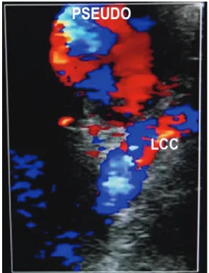
Figure 5. Doppler ultrasonography showing pseudoaneurysm (with suspected origin at the level of the left common carotid artery – LCC) with turbulent flow.
DISCUSSION
Vascular complications related to percutaneous
procedures in the cervicothoracic region can be devastating.4 Iatrogenic traumas to carotid and
subclavian arteries can provoke intense bleeding, hematoma, dissection, emboli, or thrombosis, and cases of airway obstruction secondary to expanding cervical hematomas, shock from hemothorax, stroke
caused by emboli, PA, and arteriovenous fistula
have all been described in the medical literature.4
Pseudoaneurysm is probably the rarest complication
of vertebral artery injuries and there are few cases described in the literature.3,5-8 Incidence is 0.2%, as
reported by Elias et al.2
Pseudoaneurysms of the vertebral artery can be
caused by penetrating or blunt traumas to the cervical region, arterial dissection, and surgical procedures.
Penetrating injuries primarily involve the first portion
of the artery, before it enters through the transverse foramen, whereas blunt cervical and cranial traumas involve more distal portions.2,7 Rapid identification
and management of these lesions is fundamental for a satisfactory result, although management is not fully understood and rarely described in the literature.1,2
While spontaneous resolution of these PAs has been
described, rupture is observed in 31 to 54% of patients.4,9
In the majority of cases, pseudoaneurysms appear after a delay and, primarily, present as a pulsating mass, with localized pain, dyspnea, heart failure, and cerebral and spinal ischemia.1-3 Ambekar et al. describe
diagnosis after 2 days, Elias et al., after 3 days,
Bernik et al., after 4 days, Amaral et al., after 15 days, and Cihangiroglu describe diagnosis after 32 days.2,3,5,7,10
The initial diagnostic approach described in the literature is duplex scanning, which shows hematoma
with arterial flow through the interior. Although this is a good method, it may be difficult to see the origin of the PA, as pointed out by Cihangiroglu et al.3,7
Proximity to the internal carotid artery, the volume
of the hematoma, and turgidity of the internal jugular
vein are factors that can make it difficult to see the origin of the PA. Computed tomography arteriography
is superior for viewing the lesion, including its origin, and for correctly identifying the site, the presence of any additional vascular traumas, and the patency of the contralateral vertebral artery, and it is also helpful for planning treatment.2,3,7
Treatments for PA include endovascular embolization,
direct repair of the vertebral artery or resection of
the PA with ligature.2,7 Bernik et al. also mention
ultrasound-guided compression and percutaneous thrombin injection as alternative options.3 It should
be pointed out that the depth of the lesion ruled out treatment with compression and because of the risk of cerebral embolization, treatment with thrombin is not recommended.3 Aoki et al. describe the case of a
patient treated with conservative management who, after 70 days, developed pain and paresthesia in the right shoulder and arm caused by compression of the
brachial plexus by hematoma. The PA ruptured and
a direct repair was performed during an emergency operation, but the patient died 11 days later.8
Amaral et al. suggest open treatment as the first line
option when the lesion is symptomatic and located at the origin of the vertebral artery.10 A median sternotomy,
right-side hematomas.3,7,10 Another access option is a
single incision along the sternocleidomastoid muscle or a single transclavicular incision, combined with temporary placement of a balloon at the origin of the vertebral artery.10 Bernik et al. stress the need for
excellent proximal control to provide a good view of the lesion and surgical treatment.3 On the left, access
requires an anterior thoracotomy for proximal control of the subclavian artery, since its posterior position
means that sternotomy does not offer good control.11
All of these approaches are associated with elevated
morbidity.11,12
According to the literature, endovascular treatment offers good results.4,13-16 This option has changed
management of these injuries, which previously was often achieved by ligature, to strategies for exclusion of
the PA, maintaining the vessel patent. Ambekar et al. and
Kerolus et al. describe using embolization devices
such as the Pipeline (Covidien Vascular Therapies,
Mansfield, MA, United States), which divert the flow,
preserving vessel patency. In a systematic review, they observed an 82.9% rate of aneurysm elimination at 6 months.5 Pérez et al. used a chrome-cobalt coated
balloon-expandable stent (Papyrus PK Biotronik) to
successfully exclude a vertebral artery PA, maintaining
patency of the vessel.13 In addition to fitting a covered
stent, Kwon et al. also successfully embolized a PA
using coils.16 Yamamoto et al. describe using coils
and N-butyl cyanoacrylate, resulting in occlusion of the lesion, but also of the vertebral artery.17 We report,
probably for the first time, use of a Viabahn 5 mm
× 2.5 cm covered stent (WL Gore, Flagstaff, AZ, United States) in the vertebral artery, preserving the
vessel’s patency.
As described by Amaral et al. and Yamamato et al.,
unilateral occlusion of the vertebral artery is a common practice for treatment of these lesions and, when the contralateral artery is patent, is generally well-tolerated.10,17 The posterior inferior cerebellar
artery on the side of the lesion must be supplied by the contralateral vertebral artery; if this does not occur, then the patient may develop Wallenberg syndrome.10 This syndrome is characterized by deficit
in cranial nerves (V, IX, X, and XI), ataxia, Horner
syndrome, and contralateral loss of the ability to feel pain and temperature, among other manifestations of
vertebrobasilar insufficiency. It is important to point
out that 15% of the population has one dominant vertebral artery, with a hypoplastic contralateral artery, and 5% only have one vertebral artery.10 Occlusion
or ligature of a vertebral artery in non-emergency
situations demands confirmation of adequate blood flow to the posterior circulation.10,18,19
CONCLUSIONS
While routine in modern medicine, deep venous punctures are not free from complications. Endovascular treatment has an important role to play in management of patients with iatrogenic vascular traumas. The traditional role of the vascular surgeon
is thus being challenged. As is the case with the
open repair technique, the vascular surgeon must be acquainted with technological advances, both for workup and for treatment. Endovascular techniques
generally offer reduced morbidity and mortality for management of difficult-to-access injuries.
While introduction of new technologies, such as ultrasound-guided puncture, has reduced the occurrence of complications related to this procedure, it has not eliminated iatrogenic complications. Even with training and experience, venous punctures are not risk-free. Knowledge of the cervical anatomy and its anatomic variations is as important as judicious
use of ultrasonography. Prevention of iatrogenic
injuries is the most important point to be taught in hospital settings.6
REFERENCES
1. Inamasu J, Guiot BH. Iatrogenic vertebral artery injury. Acta Neurol Scand. 2005;112(6):349-57. http://dx.doi.org/10.1111/j.1600-0404.2005.00497.x. PMid:16281916.
2. Elias M. Pulsating mass in the neck following attempted internal jugular vein catheterisation. Anaesthesia. 1999;54(9):914-5. http:// dx.doi.org/10.1046/j.1365-2044.1999.01089.x. PMid:10460719.
3. Bernik TR, Friedman SG, Scher LA, Safa T. Pseudoaneurysm of the subclavian-vertebral artery junction – case report and review of the literature. Vasc Endovascular Surg. 2002;36(6):461-4. http:// dx.doi.org/10.1177/153857440203600607. PMid:12476236.
4. Guilbert MC, Elkouri S, Bracco D, et al. Arterial trauma during central venous catheter insertion: case series, review and proposed algorithm. J Vasc Surg. 2008;48(4):918-25. http://dx.doi.org/10.1016/j. jvs.2008.04.046. PMid:18703308.
5. Ambekar S, Sharma M, Smith D, Cuellar H. Successful treatment of iatrogenic vertebral pseudoaneurysm using pipeline embolization device. Case Rep Vasc Med. 2014;2014:341748. http://dx.doi. org/10.1155/2014/341748. PMid:25276469.
6. Yu NR, Eberhardt RT, Menzoian JO, Urick CL, Raffetto JD. Vertebral artery dissection following intravascular catheter placement: a case report and review of the literature. Vasc Med. 2004;9(3):199-203. http://dx.doi.org/10.1191/1358863x04vm565cr. PMid:15675185.
7. Cihangiroglu M, Rahman A, Yildirim H, Burma O, Uysal H. Iatrogenic vertebral artery pseudoaneurysm: US, CT and MRI findings. Eur J Radiol. 2002;43(1):14-8. http://dx.doi.org/10.1016/ S0720-048X(01)00451-X. PMid:12065115.
8. Aoki H, Mizobe T, Nozuchi S, Hatanaka T, Tanaka Y. Vertebral artery pseudoaneurysm: a rare complication of internal jugular vein catheterization. Anesth Analg. 1992;75(2):296-8. http://dx.doi. org/10.1213/00000539-199208000-00027. PMid:1632547.
penetrating vertebralartery injury: case report and review of the literature. Turk Neurosurg. 2015;25(1):141-5. PMid:25640560.
10. Amaral JF, Grigoriev VE, Dorfman GS, Carney WI Jr. Vertebral artery pseudoaneurysm. A rare complication of subclavian artery catheterization. Arch Surg. 1990;125(4):546-7. http://dx.doi. org/10.1001/archsurg.1990.01410160134026. PMid:2322123.
11. Perissé RSP, Ristow AV, Palazzo JCC. Traumatismos vasculares da região cérvico-torácica e cervical. In: Ristow AV, Moreira RSP, editors. Urgências vasculares. Rio de Janeiro: Editora Cultura Médica; 1983. p. 76-101.
12. Meirelles SSL, Ristow AV. Trauma dos vasos cervicais. In: Freire E, editor. Trauma. Rio de Janeiro: Atheneu; 2001. p. 1341-1364.
13. Pérez GCI, Morata AR, Ortega JPR, Medialdea RG, García PC. Endovascular Treatment of a Symptomatic Vertebral Artery Pseudoaneurysm. Ann Vasc Surg. 2015;29(5):1018.e5-8. http:// dx.doi.org/10.1016/j.avsg.2015.01.022. PMid:25770383.
14. Guan Q, Chen L, Long Y, Xiang Z. Iatrogenic vertebral artery injury during anterior cervical spine surgery. A systematic review. World Neurosurg. 2017;106:715-22. http://dx.doi.org/10.1016/j. wneu.2017.07.027. PMid:28712898.
15. Kerolus M, Tan LA, Chen M. Treatment of a giant vertebral artery pseudoaneurysm secondary to gunshot wound to the neck using pipeline embolization device. Br J Neurosurg. 2016;1-2. http:// dx.doi.org/10.1080/02688697.2016.1265087. PMid:27927019.
16. Kwon SH, Oh JH. A large vertebral artery pseudoaneurysm due to percutaneous internal jugular vein cannulation. Intensive Care Med. 2014;40(12):1934. http://dx.doi.org/10.1007/s00134-014-3441-2. PMid:25118869.
17. Yamamoto A, Suzuki K, Sakaida H, Suzuki H, Imai H. Management of inadvertent vertebral artery injury due to central venous catheterization in a coagulopathic patient. Acute Med Surg. 2016;3(3):265-7. http://dx.doi.org/10.1002/ams2.177. PMid:29123795.
18. Aydin E, Gok M, Esenkaya A, Cinar C, Oran I. Endovascular Management of Iatrogenic Vascular Injury in the Craniocervical Region. Turk Neurosurg. 2018;28(1):72-8. PMid:27593845. 19. Abeysinghe V, Xu JH, Sieunarine K. Iatrogenic injury of vertebral
artery resulting in stroke after central venous line insertion. BMJ
Case Rep. 2017;2017. http://dx.doi.org/10.1136/bcr-2017-222429. PMid:29167220.
*
Correspondence
Victor Bilman Centro Integrado para a Pesquisa, Prevenção, Diagnóstico e Terapia das Doenças Vasculares – Centervasc Rua Sorocaba, 477, 8º andar, Botafogo CEP 22271-110 - Rio de Janeiro (RJ), Brasil Tel.: +55 (21) 2226-5858 E-mail: [email protected]
Author information
VB and BD - Postgraduates in Vascular and Endovascular Surgery, Centro Integrado para a Pesquisa, Prevenção, Diagnóstico e Terapia das Doenças Vasculares (CENTERVASC Rio), Pontifícia Universidade Católica do Rio de Janeiro (PUC-Rio). BM, AV, DL and PV - Vasculars surgeons and chiefs, Centro Integrado para a Pesquisa, Prevenção, Diagnóstico e Terapia das Doenças Vasculares (CENTERVASC Rio), Pontifícia Universidade Católica do Rio de Janeiro (PUC-Rio). AVR - Vascular surgeon and coordinator chief, Serviço de Cirurgia Vascular e Endovascular, Pontifícia Universidade Católica do Rio de Janeiro (PUC-Rio); Scientific director, Centro Integrado para a Pesquisa, Prevenção, Diagnóstico e Terapia das Doenças Vasculares (CENTERVASC Rio); Member, Academia Nacional de Medicina (ANM).
Author contributions
Conception and design: AVR, VB, BM Analysis and interpretation: AVR, VB, BD Data collection: VB, BM, PV Writing the article: AVR, VB Critical revision of the article: AVR, AV, DL, PV, BD Final approval of the article*: AVR Statistical analysis: N/A Overall responsibility: AVR