Laparoscopic treatment of celiac axis compression by the
median arcuate ligament and endovascular repair of a
pancreaticoduodenal artery aneurysm: case report
Tratamento laparoscópico da compressão do tronco celíaco pelo ligamento
arqueado do diafragma associado a correção endovascular do aneurisma de artéria
pancreatoduodenal: relato de caso
Marcio Miyamotto1,2,3
*
, Cecilia Naomi Kanegusuku4, Carla Mariko Okabe4, Christiano Marlo Paggi Claus5,
Fernanda Zandavalli Ramos3, Ágata Rothert4, Ana Paula Nudelmann Gubert6, Ricardo César Rocha Moreira3
Abstract
Compression of the celiac axis by the median arcuate ligament of the diaphragm can cause nonspecific symptoms such as abdominal pain, vomiting, and weight loss. There is a known association between stenosis or occlusion of the celiac trunk and aneurysms of the pancreaticoduodenal artery. Treatment strategies for patients who have this association should be selected on a case-by-case basis. We describe the case of a patient with pancreaticoduodenal artery aneurysm associated with compression of the celiac trunk by the arcuate ligament, which were managed with endovascular and laparoscopic techniques, respectively.
Keywords: median arcuate ligament syndrome; pancreaticoduodenal artery aneurysm; celiac plexus compression.
Resumo
A compressão do tronco celíaco pelo ligamento arqueado mediano do diafragma pode causar sintomas inespecíficos como dor abdominal, vômitos e emagrecimento. Existe uma associação comprovada entre estenoses ou oclusões do tronco celíaco e aneurismas da artéria pancreatoduodenal. Nas situações em que essa associação ocorre, a estratégia de tratamento deve ser individualizada. Relatamos o caso de uma paciente com aneurisma de artéria pancreatoduodenal associado à compressão do tronco celíaco pelo ligamento arqueado, manejados, respectivamente, por técnicas endovasculares e laparoscópicas.
Palavras-chave: síndrome do ligamento arqueado mediano; aneurisma de artéria pancreatoduodenal; compressão do plexo celíaco.
1 Pontifícia Universidade Católica do Paraná – PUC-PR, Hospital Universitário Cajuru – HUC, Serviço de Cirurgia Vascular e Endovascular, Curitiba, PR, Brasil. 2 Instituto VESSEL de Aperfeiçoamento Endovascular de Curitiba, Curitiba, PR, Brasil.
3 Hospital Nossa Senhora das Graças – HNSG, Serviço de Cirurgia Vascular e Endovascular Elias Abrão, Curitiba, PR, Brasil.
4 Pontifícia Universidade Católica do Paraná – PUC-PR, Hospital Universitário Cajuru – HUC, Liga Acadêmica de Medicina Vascular – LAMEV, Curitiba, PR, Brasil. 5 Hospital Nossa Senhora das Graças – HNSG, Serviço de Cirurgia Geral, Curitiba, PR, Brasil.
6 Hospital Santa Cruz, Serviço de Cirurgia Vascular, Curitiba, PR, Brasil. Financial support: None.
Conflicts of interest: No conflicts of interest declared concerning the publication of this article. Submitted: January 03, 2018. Accepted: June 08, 2018.
INTRODUCTION
The median arcuate ligament is formed by fibrous
bands that connect the right and left crura of the diaphragm around the aortic hiatus. The ligament may exert extrinsic compression on the celiac trunk if its location is low, or if the origin of the vessel is high.1
The association between stenosis or occlusions of the celiac trunk (irrespective of whether or not they are caused by extrinsic compression by the arcuate ligament) and aneurysms of the pancreaticoduodenal
arcade is well defined in the literature.2 Regardless
of the association, these aneurysms account for less than 2% of all visceral aneurysms. It is estimated that 63 to 80% of patients with pancreaticoduodenal artery aneurysm have stenosis or occlusion of the celiac trunk3 and the majority of these aneurysms (around
80%) are diagnosed after rupture.4
We describe the case of a patient with saccular pancreaticoduodenal artery aneurysm associated with stenosis of the celiac trunk secondary to compression by the median arcuate ligament.
CASE DESCRIPTION
A 39-year-old woman with hepatitis C was being seen by the gastroenterology service to monitor a
liver nodule. Abdominal ultrasonography identified a visceral artery aneurysm as an incidental finding.
Angiotomography revealed that it was a saccular aneurysm of the pancreaticoduodenal artery, with a diameter of 40 mm, and showed subocclusive stenosis of the celiac trunk compatible with extrinsic compression (Figure 1).
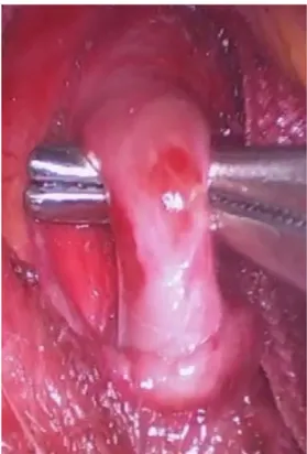
The patient underwent laparoscopic relief of celiac trunk compression (Figure 2), thereby averting the possibility of mesenteric ischemia, as the pancreaticoduodenal artery is an important collateral route between the celiac trunk and the superior mesenteric artery and an undiscovered occlusion of this artery can cause visceral ischemia. The laparoscopic procedure was performed using a 10 mm trocar for the camera, in an umbilical position, and a further four trocars; in
the right and left hypochondrium, the left flank, and
a subxiphoid position. The gastrohepatic ligament, phrenoesophageal membrane, esophagus, and crura of the diaphragmatic were dissected, with inferior sectioning of the crura to enable the arcuate ligament to be viewed. Relief of celiac trunk compression was achieved by sectioning the arcuate ligament by electrocautery and the crura were drawn back
together to prevent gastroesophageal reflux. Doppler
ultrasonography conducted before hospital discharge showed that there was no longer compression of the celiac trunk and revealed some residual stenosis
and post-stenotic dilation (the pre-stenotic celiac trunk diameter was 10 mm and at the stenosis it was 3.5 mm) (Figure 3).
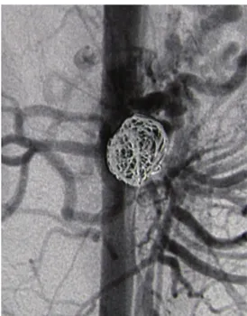
The patient returned 2 months later for pancreaticoduodenal artery aneurysm repair, which was performed under local anesthesia and sedation,
Figure 1. Angiotomography showing a pancreaticoduodenal
artery aneurysm and compression of the origin of the celiac trunk by the arcuate ligament of the diaphragm, causing stenosis exceeding 90%.
Figure 2. Relieving compression of the celiac trunk by sectioning
via a left brachial access with selective catheterization of the superior mesenteric artery and selective embolization of the aneurysm sac with microcoils, with no intercurrent conditions (Figure 4). Four 20 mm to
25 mm x 50 cm Axium 3D microcoils and two Axium
Helical microcoils 18 mm x 40 cm and 12 mm x 40 cm
were used. Follow-up Doppler ultrasonography after
3 months showed thrombosis of the aneurysm and a patent pancreaticoduodenal artery, in addition to absence of extrinsic compression of the celiac trunk.
DISCUSSION
Compression of the celiac trunk by the median arcuate ligament is not an uncommon situation, but arcuate ligament syndrome is a rare entity with varied
and nonspecific clinical presentation, so diagnosis
is by exclusion.1 One of the first descriptions of
compression of the celiac trunk by the arcuate ligament was in 1917, observed during cadaveric dissections.1,5
In 1963, the syndrome was described in a patient whose symptoms were relieved after surgical section of the ligament.6 In 1967, a series of cases of this syndrome
with similar symptoms was published.7
Clinical presentation can include postprandial or post exercise abdominal pain, nausea, vomiting, weight loss, and epigastric bruit.1,5,8 Occurrence of
symptoms may be caused by restricted blood flow
in situations of greater demand and by concomitant
compression of the fibers of the periaortic celiac
plexus.1,8 Differential diagnosis should be conducted to
rule out gastrointestinal diseases such as peptic ulcer, cholecystitis, pancreatitis, and chronic mesenteric ischemia.
Celiac trunk compression can be diagnosed by
Doppler ultrasonography, which shows compression of the vessel and reverse flow in the hepatic artery,
suggesting proximal stenosis or occlusion. An elevated systolic peak velocity in the celiac trunk only during expiration is indicative of dynamic compression.1,5,8
Angiography is the gold standard for diagnosis
and the classic findings are an asymmetrical focal
narrowing of the celiac trunk, more pronounced during expiration, with or without post-stenotic dilation.1,5
Even though angiotomography is not a dynamic
examination, it offers the possibility of assessing
adjacent, non-vascular structures.1
The association between compression of the celiac trunk by the arcuate ligament (or stenosis or occlusion of any other etiology) and aneurysms of the
pancreaticoduodenal artery was first described in the
1970s.9,10 Pathophysiology is related to the increased
blood flow through the pancreaticoduodenal arteries,9
at the stenosis or occlusion of the celiac trunk, since
Figure 4. Embolization of the pancreaticoduodenal artery
aneurysm with controlled release coils.
Figure 3. Doppler ultrasonography conducted after sectioning
flow through the territory of the superior mesenteric
artery is diverted through collaterals to those with
reduced flow.1,8
These aneurysms may be asymptomatic or may manifest symptoms related to extrinsic compression of the gastrointestinal or biliary tracts.9 Intestinal
bleeding can occur if the aneurysm ruptures into the duodenum and/or pancreatic ducts.2,11 Diagnosis can
be made by angiotomography.6
The risk of rupture does not appear to be related to size with these types of aneurysm.2 The rupture-related
mortality rate is high and can range from 50 to 90%.2,12
Considering these two facts, there is no doubt of the need for treatment in the case described, despite the lack of a consensus on the minimum size at which treatment is indicated.
There is also no consensus on the need for treatment of celiac trunk compression in asymptomatic patients with pancreaticoduodenal artery aneurysms.5 However,
it seems logical that it would be necessary to relieve the compression of the celiac trunk before attempting to treat the aneurysm, in case embolization of the aneurysm is planned, in order to avoid the possibility of ischemia and recurrence of the aneurysm because
the flow remains elevated.13 However, there are no
reports of recurrence of a pancreaticoduodenal artery aneurysm after embolization, even in the absence of prior treatment of the celiac trunk.2
Traditionally, treatment for arcuate ligament syndrome, via a midline surgical access or laparoscopy, consists of sectioning the ligament to relieve compression of the celiac trunk and eliminate irritation caused by
compression of nerve fibers.8 More recently, there has
been a trend to use endovascular and laparoscopic techniques.1
The open procedure for treatment of this syndrome is well-documented,14 and the patients that most benefit
from the treatment are those with postprandial pain,
age between 40 and 60 years, and with significant
weight loss.14 A group of 18 patients with arcuate
ligament syndrome underwent open surgical treatment to section the ligament and resection of the adjacent periaortic tissues. After three and a half years of follow-up, 73.3% of the patients were asymptomatic.5
Treatment of arcuate ligament syndrome with videolaparoscopy was documented in 16 patients. Just two of these patients did not exhibit relief from symptoms during the postoperative period (improvement
in 87.5%), because of fixed stenosis of the celiac
trunk, which was managed with balloon angioplasty and stent placement. Even so, in one of these cases an aortoceliac bypass was necessary.8
The difficulty in treating arcuate ligament syndrome resides in the patients with nonspecific gastrointestinal
symptoms. The difficulty in establishing a causal link
between the anatomic condition and the presence of symptoms can result in a low level of treatment
effectiveness.
Recent publications have demonstrated improvement in videolaparoscopic techniques, such as introduction of an ultrasonography probe, as a means of documenting
the increased blood flow after the ligament is resected,
and use of robots.15,16 It has been demonstrated that
treatment of the vascular injury in isolation does not produce good long-term results, and it is necessary
to lyse the ligament fibers.17
As for the pancreaticoduodenal artery aneurysms, endovascular treatment tends to be indicated when the diameter exceeds two centimeters, there is rapid growth and symptoms. Other factors that should be considered are a saccular shape and location in collateralization arteries. Aneurysms that are morphologically favorable for endovascular techniques are those with narrow
necks, adequate collateral flow and non-terminal
vessels. Endovascular management is the preferred option for pancreaticoduodenal aneurysms.11 There
are still indications for open surgical treatment of visceral aneurysms, but the endovascular approach
offers several advantages, such as being less invasive,
having fewer serious complications, and enabling selective embolization.
Embolization can be achieved with a variety of
different materials, although microcoils are the most
widely used.2 There are certain limitations related to
the technique when using covered stents to exclude
aneurysms, such as to the release system and difficulty of fitting in more tortuous arteries, and to the risk of
intra-stent thrombosis. Use of covered stents is more appropriate in arteries with diameters exceeding six millimeters and to prevent migration of microcoils in saccular aneurysms with wide necks.2
It is also important to point out that endovascular treatment can be used with patients who have a ruptured aneurysm.13 Open surgical treatment is subject to
technical difficulties primarily related to access to the
pancreaticoduodenal arcade and to bleeding control.
These difficulties have stimulated development of
endovascular techniques. Notwithstanding, there are reports of successful open treatment.18
CONCLUSIONS
When a patient has both compression of the celiac trunk by the arcuate ligament and a pancreaticoduodenal artery aneurysm, the treatment of both conditions is necessary. However, it is clear that less invasive treatments such as videolaparoscopy and endovascular
considering the morbidity and mortality related to the procedure.
REFERENCES
1. Duffy AJ, Panait L, Eisenberg D, Bell RL, Roberts KE, Sumpio B. Management of median arcuate ligament syndrome: a new paradigm. Ann Vasc Surg. 2009;23(6):778-84. http://dx.doi. org/10.1016/j.avsg.2008.11.005. PMid:19128929.
2. Kallamadi R, DeMoya MA, Kalva SP. Inferior pancreaticoduodenal artery aneurysms in association with celiac stenosis/occlusion. Semin Intervent Radiol. 2009;26(3):215-23. http://dx.doi. org/10.1055/s-0029-1225671. PMid:21326566.
3. Murata S, Tajima H, Fukunaga T, et al. Management of pancreaticoduodenal artery aneurysms: results of superselective transcatheter embolization. AJR Am J Roentgenol. 2006;187(3):290-8. http://dx.doi.org/10.2214/AJR.04.1726. PMid:16928907.
4. Thevenet A, Domergue J, Joyeux A. Surgical treatment of stenoses of the celiac trunk caused by the arcuate ligament of the diaphragm. Long-term results. Chirurgie. 1985;111(10):851-6. PMid:3836803.
5. Grotemeyer D, Duran M, Iskandar F, Blondin D, Nguyen K, Sandmann W. Median arcuate ligament syndrome: vascular surgical therapy and follow-up of 18 patients. Langenbecks Arch Surg. 2009;394(6):1085-92. http://dx.doi.org/10.1007/s00423-009-0509-5. PMid:19506899.
6. Harjola PT. A rare obstruction of the coeliac artery. Report of a case. Ann Chir Gynaecol Fenn. 1963;52:547-50. PMid:14083857.
7. Dunbar JD, Molnar W, Beman FF, Marable SA. Compression of the celiac trunk and abdominal angina. Am J Roentgenol Radium Ther Nucl Med. 1965;95(3):731-44. http://dx.doi.org/10.2214/ ajr.95.3.731. PMid:5844938.
8. Baccari P, Civilini E, Dordoni L, Melissano G, Nicoletti R, Chiesa R. Celiac artery compression syndrome managed by laparoscopy. J Vasc Surg. 2009;50(1):134-9. http://dx.doi.org/10.1016/j.jvs.2008.11.124. PMid:19563961.
9. Sutton D, Lawton G. Coeliac stenosis or occlusion with aneurysm of the collateral supply. Clin Radiol. 1973;24(1):49-53. http://dx.doi. org/10.1016/S0009-9260(73)80114-X. PMid:4723494.
10. Kadir S, Athanasoulis CA, Yune HY, Wilkov H. Aneurysms of the pancreaticoduodenal arteries in association with celiac axis occlusion. Cardiovasc Radiol. 1978;1(3):173-7. http://dx.doi. org/10.1007/BF02552029. PMid:743713.
11. Chadha M, Ahuja C. Visceral artery aneurysms: diagnosis and percutaneous management. Semin Intervent Radiol. 2009;26(3):196-206. http://dx.doi.org/10.1055/s-0029-1225670. PMid:21326564.
12. Hildebrand P, Esnaashari H, Franke C, Bürk C, Bruch HP. Surgical management of pancreaticoduodenal artery aneurysms in association with celiac trunk occlusion or stenosis. Ann Vasc Surg. 2007;21(1):10-5. http://dx.doi.org/10.1016/j.avsg.2006.05.001. PMid:17349329.
13. Iwazawa J, Hamuro M, Sakai Y, Nakamura K. Successful embolization of a ruptured pancreaticoduodenal artery aneurysm associated with the median arcuate ligament syndrome. Indian J Radiol Imaging. 2008;18(2):171-4. http://dx.doi.org/10.4103/0971-3026.40305.
14. Reilly LM, Ammar AD, Stoney RJ, Ehrenfeld WK. Late results following operative repair for celiac artery compression syndrome. J Vasc Surg. 1985;2(1):79-91. http://dx.doi.org/10.1016/0741-5214(85)90177-6. PMid:3965762.
15. Roayaie S, Jossart G, Gitlitz D, Lamparello P, Hollier L, Gagner M. Laparoscopic release of celiac artery compression syndrome facilitated by laparoscopic ultrasound scanning to confirm restoration of flow. J Vasc Surg. 2000;32(4):814-7. http://dx.doi. org/10.1067/mva.2000.107574. PMid:11013046.
16. Jaik NP, Stawicki SP, Weger NS, Lukaszczyk JJ. Celiac artery compression syndrome: successful utilization of robotic-assisted laparoscopic approach. J Gastrointestin Liver Dis. 2007;16(1):93-6. PMid:17410294.
17. Cina CS, Safar H. Successful treatment of recurrent celiac axis compression syndrome. A case report. Panminerva Med. 2002;44(1):69-72. PMid:11887094.
18. Golarz SR, Hohmann S. Obstruction of the celiac axis resulting in a pancreaticoduodenal artery aneurysm. Proc Bayl Univ Med Cent. 2009;22(4):330-1. http://dx.doi.org/10.1080/08998280.200 9.11928548. PMid:19865503.
*
Correspondence
Marcio Miyamotto Rua Francisco Juglair, 77/505 - Mossunguê CEP 81200-230 - Curitiba (PR), Brasil Tel.: +55 (41) 99961-0486 E-mail: miyamotto@gmail.com
Author information
MM - Vascular surgeon and chief, Serviço de Cirurgia Vascular, Hospital Universitário Cajuru (HUC), Pontifícia Universidade Católica do Paraná (PUC-PR); Vascular and endovascular surgeon, Serviço de Cirurgia Vascular e Endovascular Elias Abrão, Hospital Nossa Senhora das Graças, Curitiba; Tutor, Liga Acadêmica de Medicina Vascular, Hospital Universitário Cajuru (LAMEV); Director, Instituto VESSEL de Aperfeiçoamento Endovascular. CNK and CMO - Medical students, Pontifícia Universidade Católica do Paraná (PUC-PR); Members, Liga Acadêmica de Medicina Vascular, Hospital Universitário Cajuru, Pontifícia Universidade Católica do Paraná (LAMEV). CMPC - General and laparoscopic surgeon, Serviço de Cirurgia Geral, Hospital Nossa Senhora das Graças (HNSG). FZR - Vascular surgeon and former resident physician, Serviço de Cirurgia Vascular e Endovascular Elias Abrão, Hospital Nossa Senhora das Graças (HNSG). AR - Medical student, Universidade Federal do Paraná (UFPR); Member, Liga Acadêmica de Medicina Vascular, Hospital Universitário Cajuru, Pontifícia Universidade Católica do Paraná (LAMEV). APNG - Vascular surgeon, Serviço de Cirurgia Vascular, Hospital Santa Cruz. RCRM - Vascular surgeon and chief, Serviço de Cirurgia Vascular e Endovascular Elias Abrão, Hospital Nossa Senhora das Graças (HNSG); PhD in Surgical Medicine, Universidade Federal do Paraná (UFPR).
Author contributions
Conception and design: MM, RCRM Analysis and interpretation: MM, RCRM Data collection: MM, CNK, CMO, CMPC, FZR, AR, APGN Writing the article: MM, CNK, CMO, CMPC, FZR, AR, APGN Critical revision of the article: MM Final approval of the article*: MM, CNK, CMO, CMPC, FZR, AR, APNG, RCRM Statistical analysis: N/A. Overall responsibility: MM