1 Rheumatology Unit, Centro Hospitalar e Universitário de Coimbra, Portugal. Faculdade de Medicina da Universidade Coimbra, Portugal
2. Department of Rheumatology and Clinical Immunology, University Medical Center Utrecht, The Netherlands
3. Division of Clinical Immunology and Rheumatology, University of Alabama, Birmingham, USA
4. Department of Rheumatology and Clinical Immunology, Charité Universitätsmedizin, Berlin, Germany
Balancing the benefits and risks of low-dose
glucocorticoid in rheumatoid arthritis
ACTA REUMATOL PORT. 2015;40:10-22
ABstrAct
Glucocorticoids (GCs) have potent anti-inflammatory and immunomodulatory effects and are widely use in the management of rheumatoid arthritis in combina-tion with other synthetic and with biological disease- -mo difying anti-rheumatic drugs. Concerns about the risk of adverse effects of glucocorticoids, especially if they are given at higher dosages and for a longer time, hamper their use, despite the clear symptomatic and disease-modifying benefits. However, the evidence base for these concerns for low dose glucocorticoid therapy is quite limited due to the scarcity of quality literature on its safety in rheumatoid arthritis. This review discu sses: 1) the current understanding about its disease-modi fying effects, 2) toxicity data from recent trials and observa tio -nal studies, 3) recommendations for its management and the current efforts to improve the therapeutic ratio of glucocorticoid through the deve lopment of new formu-lations, such as modified-release prednisone.
Keywords: Rheumatoid arthritis; Glucocorticoids; Benefits; Risks.
introduction
With over 60 years of experience, the number of pa-tients treated with glucocorticoids (GCs), and the range of clinical applications are more extensive than with any other treatment. GCs represent a true anchor
treat-Santiago T1, Jacobs JW2, Saag KG3, Buttgereit F4, Pereira da Silva JA1
ment for rheumatoid arthritis (RA). This is supported by data indicating that up to 60% of all patients with RA in Germany is treated with GCs at some time du ring the course of their disease1. Similarly, 34 to 93% of all
patients entering recent clinical trials of biologics, and thus with active disease, were receiving GCs at base-line2. Such data are in contrast with the literature
pre-dominantly addressing the risks of these agents and the caution advised by every treatment recommendations. Cli nicians, and presumably patients, seem thus to va lue the antiinflammatory, immunosuppressive and di -sease-modifying therapeutic effects of GCs. In contrast, both the literature and the medical community seem to be split between those in favour and those against the use of low-dose GCs in RA. This contention cannot be resolved without a clear understanding of the potential risks of adverse effects of these drugs. Unfortunately, currently available evidence is limited, but the need to optimize benefitrisk ratio of GCs represents a conti -nuous challenge to the practicing clinician. This review reinforces the need for balancing risks and benefits of GCs use in RA, with a focus on chronic oral therapy. First, we address the rapid effect of GCs on disease acti vity and their longterm effects on radiographic dama ge, followed by their prolonged use in controlled di -sease. Next, we discuss the toxicity of low- dose GC therapy. Finally, we describe strategies and re commen -dations for its safe use and new therapeutic approa ches, including chronotherapy, which may improve our abi -lity to tailor treatment to the individual patients’ needs. A. Benefits
S
YMPTOMATIC BENEFITIn active RA, prednisone is frequently added for a short period to the treatment regimen to rapidly minimize disease activity while awaiting a clinical response to a slower-acting disease-modifying antirheumatic drug (DMARD). A review3provided evidence of short-term
more effective than either placebo or a nonsteroidal anti-inflammatory drug (NSAID), with a large effect size of 1.75 on pain. In 2000, a Cochrane metaanaly -sis4including 7 studies (253 patients in total) evalua
-ting the symptomatic effect of GC treatment in RA, concluded that prednisone (or a comparable GC preparation) at a mean dosage of less than or equal to 15 mg/day for a period of 6 months, was significantly more effective than placebo, with an effect size for pain of 0.43. Significant improvement was also document-ed in other outcomes measures: standardi zdocument-ed mean difference for tender joints = -0.37 (95%CI: -0. 59 to -0.14), swollen joints = -0.41 (-0.67 to -0.16) and functional status = -0.57 (-0.92 to -0.22). Ano ther meta-analysis of short-term (median length of treat-ment was one week in the ten studies included) found that prednisolone (<15 mg/day) had a clear superior ef-fect on pain compared to placebo (standardized efef-fect size 1.75; 0.87 to 2.64) and compared to NSAIDs (1.25; 0.26 to 2.24)3. Recently, numerous clinical
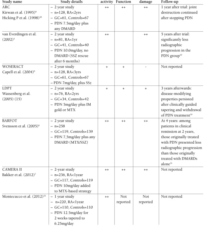
trials in early RA have demonstrated significant sympto matic benefit and clinical improvement, al-ready at 3 months after the start therapy, which is maintained at all time points thereafter, until 2 years of follow-up5-7 (Table I). In the Arthritis Research
Council (ARC) trial8, the patients in the prednisolone
group had greater reductions than the patients in the placebo group in scores on an articular index and for pain and disability at 3 months; for pain at 6 months; and for disability at 6, 12, and 15 months (all p < 0.05). In 2002, van Everdingen et al.5found a greater
clini-cal improvement in multiple measures, particularly in the first 6 months, in the 10 mg/day prednisone treat-ed group. However, this additional benefit was sus-tained only for joint tenderness at 24 months. The Better Anti-Rheumatic FarmacOTherapy (BARFOT) study6compared the addition of 7.5mg/day predni
-solone to me thotrexate (MTX) or sulfasalazine (SSZ) with DMARD alone in 250 patients with a disease du-ration of less that one year. The patients treated with DMARD plus predni solone had a significant reduction of DAS28 and HAQ score. These differences were al-ready seen at 3 months, and were present at all time points thereafter, during the 2-years of follow-up. In the prednisolone group, the mean (SD) DAS28 de-creased from 5.3 (1.1) to 2.7 (1.5) versus 5.4 (1.0) to 3.3 (1.5), (p<0.001) after 1 year, and to 2.7 (1.3) ver-sus 3.2 (1.4), (p<0.005) after 2 years. After 1 year, 51% of patients in the pre-dnisolone group had achieved disease remission compared with 39% of patients in
the no-prednisolone group (P = 0.06). After 2 years, this difference had increased to 56% in the predni -solone group compared with 33% in the placebo group (P = 0.0005). The Computer Assisted Management in Early Rheumatoid Arthritis trial-II (CAMERA-II)7
evalua ted the effect of prednisolone 10 mg/day from start, added to an MTX-based strategy with computer-assisted dose adjustments based on the level of disease activity versus the effect of the MTX-based strategy with placebo-pre dnisone in 236 patients with symptoms duration <1 year. Also this treat-to-target (target de-fined as remission) study7 confirmed the efficacy of
prednisone 10 mg/day added to DMARD in reducing disease activity and physical disability at 24 months. The patients treated with prednisone at 10 mg/day had more symptomatic benefit during the first three months - mean difference of DAS28 was -1.56 (CI, -1.88 to 1.25). This difference was sustained but was not signi -ficantly different anymore at the end of two years: -0.26 (CI, -0.68 to 0.16), because both step-up strategy arms were aimed at remission. The response rates after 1 year of treatment for the MTXbased strategy plus predni -sone and the MTX-based strategy plus placebo were respectively for the ACR20 70% versus 66% (P = 0.45); for the ACR50 56% versus 43% (P= 0.037), and for the ACR70 27% versus 26% (P = 0.82). Similar differences were seen at 2 years.
Regarding efficacy seen with 5 to 7 mg/day, in the study of Capell et al.9a dose of 7 mg/day prednisolone
was associated with a non-significant improvement (p=0.07) in individual clinical measures and a “modi-fied” ACR 20% response (20% improvement in Ritchie articular index, erythrocyte sedimentation rate, pain scores, physician global, patient global, HAQ) in the prednisolone group versus placebo.
In intensive (tight-control, treat-to-target) treatment strategies in early RA, GCs are mainly used to achieved fast symptomatic improvement and disease-control. As an alternative to initial high oral doses such as in Com-binatietherapie Bij Reumatoïde Artritis (COBRA), re-cently trials have shown positive effects of either one in-tramuscular 120 mg methylprednisolone administra-tion10, and even intra-articular GC injections into
in-flamed joints11.
L
ONG-
TERM SYMPTOMATIC EFFICACYSeveral papers and textbooks state that the beneficial effects of GCs upon symptoms tend to wear off or di sappear after one or two years of treatment. Such opi -nions are contradicted by studies demonstrating that
tABle i. effects of low- to medium-dose of Gcs durinG rcts, And follow-up thereAfter, in rA.
Disease Radiographic
Study name Study details activity Function damage Follow-up
ARC – 2 year study ++ ++ ++ 1 year after trial: joint
Kirwan et al. (1995)8 – n=128, RA<2yrs destruction continued
Hicking P et al. (1998)30 – GC=61, Controls=67 after stopping PDN
– PDN 7.5mg/day plus any DMARD
van Everdingen et al. – 2 year study ++ ++ 5 years after trial:
(2002)5 – n=81, RA<1yr significantly less
– GC=41, Controls=40 radiographic
– PDN 10.0mg/day, no progression in the
DMARD (SSZ rescue PDN group19
after 6 months)
WOSERACT – 2 year study + + - Not reported
Capell et al. (2004)9 – n=128, RA<3yrs
– GC=61, Controls=67 – PDN 7mg/day, plus SSz
LDPT – 2 year study + + + 3 years afterwards:
Wassenberg et al. – n=76, RA<2yrs disease-modifying
(2005) (15) – GC=34, Controls=42 properties persisted
– PDN 5mg/day plus IM after clinically guided
gold or MTX tapering and withdrawal
of PDN treatment19
BARFOT – 2 year study ++ ++ ++ At 4 years: among
Svensson et al. (2005)6 – n=258 patients in clinical
– GC=119, Controls=139 remission at 2 years, – PDN 7,5mg/day plus any those originally treated
DMARD (MTX/SSZ) with PDN presented less
radiographic progression than those originally treated with DMARDs alone31
CAMERA II – 2-year study ++ ++ ++ Not reported
Bakker et al. (2012)7 – n=236, RA<1year
– GC=117, Controls=119 – PDN 10mg/day added
to MTX-based strategy
Montecucco et al. (2012)32 – 1 year study ++ Not Not Not reported
– n=220, RA<1year reported reported – GC=110, Controls=110
– PDN 12.5mg/day for 2 weeks tapered to 6.25mg/day
In total, 1127 patients were included (GC = 543; controls = 584). In most cases, the comparator was placebo with DMARD ++, Statistically significant effect; +, non-statistically significant trend; -, no effect. PDN: prednisone or prednisolone
the withdrawal of even very low doses of GCs, in pa-tients with stable disease under long-term therapy, is followed by disease flares in a high percentage of pa-tients.
A randomized double-blind placebo controlled withdrawal trial of prednisone included 31 patients with RA in remission with stable doses of 1 to 4 mg/day prednisone for at least 12 weeks. Patients were ran-domized to the same dose prednisone in 1 mg tablets or identical placebo tablets, for 24 weeks12. Patients
who were switched from stable doses of prednisone to identical placebo tablets were significantly more like-ly to withdraw the study due to lack of efficacy over a subsequent 6–9-month period (11 out of 15) than those who were randomized to continued prednisone (3 out of 13, p=0.02). Another study by Tengstrand et al.13, with a similar design, included 58 RA patients
treated with 5 to 7.5 mg prednisolone/day for at least 2 years. Of the 26 patients randomized to stop pre -dnisolone treatment (median DAS28 at baseline 3.8), 11 (42%) succeeded to stop treatment and 15 (58%) failed withdrawal of GCs because of increased joint symptoms. Finally, in a withdrawal study among pa-tients, whose RA had been controlled for at least 3 months mean GCs treatment duration, 7.5 years; (mean daily dose, 8.6mg/d), the GCs dose was decrea -sed each month by steps of 1 mg/day14. At study end,
10 patients successfully has stopped prednisolone but 23 patients had experienced a flare of the disease (1 pa-tient stopped the study because of adrenal insufficien-cy and 4 were lost to follow-up). However, only one patient had been able to decrease the dose by 1 mg/month steps as planned; the mean decrease for all patients was 1 mg/3.5 months. Successful withdrawal was more common among patients who had been on GC treatment for less than 5 years14.
Taken together, notwithstanding the relatively small number of patients included, these results suggest that GCs may retain favourable symptomatic effects for a long time, even in patients in remission. This would suggest that GC withdrawal should not be considered an obligatory path but should rather depend on the evaluation of risks and benefits in the individual pa-tient. Interestingly, some authors tend to refer to the aggravation of disease after GC withdrawal as “re-bound effect” or physical dependency. However, none of the withdrawal studies we revised suggests that the di sease gets worse or more difficult to control after GC treatment is stopped. This concept of “rebound” is ex-clusively used in respect to GCs – when observed with
other medications, it is taken as evidence of efficacy. Furthermore, aggravation of disease activity after stop-ping another DMARD than prednisone is never seen as physical dependency, so why then should it be physical dependency for prednisone? These, at least appa -rent, biases deserve reflection.
DMARD
PROPERTIES IN EARLYRA
The disease-modifying effects of GCs have been well established in a number of randomized controlled trials of up to 2 years duration, using 5-10 mg/day of prednisolone or equivalent, in early RA6, 9, 15and have
been confirmed in two meta-analyses16, 17(Table I).
In 1995, in the ARC trial, after two-year treatment, prednisolone therapy had resulted in significantly few-er hand few-erosions (22 vfew-ersus 46%)18.
In 2002, van Everdingen et al.5found that
radio-graphic progression was less in the prednisone-treat-ed group, and this advantage persistprednisone-treat-ed for at least three years following the completion of two years of pre -dnisone treatment study19.
Also in the CAMERA-II study7, the MTX-based
strategy plus prednisolone was more effective than MTX-based strategy plus placebo in reducing the pro-gression of erosive joint damage as assessed at 104 weeks. This study additionally demonstrated the joint sparing benefit of prednisone even if added to an MTX-based tight-control strategy. After 2-year treatment, the prednisone group showed a statistically significantly lower progression of radiologic scores than the place-bo group, although the absolute difference was small (difference in Sharp-van der Heijde score of 0.87 units) because of low radiologic scores in both strategy groups due to intensive therapy7.
In the study of Capell et al.9a dose of 7 mg/day of
pre dnisone was not better than placebo in inhibiting radiographic joint damage. Authors attributed this ne -gative result to factors such as different therapeutic protocols, different baseline populations and a diffe -rent radiographic scoring system (Larsen score), com-pared to studies that demonstrated a DMARD-effect of prednisone.
In 2007, a Cochrane meta-analysis included ran-domized controlled trials with at least one arm including GC treatment and one arm without this thera -py. All studies measured radiographic change in joints of the hand and/or feet. Fifteen studies, with approxi-mately 1400 patients mostly with early RA, provided clear evidence that GCs substantially reduce the rate of radiographic progression in early RA16. If including
studies with very low doses of GCs and patients not taking DMARDs, the average reduction in the rate of progression was almost 70%.
In 2014, another systematic review17assessing the
efficacy of GCs in early RA (<2 years of duration) confirmed that initial treatment with lowdose pre -dnisolone plus MTX results in better structural out-comes compared with MTX alone.
T
HE JOINT-
SPARING EFFECTS PERSIST AFTER GLUCOCORTICOID TREATMENT DISCONTINUATIONFinally, some studies report enduring effects of GC upon structural damage progression long after GCs have been discontinued6, 19, 20. In the Utrecht trial, the
radiographic scores showed significantly less progres-sion over 3 years of follow-up after study closure in the former prednisolone group than in the former placebo group19. However, the inhibition effect of GCs
on formation of erosions, is only partial and additio -nal DMARD the rapy is required for control of radio-graphic joint da mage19.
In the COBRA trial, patients with early RA treated with prednisolone (initially 60 mg/day, rapidly reduced to 7.5 mg/day during weeks 7–28 and subsequently stopped) together MTX and SSZ showed significantly less radiographic progression compared with the group treated with SSZ alone. The benefits of short-term com-bination therapy on disease progression were still evi-dent at 5-year and 11-year follow-up20, 21. Can these
dif-ferences be attributed to GCs? In fact, two randomi zed controlled 52-week trials, in patients with early RA22, 23,
treated with SSZ or MTX or MTX plus SSZ (with no GC use in any of the three groups) have proven that MTX plus SSZ was not more effective than either drug alone. These observations suggest that the superiority of com-bination therapy over monotherapy in COBRA may pri-marily rest upon the effects of pre dnisolone.
Recently, a non-inferiority trial comparing COBRA and COBRA-light therapy (initial prednisolone 30 mg/day plus MTX increased to 25 mg/week) revealed similar efficacy in suppression of clinical disease acti -vity and improvement of function. Both groups showed major improvements in DAS44 at 52 weeks: mean (SD) −2.41 (1.2) in the COBRA and −2.02 (1.0) in the COBRA-light group (p=ns). In addition, both strategies have been shown to effectively suppress pro-gression of joint damage at 52 weeks of treatment24.
Although these benefits on disease progression have been shown, it is difficult to distinguish the contribu-tions made by different individual components in a
combination strategy that includes GCs and synthetic DMARD. It may also be that the COBRA trials, as o ther intensive treatment strategy studies25,26, provide
sup-port for an intensive target-oriented strategy rather than for the use of specific individual disease-modify-ing drugs, as they are strategy trials, not drug trials27.
A
REG
LUCOCORTICOIDSDMARD
S?
Drugs are considered DMARDs in RA if they reduce inflammation and pain, limit joint destruction and im-prove long-term disease outcome. Based on the evi-dence available, the question above can only receive a clear and solid YES, as a response, at least in early di -sease and when GCs are used in combination with other DMARDs. The evidence to support it is, actual-ly, much more robust than that available to support the DMARD quality of agents typically included in this category, such as hydroxychloroquine or SSZ.
It must be recognized that structural effects of GCs have not been adequately investigated in patients with longstanding disease: they should not be presumed to be present but they cannot be excluded to exist, either. The inclusion of patients with longstanding di -sease may have contributed to the lack of structural ef-fect of GCs reported by the study of Capell et al.9, but
this was not been specifically investigated. The same may have occurred in the study reported by Hansel et al.28where 102 patients with active RA were
random-ly allocated to treatment with DMARD alone or DMARD and prednisolone, and followed-up for 1 year. Pre dnisolone was given in a dose regimen adapted to the disease activity of the individual patient with a mean daily dose of 6mg during the trial. The authors reported that there were no benefits of prednisolone use with regard to radiological damage (Larsen score). However, the intention-to-treat analysis revealed a sig-nificantly higher rate of progression (delta Larsen score) in the group on DMARD alone (3.5 versus 1.8; p<0.03). Unfortunately, the disease duration at base-line was significantly higher in the DMARD alone group (8.5 versus 2.8 years; p<0.05) and no efforts were done to elucidate the impact of this parameter. Structural outcomes in RA seem to get less emphasis in the current literature because of the use of effective tightcontrol strategies, relatively low rates of radiogra -phic damage observed in recent clinical trials and the estimation that 1 Sharp/van der Heijde unit of radio-logical damage corresponds to 0.01 unit in deteriora-tion on the HAQ29. However, prevention of structural
B. risks
In 2006, a comprehensive review on the safety of low-dose GC treatment in RA30combining data from avai
-lable long-term RCTs5,8,9,15,31 concluded that adverse
ef-fects associated with this treatment in clinical trials are modest, and often not statistically different from those of placebo. In a recently published update of this review32,
with three additional RCTs6,7,33scarce evidence was
added to the previous conclusions. The authors con-cluded that the safety profile of low-dose GC, as demonstrated by RCTs, seems mild and hardly diffe -rent from that described for placebo, except for weight gain and glaucoma34.
The risks for adverse effects of low-dose GCs seem, therefore, to be often overestimated and this may be due to several reasons. It may be that the tolerability profile of high-dose GCs excessively influences per-ceptions regarding low-dose GCs, possibly exacerbat-ed by a lack of literature on the risks at low-doses as well as bias by indication. Patients with severe disease are more likely to be prescribed GCs and also more likely to experience adverse events associated with the
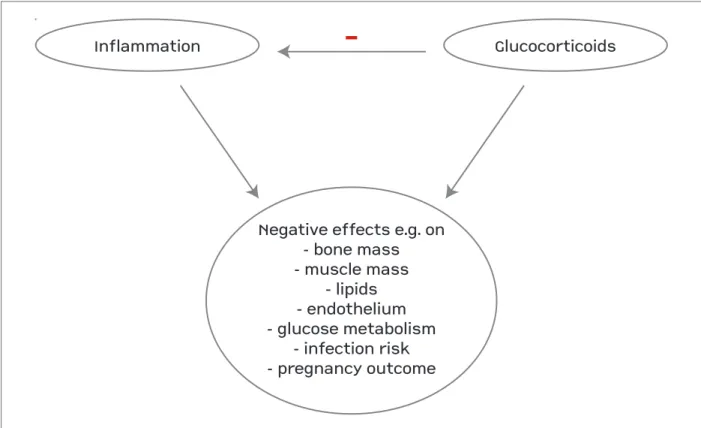
disease itself. Negative effects arising as a consequence of both RA and GC treatment may be attributed only to GC therapy. Examples include negative effects upon bone mineral density, lipids, endothelium, glucose metabolism and infection risk. It is conceivable that low-doses of GCs may actually inhibit or balance these negative effects of the disease process by reducing di sease activity. Discriminating the negative effects of di -sease and adverse-effects of its treatment is impossible in the absence of randomization. This is well illus trated in Figure 1, the so-called “magic triangle”. This dy-namic process was well recognized in the CAMERA-II, in the analysis of changes in bone mineral density (BMD) between the treatment groups35. BMD increased
significantly over time in both treatment groups at the lumbar spine with a mean of 2.6 % during the first year (p<0.001), but not at the hip; at none of the time points did BMD differ significantly between the pre -dnisone and placebo group. Higher age and lower weight at baseline and higher disease activity scores during the trial, but not GC therapy, were associated with lower BMD at both the lumbar spine and the hip
–
InflammationGlucocorticoidsNegative effects e.g. on- bone mass- muscle mass- lipids- endothelium- glucose metabolism- infection risk- pregnancy outcome
–
Inflammation Glucocorticoids
Negative effects e.g. on - bone mass - muscle mass - lipids - endothelium - glucose metabolism - infection risk - pregnancy outcome
fiGure 1.Association between the inflammatory disease, glucocorticoid treatment, and specific negative effects.
in mixed-model analyses. This was attributed to the effective dampening of the inflammatory process by GCs in early RA, especially of pro-inflammatory cy-tokines such as IL-1 and TNF. Of note, all patient re-ceived calcium, vitamin D and a bisphosphonate.
Regarding the impact of GCs upon glucose metabolism, similar perspectives can be drawn from the study by Hoes et al36. The authors measured
glu-cose tolerance, insulin sensitivity and b-cell function in two RA populations (58 chronic GC-users and 82 GC-naive) and in 50 healthy controls, with no known type 2 diabetes mellitus. Chronic users and GCnaive RA patients presented similar metabolic para -meters, with decreased insulin sensitivity and b-cell function in comparison to controls. Cumulative do ses of GCs had a negative impact on glucose tolerance state and insulin sensitivity. The results highlight a complex interplay of three factors. First, the pro-inflammatory state in RA has a negative impact on glucose metabolism. Second, GCs down regulate disease acti -vity, which may reduce this effect, but, third, GCs themselves, especially at higher dosages, impair glu-cose metabolism. These could be the reasons that data arising from observational studies and RCTs are quite different and contradictory.
This was clearly demonstrated by a recent systema -tic-review by Dixon et al.37. The authors collected data
from 21 RCTs (including 1026 GC-treated patients with RA) and 42 observational studies. The estimated relative risk (RR) of infection associated with GC thera -py was not significantly different from placebo in the RCTs (RR 0.97 (95%CI, 0.69-1.36). In contrast, the observational studies suggested an excess risk of in-fection of 67% in association with GCs (RR: 1.67, 95%CI: 1.491.87). There was significant heterogenei -ty of results between the observational studies, which the authors attributed to GC dose, cumulative expo-sure, time-varying expoexpo-sure, co-therapy, comorbidity, recruitment methods, outcome and bias (in particular publication bias)37.
A study based on the German biologics register -Rheumatoid Arthritis Observation of Biologic Thera-py (RABBIT) - enrolled 5044 RA patients, in whom 392 serious infections occurred, to evaluate the risk of serious infection associated with TNF inhibitors38. A
clear dose–response relationship was seen for treat-ment with GCs: the adjusted incidence rate ratios (IRR) of serious infections increased from 2.1 to 4.7 as pre -dnisone-equivalent GC dose increased from 7.5-14 to ≥15 mg/day. No significant increase in risk was obser
-ved for treatment with <7.5 mg/day (IRR=1.1 (95%CI 0.8; 1.7). These rates may reflect that GCs enhance in-fection risk in a dose-dependent way, but the impor-tance of the underlying disease cannot be fully ex-cluded in this study design.
Another adverse effect often overestimated is the weight gain. In a sub-analysis of the CAMERA-II study, Jurgens et al. showed that at least part of the difference in weight gain between groups was due to an earlier and better control of disease activity with predni -solone39. Weight gain has also been reported as a result
of TNF-inhibitors in RA40, 41.
Thus, weight gain under GCs and antiTNFinhibitors may, at least in part, be explained by normali -zation of body composition through control of in-flammation - the recovery of weight lost due to the catabolic state associated with high disease activity. Conversely, decreasing disease activity might be ex-pected to result in increased physical mobility, which could promote weight loss.
Additionally, data arising from observational stu dies and RCTs deserve serious reflection.
RCTs are considered to be the gold standard to eva -lua te the effectiveness of treatment, and are often de-signed to ensure a good internal validity of results, i.e., the potential of confounding by indication is removed by randomization. However, they also have their limi -tations, with emphasis on the strict inclusion/exclu-sion criteria, which preclude generalizability of the re-sults and their direct extrapolation to daily clinical practice. The typical patient in daily clinical practice will commonly be older, have less severe disease and more comorbidities than patients included in trials42.
None of the RCTs included in Table I was designed to assess the toxicity of GCs and the assessment of adver seeffects was frequently poorly structured or des cribed. Reporting of adverse effects is also highly varia -ble and potential confounders, such as concomitant therapies, are not systematically reported or ac counted for. Furthermore, the available studies on GCs are re -latively small and of short duration, thus limiting their ability to exclude all potentially significant adverse effect s.
Observational studies, on the other hand, are inex-tricably exposed to the risk of bias by indication. Huscher et al.43, provided some real-life data on the
adverse effects of GCs in RA. The authors describe self-reported health problems related to dose and duration of GC intake in unselected patients from routine prac-tice. Two distinct patterns were identified: A “linear
pattern” - approximately linear rising in the frequen-cy of adverse events with increasing dose – was seen in relation with Cushingoid phenotype, ecchymosis, leg edema, mycosis, parchment-like skin, shortness of breath and sleep disturbance. And a “threshold pat-tern” - an elevation in the frequency of health problems beyond a certain threshold value. From the study data, glaucoma, depression/listlessness and an increase in blood pressure only became issues at dosages above 7.5 mg/day. Dosages of 5 mg/day or above turned out to be relevant for epistaxis and weight gain; and <5 mg/day for eye cataract. These data may guide the clini cian in adapting therapy with GCs accordingly and improve the benefit–risk ratio. Again, correction for the characteristics of the underlying disease and its inherent risk of negative effects was limited by the obser -vational nature of the data.
In summary, given the limitations of currently avai -la ble RCTs and the inherent problems of observatio nal data, all we can state is that there is no evidence that low-dose GCs are associated with significant toxicity in early RA for over two years. Definite conclusions about the safety of GCs require randomized clinical trials with sufficient dimension and duration and with appropriate standardization in the definition and moni toring of adverse effects.
Clearly, the conclusion would be totally different if we considered medium and high doses of GCs, but these are not typically used chronically in the treat-ment of RA.
c. the BAlAnce
How can the physician achieve the best possible bene -fit over risk in GC therapy? Which adverse-effects should be monitored in clinical practice and how?
The EULAR recommendations on monitoring ad-verse events of low-dose glucocorticoid therapy pro-vide the best available guidance. Epro-vidence on moni-toring proved to be scarce and most recommendations were based on consensus44. Monitoring for an adverse
effect was considered especially useful if it is common or severe, the cost of screening is low, the monitoring is feasible in daily practice, and the adverse event is preventable and/or treatable and/or reversible after dose reduction or stopping the GC, if possible.
For clinical practice, the EULAR Task Force recom-mended that, for most potential adverse-effects, physi-cians may adhere to “standard care” ie, the same prac-tice advised as good clinical care in all patients with an inflammatory rheumatic disease. This standard care
applies to hypertension, cardiovascular disease, pep-tic ulcer disea se, diabetes and body weight. Adherence to national guidelines is recommended regarding GC-induced osteoporosis. Baseline assessments of ankle edema, fasting blood glucose and risk factors for glau-coma (family history, high myopia and diabetes) are also recommended. Patients at risk for glaucoma should undergo an ophthalmologic observation. The EULAR Task Force made no recommendations for monitoring in clinical practice in the context of low-dose GC-the rapy regarding lipids, electrolyte distur-bances, infections, mood disturdistur-bances, psychosis, sex or adrenal hormone changes, skin changes, os-teonecrosis or myo pathy, because these did not satis-fy one or more of the criteria for monitoring describe above.
Monitoring should be expanded and/or intensified in patients with GC-related adverse effects45.
It is crucial that patients with, or at risk of, GC-in-duced osteoporosis receive appropriate preventive and/or therapeutic interventions46. According to Van
Staa et al., fracture risk increases even with small do ses of GCs between 2.5 and 7.5 mg prednisone equi -valent daily47.The relative fracture risks during the first
year of therapy were 1.77 (95%CI: 1.55–2.02) and 2.27 (95%CI: 1.94–2.66) for patients taking predni solone in doses of 2.5–7.5 mg and > 7.5 mg daily, res -pectively47. Several meta-analyses showed efficacy of
therapy with calcium, vitamin D and bisphosphonates in preventing and treating GC-induced
osteoporo-sis48,49. Bisphosphonates have proven efficacy in
in-creasing bone mineral density and in reducing fre-quency of vertebral fractures50-52.
Furthermore, appropriate timing of GC adminis-tration might influence its efficacy, as signs and symp-toms, as well as serum levels of pro-inflammatory cy-tokines show a circadian rhythm53, 54. Thus,
adminis-tration of GC in the early morning53, or the use of mo
-dified-release prednisone (MR-pred) at bedtime (see below) may result in improved efficacy and, thus, low-er doses and less risk of advlow-erse effects. Patients should be adequately informed about the risks and benefits of GC therapy and be advised about the danger of abrupt cessation of the medication after long-term use55.
It is generally recommended that GCs should be used in the lowest dose for the shortest period of time to achieve the treatment goals. This statement is based on the current evaluation of the risks of GC and on the fact that DMARDproperties have only been demons
-trated in early disease and for up to 2 years of treat-ment. As knowledge on these aspects expands and im-proves, this recommendation may need revision in the future.
Of course it is generally true that a drug should be used in the lowest dose for the shortest period of time to achieve the treatment goals, but this statement is predominantly made and repeated regarding GCs. This may be seen as a reflection of the (over-?) evaluation of the risks of GC and (under-?) evaluation of their DMARD-properties, and deserves contemplation if we want to base our decisions on evidence and not on “common wisdom”.
D
EFLAZACORTDeflazacort, an oxazoline derivative of prednisolone, has been proposed to have similar anti-inflammatory and immunosuppressive effects but fewer adverse events than prednisolone, especially with regards to glucose and bone metabolism56-58. However, the data to
support this concept are inconsistent and come from small and relatively short duration trials59, 60. A
dou-ble-blind controlled randomized one-year study with 76 RA patients suggested that deflazacort has equiva-lent efficacy to prednisolone only in a dose ratio of 1.2:157. However, other studies indicated that defla
-zacort may be actually less potent, its equivalent dose to prednisolone being more in the range of 1.5 to 1.6:161,62. This completely abrogates the presumed
advan tage of deflazacort in terms of safety. Taken to-gether, there is no scientific evidence to support that deflazacort in equipotent doses is safer than other GCs. new Glucocorticoid developments
T
IMING THERAPYAs describe above, the EULAR recommendations no -ted the importance of timing of GC administration with respect to the circadian rhythms of both the natu -ral cortisol secretion and the disease processes45. A
re-cent approach, already licensed for clinical use, is modi fied-release prednisone (MR-pred), and has shown a clinically relevant reduction in early morning stiffness compared with conventio nal prednisone54, 63.
The efficacy and safety of MR-pred were examined in the CAPRA (Circadian Administration of Prednisone in Rheumatoid Arthritis) studies (Table II). In CA PRA-1, the MR-pred tablet was taken at bedtime, to be released with a delay of 4 hour after ingestion. This new for-mulation was shown to be clinically superior to the conventional prednisone with respect to reducing mor ning stiffness (primary endpoint of this study)54.
MR-pred reduced the duration of morning stiffness
tABle ii. overview of cAprA-1, cAprA-1 extension And cAprA-2 clinicAl studies in rheumAtoid Arthritis. AdApted from (57)
CAPRA-1 CAPRA-1 extension CAPRA-2
Buttgereit et al. (2008) (54) Buttgereit et al. (2010) (58) Buttgereit et al. (2013) (56)
Design Randomized Open label Randomized
Double-blind Double-blind
Double-dummy Placebo-controlled
Active control
Patients N= 288, on stable low-dose N= 249, from CAPRA-1 N= 350, not on
GC (2.5-10mg/day) glucocorticoid
Stable DMARD allowed Stable DMARD allowed
Study Continue same conventional All patients (n= 249) continue Placebo OR (n= 119) treatments prednisone (morning dose) on stable dose, taken as MR-pred MR-pred 5mg/day (n= 119)
(n= 144) OR same dose MR-pred (evening dose) (both evening doses) (evening dose) (n= 144)
Primary Change in duration of morning Change in duration of morning ACR20 response endpoint joint stiffness stiffness
Change in IL-6, DAS28, pain, ACR20
Duration 12 weeks 9 months 12 weeks
(when patients used prednisone) by 22.7% compared with 0.4% reduction with continuation of conven-tional prednisone (p = 0.045) from baseline to 12-week of treatment. The safety profile showed no dif-ferences between the two preparations.
The CAPRA-1 trial was followed by an open-label 9-month extension study, with 249 participants64.
Pa-tients on conventional prednisone switched to MR-pred (MR-pred/MR-MR-pred), while patients on MR-MR-pred maintained their treatment (MR-pred/MR-pred). Thus, during this extension trial a reduction in morning stiff-ness was reported after 3-months (33.1% versus no change), after 6 months (56% versus 54%); and after 12 months (55% versus 45%), respectively in the MR-pred/MR-pred and in the MR-pred/MR-pred group.
Additionally, IL-6 levels showed a 50% reduction in patients who switched from conventional to MR-pred: from baseline 1110 IU/l (of the double-blind study) to 515 IU/l, median value (end of the open-la-bel extension phase). DAS28 and pain intensity showed important improvements, however with no differences between the treatment groups over the 12 months.
To evaluate the impact of MR-pred on the hypotha-lamic–pituitary–adrenal axis (HPA), cortisol response to corticotropin-release hormone (CRH test) was de-termined in a subgroup of 28 patients from the CAPRA-1 at 3 time-points: baseline, 3 months and 9 months. This study found no evidence for increased suppression of the HPA by MR-pred as compared with standard prednisone64; rather, there was some
indication that GC administraindication in accordance with phy siological circadian rhythms reduced the hypothala -mic–pituitary–adrenal suppression when compared to conventional prednisone65.
To further confirm efficacy and safety of the MR-pred, a second trial, the CAPRA-2 study, was designed including 350 patients with active RA and morning stiffness of more than 45 minutes. The group recei ving MR-pred 5 mg/day plus traditional DMARDs (e.g., MTX) presented a significantly greater clinical im-provement in composite measures of disease acti vity compared with placebo + DMARDs (ACR20 of 48 ver-sus 29% and ACR50 of 22 verver-sus 10%) and in reduc-tion of morning stiffness from baseline (55 versus 35%). Significantly greater reductions in severity of RA and fatigue, as well as a greater improvement in evening pain and physical function were seen at week 12 with MR-pred compared with placebo. The inci-dence of adverse effects was similar for MR-pred and
placebo.
In conclusion, the data from CAPRA-1 suggests that MR-pred may have a superior effect to conventional prednisone in reducing early morning stiffness, and this is a relevant development for many patients, gi ven the impact of early morning dysfunction in the lives of patients with RA66.
Certainly, a head to head comparison trial with con-ventional formulation of GCs is needed to determine if these agents add enough benefit over standard GCs and are ultimately cost effective. In addition, these findings need to be extended to include large numbers of patients, ideally from real-world, in different clini-cal stages of their inflammatory disease, to demonstrate use of MR-pred in routine clinical practice67. Further
studies are needed to establish its use over longer pe-riods of time and especially to explore the potential benefits of this strategy upon structural damage and disease progression, based on changes in cytokine le -vels.
new selective AGents
Development research efforts into glucocorticoidreceptor ligands are based on the hypothesis that se -lective glucocorticoid receptor agonists (SEGRAs) may retain the anti-inflammatory properties of GCs – mecha nism of transrepression – with fewer or no metabolic adverse effects - mechanism of transactiva-tion. The results are, overall, promising and support further research68. However, in mouse studies some
SEGRAs have failed to exert a full inflammatory res -ponse and unexpectedly retained some classic adverse effects of GCs69. Further studies are underway.
tArGetinG therApy
Targeting therapy to the site of inflammation is possi-ble through encapsulating GCs in liposomes70.
Re-cently, nano-liposomes administered intravenously and subcutaneously have demonstrated a powerful suppression of the secretion of pro-inflammatory cy-tokines in rat models of arthritis71. Preclinical human
studies are awaited to see whether liposomal GCs will be effective in clinical practice.
AlternAtive reGimes
The promising results of recent trials using either a single intramuscular injection of 120mg of methylpre -dnisolone (Treatment in the Rotterdam Early Arthritis Cohort – tREACH trial)10or intra-articular injection of
Algorithm for Patients With Early Rheumatoid Arthri-tis - OPERA trial)11, both from the efficacy and the
safe-ty perspectives, indicate that there is room to improve our current strategies, seeking for the best possible use of classical GCs.
overAll summAry
GCs have been a cornerstone in the treatment of RA for many decades. GCs can, beyond any doubt, successfully suppress disease activity and, at least in early di -sease, significantly reduce structural damage accrual. Although these effects have been demonstrated even in the absence of other DMARDs5, there is general
agree-ment that GCs should not be used as monotherapy in RA. Safety concerns, often without firm evidence, li mit their widespread and long-term use.
Over the last decade, more attention has been gi ven to monitoring and reporting adverse effects in cli -nical trials, although only scarce evidence could be added to that described in the comprehensive review of Da Silva et al. published in 200630.
Given their low cost, the accumulated experience and the flexibility of their use, GCs will surely conti -nue to play an important role in the treatment of RA for the foreseeable future, despite the development of biological and targeted small molecules.
New GC formulations may offer significant advantages over conventional GC drugs, and thus more stu -dies are warranted in this field to investigate the bene fit of low-dose MR-pred chronotherapy and other alterna-tives. New regimes of therapy deserve consideration.
In the meantime, evidence-based recommendations for patient education, monitoring and prevention of GC-related adverse effects in RA have been published44-46,72.
Adherence to the standardized interventions and as-sessments described in these documents might signifi-cantly contribute to our ability to reduce the GC-rela ted adverse effects in RA and optimize GC use to the bene-fit of patients.
correspondence to
Tânia Santiago
Rheumatology Unit, Centro Hospitalar e Universitário de Coimbra Coimbra, Portugal
E-mail: [email protected]
references
1. Thiele K, Buttgereit F, Huscher D, Zink A. Current use of glu-cocorticoids in patients with rheumatoid arthritis in Germany.
Arthritis Rheum. 2005;53(5):740-747.
2. Buttgereit F. Do the treatment with glucocorticoids and/or the disease itself drive the impairment in glucose metabolism in patients with rheumatoid arthritis? Ann Rheum Dis. 70. En-gland 2011. p. 1881-1883.
3. Gotzsche PC, Johansen HK. Meta-analysis of short-term low dose prednisolone versus placebo and non-steroidal anti-in-flammatory drugs in rheumatoid arthritis. BMJ. 1998;316 (7134):811-818.
4. Criswell LA, Saag KG, Sems KM, Welch V, Shea B, Wells G, et al. Moderate-term, low-dose corticosteroids for rheumatoid arthritis. Cochrane Database Syst Rev. 2000(2):Cd001158. 5. van Everdingen AA, Jacobs JW, Siewertsz Van Reesema DR,
Bij lsma JW. Low-dose prednisone therapy for patients with ear-ly active rheumatoid arthritis: clinical efficacy, disease-modi-fying properties, and side effects: a randomized, double-blind, placebo-controlled clinical trial. Ann Intern Med. 2002;136 (1):1--12.
6. Svensson B, Boonen A, Albertsson K, van der Heijde D, Keller C, Hafstrom I. Low-dose prednisolone in addition to the initial disease-modifying antirheumatic drug in patients with early active rheumatoid arthritis reduces joint destruction and in-creases the remission rate: a two-year randomized trial. Arth-ritis Rheum. 2005;52(11):3360-3370.
7. Bakker MF, Jacobs JW, Welsing PM, Verstappen SM, Tekstra J, Ton E, et al. Low-dose prednisone inclusion in a methotrexa-te-based, tight control strategy for early rheumatoid arthritis: a randomized trial. Ann Intern Med. 2012;156(5):329-339. 8. Kirwan JR. The effect of glucocorticoids on joint destruction in
rheumatoid arthritis. The Arthritis and Rheumatism Council Low-Dose Glucocorticoid Study Group. N Engl J Med. 1995;333(3):142-146.
9. Capell HA, Madhok R, Hunter JA, Porter D, Morrison E, Lar-kin J, et al. Lack of radiological and clinical benefit over two years of low dose prednisolone for rheumatoid arthritis: results of a randomised controlled trial. Ann Rheum Dis. 2004;63(7): 797-803.
10. de Jong PH, Hazes JM, Han HK, Huisman M, van Zeben D, van der Lubbe PA, et al. Randomised comparison of initial triple DMARD therapy with methotrexate monotherapy in combina-tion with low-dose glucocorticoid bridging therapy; 1-year data of the tREACH trial. Ann Rheum Dis. 2014;73(7):1331-1339. 11. Axelsen MB, Eshed I, Horslev-Petersen K, Stengaard-Pedersen
K, Hetland ML, Moller J, et al. A treat-to-target strategy with methotrexate and intra-articular triamcinolone with or without adalimumab effectively reduces MRI synovitis, osteitis and te-nosynovitis and halts structural damage progression in early rheumatoid arthritis: results from the OPERA randomised con-trolled trial. Ann Rheum Dis. 2014.
12. Pincus T, Swearingen CJ, Luta G, Sokka T. Efficacy of predni-sone 1-4 mg/day in patients with rheumatoid arthritis: a ran-domised, double-blind, placebo controlled withdrawal clini-cal trial. Ann Rheum Dis. 2009;68(11):1715-1720.
13. Tengstrand B, Larsson E, Klareskog L, Hafstrom I. Randomized withdrawal of long-term prednisolone treatment in rheuma-toid arthritis: effects on inflammation and bone mineral densi-ty. Scand J Rheumatol. 2007;36(5):351-358.
14. Bacon PA, Myles AB, Beardwell CG, Daly JR. Corticosteroid with-drawal in rheumatoid arthritis. Lancet. 1966;2(7470):935-937. 15. Wassenberg S, Rau R, Steinfeld P, Zeidler H. Very low-dose pred-nisolone in early rheumatoid arthritis retards radiographic
pro-gression over two years: a multicenter, double-blind, placebo-controlled trial. Arthritis Rheum. 2005;52(11):3371-3380. 16. Kirwan JR, Bijlsma JW, Boers M, Shea BJ. Effects of
glucocorti-coids on radiological progression in rheumatoid arthritis. Coch-rane Database Syst Rev. 2007(1):Cd006356.
17. Gaujoux-Viala C, Nam J, Ramiro S, Landewe R, Buch MH, Smo-len JS, et al. Efficacy of conventional synthetic disease-modi-fying antirheumatic drugs, glucocorticoids and tofacitinib: a systematic literature review informing the 2013 update of the EULAR recommendations for management of rheumatoid arth-ritis. Ann Rheum Dis. 2014;73(3):510-515.
18. Strand V. Steroid withdrawal favours joint erosion in rheuma-toid arthritis. Clin Exp Rheumatol. 1999;17(5):519-520. 19. Jacobs JW, van Everdingen AA, Verstappen SM, Bijlsma JW.
Fol-lowup radiographic data on patients with rheumatoid arthritis who participated in a two-year trial of prednisone therapy or placebo. Arthritis Rheum. 2006;54(5):1422-1428.
20. Landewe RB, Boers M, Verhoeven AC, Westhovens R, van de Laar MA, Markusse HM, et al. COBRA combination therapy in patients with early rheumatoid arthritis: long-term structural benefits of a brief intervention. Arthritis Rheum. 2002;46(2):347-356.
21. van Tuyl LH, Boers M, Lems WF, Landewe RB, Han H, van der Linden S, et al. Survival, comorbidities and joint damage 11 years after the COBRA combination therapy trial in early rheu-matoid arthritis. Ann Rheum Dis. 2010;69(5):807-812. 22. Dougados M, Combe B, Cantagrel A, Goupille P, Olive P,
Schat-tenkirchner M, et al. Combination therapy in early rheumatoid arthritis: a randomised, controlled, double blind 52 week cli-nical trial of sulphasalazine and methotrexate compared with the single components. Ann Rheum Dis. 1999;58(4):220-225. 23. Haagsma CJ, van Riel PL, de Jong AJ, van de Putte LB. Combi-nation of sulphasalazine and methotrexate versus the single components in early rheumatoid arthritis: a randomized, con-trolled, double-blind, 52 week clinical trial. Br J Rheumatol. 1997;36(10):1082-1088.
24. Ter Wee MM, den Uyl D, Boers M, Kerstens P, Nurmohamed M, van Schaardenburg D, et al. Intensive combination treatment regimens, including prednisolone, are effective in treating pa-tients with early rheumatoid arthritis regardless of additional etanercept: 1-year results of the COBRA-light open-label, ran-domised, non-inferiority trial. Ann Rheum Dis. 2014 May 12. doi: 10.1136/annrheumdis-2013-205143.
25. Goekoop-Ruiterman YP, de Vries-Bouwstra JK, Allaart CF, van Zeben D, Kerstens PJ, Hazes JM, et al. Comparison of treatment strategies in early rheumatoid arthritis: a randomized trial. Ann Intern Med. 2007;146(6):406-415.
26. Mottonen T, Hannonen P, Leirisalo-Repo M, Nissila M, Kau-tiainen H, Korpela M, et al. Comparison of combination the-rapy with single-drug thethe-rapy in early rheumatoid arthritis: a randomised trial. FIN-RACo trial group. Lancet. 1999;353 (9164):1568-1573.
27. Bijlsma JW. Disease control with glucocorticoid therapy in rheumatoid arthritis. Rheumatology (Oxford). 2012;51 Suppl 4:iv9-13.
28. Hansen M, Podenphant J, Florescu A, Stoltenberg M, Borch A, Kluger E, et al. A randomised trial of differentiated prednisolo-ne treatment in active rheumatoid arthritis. Clinical beprednisolo-nefits and skeletal side effects. Ann Rheum Dis. 1999;58(11):713-718. 29. Smolen JS, Aletaha D, Grisar JC, Stamm TA, Sharp JT.
Estima-tion of a numerical value for joint damage-related physical
di-sability in rheumatoid arthritis clinical trials. Ann Rheum Dis. 2010;69(6):1058-1064.
30. Da Silva JA, Jacobs JW, Kirwan JR, Boers M, Saag KG, Ines LB, et al. Safety of low dose glucocorticoid treatment in rheumatoid arthritis: published evidence and prospective trial data. Ann Rheum Dis. 2006;65(3):285-293.
31. Hafstrom I, Albertsson K, Boonen A, van der Heijde D, Lande-we R, Svensson B. Remission achieved after 2 years treatment with low-dose prednisolone in addition to disease-modifying anti-rheumatic drugs in early rheumatoid arthritis is associated with reduced joint destruction still present after 4 years: an open 2-year continuation study. Ann Rheum Dis. 2009;68(4):508-513.
32. Santiago T, da Silva JA. Safety of glucocorticoids in rheumatoid arthritis: evidence from recent clinical trials. Neuroimmuno-modulation. 2015;22(1-2):57-65.
33. Montecucco C, Todoerti M, Sakellariou G, Scire CA, Caporali R. Low-dose oral prednisone improves clinical and ultrasono-graphic remission rates in early rheumatoid arthritis: results of a 12-month open-label randomised study. Arthritis Res Ther. 2012;14(3):R112.
34. Santiago T, da Silva JA. Safety of low- to medium-dose gluco-corticoid treatment in rheumatoid arthritis: myths and reality over the years. Ann N Y Acad Sci. 2014;1318(1):41-49. 35. van der Goes MC, Jacobs JW, Jurgens MS, Bakker MF, van der
Veen MJ, van der Werf JH, et al. Are changes in bone mineral density different between groups of early rheumatoid arthritis patients treated according to a tight control strategy with or without prednisone if osteoporosis prophylaxis is applied? Os-teoporos Int. 2013;24(4):1429-1436.
36. Hoes JN, van der Goes MC, van Raalte DH, van der Zijl NJ, den Uyl D, Lems WF, et al. Glucose tolerance, insulin sensitivity and beta-cell function in patients with rheumatoid arthritis treated with or without low-to-medium dose glucocorticoids. Ann Rheum Dis. 2011;70(11):1887-1894.
37. Dixon WG, Suissa S, Hudson M. The association between sys-temic glucocorticoid therapy and the risk of infection in pa-tients with rheumatoid arthritis: systematic review and meta-analyses. Arthritis Res Ther. 2011;13(4):R139.
38. Strangfeld A, Eveslage M, Schneider M, Bergerhausen HJ, Klopsch T, Zink A, et al. Treatment benefit or survival of the fit-test: what drives the time-dependent decrease in serious infec-tion rates under TNF inhibiinfec-tion and what does this imply for the individual patient? Ann Rheum Dis. 2011;70(11):1914-1920. 39. Jurgens MS, Jacobs JW, Geenen R, Bossema ER, Bakker MF, Bi-jlsma JW, et al. Increase of body mass index in a tight controlled methotrexate-based strategy with prednisone in early rheumatoid arthritis: side effect of the prednisone or better control of disea-se activity? Arthritis Care Res (Hoboken). 2013;65(1):88-93. 40. Stagakis I, Bertsias G, Karvounaris S, Kavousanaki M, Virla D,
Raptopoulou A, et al. Anti-tumor necrosis factor therapy im-proves insulin resistance, beta cell function and insulin signa-ling in active rheumatoid arthritis patients with high insulin resistance. Arthritis Res Ther. 2012;14(3):R141.
41. Metsios GS, Stavropoulos-Kalinoglou A, Douglas KM, Koute-dakis Y, Nevill AM, Panoulas VF, et al. Blockade of tumour ne-crosis factor-alpha in rheumatoid arthritis: effects on compo-nents of rheumatoid cachexia. Rheumatology (Oxford). 2007;46(12):1824-1827.
42. Pincus T, Sokka T. Quantitative measures for assessing rheu-matoid arthritis in clinical trials and clinical care. Best Pract Res
Clin Rheumatol. 2003;17(5):753-781.
43. Huscher D, Thiele K, Gromnica-Ihle E, Hein G, Demary W, Dreher R, et al. Dose-related patterns of glucocorticoid-indu-ced side effects. Ann Rheum Dis. 2009;68(7):1119-1124. 44. van der Goes MC, Jacobs JW, Boers M, Andrews T, Blom-Bakkers
MA, Buttgereit F, et al. Monitoring adverse events of low-dose glu-cocorticoid therapy: EULAR recommendations for clinical trials and daily practice. Ann Rheum Dis. 2010;69(11): 1913-1919. 45. Hoes JN, Jacobs JW, Boers M, Boumpas D, Buttgereit F, Caeyers
N, et al. EULAR evidence-based recommendations on the ma-nagement of systemic glucocorticoid therapy in rheumatic di-seases. Ann Rheum Dis. 2007;66(12):1560-1567.
46. Duru N, van der Goes MC, Jacobs JW, Andrews T, Boers M, Butt-gereit F, et al. EULAR evidence-based and consensus-based re-commendations on the management of medium to high-dose glu-cocorticoid therapy in rheumatic diseases. Ann Rheum Dis. 2013. 47. Van Staa TP, Leufkens HG, Abenhaim L, Zhang B, Cooper C. Use of oral corticosteroids and risk of fractures. J Bone Miner Res. 2000;15(6):993-1000.
48. Richy F, Schacht E, Bruyere O, Ethgen O, Gourlay M, Regins-ter JY. Vitamin D analogs versus native vitamin D in preventing bone loss and osteoporosis-related fractures: a comparative meta-analysis. Calcif Tissue Int. 2005;76(3):176-186. 49. Homik J, Cranney A, Shea B, Tugwell P, Wells G, Adachi R, et
al. Bisphosphonates for steroid induced osteoporosis. Cochra-ne Database Syst Rev. 2000(2):Cd001347.
50. Saag KG, Shane E, Boonen S, Marin F, Donley DW, Taylor KA, et al. Teriparatide or alendronate in glucocorticoid-induced os-teoporosis. N Engl J Med. 2007;357(20):2028-2039. 51. Reid DM, Devogelaer JP, Saag K, Roux C, Lau CS, Reginster JY,
et al. Zoledronic acid and risedronate in the prevention and treatment of glucocorticoid-induced osteoporosis (HORIZON): a multicentre, double-blind, double-dummy, randomised con-trolled trial. Lancet. 2009;373(9671):1253-1263.
52. Saag KG, Zanchetta JR, Devogelaer JP, Adler RA, Eastell R, See K, et al. Effects of teriparatide versus alendronate for treating glucocorticoid-induced osteoporosis: thirty-six-month results of a randomized, double-blind, controlled trial. Arthritis Rheum. 2009;60(11):3346-3355.
53. Arvidson NG, Gudbjornsson B, Larsson A, Hallgren R. The ti-ming of glucocorticoid administration in rheumatoid arthritis. Ann Rheum Dis. 1997;56(1):27-31.
54. Buttgereit F, Doering G, Schaeffler A, Witte S, Sierakowski S, Gromnica-Ihle E, et al. Efficacy of modified-release versus stan-dard prednisone to reduce duration of morning stiffness of the joints in rheumatoid arthritis (CAPRA-1): a double-blind, ran-domised controlled trial. Lancet. 2008;371(9608):205-214. 55. van der Goes MC, Jacobs JW, Boers M, Andrews T,
Blom-Bak-kers MA, Buttgereit F, et al. Patient and rheumatologist pers-pectives on glucocorticoids: an exercise to improve the imple-mentation of the European League Against Rheumatism (EU-LAR) recommendations on the management of systemic glu-cocorticoid therapy in rheumatic diseases. Ann Rheum Dis. 2010;69(6):1015-1021.
56. Deflazacort--an alternative to prednisolone? Drug Ther Bull. 1999;37(8):57-58.
57. Eberhardt R, Kruger K, Reiter W, Gross W, Zwingers T. Long-term therapy with the new glucocorticosteroid deflazacort in rheumatoid arthritis. Double-blind controlled randomized 12-months study against prednisone. Arzneimittelforschung. 1994;44(5):642-647.
58. Gray RE, Doherty SM, Galloway J, Coulton L, de Broe M, Ka-nis JA. A double-blind study of deflazacort and predKa-nisone in patients with chronic inflammatory disorders. Arthritis Rheum. 1991;34(3):287-295.
59. Messina OD, Barreira JC, Zanchetta JR, Maldonado-Cocco JA, Bogado CE, Sebastian ON, et al. Effect of low doses of deflaza-cort vs prednisone on bone mineral content in premenopausal rheumatoid arthritis. J Rheumatol. 1992;19(10):1520-1526. 60. Montecucco C, Baldi F, Fortina A, Tomassini G, Caporali R,
Cherie-Ligniere EL, et al. Serum osteocalcin (bone Gla-protein) following corticosteroid therapy in postmenopausal women with rheumatoid arthritis. Comparison of the effect of predni-sone and deflazacort. Clin Rheumatol. 1988;7(3):366-371. 61. Saviola G, Abdi Ali L, Shams Eddin S, Coppini A, Cavalieri F,
Campostrini L, et al. Compared clinical efficacy and bone meta-bolic effects of low-dose deflazacort and methyl prednisolone in male inflammatory arthropathies: a 12-month open randomized pilot study. Rheumatology (Oxford). 2007;46(6):994-998. 62. Krogsgaard MR, Lund B, Johnsson B. A longterm prospective
study of the equipotency between deflazacort and prednisolo-ne in the treatment of patients with polymyalgia rheumatica. J Rheumatol. 1995;22(9):1660-1662.
63. Buttgereit F, Mehta D, Kirwan J, Szechinski J, Boers M, Alten RE, et al. Low-dose prednisone chronotherapy for rheumatoid arth-ritis: a randomised clinical trial (CAPRA-2). Ann Rheum Dis. 2013;72(2):204-210.
64. Buttgereit F, Doering G, Schaeffler A, Witte S, Sierakowski S, Gromnica-Ihle E, et al. Targeting pathophysiological rhythms: prednisone chronotherapy shows sustained efficacy in rheu-matoid arthritis. Ann Rheum Dis. 2010;69(7):1275-1280. 65. Kirwan JR, Buttgereit F. Symptom control with low-dose
glu-cocorticoid therapy for rheumatoid arthritis. Rheumatology (Oxford). 2012;51 Suppl 4:iv14-20.
66. da Silva JA, Phillips S, Buttgereit F. Impact of impaired morning function on the lives and well-being of patients with rheuma-toid arthritis. Scand J Rheumatol Suppl. 2011;125:6-11. 67. Buttgereit F, Gibofsky A. Delayed-release prednisone - a new
ap-proach to an old therapy. Expert Opin Pharmacother. 2013;14(8):1097-1106.
68. Strehl C, Buttgereit F. Optimized glucocorticoid therapy: tea-ching old drugs new tricks. Mol Cell Endocrinol. 2013;380(1-2):32-40.
69. Baschant U, Lane NE, Tuckermann J. The multiple facets of glucocorticoid action in rheumatoid arthritis. Nat Rev Rheu-matol. 2012;8(11):645-655.
70. Schiffelers RM, Banciu M, Metselaar JM, Storm G. Therapeutic application of long-circulating liposomal glucocorticoids in auto-immune diseases and cancer. J Liposome Res. 2006;16(3):185-194.
71. Ulmansky R, Turjeman K, Baru M, Katzavian G, Harel M, Sigal A, et al. Glucocorticoids in nano-liposomes administered in-travenously and subcutaneously to adjuvant arthritis rats are superior to the free drugs in suppressing arthritis and inflam-matory cytokines. J Control Release. 2012;160(2):299-305. 72. Hoes JN, Jacobs JW, Verstappen SM, Bijlsma JW, Van der
Heij-den GJ. Adverse events of low- to medium-dose oral glucocor-ticoids in inflammatory diseases: a meta-analysis. Ann Rheum Dis. 2009;68(12):1833-1838.
73. Jacobs JW. Lessons for the use of non-biologic anchor treat-ments for rheumatoid arthritis in the era of biologic therapies. Rheumatology (Oxford). 2012;51 Suppl 4:iv27-33.