Universidade de Lisboa
Faculdade de Farmácia
Cell based studies: Identification of effective
and powerful drugs to treat aggressive
pancreatic cancer
Carolina Maria Temóteo Pedro
Mestrado Integrado em Ciências Farmacêuticas
Universidade de Lisboa
Faculdade de Farmácia
Cell based studies: Identification of effective
and powerful drugs to treat aggressive
pancreatic cancer
Carolina Maria Temóteo Pedro
Monografia de Mestrado Integrado em Ciências Farmacêuticas apresentada à Universidade de Lisboa através da Faculdade de Farmácia
Orientador: Professora Doutora Noélia Duarte
Co-Orientador: Professor Doutor Stephen Neidle
2
The research project presented in this thesis was developed from
February to May 2017 in the Drug Discovery Department, at University
College London (UCL) School of Pharmacy, under the supervision of
Professor Stephen Neidle.
3
Abstract
Pancreatic cancer remains one of the most aggressive malignancies worldwide, with an extremely high mortality rate. Due to its late symptoms, patients are often diagnosed at an advanced stage, when few effective therapeutic options are available.
Gemcitabine based treatment is currently the standard of care for locally advanced and metastatic pancreatic cancer. However, it provides only modest improvements in survival due to the rapid development of chemotherapeutic resistance. Therefore, new therapeutic strategies are desperately needed, in order to overcome gemcitabine resistance and, ultimately, improve patients’ outcome.
Recent studies revealed that compounds selectively binding and stabilizing G-quadruplex structures could inhibit telomerase, acting as anticancer agents. In this context, research scientists from UCL School of Pharmacy have designed a new chemical compound, named CMO3, which targets a G-quadruplex located in a gene involved in enhancing resistance in pancreatic cancer.
In this research project, the main aim was to evaluate the cytotoxic activity of the experimental drug CMO3 in different pancreatic cancer cell lines. Results, provided by a sulforhodamine B (SRB) assay, revealed that this new compound stops tumor growth effectively, whereas the standard treatment, gemcitabine, is effective only for a short period of time, before the development of resistance. Moreover, data obtained from molecular modelling confirmed that CMO3 binds efficiently to a quadruplex involved in the development of gemcitabine-resistance in pancreatic cancer, promoting its stabilization. This stabilized complex showed a significant anticancer activity, due to its ability to inhibit the maintenance of telomerase integrity.
Taken together, these results demonstrated that the experimental drug CMO3 is especially promising, showing an exceptional anti-proliferative activity in pancreatic cancer cell lines. In the near future, CMO3 will eventually be taken into clinical human trials and this approach will be extended to other human cancers.
Keywords: Pancreatic cancer, gemcitabine, chemotherapeutic resistance, G-quadruplex,
4
Resumo
O cancro do pâncreas continua a ser considerado como uma das neoplasias malignas mais agressivas em todo o mundo, possuindo uma taxa de mortalidade extremamente elevada. Esta doença desenvolve-se de forma silenciosa, sendo os seus sintomas pouco especifícos e tardios. Deste modo, os doentes são frequentemente diagnosticados num estadio já avançado, quando escassas opções terapêuticas estão disponíveis.
Atualmente, a quimioterapia com gencitabina constitui o tratamento standard para o cancro do pâncreas localmente avançado ou metastático. Contudo, este fármaco apresenta um aumento da taxa de sobrevida limitado devido ao rápido desenvolvimento de resistência quimioterapêutica, pelas células cancerígenas. Assim, torna-se essencial a descoberta de novas estratégias terapêuticas, por forma a superar a resistência à gencitabina e, por fim, melhorar o prognóstico dos doentes.
Estudos recentes revelaram que compostos que se ligam seletivamente a quadruplexos-G, estabilizando-os, podem inibir a enzima telomerase, atuando como agentes anticancerígenos.
Neste contexto, cientistas investigadores da UCL School of Pharmacy desenvolveram um novo composto químico, designado CMO3, que tem como alvo um quadruplexo-G presente num gene envolvido no aumento de resistência no cancro do pâncreas.
Neste projeto de investigação, o principal objetivo consistiu na avaliação da atividade citotóxica do fármaco experimental CMO3, em diferentes linhagens celulares do cancro pancreático. Resultados, obtidos através do ensaio da sulforodamina B (SRB), demonstraram que este novo composto inibe o crescimento tumoral de forma efetiva, contrariamente ao tratamento standard, gencitabina, que apenas é efetivo durante um curto período de tempo, antes do desenvolvimento de resistência. Por outro lado, o processo de modelação molecular comprovou que o fármaco CMO3 se liga, de forma eficiente, a um quadruplexo responsável pelo desenvolvimento de resistência à gencitabina no cancro de pâncreas, promovendo a sua estabilização. Este complexo estabilizado apresentou uma atividade anticancerígena significativa, devido à sua capacidade de inibir a manutenção da integridade da enzima telomerase.
5
Em conjunto, estes resultados comprovam que o fármaco experimental CMO3 é especialmente promissor, demonstrando uma atividade antiproliferativa excecional nas linhagens celulares do cancro do pâncreas. Num futuro próximo, o fármaco CMO3 integrará, eventualmente, ensaios clínicos em humanos e esta abordagem será alargada a outros tipos de cancro.
Palavras-chave: Cancro do pâncreas, gencitabina, resistência quimioterapêutica,
6
Acknowledgements
This work would not have been possible without the advice and support of some really important people.
At first, I would like to express my deep gratitude to Professor Stephen Neidle, for providing me the opportunity of entering the world of pancreatic cancer research. I would also like to thank to Doctor Ahmed Ahmed for his patient guidance, enthusiastic encouragement and advice from the very early stage of this research project.
Secondly, I would like to offer my special thanks to Professor Noélia Duarte for her dedication, total availability and support in the redaction of this thesis.
I present my biggest gratitude to my family, especially to my parents and brother, who never doubt my abilities and always supported and encouraged me to pursuit my dreams.
Also, I want to express my thanks to Diogo, my life partner and best friend, for believing in me even when I was doubtful and for giving me the strength to improve every day.
7
Table of contents
Abstract ... 3 Resumo ... 4 Acknowledgements ... 6 Index of figures ... 9 Index of tables ... 10 Acronyms ... 11 I. Introduction ... 141. Pancreas: Anatomy and Functions ... 14
2. Pancreatic Cancer ... 15
2.1. Incidence and Mortality ... 15
2.2. Types of Pancreatic Cancer ... 16
2.3. Etiology and Risk Factors ... 17
2.4. Signs and Symptoms ... 18
2.5. Diagnosis and Biomarkers ... 19
2.6. Progression Stages ... 20 2.7. Treatment ... 21 2.7.1. Surgery ... 22 2.7.2. Radiation therapy ... 22 2.7.3. Chemotherapy ... 23 2.7.4. Targeted therapy ... 24
3. First-line treatment: gemcitabine ... 25
4. Suberoylanilide hydroxamic acid (Vorinostat, SAHA) ... 29
5. G-quadruplex-binding compounds ... 32
II. Aim of this research project ... 34
III. Experimental Procedure ... 35
1. Materials ... 35
1.1. Chemical compounds ... 35
1.2. Equipment ... 35
2. Methods ... 36
2.1. Cell lines and culture ... 36
2.2. Drugs ... 36
2.3. Establishment of gemcitabine-resistant pancreatic cancer cells ... 36
8
2.5. Statistical analysis ... 37
2.6. Molecular modelling ... 37
IV. Results and discussion ... 38
1.1. Gemcitabine-resistant pancreatic cancer cell lines (GR)... 38
1.2. Sulforhodamine B (SRB) assay for chemosensitivity ... 38
1.3. Molecular Modelling ... 43
V. Conclusion ... 47
9
Index of figures
Figure 1- Anatomy of the pancreas ... 15
Figure 2 - Age-standardized incidence rates for pancreatic cancer (GLOBOCAN 2012) ... 16
Figure 3 - Differences in relative survival rates (%) for endocrine and exocrine pancreatic cancer ... 17
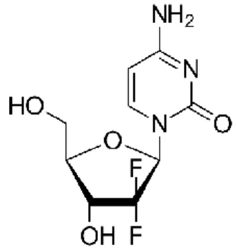
Figure 4 – Gemcitabine´s chemical structure ... 25
Figure 5 – Gemcitabine´s cellular metabolism ... 27
Figure 6 - Epithelial-mesenchymal transition (EMT) ... 29
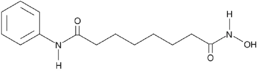
Figure 7 – SAHA´s chemical structure ... 30
Figure 8 - Organization and packing of genetic material ... 30
Figure 9 - SAHA´s mechanism of action ... 32
Figure 10 - G-quadruplex structure ... 33
Figure 11 - SRB colorimetric assay for determination of chemosensitivity to gemcitabine on pancreatic cancer cell lines ... 39
Figure 12 - SRB colorimetric assay for determination of chemosensitivity to SAHA on pancreatic cancer cell lines ... 41
Figure 13 - SRB colorimetric assay for determination of chemosensitivity to CMO3 on pancreatic cancer cell lines ... 42
Figure 14 - Effect on CMO3 on tumor growth over time... 43
Figure 15 - Experimental drug CMO3 bound to a specific G-quadruplex involved in the development of gemcitabine-resistance... 44
Figure 16 - CMO3 chemical structure, emphasizing its side-chain end groups (two morpholino and one pyrrolidino chains) ... 44
10
Index of tables
Table 1 – TNM classification for pancreatic cancer………...21 Table 2 – TNM staging of pancreatic cancer………..21 Table 3 – Chemotherapy regimens for pancreatic cancer………...24 Table 4 – Comparison between the IC50 values of gemcitabine, SAHA and CMO3 in
pancreatic cancer cell lines………...40
11
Acronyms
ALT – Alanine aminotransferase
AJCC - American Joint Committee on Cancer AST – Aspartate aminotransferase
CSC - cancer stem cell
CA 19-9 - Carbohydrate 19-9 CEA - Carcinoembryonic antigen
CHMP - Committee for Medicinal Products for Human Use CT - Computed tomography dCK - Deoxycytidine kinase dFdC - 2´, 2 ´-Difluoro-2´-deoxycytidine dFdCDP - 2´, 2 ´-Difluoro-2´-deoxycytidine diphosphate dFdCMP - 2´, 2 ´-Difluoro-2´-deoxycytidine monophosphate dFdCTP - 2´, 2 ´-Difluoro-2´-deoxycytidine triphosphate DMSO - Dimethyl sulfoxide
DMEM - Dulbecco´s modified eagle medium DPBS - Dulbecco´s phosphate-buffered saline EDTA - Ethylenediaminetetraacetic acid
ERCP - Endoscopic retrograde cholangiopancreatography EUS - Endoscopic ultrasound
EGFR - Epidermal growth factor receptor EMT - Epithelial-mesenchymal transition EMA - European Medicines Agency EBRT - External beam radiation therapy
12
FBS - Foetal bovine serum
FDA - Food and Drug Administration
GR - Gemcitabine-resistant pancreatic cancer cells G4 - G-quadruplexes
IC50 - Half maximal inhibitory concentration
Hh - Hedgehog
HMGA1 - High motility group A1 HAT - Histone acetyltransferase HDAC - Histone deacetylase
hNT - Human nucleoside transporter HIF-1α - Hypoxia inducible factor-1α HS - Horse serum
MRI - Magnetic resonance imaging MDACC - MD Anderson Cancer Center MUC4 - Mucin-4
NDPK - Nucleoside diphosphate kinase hENT1 - Nucleoside transporter-1 NF-kB - Nuclear factor-kB OD - Optical density
PDAC - Pancreatic ductal adenocarcinoma NET - Pancreatic neuroendocrine tumor MP - Parental MIA PaCa-2 cell line PET - Positron emission tomography
13
RR - Ribonucleotide reductase
SAHA - Suberoylanilide hydroxamic acid SRB - Sulforhodamine B
TCA - Trichloroacetic acid
TRAIL - Tumor necrosis factor-related apoptosis inducing ligand TNM - Tumor-node-metastasis
UCL - University College London
VEGF - Vascular endothelial growth factor VIP - Vasoactive intestinal peptide
14
I.
Introduction
1. Pancreas: Anatomy and Functions
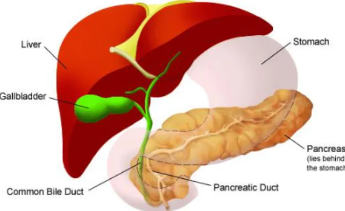
The pancreas is an elongated, spongy organ located behind the stomach in the upper left abdomen. In adults, it is about fifteen centimeters long and has a rich blood supply, not only from the superior mesenteric artery and vein, but also from the portal vein and the celiac axis.
Anatomically, the pancreas is composed by four different parts: head, neck, body and tail. The head of the pancreas is on the right side of the abdomen and lies where the stomach meets the first section of the duodenum. The neck is directed upward to join the body, which is the largest part of the pancreas. The thin end, on the left side of the abdomen next to the spleen, is called the tail (1,2) (Figure 1).
This organ contains both and endocrine cells. The predominant exocrine cells (representing 95% of the pancreas) form glands that produce enzymes important to digestion. These enzymes include amylase, lipase, trypsin and chymotrypsin, responsible for the digestion of carbohydrates, fats and proteins, respectively. They are released into the pancreatic duct, which then joins the common bile duct to form the ampulla of Vater, at the first portion of the small intestine (duodenum). An aqueous alkaline solution, rich in bicarbonate, is also produced by the exocrine tissue to neutralize the acidity of the duodenum.
The smaller percentage of the pancreas consists of endocrine cells that form clusters called islets of Langerhans. These clusters produce hormones that are released directly into the bloodstream. The main hormones secreted are insulin and glucagon, which maintain blood glucose at stable levels. Somatostatin prevent the release of the first two hormones (3–5).
15
2. Pancreatic Cancer
The term “cancer” was used for the first time by Hippocrates, the father of the modern medicine, who applied the words “carcinoma” and “Karakinos” to describe a tumor. Currently, and according to World Health Organization (WHO), cancer is defined as a “generic term for a large group of diseases characterized by the growth of abnormal cells beyond their usual boundaries that can then invade adjoining parts of the body and/or spread to other organs”(6).
2.1. Incidence and Mortality
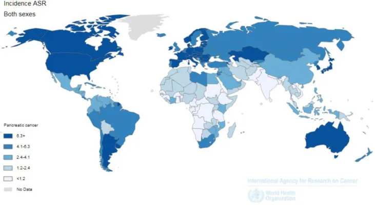
Pancreatic cancer is a highly aggressive solid tumor with an annual mortality identical to its annual incidence (7,8). Due to its early systemic progression and late symptoms, most of patients are diagnosed at an advanced stage, with an overall 5-year survival of only 2% (9–12). According to 2012 Global Cancer Statistics, pancreatic cancer was the seventh most lethal type of cancer worldwide, causing more than 331000 deaths per year (7,13). Over the past decades, its incidence rate has risen significantly in developed countries, and it is expected to become the second leading cause of cancer-related death in the United States, by 2030 (9,13,14) (Figure 2).
Figura 1- Anatomy of the pancreas
Figura 2- Anatomy of the pancreas
16 2.2. Types of Pancreatic Cancer
More than 95% of pancreatic cancers are classified as exocrine tumors. Among these, pancreatic ductal adenocarcinoma (PDAC) is the most common. Less common exocrine pancreatic cancers include acinar cell carcinoma, pancreato blastoma, solid pseudopapillary neoplasm and serous cystadenoma.
Pancreatic neuroendocrine tumors (NETs), also known as islet cell tumors, account for less than 5% of all pancreatic tumors, and can be either functional or non-functional. Functional NETs produce a significant amount of hormones that are released into the bloodstream and cause specific symptoms. On the other hand, non-functional NETs don´t produce enough excess hormones to cause symptoms, leading to a difficult and late diagnosis, sometimes only possible when the cancer has spread beyond the pancreas (9,15).
17
Pancreatic neuroendocrine tumors (NETs) tend to be less aggressive than exocrine tumors, with a much better prognosis. As shown in Figure 3, the 5-year survival rate for NETs is around 50%, compared with less than 5% for exocrine tumors (16).
2.3. Etiology and Risk Factors
The etiology of pancreatic cancer is not yet elucidated, although some factors have been associated with increased risk. Modifiable risk factors include cigarette smoking, heavy alcohol use, increased body mass index, dietary fat and physical inactivity. Also, occupational exposure to certain chemicals, in dry cleaning or metal working industries, has been shown to raise the risk. Non-modifiable risk factors comprise increasing age, with most cases occurring between the ages of 60 and 80 years, male gender and diabetes mellitus. Moreover, some studies have shown that African American population is more likely to develop pancreatic cancer than white population. (7,18,19). There is also some evidence that pathologies like chronic pancreatitis, cirrhosis and
Figure 4 - Differences in relative survival rates (%) for endocrine and exocrine pancreatic cancer (17)
18
Helicobacter pylori infection are strongly associated with elevations in the risk of
this type of cancer (7).
Family history is a strong predictor of pancreatic cancer risk. Some findings suggest that 5-10% of pancreatic cancers are related to genetic factors. Therefore, mutations in genes BRCA2 (hereditary breast and ovarian cancer syndrome), PRSS1 (familial pancreatitis), p16 (familial melanoma), p53 and k-ras have an increased risk. Other inherited genetic disorders that may be linked to pancreatic cancer include Lynch syndrome, Peutz-Jeghers syndrome and Familial adenomatous syndrome (7,19,20).
2.4. Signs and Symptoms
Exocrine pancreatic cancer development is usually silent and signs and symptoms only occur when the disease is already advanced and difficult to treat (9,10). The initial symptoms are unspecific and will depend on the tumor location within the gland. The most common early disease symptoms are weight loss, nausea and vomiting, pain in the upper abdomen that radiates to the back and dyspepsia. Some patients can develop diabetes as the tumor impairs pancreas` ability to produce insulin. Jaundice (yellowing of the skin and eyes) is also common in patients with tumors in the head of the pancreas, which can obstruct adjacent biliary system. Late symptoms, when the tumor is spread, can include gastrointestinal obstruction and bleeding. Anemia, depression and ascites can also be reported in advanced pancreatic cancer (1,14,21).
Pancreatic neuroendocrine tumors (NETs) are rare and, as described above in section 2.2., may be functional or non-functional. Non-functional tumors don´t secrete hormones, so signs and symptoms are unspecific and generally caused by the tumor as it spreads and grows. These unspecific symptoms are similar to the ones described for exocrine pancreatic cancer and may include jaundice, abdominal pain, weight loss, nausea and vomiting. Functional tumors produce excess of certain hormones, causing different symptoms depending on the hormone released. Therefore, functional NETs are named after the type of hormone they overproduce. Insulinomas are the most common type of NETs and are usually benign. They produce excess of insulin, leading to low blood glucose levels, which can cause heart palpitations, weakness, diplopia, shakiness,
19
confusion and seizures. Gastrinomas overproduce gastrin (a hormone that helps to digest food by promoting gastric acid secretion), causing burning abdominal pain, acid reflux, weight loss, severe diarrhea and stomach ulcers. Frequently, gastrinomas occur in a rare disorder called Zollinger-Ellison syndrome. Most of them are malignant. Glucagonomas are rare and half of them are cancerous. They cause overproduction of glucagon, a hormone that causes increased blood glucose levels. This leads to diarrhea, weight loss, anemia, severe swelling or irritation of the skin and mouth sores. Somatostatinomas are extremely rare malignant tumors that produce an excess amount of the hormone somatostatin. Increased levels of somatostatin inhibit the production of other pancreatic and gastrointestinal hormones. Thus, its symptoms are unspecific and include diabetes (due to inhibition of insulin), gallstones and steatorrhea (due to inhibition of cholecystokinin), achlorhydria (due to inhibition of gastrin), weight loss, diarrhea, nausea and vomiting. Vasoactive intestinal peptide releasing tumor (VIPoma), also called Verner-Morrison syndrome, is an uncommon malignant tumor that causes overproduction of vasoactive intestinal peptide (VIP). Excess of VIP may lead to the development of certain symptoms, such as watery diarrhea, dehydration, weight loss, muscle weakness, aching and cramps (22–25).
2.5. Diagnosis and Biomarkers
The early clinical diagnosis of pancreatic cancer is challenging, as there is no reliable test currently available to screen general population. In addition to a physical exam and a medical history assessment, imaging tests are performed. Computed tomography (CT) scan is usually the first approach, as it can pinpoint the location and evaluate the extent of the tumor. Other tests, such as magnetic resonance imaging (MRI), endoscopic ultrasound (EUS), positron emission tomography (PET) and endoscopic retrograde cholangiopancreatography (ERCP), provide complementary information (1,26–29).
Serum biochemistry also plays an important role in patients with suspected pancreatic cancer. These include hepatobiliary tests and biomarkers. In hepatobiliary tests, bilirubin (conjugated and total), alkaline phosphatase and α-glutamyltransferase tend to be raised in obstructive jaundice. Also,
20
aminotransferases (ALT and AST) may be associated with hepatocellular problems. Biomarkers seem to play an important role in therapeutic monitoring and surveillance of disease recurrence. Carbohydrate 19-9 (CA 19-9) is widely used, followed by carcinoembryonic antigen (CEA) (1,7,21,30).
However, a definitive diagnosis of pancreatic cancer can only come from a biopsy, where a small sample of the tumor is removed and examined (31).
2.6. Progression Stages
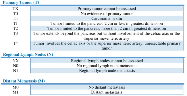
The classification system tumor-node-metastasis (TNM), by the American Joint Committee on Cancer (AJCC), is used to stage pancreatic cancer. This system describes: the size of a primary tumor and whether it has grown beyond the pancreas and into nearby organs (TX to T4); the spread to regional lymph nodes (NX to N1); and whether the cancer has metastasized to distant organs (M0 or M1) (Table 1). Once T, N and M categories have been determined, this information is combined to assign different stages (Table 2).
Although AJCC staging system is really useful as a prognostic tool, it isn´t completely accurate in determining which patients are eligible for surgical resection. As such, more information is needed during the initial phase of treatment. Other clinical staging systems have been studied in order to categorize pancreatic cancer based on surgical resectability. MD Anderson Cancer Center (MDACC) classifies pancreatic cancer into three groups: resectable, borderline resectable and unresectable (locally advanced or metastatic). If the cancer is confined to the pancreas, it is called resectable disease. The term borderline resectable is used to describe some cancers that have reached nearby blood vessels without extrahepatic disease. Unresectable disease is characterized by cancers that have spread to distant organs and, consequently, can´t be entirely removed by surgery (21,26).
21
Table 1 – TNM classification for pancreatic cancer
Primary Tumor (T)
TX Primary tumor cannot be assessed
T0 No evidence of primary tumor
Tis Carcinoma in situ
T1 Tumor limited to the pancreas, 2 cm or less in greatest dimension T2 Tumor limited to the pancreas, more than 2 cm in greatest dimension T3 Tumor extends beyond the pancreas but without involvement of the celiac axis or the
superior mesenteric artery
T4 Tumor involves the celiac axis or the superior mesenteric artery; unresectable primary tumor
Note: Adapted from National Cancer Institute (2014)
Table 2 – TNM staging of pancreatic cancer
Stage T N M 0 Tis N0 M0 IA T1 N0 M0 IA T2 N0 M0 IIA T3 N0 M0 IIB T1, T2, T3 N1 M0 III T4 N0 or N1 M0 IV T1, T2, T3, T4 N0 or N1 M1
Note: Adapted from National Cancer Institute (2014)
2.7. Treatment
Treatment options and recommendations for pancreatic cancer depend on several features related with not only the stage and location of the cancer, but also personal preferences and overall health. Generally, the first aim is to remove the tumor and surrounding cancerous cells, when possible. If that´s not possible, the second approach relies on improving the quality of life and preventing the tumor from growing. Sometimes, none of these options are feasible. In that case, treatment will aim to relieve symptoms (palliative care) to provide the best comfort possible (9,32).
Regional Lymph Nodes (N)
NX Regional lymph nodes cannot be assessed
N0 No regional lymph node metastasis
N1 Regional lymph node metastasis
Distant Metastasis (M)
M0 No distant metastasis
22
The current treatment options for pancreatic cancer may include surgery, radiation therapy, chemotherapy, targeted therapy or a combination of these (9,33).
2.7.1. Surgery
Pancreatic cancer surgery is a complex procedure and only 15-20% of patients are eligible due to the late diagnosis and early systemic spread. Depending on the location and size of the tumor, three different types of surgery can be performed (29,33).
The Whipple procedure, also called pancreatoduodenectomy, is the most commonly performed surgery when the tumor is confined to the head of the pancreas. The surgeon removes the head of the pancreas, the first part of the duodenum, the gallbladder, part of the bile duct and, sometimes, a portion of the stomach. Nearby lymph nodes can also be removed to test for cancer cells. Once the surgery is performed, the digestive tract and biliary system have to be reconnected to allow food digestion (33–35). A distal pancreatectomy involves the removal of the left side (body and tail) of the pancreas and some nearby lymph nodes. The spleen may also be removed, as well as its blood vessels (33–35).
In some problematic cases, when the tumor has spread throughout the pancreas, a total pancreatectomy may be needed. This surgery includes the removal of the entire pancreas, as well as the gallbladder, the spleen, part of the duodenum, stomach and common bile duct (33–35).
Side effects of surgery depend on the extent of the operation, the patient´s overall health and other factors. During the first few days after surgery, patients may feel weakness, pain and tiredness. Other common side effects may include diabetes, difficult in digestion, surgical scars and fistulas, with leakage of pancreatic fluids (33,36).
2.7.2. Radiation therapy
Radiation therapy uses high energy X-rays to destroy cancerous cells. It can be performed before (neo-adjuvant therapy) or after surgery (adjuvant therapy), or even in
23
combination with chemotherapy (chemoradiation). Chemoradiation is mostly used to treat cancers that have spread throughout the pancreas, but only to nearby organs (33,37). There are two main ways to deliver radiation: externally (external beam radiation therapy – EBRT) or internally (brachytherapy). For pancreatic cancer, external beam radiation therapy (EBRT) is the most commonly used. In EBRT, a machine, called linear accelerator, direct beams (high energy X-rays) from outside the body into the pancreatic tumor. Less frequently, internal radiation therapy can also be performed during surgery. It involves the placement of a small radioactive object near the tumor (38,39).
Although radiotherapy should only destroy cancerous cells, it can also damage some healthy cells and, consequently, cause some side effects. These may include fatigue, skin rashes, nausea or vomiting, diarrhea, loss of appetite and, in some circumstances, lower blood counts (33,40).
2.7.3. Chemotherapy
Chemotherapy consists in a type of cancer treatment that uses cytotoxic drugs to damage and destroy cancerous cells. These drugs are given by a medical oncologist and have two main administration routes: oral, where the pill or capsule is swallowed; and intravenous, where a liquid is slowly injected into a vein. Either way, the drugs enter the bloodstream and travel throughout the body to reach cancerous cells (33,41).
Chemotherapy may be given at any stage of pancreatic cancer, including: neoadjuvant treatment (before surgery), to try to shrink the tumor; adjuvant treatment (after surgery), to reduce the risk of relapses; and advanced or metastatic disease, to relieve the symptoms (palliative chemotherapy) (42).
In general, chemotherapy treatment is given in cycles (14, 21 or 28 days long) with a rest period between them. This allows the attack of cancerous cells at their most vulnerable times and, at the same time, allows healthy cells to recover from the damage. The length of chemotherapy cycles is based on the type and extent of cancer, the type of drugs used, as well as the body reaction to the treatment (33).
When chemotherapy regimen uses only one drug at a time, it is called a single agent. However, combination treatments with two or more drugs are usually best for people who are able to carry their daily activities autonomously. The main chemotherapy
24
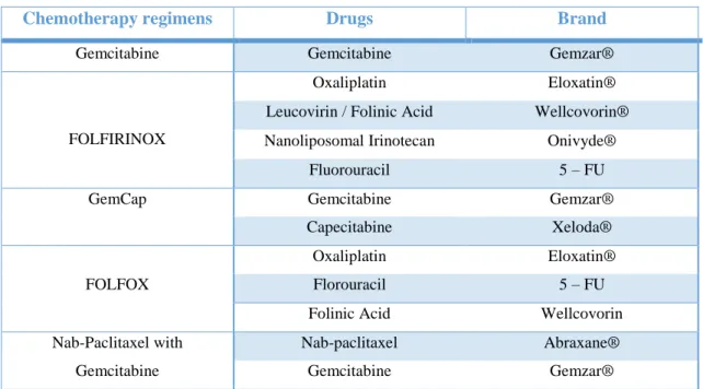
drug combinations approved by the US Food and Drug Administration (FDA) for pancreatic cancer are listed in Table 3 (33,43).
Table 3 – Chemotherapy regimens for pancreatic cancer
Chemotherapy regimens Drugs Brand
Gemcitabine Gemcitabine Gemzar®
FOLFIRINOX
Oxaliplatin Eloxatin®
Leucovirin / Folinic Acid Wellcovorin® Nanoliposomal Irinotecan Onivyde®
Fluorouracil 5 – FU
GemCap Gemcitabine Gemzar®
Capecitabine Xeloda®
FOLFOX
Oxaliplatin Eloxatin®
Florouracil 5 – FU
Folinic Acid Wellcovorin
Nab-Paclitaxel with Gemcitabine
Nab-paclitaxel Abraxane®
Gemcitabine Gemzar®
Gemcitabine, FOLFIRINOX or nab-paclitaxel with gemcitabine are recommended as first-line therapy for pancreatic cancer. When these chemotherapy regimens don´t work, patients may benefit from different drugs to control the cancer. This is called second-line therapy and there are many options considering the patient overall health. Gemcitabine with capecitabine may be used, as well as florouracil with oxaliplatin (FOLFOX) as final option (44,45).
Common chemotherapy side effects include nausea and vomiting, diarrhea, mouth sores, fatigue, hair loss, swelling and dry skin. People receiving this type of treatment have also an increased risk of infection, due to the decrease of white blood cells (neutropenia). Most of these side effects are only temporary and should diminished once treatment finishes (46,47).
2.7.4. Targeted therapy
Recent studies in cancer biology have found unique aspects of cancer cells that contribute to their growth and survival. This led to the development of targeted therapy,
25
where drugs specifically identify and attack cancer cells. Therefore, targeted therapy has fewer side effects than other available treatments (33,48).
In 2005, the targeted therapy drug erlotinib (Tacreva®) was approved by the FDA, in combination with the chemotherapy drug gemcitabine, for patients with unresectable advanced pancreatic cancer. Three years later, in 2007, it has also won the approval from the European Medicines Agency´s (EMA) Committee for Medicinal Products for Human Use (CHMP) for the same therapeutic indication. Erlotinib is an orally available drug that targets and inhibits the epidermal growth factor receptor (EGFR). The EGFR is highly expressed in cancer cells and allows them to grow and spread. Side effects of erlotinib include acneiform skin rash, diarrhea, loss of appetite and tiredness (33,49–52).
3. First-line treatment: gemcitabine
Gemcitabine (Figure 4), a synthetic pyrimidine nucleoside analog, is an antineoplastic drug with a broad-spectrum activity in several types of cancer, such as ovarian, breast, pancreatic, bladder and lung cancers. In locally advanced and metastatic pancreatic cancer (stages II, II and IV), it has become the standard treatment choice (10,53).
Gemcitabine belongs to a family of chemotherapy drugs called antimetabolites. These compounds are structurally similar to normal substances present within the cell, which facilitates its entrance. Antimetabolite agents are cell-cycle specific, acting as
26
inhibitors of DNA synthesis, predominantly in the S phase, and inducing cancer cells apoptosis (53,55).
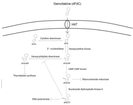
Gemcitabine (2´, 2 ´-difluoro-2´-deoxycytidine; dFdC) is a prodrug that, once inside the cell, needs to be metabolized into its active diphosphate (dFdCDP) and triphosphate (dFdCTP) forms. Since dFdC is a hydrophilic compound, its cellular uptake is mediated by a family of membrane proteins named as human nucleoside transporters (hNTs). When gemcitabine enters the cell, it is first phosphorylated by deoxycytidine kinase (dCK) to the monophosphate (dFdCMP), and then by pyrimidine nucleoside monophosphate kinase (UMP-CMP kinase) and nucleoside diphosphate kinase (NDPK) to give gemcitabine diphosphate (dFdCDP) and triphosphate (dFdCTP), respectively. The phosphorylation by dCK is considered to be the rate limiting step for the dFdCDP and dFdCTP formation (53,55–58) (Figure 5).
The diphosphate form (dFdGDP) inhibits ribonuclease reductase, interfering with subsequent de novo nucleotide production. It also enhances the incorporation of triphosphate form (dFdGTP) into DNA, by reducing intracellular concentrations of deoxycytidine triphosphate. This process is called “self-potentiation” (53,55–58).
Triphosphate gemcitabine (dFdCTP) competes with the natural substract (deoxycytidine triphosphate) for DNA polymerase. When dFdCTP is incorporated into DNA, a single nucleotide is added, making DNA polymerases unable to proceed. This leads to the inhibition of DNA synthesis and consequent apoptosis, in a process called “mask-chain termination” (53,55–58).
27
Gemcitabine is a chemotherapy drug that acts by destroying any cell in rapid division. Therefore, it may harm some healthy cells, leading to the development of some adverse effects (53).
Side effects that have been reported in more than 10% of patients receiving gemcitabine include, among others, flu-like symptoms (fever, muscle aches), alopecia, nausea and vomiting, diarrhea, rashes and itchy skin, proteinuria, hematuria, elevated transaminases (ALT and AST), pain, fever, myelosuppression (low blood cells), increased risk of infection, constipation, allergic reactions (53,59).
As referred above, gemcitabine remains the standard of care therapy for pancreatic cancer patients. However, its long term benefits are modest, with an improvement in the overall survival of only 5 months (60,61). This lack of significant clinical response is largely due to the development of drug resistance mechanisms (62). Therefore, it becomes essential to understand these mechanisms in order to develop new effective treatments and increase patients survival (55,63).
Drug resistance can be either intrinsic (de novo) or acquired during treatment cycles (therapy-induced). Several mechanisms of gemcitabine resistance have been
28
identified and are described below by the following order: tumor microenvironment (A), metabolic proteins (B), deregulations of key signaling pathways (C), epithelial-mesenchymal transition (D) and cancer stem cells (E) (64,65).
A) Among all epithelial tumors, pancreatic cancer is the only one characterized by a dense desmoplastic stroma, promoted by Hedgehog (Hh) signaling. This feature combined with its hypoxic microenvironment, due to poor vascularization, result in difficult penetration and delivery of chemotherapeutic drugs (55,62,66).
B) Gemcitabine uptake and metabolism are crucial to its therapeutic effect. Thus, cancer cells develop mechanisms for modifying the expression or activity of proteins that participate in gemcitabine metabolism pathways. These include the decrease in nucleoside transporter-1 (hENT1), responsible for gemcitabine uptake; the down-regulation of rate-limiting enzyme deoxycytidine kinase (dCK); and the increase in ribonucleotide reductase (RR), as well as in the detoxifying enzyme cytidine deaminase (CDA) (63,64,67).
C) Previous studies have suggested that the activity of some transcription factors can also contribute to gemcitabine resistance (55). For example, nuclear factor-kB (NF-kB) is a transcription factor that is involved in the control of a large number of cellular events, including inflammation, tumorigenesis and apoptosis. In pancreatic cancer, it is overexpressed, resulting in tumor proliferation and inhibiting apoptosis (62,68). High motility group A1 (HMGA1) protein, hypoxia inducible factor-1α (HIF-1α) and mucin-4 (MUC4) pathways have also been implicated in gemcitabine resistance (55,67).
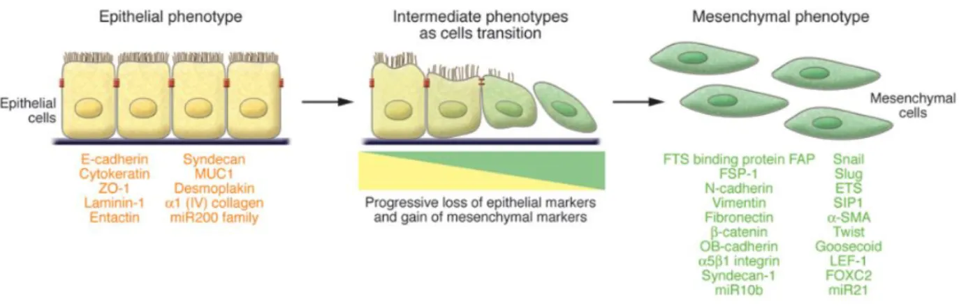
D) Epithelial-mesenchymal transition (EMT, Figure 6) is a biologic process where cells suffer a phenotypic change. It is essential in development and wound healing, but also plays an important role in cancer progression and fibrosis (69). During this process, polarized epithelial cells lose their intercellular adhesion and gain invasiveness capacity and elevated
29
resistance to apoptosis, becoming mesenchymal, fibroblast-like cells. This new elongated mesenchymal phenotype is thought to be induced by an imbalance between epithelial (e.g., E-cadherin, Laminin-1) and mesenchymal (e.g. , N-cadherin, Snail) factors (12,70,71).
E) Pancreatic cancer is composed by a heterogeneous population of cells with different properties. The cancer stem cells (CSCs), also called “tumor-initiating cells”, belong to a small population. These cells are characterized by their ability to self-renew and to produce all cell lines, including those with invasive properties. Therefore, CSCs seem to play a critical role in chemoresistance and cancer progression (62,72,73).
4. Suberoylanilide hydroxamic acid (Vorinostat, SAHA)
Suberoylanilide hydroxamic acid (SAHA) (Figure 7) is an oral histone deacetylase (HDAC) inhibitor with anti-tumor activity (74). In 2006, it was approved by the FDA for the treatment of advanced cutaneous T cell lymphoma (75,76). Over the past few years, clinical studies with SAHA have been performed and it has been demonstrated to be significantly effective in several types of malignancies, including pancreatic ductal adenocarcinoma.
30
Histones are small positively charged proteins, located in the cell nucleus, that complex tightly with the DNA to form nucleosomes. Each nucleosome is composed by a central histone octamer, comprising two molecules of each of the core histones (H2A, H2B, H3 and H4), and about 146 base pairs of DNA. Repetitive units of these nucleosomes constitute the chromatin, which undergoes further condensation to form chromosomes (78–80) (Figure 8).
Histones go through several posttranslational modifications in their N-terminal, altering chromatin structure and gene expression (78,81). These modifications include acetylation, phosphorylation, methylation, ubiquitination and ADP-ribosylation (79).
Histone acetylation is involved in the regulation of many cellular pathways, such as differentiation, inflammation, proliferation and apoptosis (82). It is a dynamic process controlled by two enzymes with opposing activities: histone acetyltransferases
Figure 8 – SAHA´s chemical structure (77)
31
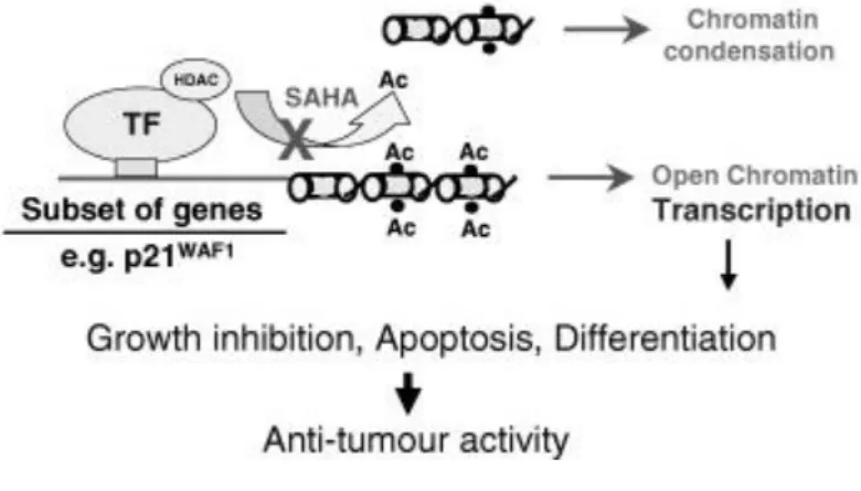
(HATs) and histone deacetylases (HDACs) (76). HAT catalysis the addition of an acetyl group to histone lysine residues, neutralizing its positive charge. Therefore, the interaction between histone and DNA decreases and chromatin structure becomes relaxed, facilitating the access of RNA polymerase and other transcriptional factors, resulting in transcriptional activation. On the other hand, HDAC promotes chromatin condensation by removing the acetyl group, inducing transcriptional repression (78,79,83).
An imbalance in histone acetylation has been associated with transcriptional deregulation of certain genes involved in cell cycle and, subsequent, development of tumors. Indeed, HDACs have been shown to be overexpressed in different types of human cancers, including pancreatic ductal adenocarcinoma. As a result, inhibition of HDAC is a promising therapeutic target for the development of anticancer drugs (76,79).
HDAC inhibitors (HDACi) are a relatively recent group of antineoplastic drugs, which are still under clinical trials. Among these, SAHA is the most promising. It has shown significant antitumor activity, but also safety and poor toxicity (82).
SAHA is a synthetic hydroxamic acid, with high affinity to biometals, including Fe3+, Ni2+ and Zn2+. Thus, it has the ability to chelate zinc ion (Zn2+) located in the catalytic site of HDAC, inhibiting deacetylation. Hyperacetylation of histone proteins results in upregulation of cyclin dependent-kinase inhibitor p21, which antagonizes cyclin-CDK complexes with G1 cell cycle arrest. Hyperacetylation of other non-histone proteins, such as tumor suppressor p53, α-tubulin and heat shock protein-90, promotes tumor cell proliferation blockage (78,83).
SAHA also induces apoptosis by restoring the tumor necrosis factor-related apoptosis inducing ligand (TRAIL) and changing the balance between pro and anti-apoptotic proteins. Pro-anti-apoptotic proteins, like Bim, Bak and Bax, are upregulated, whereas anti-apoptotic proteins, including Bcl-1 and Bcl-2, are downregulated (84).
Angiogenesis might be also inhibited by SAHA. Under hypoxia environments, it has the capacity to suppress hypoxia inducible factor-1 (HIF-1) and vascular endothelial growth factor (VEGF) (84) (Figure 9).
32
SAHA is generally well tolerated and common side effects usually occur when normal dose (400 mg per day) is exceeded. Commonly, these side effects are mild to moderate with no need for intervention, as they will disappear by their own after the treatment is completed (84,85).
Major adverse effects, reported in more than 30% of patients taking SAHA, include fatigue, diarrhea, low platelet count (thrombocytopenia) and nausea and vomiting. Hyperglycemia may also occur, so patients with diabetes should be carefully monitored. Rarely, in about 10-29% of patients receiving this drug, other adverse effects like anorexia, dehydration, muscle spasms and upper respiratory infection may be reported (85,86).
5. G-quadruplex-binding compounds
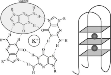
G-quadruplexes (G4) are four-stranded DNA secondary structures formed by rich sequences. The basic unit of the G-quadruplex structure is the guanine-quartet, built by the linear association of four guanine bases through cyclic Hoogsteen hydrogen bonding. This G-quartet is further stabilized by an alkali metal ion, such as Na+ and K+, which is located in the central channel. The planar G-quartets stack one above the other to form a helical G-quadruplex (Figure 10) (87,88) .
33
Recent studies have proven that these G-quadruplex (G4) structures are over-represented in primary tumors, when compared to normal cell. They have also demonstrated to play an important role in certain biological events, with a regulatory function within the cell. Its formation may occur in key regions of the human genome, such as telomeres, ribosomal DNA, oncogene promoter regions and mutational hot spots. Therefore, stabilization of G-quadruplex structures has been associated with the regulation of gene transcription and telomerase activity, emerging as a promising new therapeutic strategy in oncology (87,90,91).
In the case of oncogene promoter regions, G4 can be stabilized by quadruplex-specific small molecules, resulting in an effective decrease in the transcription of the targeted gene (92).
Telomerase is an enzyme responsible for maintaining telomeres integrity by the addition of guanine-rich repetitive sequences. This enzyme is especially up-regulated in the majority of tumors, allowing cellular immortalization and tumor progression. Thus, G-quadruplex selective ligands are being assessed due to their ability to interfere with telomerase enzyme complex and, consequently, destabilize telomere end-capping. As a result, these ligands can inhibit the maintenance of telomere integrity, resulting in an eventual induction of apoptosis in cancerous cells (87,93,94).
34
II. Aim of this research project
Pancreatic cancer remains one of the most devastating human malignancies, with a median overall survival of only 2-8 months after diagnosis. Current standard treatment, gemcitabine, offers limited benefit due to the rapid development of chemoresistance. Therefore, there is an urgent need for new therapeutic strategies with the ability of overcoming gemcitabine resistance and, ultimately, improve patients´ outcome.
Recently, studies at University College London (UCL) School of Pharmacy have revealed that G-quadruplex (G4) structures are associated with cancer cell proliferation. As it was referred previously, a G-quadruplex structure is a guanine rich four-stranded form of DNA that is fundamentally different from normal double-helical DNA. Its formation may occur in telomeres and in oncogene promoter regions, leading to the inhibition of telomerase and RNA polymerase activities. Thus, G-quadruplexes have emerged as novel molecular targets for anticancer therapies.
Research scientists from UCL School of Pharmacy have designed a new chemical compound, named CMO3, which shows promising anticancer activity in pancreatic cancer. It targets a G-quadruplex located in a specific gene involved in the development of gemcitabine chemoresistance. Furthermore, it is currently being assessed for future inclusion in human clinical trials.
The main aim of this research project was to evaluate the anticancer activity of the experimental drug CMO3 in different pancreatic cancer cell lines. For that purpose, the half maximal inhibitory concentration (IC50) of CMO3 was determined and then
compared with gemcitabine and SAHA IC50s. Moreover, the chemical structure of CMO3
was built and optimized using molecular modelling, in order to enhance its docking with the specific G-quadruplex mentioned above.
35
III. Experimental Procedure
1. Materials
1.1. Chemical compounds
All reagents and solvents used in this study were purchased from Lonza, Sigma-Aldrich or Thermo Fisher Scientific companies and used without further purification. Dulbecco´s phosphate-buffered saline (DPBS), Dulbecco´s modified eagle medium (DMEM) and trypsin-EDTA 10x were acquired from Lonza (Visp, Swiss). Penicillin-streptomycin solution (P/S), dimethyl sulfoxide (DMSO), trichloroacetic acid 10% (TCA), acetic acid % (v/v), sulforhodamine B 0.4% (SRB) and tris-base 10mM were supplied by Sigma-Aldrich (St. Louis, MO, USA). Foetal bovine serum (FBS) and horse serum (HS) were provided by Thermo Fisher Scientific (Rockford, IL, USA).
Drugs gemcitabine and vorinostat/suberoylanilide hydroxamic acid (SAHA) were obtain from Tokyo Chemical Industry Co. Ltd. (TCI) (Chuo, Tokyo) and Cayman Chemical (Ann Harbor, MI, USA), respectively. Drug CMO3 was prepared by chemists in the lab.
1.2. Equipment
Incubation of cells was performed using Corning tissue-culture flasks and plates and their confluence was observed with a Nikon TMS inverted phase contrast microscope. Cells were, then, seeded into Corning 6-well clear flat-bottom polystyrene tissue-culture plates, using a Gilson multichannel pipette. After treatment with the intended drug, cells were fixed and stained. Subsequently, cells were solubilized with a Lab-Line gyratory plate shaker, in order to allow measurement at 540nM in a BMG, Labtech-96 microplate reader.
36
2. Methods
2.1. Cell lines and culture
Pancreatic cancer cell line MIA-PaCa-2 (ATCC CRL-1420) was obtained from the American Type Culture Collection and maintained in Dulbecco’s modified Eagle’s medium (DMEM; Lonza) supplemented with 10% (v/v) foetal bovine serum (FBS), 100 U/mL penicillin and 0.1 mg/mL streptomycin (P/S), 2.5% horse serum (HS) and 2 mM L-glutamine. Cells were incubated in 75cm2 culture flasks in a
humidified incubator at 37°C with 5% CO2, and when cell confluence reaches 60–
80%, they are passaged at 1:3 or 1:5 ratio, 2 to 3 times a week.
2.2. Drugs
Gemcitabine was obtained from Tokyo Chemical Industry Co. Ltd. (TCI) and vorinostat/SAHA was supplied by Cayman Chemical. Both compounds were dissolved in DMSO and a stock of 1mM gemcitabine and 10mM vorinostat/SAHA were prepared. CM03 was synthesized by the chemist and 1mM stock was prepared in saline solution (PBS).
All these drugs were stored at -20C and diluted in culture medium immediately before use.
2.3. Establishment of gemcitabine-resistant pancreatic cancer cells
Previously, in the lab, gemcitabine-resistant pancreatic cancer cells (GR) were established by incubating the parental MIA PaCa-2 cell line (MP) with increasing concentrations of gemcitabine, reaching 150nM.
This was carried on reaching 500nM of gemcitabine and three established gemcitabine resistant cell lines (GR150, GR250 and GR350) were selected to be used in a chemosensitivity study.
37 2.4. Sulforhodamine B (SRB) assay for chemosensitivity
Parental MIA PaCa-2, GR150, GR250 and GR350 cells were seeded into 96-well plates at pre-established densities (2000 cells per well) in 0.1 mL of culture medium and allowed to attach overnight in the incubator. The following day, the drug (gemcitabine, SAHA or CM03) was added to cells, at increasing concentrations, in four replicas. Two columns of cells were left as untreated controls, one for background and the other for 100% viability. After 96 hours of incubation, cells were fixed with 10% trichloroacetic acid (TCA) for 30 minutes at 4ºC, washed five times with deionized water, dried in an oven at 80ºC for 1 hour and stained with 0.4% SRB solution for 15 minutes at room temperature. The excess unbounded SRB was removed by rinsing with 1% acetic acid. Afterwards, stained cell proteins were dried and dissolved with 10 mM Tris-base solution. The optical density value was measured using a microplate reader (Microplate BMG Labtech-96) at 540 nm. All experiments were carried out three times for each cell line.
2.5. Statistical analysis
All experiments were performed a minimum of three times. Data were presented as mean ±SEM and compared using Student´s t-test. A *= P-value <0.05 was considered to be statistically significant.
2.6. Molecular modelling
The chemical structure of the drug CM03 was built and docked using Molsoft software in Linux computer operating system. Afterwards, the three lateral chains of CM03 (two morpholino chains and one pyrrolidino chain) were extended and its score evaluated to study the improvement of ligand-receptor interaction.
38
IV. Results and discussion
1.1. Gemcitabine-resistant pancreatic cancer cell lines (GR)
Concerning the rapid development of resistance to gemcitabine, the standard first line treatment against pancreatic cancer, establishment of resistant cell lines was performed for further use in this chemosensitivity study. Therefore, the parental MIA-PaCa-2 (MP) cell line was selected for survival under continuous exposure to increasing concentrations of gemcitabine, reaching 500 nM. To investigate the chemosensitivity to SAHA and CMO3, two promising therapeutic strategies for pancreatic cancer, three gemcitabine-resistant cell lines (GR150, GR250 and GR350) were used.
1.2. Sulforhodamine B (SRB) assay for chemosensitivity
Sulforhodamine B (SRB) assay was developed to investigate drug-induced cytotoxicity and cell viability in cell based studies. This method is based on the ability of SRB, a fluorescent dye, to bind electrostatically to basic amino acid residues of proteins, under moderately acid conditions. Under mild basic conditions, SRB can be extracted from cells. Thus, the amount of bound dye can be used as a predictor of cell mass and subsequently extrapolated to measure cell proliferation (95,96).
The protocol started with the preparation of treatment, after which cells were seeded and incubated with drug of choice (gemcitabine, SAHA or CMO3) for 96 hours. Cellular proteins were, then, fixed with 10% trichloroacetic acid (TCA) and stained by the addition of 0.4% SRB solution. Afterwards, a mild basic environment was established by addition of Tris-base solution to allow for optical density (OD) determination at 540 nm.
This assay provides a sensitive linear response and a higher sensitivity. Also, it possesses a stable colorimetric endpoint, which is readily measured at a 540 nm absorbance, and is defined as accurate, simple, nondestructive and reliable (97).
39
The cytotoxic effect of gemcitabine, SAHA and CMO3 in four pancreatic cancer cell lines (parental MIA-PaCa-2, GR150, GR250 and GR350) was determined by SRB colorimetric assay (Figures 11, 12 and 13). Subsequently, the concentrations at which cell growth was inhibited by 50% (IC50) were evaluated and statically analyzed using
Student´s t-test (Table 4).
Parental MIA-PaCa-2 and gemcitabine-resistant 150nM, 250nM and 350nM pancreatic cancer cells lines were exposed to different gemcitabine concentrations for 96 hours. As expected, cell resistance increased significantly with increasing gemcitabine selection pressure (p<0.05 for all GR cell lines). Parental MIA-PaCa-2 cell line displayed the highest sensitivity to gemcitabine, with an IC50 value of 18.38nM, while
gemcitabine-resistant 350nM cell line was the most gemcitabine-resistant, with an IC50 value of 1238.81nM (Table
4). These results are consistent with the limited therapeutic benefit of gemcitabine due to the rapid development of resistance mechanisms by pancreatic cancer cells.
Figure 12 - SRB colorimetric assay for determination of chemosensitivity to gemcitabine on pancreatic cancer cell lines
40
Table 4 – Comparison between the IC50 values of gemcitabine, SAHA and CMO3 in
pancreatic cancer cell lines
*= P-value <0.05 (statistically significant) ** = P-value >0.05 (not statistically significant)
As shown above, gemcitabine-resistant cell lines were very insensitive to gemcitabine apoptosis effect. Therefore, new promising therapeutic strategies are needed in order to improve patients’ outcome and overall survival.
In this study, SAHA, also called vorinostat, was assessed to determine its effect in gemcitabine-resistant pancreatic cancer cell lines. As shown in Figure 12, the IC50
declined slightly as the resistance to gemcitabine increased. MIA-PaCa-2 parental cell line exhibited an IC50 value of 1558.65nM, which gradually decreased to 1060.07nM in
gemcitabine-resistant 350nM cell line (Table 4). However, this decrease was not significant, as p value was > 0.05 in all studied cell lines. To work around this issue, one option could be the use of a more broad range in future tests (for example, with SAHA concentrations from 200 to 1200nM).
Drug Parental GR150 GR250 GR350
Gemcitabine 18.38 117.52* 111.08* 1238.81*
SAHA 1558.65 1484.41** 1227.15** 1060.07**
41
As demonstrated in Figure 13, gemcitabine-resistant cell lines revealed a greater sensitivity to CMO3 than the MIA-PaCa-2 parental cell line, which was considered to be statically significant (p<0.05 for all GR cell lines). In fact, MIA-PaCa-2 parental cell line expressed an IC50 value of 22.75nM, which is around 2.5 fold higher than the IC50 value
of 9.13nM observed in gemcitabine-resistant 350nM cell line (Table 4).
Moreover, when compared with the results obtain from SAHA´s SRB assay, resistant cell lines evidenced a much higher sensitivity to CMO3. Indeed, in SAHA´s assay, the IC50 values for gemcitabine-resistant cell lines ranged from 1060.07 to
1484.41nM, whereas in CMO3´s assay, these values ranged from 9.13 to 10.50nM.
Figure 13 - SRB colorimetric assay for determination of chemosensitivity to SAHA on pancreatic cancer cell lines
42
These results confirm that CMO3 showed a promising anticancer activity, with the ability of stopping tumor growth effectively, in contrast to gemcitabine, which was effective only for a short period of time due to the rapid development of resistance (Figure 14).
However, in the gemcitabine-resistant cell lines used (GR150, GR250 and GR350), the IC50 didn´t reflect accurately the cells resistant concentrations, with values
ranging from 10.50nM (GR150) to 9.13nM (GR350). This similarity between the IC50
values of the gemcitabine-resistant cell lines may be due to two main reasons. One of them is the maintenance of gemcitabine selection pressure just before performing the assay. The other reason could be the closeness between the resistant concentrations used. Therefore, more studies should be done, on one hand, without the maintenance of gemcitabine selection pressure and, on the other hand, with a more broad range in the resistant concentrations used.
Figure 114 - SRB colorimetric assay for determination of chemosensitivity to CMO3 on pancreatic cancer cell lines
43
Figure 14 – Effect on CMO3 on tumor growth over time
1.3. Molecular Modelling
In the last few years, molecular modelling has become an important tool in the drug discovery field, allowing, not only, the design of new chemical compounds, but also its optimization. In this study, the aim was to evaluate the binding affinity between the experimental drug CMO3 and a G-quadruplex involved in the development of gemcitabine resistance (Figure 15). Afterwards, the three later chains of CMO3 were extended and its score calculated to assess the improvement of ligand-G-quadruplex interaction. Gemcitabine CMO3 Control Time (days) T u m o r v o lu m e (c m 3)
44
The experimental drug CMO3 was synthesized by the chemists Stephan Ohnmacht and Chiara Marchetti, from UCL. As shown in Figure 16, its structure is composed by three side-chain end groups, two morpholino and one pyrrolidino chains. These side-chain end groups have revealed a high affinity and selectivity for G-quadruplexes, in particular the telomeric ones. Moreover, morpholino oligomers are being assessed as research tools by knocking down gene function.
Pyrrolidino side chains
Morpholino side chains
Figure 17 - Experimental drug CMO3 bound to a specific G-quadruplex involved in the development of gemcitabine-resistance
Figure 118 - CMO3 chemical structure, emphasizing its side-chain end groups (two morpholino and one pyrrolidino chains
45
The molecular modelling and simulation of the interaction between the G-quadruplex structure and its ligands, CMO3 and extended analogues, was performed using a variety of tasks, including model building, ligand docking, dynamics simulation and energetic calculations. Primarily, ligand structure was designed, minimized, and partial charges calculated semi-empirically. The ligand was then docked in the active site of the quadruplex and, subsequently, ligand-quadruplex interaction energies (scores) were calculated (Table 5). A low (negative) energy value indicates a stable complex and, therefore, an optimized binding interaction.
Table 5 – Molecular modelling study on CMO3 and its analogues
Ligand Nº of carbons in morpholino chains Nº of carbons in pyrrolidino chain Binding energy (kcal/mol) 1 7 2 -27.7 2 8 2 -25.1 CM03 3 2 -24.4 4 7 3 -22.5 5 4 3 -21.9 6 6 4 -21.3 7 5 2 -20.9 8 6 2 -19.8 9 7 2 -19.5 10 6 5 -19.3 11 6 6 -19.1 12 5 3 -17.9 13 7 6 -17.7 14 5 5 -17.7 15 5 4 -17.6 16 7 5 -17.6 17 5 6 -17.6 18 5 6 -17.1 19 4 2 -16.3 20 6 3 -16.1 21 8 3 -15.5 22 7 4 -12.2
The extension of the side-chain end groups of CMO3 gave rise to a small library of 21 new compounds, which were then evaluated for their ability to bind a G-quadruplex structure. The calculated binding energies for these compounds, including CMO3, are shown in Table 5 sorted by decreasing order of quadruplex-binding affinity.
46
All of the CMO3 analogues were found to dock into the active site of the quadruplex, with binding energy values ranging from -12.2 to -27.7 kcal/mol. However, only two of them (compounds 1 and 2) revealed a higher binding energy, when compared to CMO3. Compounds 1 and 2 showed binding energy values of -27.7 and -25.1 kcal/mol, whereas CMO3 revealed a value of -24.4 kcal/mol. The higher binding energy value of these analogues may be due to the increased length of the morpholino chains, which seems to optimize the van der Waals interactions with the quadruplex structure. It was also verified that a shorter pyrrolidino chain, only composed by two carbon atoms, enhances ligand-quadruplex interaction. This may be explained by the presence of a hydrophobic site in the quadruplex that can interact with the pyrrolidino ring, only when the chain is two carbons length.
In sum, these results provide evidence that the experimental drug CMO3 binds effectively to a quadruplex involved in the development of gemcitabine-resistance in pancreatic cancer, promoting its stabilization. This stabilized complex shows a significant anticancer activity, due to its ability to inhibit the maintenance of telomere integrity. Furthermore, optimized compounds have been identified and, thus, more studies would be done in order to test their cytotoxic effect in gemcitabine-resistant pancreatic cancer cell lines.
47
V.
Conclusion
In this research project, the experimental drug CMO3 was evaluated for its ability to induce a cytotoxic effect in previously established gemcitabine-resistant pancreatic cancer cell lines. The results obtained were based not only on the sulforhodamine B (SRB) assay, but also on molecular modelling.
The results obtained from the SRB assay revealed that CMO3 has an exceptional anti-proliferative activity in gemcitabine-resistant (GR) pancreatic cancer cell lines. In fact, GR cell lines showed a 2.5 fold higher sensitivity to CMO3, when compared with the standard treatment, gemcitabine. Also, when compared to the HDAC inhibitor, SAHA, CMO3 revealed a much greater efficiency in stopping cell proliferation in GR cell lines.
Data provided by molecular modelling confirmed that CMO3 targets and binds effectively to the quadruplex in a gene involved in enhancing resistance in pancreatic cancer. Thus, the quadruplex structure was stabilized, which led to the inhibition of telomerase enzyme, with subsequent induction of apoptosis in pancreatic cancer cells. Therefore, 21 new compounds were designed through the extension of CMO3 structure, aiming the optimization of its active sites. Among these analogues, two of them displayed a higher binding energy to the quadruplex structure than CMO3, emerging as promising therapeutic strategies for human pancreatic cancer.
In the near future, further studies will be performed in order to evaluate the anticancer activity of the optimized CMO3 compounds, in GR pancreatic cancer cell lines. Furthermore, these analogues will eventually be taken into clinical human trials and this approach will be extended to other human cancers.
48
References
1. Bowles MJ, Benjamin IS. Cancer of the stomach and pancreas. :1413–6. 2. Pancreas: Function, Location & Diseases [Internet]. [cited 2017 Sep 5].
Available from: https://www.livescience.com/44662-pancreas.html
3. What Is Pancreatic Cancer? [Internet]. [cited 2017 Sep 5]. Available from: https://www.cancer.org/cancer/pancreatic-cancer/about/what-is-pancreatic-cancer.html
4. Pancreas: Anatomy and Functions | Johns Hopkins Medicine Health Library [Internet]. [cited 2017 Sep 5]. Available from:
http://www.hopkinsmedicine.org/healthlibrary/conditions/liver_biliary_and_panc reatic_disorders/pancreas_anatomy_and_functions_85,P00682/
5. Tsuchitani M, Sato J, Kokoshima H. A comparison of the anatomical structure of the pancreas in experimental animals. J Toxicol Pathol. 2016;29(3):147–54. 6. WHO | Cancer. WHO [Internet]. 2017 [cited 2017 Sep 11]; Available from:
http://www.who.int/cancer/en/
7. Ilic M, Ilic I. Epidemiology of pancreatic cancer. 2016;22(44):9694–705. 8. Xu B, Fu Z, Meng Y, Wu X, Wu B, Xu L. Gemcitabine enhances cell invasion
via activating HAb18G / CD147-EGFR-pSTAT3 signaling. 2016;7(38). 9. Polireddy K, Chen Q. J o u r n a l o f C a n c e r Cancer of the Pancreas :
Molecular Pathways and Current Advancement in Treatment. 2016;7. 10. Heinemann V. Gemcitabine : Progress in the Treatment. 2001;8–18.
11. Lee HS, Park SB, Kim SA, Kwon SK, Cha H, Lee DY, et al. A novel HDAC inhibitor , CG200745 , inhibits pancreatic cancer cell growth and overcomes gemcitabine resistance. Nat Publ Gr [Internet]. 2017;(December 2016):1–9. Available from: http://dx.doi.org/10.1038/srep41615
12. Elaskalani O, Binti N, Razak A, Falasca M, Metharom P, Elaskalani O, et al. Epithelial-mesenchymal transition as a therapeutic target for overcoming chemoresistance in pancreatic cancer. 2017;9(1):37–41.
49
13. Yue Q, Gao G, Zou G, Yu H, Zheng X. Natural Products as Adjunctive
Treatment for Pancreatic Cancer : Recent Trends and Advancements. 2017;2017. 14. Kosmidis C, Sapalidis K, Kotidis E, Mixalopoulos N, Zarogoulidis P, Tsavlis D,
et al. Pancreatic cancer from bench to bedside : molecular pathways and treatment options. 2016;4(8).
15. Manuscript A. developments. 2013;6(5):597–612.
16. Fesinmeyer MD, Austin MA, Li CI, Roos AJ De, Bowen DJ. Differences in Survival by Histologic Type of Pancreatic Cancer Differences in Survival by Histologic Type of Pancreatic Cancer. 2005;14(July):1766–73.
17. PDQ Adult Treatment Editorial Board. Pancreatic Neuroendocrine Tumors (Islet Cell Tumors) Treatment (PDQ®): Health Professional Version [Internet]. PDQ Cancer Information Summaries. 2002 [cited 2017 Oct 14]. Available from: http://www.ncbi.nlm.nih.gov/pubmed/26389309
18. Xu Z, Pothula SP, Wilson JS, Apte M V. Pancreatic cancer and its stroma : A conspiracy theory. 2014;20(32):11216–29.
19. Manuscript A. Genetic Susceptibility to Pancreatic Cancer. 2013;51(1):14–24. 20. Ying H, Dey P, Yao W, Kimmelman AC, Draetta GF, Maitra A, et al. Genetics
and biology of pancreatic ductal adenocarcinoma. 2016;355–85.
21. Reynolds RAEB, Folloder J. Clinical Management of Pancreatic Cancer. 2014; 22. Ro C, Chai W, Yu VE, Yu R. Pancreatic neuroendocrine tumors: biology,
diagnosis,and treatment. Chin J Cancer [Internet]. 2013;32(6):312–24. Available from:
http://www.pubmedcentral.nih.gov/articlerender.fcgi?artid=3845620&tool=pmce ntrez&rendertype=abstract
23. Vinik A, Casellini C, Perry RR, Feliberti E, Vingan H. Diagnosis and Management of Pancreatic Neuroendocrine Tumors (PNETS) [Internet]. Endotext. MDText.com, Inc.; 2000 [cited 2017 Oct 14]. Available from: http://www.ncbi.nlm.nih.gov/pubmed/25905300