outubro de 2013
Universidade do Minho
Escola de Engenharia
Elza Sofia da Silva da Fonseca
Resistance Mechanisms of Candida glabrata
Biofilms to Fluconazole
UMinho|20 13 Elza Sof ia da Silva da F onseca R esis tance Mechanisms of Candida glabrata Biofilms to FluconazoleDissertação de Mestrado
Mestrado em Bioengenharia
Trabalho efetuado sob a orientação da
Doutora Mariana Henriques
e co-orientação da
Doutora Sónia Silva
outubro de 2013
Universidade do Minho
Escola de Engenharia
Elza Sofia da Silva da Fonseca
Resistance Mechanisms of Candida glabrata
Biofilms to Fluconazole
ii
DECLARAÇÃO
Nome: Elza Sofia da Silva da Fonseca
Endereço eletrónico: pg18716@alunos.uminho.pt Número do Bilhete de Identidade: 13544638
Título da tese: Resistance Mechanisms of Candida glabrata Biofilms to Fluconazole Orientador: Doutora Mariana Henriques
Co-orientador: Doutora Sónia Silva Ano de conclusão: 2013
Designação do Mestrado: Mestrado em Bioengenharia
É AUTORIZADA A REPRODUÇÃO INTEGRAL DESTA DISSERTAÇÃO APENAS PARA EFEITOS DE INVESTIGAÇÃO, MEDIANTE DECLARAÇÃO ESCRITA DO INTERESSADO, QUE A TAL SE COMPROMETE;
Universidade do Minho, ___/___/______
iii
Agradecimentos
Gostaria de prestar os meus sinceros agradecimentos a todas pessoas que, de forma direta ou indireta, contribuíram para que este trabalho se concretizasse.
À minha orientadora, Doutora Mariana Henriques, agradeço a oportunidade de ter realizado este trabalho. Gostaria também de agradecer todo o seu apoio, disponibilidade e incentivo prestado ao longo deste trabalho e sobretudo a sua benevolência e humanidade neste período tão importante e também tão difícil da minha vida.
À minha Co-orientadora Sónia Silva, por toda a disponibilidade, apoio e paciência, sem os quais teria sido impossível realizar este trabalho. Os seus conselhos foram indispensáveis para a concretização deste trabalho e para a conclusão desta etapa académica.
A todos meus colegas do Laboratório de Biologia Molecular 2, em especial ao Carlos Tiago Alves que me acompanhou no início do trabalho, à Célia Rodrigues que foi preponderante na terminação deste trabalho e a todos os outros por todo o apoio, espírito de entreajuda e boa disposição demonstrados, em especial a Ana Oliveira que sempre nos animou, juntamente com a Sónia Silva, com as cantorias do Jorge Palma.
A todos os meus verdadeiros amigos, em especial à Marília Lopes, à Isabel Alves e ao Rúben Antunes, por terem estado sempre disponíveis para me ajudar e apoiar nos piores momentos e por me proporcionarem ótimos momentos.
v
Abstract
Candida glabrata has emerged as the second most prevalent fungal pathogen
and its ability to form biofilms has been considered one of the most important virulence factors, since biofilms present a high tolerance to antifungal agents used in fungal infection treatment. The mechanisms of biofilm tolerance to antifungal agents remain poorly understood. Thus, the aim of this study was to evaluate the effects of fluconazole (FLU) in the formation and control of C. glabrata biofilms, its relation with the expression of genes encoding for ABC transporters, CDR1, SNQ2, and PDR1 and how the ergosterol biosynthesis may be affected.
Additionally to the high amounts of proteins and carbohydrates detected in the extracellular matrices in the presence of FLU, this work showed that the overexpression of efflux pumps is a possible mechanism of biofilm tolerance to FLU and this phenomenon alters the structure of C. glabrata biofilms by creating cell clusters.
vii
Resumo
Candida glabrata emergiu como o segundo fungo patogénico mais prevalente e a sua capacidade para formar biofilmes tem sido considerado um dos fatores de virulência mais importante, uma vez que os biofilmes apresentam elevada tolerância a agentes antifúngicos usados no tratamento de infecções fúngicas. Os mecanismos de tolerância dos biofilmes continuam por explorar. Por isso, o objetivo deste estudo é avaliar os efeitos do fluconazol (FLU) na formação e controlo dos biofilmes de C. glabrata, a sua relação com a expressão de genes que encodam os transportadores ABC, CDR1, SNQ2 e PDR1 e como a biossíntese do ergosterol pode ser afetada.
Adicionalmente, para as grandes quantidades de proteínas e hidratos de carbono detetados nas matrizes extracelulares, na presença de FLU, este trabalho demonstrou que a sobre-expressão de bombas de efluxo é um possível mecanismo de tolerância dos biofilmes contra o FLU e este fenómeno altera a estrutura dos biofilmes de C. glabrata pela criação de agregados de células.
ix
The work presented in this thesis will be published: Elza Fonseca, Sónia Silva, Célia Rodrigues, Carlos Tiago Alves, Joana Azeredo, Mariana Henriques (2013). Submitted
xi
Index
Agradecimentos ... v Abstract ... vii Resumo………...…..ix Nomenclature... xiiivIndex of Figures ... xvxiv
Index of Tables ... xvi
Chapter I – Background and Objectives ... 1
I.1. Background ... 3
I.2. Objective ... 4
Chapter II - Introduction ... 7
II.1. Candida Discovery ... 9
II.2. Candida Characteristics ... 9
II.2.1. Cell Biology ... 10
II.2.2. Growth Forms of Candida ... 11
II.2.3. Virulence ... 11
II.3. Non-Candida albicans Candida species ... 13
II.4. Candida glabrata ... 14
II.4.1. Epidemiology... 14
II.4.2. Cell Biology ... 15
II.4.3. Virulence ... 16
II.5. Mechanisms of Resistance to Antifungal Agents ... 17
II.5.1. Antifungal Agents and Resistance ... 18
II.5.2. Biofilms ... 26
II.6. Drug Efflux pumps ... 27
II.6.1. Major-Facilitator Superfamily (MFS) ... 28
II.6.2. ATP-Binding Cassette (ABC) ... 28
II.7. Ergosterol ... 30
Chapter III – Materials and Methodology ... 33
III.1. Organisms and growth conditions ... 35
III.2. Antifungal susceptibility tests ... 35
III.3. Biofilm formation ... 36
xii
III.5. Fluconazole effect on Candida glabrata biofilm formation ... 37
III.6. Biofilm analysis ... 37
III.6.1. Biofilm biomass determination ... 37
III.6.2. Biofilm cultivable cells determination ... 37
III.6.3. Biofilm matrix composition ... 38
III.6.4. Biofilm structure ... 39
III.7. Gene expression analysis ... 39
III.7.1. Gene selection and primers design for quantitative real-time PCR ... 39
III.7.2. Biofilm and planktonic cells preparation ... 40
III.7.3. RNA extraction ... 41
III.7.4. Synthesis of cDNA ... 41
III.7.5. Quantitative Real-Time PCR (qRT-PCR) ... 41
III.8. Statistical Analysis ... 42
Chapter IV – Analysis of the results ... 43
IV.1. Fluconazole minimum inhibitory concentrations and its effect against Candida glabrata biofilms ... 455
IV.2. Biofilm matrix composition and its structure ... 488
IV.3. Gene expression of ABC transporters ... 49
IV.4. Gene expression of ERG genes ... 511
Chapter V – Discussion of the results ... 55
V. 1. Discussion of the results ... 577
Chapter VI - Conclusions ... 61
VI.1. Conclusions ... 633
Chapter VII - References ... 65
xiii
Nomenclature
ACT-Actin
ANOVA - Analysis of variance
ATCC - American Type Culture Collection cDNA - complementary Deoxyribonucleic Acid CFU - Colony Forming Units
CHROMagar - Chromogenic media agar CLSM - Confocal Laser Scanning Microscopy CV - Crystal violet
DNA - Deoxyribonucleic Acid
dNTP - desoxynucleoside triphospahte FLU - Fluconazole
h - Hour
Log - Logarithm
mRNA - messenger Ribonucleic Acid NCAC - non-Candida albicans Candida PBS - Phosphate Buffer Saline
RT- PCR – Real-time Polymerase Chain Reaction RNA - Ribonucleic Acid
rpm - rotation per minute
rRNA - ribosomal Ribonucleic Acid
SDA - Sabouraud dextrose agar
SDB - Sabouraud dextrose broth
SD - Standard deviation
xiv
Index of Figures
Figure II. 1. Mechanisms of action of antifungal agents: target molecules... 18
Figure II. 2 Structure of amphotericin B (AmB). Adapted from (Doctor Fungus 2010 [http://www.doctorfungus.org/thedrugs/Ampho_Deoxycholate.htm] ... 20
Figure II. 3. Mechanism of action of amphotericin B. Adapted from Doctor Fungus 2010 [http://www.doctorfungus.org/thedrugs/antif_pharm.htm]. ... 20
Figure II. 4. Structure of 5-fluorocytosine (5-FC). Adapted from Doctor Fungus 2010 [http://www.doctorfungus.org/thedrugs/Flucytosine.htm]. ... 21
Figure II. 5. Mechanism of action of 5-fluorocytosine. Adapted from Doctor Fungus 2010 [http://www.doctorfungus.org/thedrugs/antif_pharm.htm]. ... 22
Figure II. 6. Structure of fluconazole (FLU). Adapted from (Doctor Fungus 2010 [http://www.doctorfungus.org/thedrugs/Fluconazole.htm]) ... 22
Figure II. 7. Mechanism of action of azoles. Adapted from Doctor Fungus 2010 [ http://www.doctorfungus.org/thedrugs/antif_pharm.htm]. ... 23
Figure II. 8. Structure of caspofugin. Adapted from Doctor Funfus 2010 [http://www.doctorfungus.org/Thedrugs/Caspofungin.htm]. ... 24
Figure II. 9. Mechanism of action of echinocandins. Adapted from Doctor Fungus 2010 [http://www.doctorfungus.org/thedrugs/antif_pharm.htm]. ... 25
Figure II. 10. Representation of a MFS transporter: efflux of drugs with influx of protons into the cell. Adapted from (Richard D. Cannon, Erwin Lamping et al. 2009). ... 28
Figure II. 11. Representation of an ABC transporter: efflux of drugs with ATP hydrolyses. Adapted from (Richard D. Cannon, Erwin Lamping et al. 2009). ... 29
Figure II. 12. Representation of the hydrophilic N-terminal and the hydrophobic C-terminal domains. Adapted from (Richard D. Cannon, Erwin Lamping et al. 2009). ... 29
Figure II. 13. Ergosterol biosynthesis pathway. Adapted from Wikipathways 2010 [http://www.wikipathways.org/index.php/Pathway:WP343]. ... 30
Figure IV.1. Effect of fluconazole on C. glabrata pre-formed biofilms. Mean values of the logarithm of colony forming units normalized by unit of area (Log10 CFU cm
-2
) presented on pre-formed biofilms treated for additional 24 h with different FLU concentrations (A); Mean values of the absorbance at 570 nm normalized by unit of area (Abs570 cm
-2
xv
biofilms treated for additional 24 h with different FLU concentrations (B). Error bars indicate the standard deviations. ** Indicates P<0.01 and statistically different from the control. ... 46
Figure IV.2. Effect of fluconazole on the control of C. glabrata biofilms. Mean values of the logarithm of colony forming units normalized by unit of area (Log10 CFU cm
-2
) (A); Mean values of the absorbance at 570 nm normalized by unit of area (Abs570 cm
-2
) (B), on 24 h C. glabrata biofilms formed in the presence of different FLU concentrations. Error bars indicate the standard deviations. **and *** indicates P<0.01 and P<0.001, and consequently statistically different from its controls. ... 47
Figure IV. 3. Effect of fluconazole on matrices composition of C. glabrata biofilms. Mean values of polysaccharides quantity (A); Mean values of proteins quantity (B), in milligrams per grams of biofilm (mg/gbiofilm) of dry biofilm grown for 24 h in the presence of different FLU concentrations. Error bars indicate the standard deviations. *, ** and *** indicates P<0.05, P<0.01 and P<0.001, and consequently statistically different from its controls. ... 48
Figure IV. 4. Scanning electron microscopy images of C. glabrata. C. glabrata ATCC 2001 (I) and C. glabrata 562123 (II) biofilms formed in SDB for 24 h in the absence of FLU (A) or in the presence of 50 mg l-1 of FLU (B) or 1250 mg l-1 (C). The bar in the images corresponds to 20 μm for the magnification 1000x and 10 μm for the magnification of 3000x. ... 50
Figure IV. 5. Expression of ABC transporter genes. Mean values of n-fold expression levels of SNQ2 (I), CDR1 (II) and PDR1 (III) genes in C. glabrata ATCC 2001 grown as planktonic cells (A) and as biofilm (B) and C. glabrata 562123 grown as planktonic cells (C) and as biofilm (D) treated with 50 and 1250 mg l-1 of FLU. Comparisons are made with planktonic and biofilm grown in the absence of FLU. Error bars indicate the standard deviations. *, ** and *** correspond to P<0.05, P<0.01 and P<0.001, respectively ... 51
Figure IV. 6. Expression of ERG genes. Mean values of n-fold expression levels of ERG1, ERG3, ERG6, ERG9 and ERG11 genes in C. glabrata ATCC 2001 grown as planktonic cells (A) and as biofilm (C) and C. glabrata 562123 grown as planktonic cells (B) and as biofilm (D) treated with 50 and 1250 mg l-1 of FLU. Comparisons are made with planktonic and biofilm grown in the absence of FLU. Error bars indicate the standard deviations. *, ** and *** correspond to P<0.05, P<0.01 and P<0.001, respectively ... 52
xvi
Index of Tables
Table II. 1. Morphological characteristics of Candida albicans, Candida tropicalis, Candida parapsilosis and Candida glabrata species. Adapted from (Silva, Negri et al. 2012). ... 10
Table II. 2. Minimum Inhibitory Concentration (MIC) of amphotericin B, fluoconazole and voriconazole (mg/L) in Candida spp. ... 19
Table III. 1. Primers used for quantitative RT-PCR analysis ... 40
Table IV. 1. Minimum Inhibitory Concentrations (MICs) of fluconazole against Candida glabrata strains ... 45
Chapter I
Chapter I – Background and Objectives
3
I.1. Background
Most cases of candidiasis have been attributed to Candida albicans, but recently, non-Candida albicans Candida (NCAC) species, as Candida glabrata, have been identified as common pathogens. The incidence of systemic infections caused by C.
glabrata increased dramatically throughout the 1990s and became the most common
cause of candidiasis after C. albicans. Candida glabrata systemic infections are a subject of considerable concern due to the tendency of this species to rapidly develop resistance to azole antifungal agents, especially fluconazole, and polyenes like amphotericin B. Moreover, this species is also important due to the high mortality rates associated with C. glabrata fungemia. Adherence to host surfaces including medical devices, secretion of hydrolytic enzymes and specially biofilm formation are virulence factors that are associated with Candida pathogenicity.
Biofilms formed by Candida isolates have been associated with higher morbidity and mortality rates compared with isolates unable to form biofilms, due to the significant resistance to antifungal therapy conferred by the complex biofilm structure and composition. Despite, the lack of knowledge about the exact mechanism of biofilm resistance to antifungals, it is believed that this is a complex multifactorial phenomenon. Actually, restricted penetration of drugs through the biofilm matrix, phenotypic changes resulting from a decreased growth rate or nutrient limitation, expression of resistance genes induced by contact with a surface and the presence of a small number of “persister” cells are hypothesized as mechanisms of biofilm resistance.
At the present, little is known about C. glabrata biofilms resistance, so the aim of this project is to study the resistance mechanisms of C. glabrata biofilms to antifungal agents. It is expected that these studies will ultimately contribute towards the identification of targets for novel therapeutics against C. glabrata infections.
Chapter I – Background and Objectives
4
I.2. Objective
The comprehension of resistance mechanisms of C. glabrata biofilms is the key to succeed on the Candida infections treatment, since the biofilms present much higher MICs for the common antifungal agents used, which in most cases, results on treatment failure. Thus, the main goal of this project is to study the resistance mechanisms of C.
glabrata biofilms to fluconazole. To achieve this goal, the effects of FLU in the
formation and control of C. glabrata biofilms, in the extracellular matrix composition was evaluated, as well as on ABC transporter genes expression (CDR1, SNQ2, PDR1) and on ERG genes (ERG1, ERG3, ERG6, ERG9, ERG11) that are involved in the ergosterol biosynthesis.
Therefore, under the subject of this project it is expected to provide more knowledge for the development of new and more specific therapies for Candida infections, thus rising patients health and lowering the costs involved in the wrong application of these agents.
Chapter I – Background and Objectives
5
I.3. Structure of the thesis
This dissertation is divided in six different chapters in order to present the work done during the time of investigation:
I. Background and objectives
This chapter will present the context and the objectives that gave wings to this thesis.
II. Introduction
This chapter will be focused in the theoretical basis associated to this work.
III. Materials and methodology
In this chapter the materials and the methods and techniques used in the experiments will be presented.
IV. Analysis of the results
In this chapter all results obtained during the whole experimental work will be included.
V. Discussion of the results
In this chapter the discussion of all the results obtained will be exposed.
VI. Conclusions
In this chapter the main conclusions obtained from the realized work will be presented and some works will be suggested to the future.
VII. References
The all bibliography used to the comprehension, execution and written of this dissertation will be listed in this chapter.
The work presented in this thesis was developed in the Center of Biological Engineering, Department of Biological Engineering of the University of Minho.
Chapter I – Background and Objectives
.Chapter II
Chapter II - Introduction
9
II.1. Candida Discovery
“Thrush” was documented for the first time by Pepys in 1665. However, only in 1846, a scientific approach to the study of thrush, carry out by Berg, showed the presence and relationship of the fungus with the disease. After these studies, the idea that the organism could cause several forms of the same disease became clear. In 1792 and then in 1849, Frank and Wilkinson, respectively, observed that aphthae occurs not only in oral cavity but also in sexual organs and that a dimorphic fungus was the probable cause (Calderone 2002).
Even so, the identity of the organism that causes the disease was only approved in 1954. Langenbeck was the first one that observed the fungus, in 1839, but the identity of the organism was incorrect. Then, in 1842, Gruby studied Langenbeck’s organism and concluded that it was a species of Sporotrichum. Five years late, Robin reclassified it as Oidium albicans (Calderone 2002)
It was only in 1923, that the generic name Candida was proposed to the organism responsible for thrush, when Berkhout proved that it was not a species of
Monilia, but a fungus that grows in plant materials and clearly morphologically
different from Candida, which was associated until that time. The whitish colonies on agar or the oral lesion of aphthae or thrush was probably the reason for the name
Candida, which derived from the Latin phrase toga candida, which was used to
describe a special white robe worn by candidates for the Roma Senate (Calderone 2002).
In the past 50 years, the number of new species of Candida described increased to approximately 150 (Calderone 2002).
II.2. Candida Characteristics
Candida species are ubiquitous organisms and most of them are not human
pathogens. The genus Candida is very heterogeneous and its principal characteristics will be described below.
Chapter II - Introduction
10
II.2.1. Cell Biology
The genus Candida is a genus consisting of yeasts that do not possess a known natural sexual cycle. Most Candida species exist as spherical to ovoid budding yeast cells or blastospores, typically 4 to 6 μm in diameter. Some Candida species are capable of producing chains of elongated blastospores termed pseudohyphae both in vivo and under certain conditions in vitro. Table II.1 summarizes these properties, for some of the most relevant species (Calderone 2002).
Table II. 1. Morphological characteristics of Candida albicans, Candida tropicalis,
Candida parapsilosis and Candida glabrata species. Adapted from (Silva, Negri et al.
2012). Species Germ tube Pseudohyphae Yeast size (μm) CHROM-agar colony colour C. albicans + + 4-6 x 6-10 Blue-green
C. tropicalis - + 4-8 x 5-11 Dark blue
C. glabrata - - 1-4 White, Pink-purple
C. parapsilosis - + 2.5-4 x 2.5-9 White
The cell wall of pathogens is critical to their interaction with host cells. For pathogenic fungi the cell wall represents the primary way in which the organism interacts with its host.
Yeast and hyphal cell walls are similar qualitatively but different quantitatively in specific components, as chitin. Thus, the mycelial cell wall exhibited four- to fivefold higher level of chitin (Calderone 2002).
The cell wall of Candida is approximately 80 to 90% carbohydrate and β-glucan (branched polymers of glucose), mannan (polymers of mannose) and chitin (polymers of β-1,4 N-acetyl-D) are the primary constituents. The most abundant component of
Candida cell wall is β-glucan (β-1,3 and β-1,6 glucose polymer) that account fot 47 to
60% of the weight of the cell wall, followed by mannoproteins that account for approximately 40% of the total cell wall polysaccharides, chitin that account for 0.5 to
Chapter II - Introduction
11
3% and glycolipids that account for 1 to 7% by dry weight of the cell wall. Ultrastructural and biochemical observation revealed a layer arrangement: an inner wall composed of structural polysaccharides and an outer layer containing primarily mannan, mannoproteins or nonglycosylated proteins (Chauhan, Li et al. 2002).
The biomolecules of the cell wall that are not found in mammalian cells are potential targets for the identification of antifungal agents.
II.2.2. Growth Forms of Candida
The genus Candida is composed of an extremely heterogeneous group of organisms that grow as yeast, but most members of the genus also produce a filamentous type of growth (pseudohyphae, pseudomycelium). However, C. albicans and C. dubliniensis form true hyphae in addition to pseudohyphae. Thus, both species are considered polymorphic (Calderone 2002).
Pseudohyphae are formed from yeast cells or hyphae by budding, but the new cell remains attached to the parent one and elongates, resulting in filaments with constrictions at the cell-cell junctions of the filaments (Calderone 2002).
True hyphae are formed from yeast cells or as branches of existing hyphae. Outgrowths of the yeast cells (germ tubes) grow by apical extension and cross walls (septa) are formed behind the growing tip of the hyphae. Budding occurs laterally just behind the septa, the latter of which are perpendicular to the main axis of the hyphae (Calderone 2002).
The pseudohyphae appear to be an intermediate growth form of yeast and hyphal morphologies (Calderone 2002).
Germination can be induced in complex media, chemically defined media and serum. Temperatures greater than 35 oC, pH of 6.5 to 7.0 or slightly alkaline and inoculum of <106 mL-1 favor germination, whereas glucose as a sole carbon source, lower temperatures and an acid pH favor yeast growth (Calderone 2002).
II.2.3. Virulence
Candida is a sophisticated pathogen. Although there are about 150 species of Candida, approximately 65% of Candida species are unable to grow at a temperature of
37 oC, a prerequisite for an organism to be a successful pathogen (Calderone and Gow 2002). There are several known virulence factors contributing to Candida pathogenicity that include adherence to epithelial and endothelial cells, proteinase production, hyphae
Chapter II - Introduction
12
and pseudohyphae formation, phenotypic switching, phospholipase production and antigenic modulation as a result of pseudohyphae formation (Fidel, Vazquez et al. 1999).
Adhesion is one of the most important virulence factors of Candida species. Adhesins are cell-surface components of Candida that promote host recognition and colonization (Calderone and Gow 2002).
Most of the medically important species within the genus Candida possess the ability to produce pseudomycelium and are otherwise morphologically very similar. The interconversion of yeast forms to filamentous growth, a process named by morphogenesis, is associated with invasiveness of the organism and also contributes for virulence (Calderone and Gow 2002).
The secretion of digestive enzymes such as the SAPs, a family of secreted aspartyl proteinases, and phospholipase B (PLB), is also required for their virulence since these hydrolytic enzymes can degrade host tissues and thus contribute for their invasion (Calderone and Gow 2002).
As opportunistic pathogens, Candida species can invade every tissue of the human body, depending on the integrity of the host immune system. Its capacity to live both as a commensal and pathogen, to evade the immune system, to overcome drug therapy, to invade a variety of body location and to adjust so rapidly to changes in host physiology suggests that it has extraordinary phenotypic plasticity and can adapt rapidly to environmental changes. “High-frequency phenotypic switching” can generate a variety of general phenotypes, occurs spontaneously, moreover can be affected by environmental changes and can have a profound effect on pathogenic traits. Switching is regulated by a number of phase-specific genes in a combinatorial fashion and a high proportion of these genes directly or indirectly have impact on pathogenesis and virulence (Soll 2002).
The development of biofilms, the most prevalent growth form of microorganisms, is usually observed after initial attachment of Candida to host or/and medical devices. Biofilms are described as surface-associated communities of microorganisms embedded within an extracellular matrix and are an important virulence factors for a number of Candida species, as they confer significant resistance to antifungal therapy by limiting the penetration of substances through the matrix and protecting cells from host immune responses. Furthermore, biofilms formed by C.
Chapter II - Introduction
13
with higher morbidity and mortality rates compared with isolates unable to form biofilms (Silva, Negri et al. 2012).
II.3. Non-Candida albicans Candida species
Candidiasis remains an important clinical problem, primarily in the immunocompromised patient population. Candida albicans initially was the most important pathogen but now non-Candida albicans Candida (NCAC) species, as
Candida tropicalis, Candida parapsilosis, Candida glabrata, Candida krusei and
Candida dubliniesis, have gained clinical importance (Moran, Sullivan et al. 2002; Silva, Negri et al. 2011).
The apparent increased emergence of NCAC species in human candidiasis may be related to improvements in diagnostic methods, such as the use of chromogenic media with the ability to differentiate Candida species, as well as the introduction of molecular techniques in the routine diagnosis of fungemia. Nevertheless, the high prevalence of NCAC species in infections could also be a reflection of their inherent higher level of resistance to certain antifungal drugs compared to C. albicans, as this would promote their persistence in mixed species infections treated with traditional antifungal agents (Silva, Negri et al. 2012).
NCAC species are a very heterogeneous group of organisms that are fundamentally different from each other and from C. albicans at the biological level (Table I). The virulence of different NCAC species in human and in animal models of infection varies considerably, for example, the ability or the inability to form pseudohyphae, the family of adhesins, the kind of hydrolytic enzymes produced (Moran, Sullivan et al. 2002).
In the NCAC species, C. glabrata is considered relatively nonpathogenic in animal models, which suggests that it has few virulence attributes. However, high mortality rate has been associated to this Candida (Fidel, Vazquez et al. 1999). Few studies had been conducted on virulence of C. glabrata, that’s why great importance is given to this Candida in this work.
Chapter II - Introduction
14
II.4. Candida glabrata
Historically, C. glabrata was considered a relatively nonpathogenic saprophyte of the normal flora of healthy individuals, rarely causing serious infection in humans (Fidel, Vazquez et al. 1999). However, the incidence of systemic infections caused by
C. glabrata increased dramatically throughout the 1990s and depending on the site of
infection C. glabrata is often the second or third most common cause of candidiasis after C. albicans and is also the NCAC species most commonly recovered from the oral cavities of HIV-infected individuals (Fidel, Vazquez et al. 1999; Moran, Sullivan et al. 2002).
Candida glabrata systemic infections are a subject of considerable concern due
to the tendency of this species to rapidly develop resistance to azole antifungal agents and due to the high mortality rate associated with C. glabrata fungemia (Moran, Sullivan et al. 2002).
II.4.1. Epidemiology
Data from the 90s show that approximately 31 to 55% of the oral cavity of healthy individuals is colonized by Candida species and this colonization increases with severity of illness and duration of hospitalization. Initially, C. albicans accounted for 70 to 80% of the isolates recovered from infected patients, C. glabrata and C. tropicalis each accounted for approximately 5 to 8% of isolates, while other NCAC species occur only rarely. However, a change in epidemiology was observed. Although C. albicans is the most common fungal species isolated from blood, C. glabrata started to appear associated with an equally high mortality rate. The incidence of C. glabrata is higher in adults than in children and lower in neonates and, despite had being considered a relatively nonpathogenic saprophyte of the normal flora of healthy individuals and certainly not readily associated with serious infection in humans, it is of special importance because of its innately increased resistance to antifungal agents, specifically the azoles (Fidel, Vazquez et al. 1999; Hachem, Hanna et al. 2008; Silva, Negri et al. 2012).
More recently, in the United States, a study demonstrated that C. glabrata has increased as a cause of invasive candidiasis from 18% of all blood stream infection
Chapter II - Introduction
15
isolates in the time period of 1992-2001 to 25% in 2001- 2007 with a concomitant increase in fluconazole resistance from 9% to 14%.Another recent study demonstrated that resistance to both azoles and echinocandins was most prominent among isolates of
C. glabrata with the highest resistance rates to echinocandins (16.7%), fluconazole
(16.7%), posaconazole (5.0%) and voriconazole (11.0%) among isolates from the 20-39-year age group (Pfaller 2012).
The emergence of multidrug resistant (MDR) in C. glabrata is a real fear since that neither azoles nor amphotericin B are an optimal approach for therapy for C.
glabrata infection (Pfaller 2012). For this reason, future surveillance efforts should
focus on emergence of these potentially MDR strains of C. glabrata and the knowledge of the resistance mechanisms to antifungal agents should be a priority.
II.4.2. Cell Biology
Candida glabrata is a nondimorphic yeast that exists as small blastoconidia (1 to
4 m) under some environmental conditions as a pathogen. In fact, C. glabrata is the only Candida species that does not form pseudohyphae at temperatures above 37 °C (Table II.1) (Fidel, Vazquez et al. 1999; Calderone 2002).
On Sabouraud dextrose agar, C. glabrata forms glistening, smooth, cream-colored colonies, which are relatively indistinguishable from those of other Candida species except for their relative size, which is quite small. On Chromagar, a differential medium that distinguishes Candida species by color as a result of biochemical reactions, C. glabrata colonies appear pink to purple, in contrast to C. albicans colonies, which appear green to blue-green (Table II.1) (Fidel, Vazquez et al. 1999; Calderone 2002). Among the critical distinguishing characteristics of C. glabrata are its haploid genome, in contrast to the diploid genome of C. albicans and several other NCAC species and its small-subunit rRNA (Fidel, Vazquez et al. 1999; Ernst and Bockmuhl 2002).
The biochemical reactions of C. glabrata are also quite distinct. Candida
glabrata ferments and assimilates only glucose and trehalose, while C. albicans
ferments and assimilates a high number of sugars (Fidel, Vazquez et al. 1999; Calderone 2002).
Chapter II - Introduction
16
II.4.3. Virulence
Candida glabrata is considered less pathogenic than C. albicans and other
NCAC species, particularly in animal models of infection, although it is being associated with virulent infection in several immunocompromised individuals. C.
glabrata adheres poorly to host surfaces and produces less proteinases than C. tropicalis
and C. parapsilosis. Adhesins, cell surface proteins that are involved in specific adherence, encoded by EPA gene family are major group of adhesins in C. glabrata and it is known that EPA1p is a calcium-dependent lectin (Fidel, Vazquez et al. 1999; Moran, Sullivan et al. 2002; Silva, Negri et al. 2011; Silva, Negri et al. 2012).
Haemolysins are considered key virulence factors since they enable pathogen grow in the host using haemin or haemoglobin as a source of iron. Luo (Silva, Negri et al. 2012) observed that C. glabrata is able to produce haemolysins in vitro, inducing partial or total erythrocyte lyses and showed that a haemolysinlike protein (HLP) gene was associated with the haemolytic activity of C. glabrata. But other authors only observed production of haemolysins by C. albicans (Silva, Negri et al. 2012).
It is known that C. glabrata is unable to produce filamentous forms (hyphae or pseudohyphae) in vivo an important virulence factor required for tissue invasion (Fidel, Vazquez et al. 1999; Moran, Sullivan et al. 2002).
Switching was firstly reported in C. albicans, but it has been demonstrated in other Candida species such as C. glabrata, C. tropicalis and C. parapsilosis. Using an indicator agar 1 mM CuSO4, reversible switching in C. glabrata is demonstrated at high
frequency between a white to light brown and dark brown colony phenotype. As in C.
albicans, switching in C. glabrata is accompanied by the differential expression of
genes: MT-II metallothionein gene and HLP gene that encodes a hemolysis-like protein. These genes are expressed in a graded fashion that correlates with the intensity of pigmentation. One of the genes is involved in copper detoxification while the other may be involved in red blood cell lysis which suggests that just as in the case of C. albicans, switching in C. glabrata is pleiotropic and again may represent a high order of virulence trait and play a role in causing symptomatic infections (Fidel, Vazquez et al. 1999; Soll 2002).
Biofilm formation is another virulence factor of C. glabrata, since biofilms limit the penetration of substances through the matrix and protect cells from host immune responses. The formation of mature biofilms and subsequent production of extracellular
Chapter II - Introduction
17
matrix is strongly dependent on species, strain and environmental conditions (pH, medium composition, oxygen) and, in the case of C. glabrata, it was recently showed that a higher biofilm biomass is produced on silicone surfaces in the presence of urine, compared to C. parapsilosis and C. tropicalis. The opposite was found for biofilms formed in Sabouraud dextrose broth. Thus, biofilm formation by C. glabrata is lower compared with other NCAC species, when grown in rich culture media. Candida
albicans biofilm matrix is mainly composed of carbohydrates, proteins, phosphorus and
hexosamines. However, Silva (Silva, Negri et al. 2012) reported that the extracellular matrix of C. glabrata biofilm is characterized by a high level of both proteins and carbohydrates, while the matrix of C. parapsilosis biofilm is mostly composed by carbohydrates and the matrix of C. tropicallis biofilm exhibits low levels of both proteins and carbohydrates (Silva, Negri et al. 2012).
II.5. Mechanisms of Resistance to Antifungal Agents
Both the frequency of invasive fungal infections (IFIs) and the resistance to antifungal therapy continue to increase, despite the introduction of new antifungal agents. In vitro susceptibility testing is often used to study resistance/sensibility to specific agents against microorganisms. Standardized methods for reliable in vitro antifungal susceptibility testing are now available from the European Committee on Antimicrobial Susceptibility Testing (EUCAST) in Europe. Epidemiologic surveys that examine local and regional data can be used to develop empiric treatment strategies and are essential in tracking resistance trends (Pfaller 2012).
Various mechanisms can lead to the acquired resistance of Candida species to antifungal agents, like the induction of the efflux pumps encoded by the MDR or CDR genes and the acquisition of point mutations in the genes encoding for the targeted enzymes (Pfaller 2012). Moreover, it has been reported that biofilm formation confers significant resistance to the antifungal therapies (Baillie and Douglas 2000).
Antifungal resistance is associated with elevated minimum inhibitory concentrations, poorer clinical outcomes, and breakthrough infections during antifungal treatment and prophylaxis (Pfaller 2012).
Chapter II - Introduction
18
II.5.1. Antifungal Agents and Resistance
The identification of antifungal drugs began in the late 1940s and continues today.
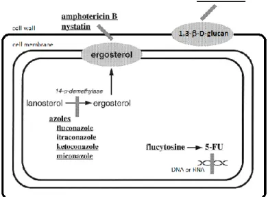
Antifungal treatments against Candida infections are hampered by several factors including the limited number of active agents, the emergence of refractory fungal species and the development of resistance. This situation has triggered the search for new antifungal agents with novel modes of action. Different cellular processes involved in the biosynthesis of components required for the growth of fungal cells have been targeted by antifungal agents (Sanglard and Bille 2002). Actually, the classification of these antifungal agents is based on their target of activity (Figure II.1).
Ergosterol biosynthesis is specific to fungi and is necessary for their growth and this feature has been largely exploited for the design and isolation of antifungal agents such as polyenes and azoles (Sanglard and Bille 2002). Moreover, components of the fungi cell wall are also targets for some antifungal agents as for example echinocandins (Silva, Negri et al. 2012). 5-fluorocytosine (5-FC) is another drug currently used against
Candida, which can be incorporated into RNA molecules and subsequently interferes
with the synthesis of proteins (Sanglard and Bille 2002) (Figure II.1). Figure II. 1. Mechanisms of action of antifungal agents: target molecules.
Chapter II - Introduction
19
Antifungal resistance can be defined as in vitro or clinical resistance. In vitro resistance can be subdivided into primary resistance and secondary resistance. Primary resistance (intrinsic or innate resistance) occurs when the organism is naturally resistant to the antifungal agent (e.g., C. krusei, which is known to be universally resistant to fluconazole). Secondary resistance (acquired resistance) is said to occur when the infecting organism or pathogen becomes resistant to the antifungal agent, in others words, its growth is inhibited by an antimicrobial agent concentration higher than the range seen for wild-type strains (Fidel, Vazquez et al. 1999). Clinical resistance is defined by the situation in which the infecting organism is inhibited by an antimicrobial concentration that is higher than could be safely achieved with normal dosing (Pfaller 2012).
Table II.2 presents the Minimum Inhibitory Concentrations (MICs) of amphotericin B, fluoconazole and voriconazole in C. albicans, C. glabrata, C. krusei,
C. parapsilosis and C. tropicalis, data collected from EUCAST.
Table II. 2. Minimum Inhibitory Concentration (MIC) of amphotericin B, fluoconazole and voriconazole (mg/L) in Candida spp.
C. albicans C. glabrata C. krusei C. parapsilosis C. tropicalis
Amphotericin B 0.032 – 1 0.032 – 2 0.12 – 2 0.032 – 2 0.032 – 2
Fluoconazole 0.12 – 128 2 – 128 8 – 128 0.12 – 8 0.12 – 128
Voriconazole 0.004 – 0.25 0.016 – 4 0.016 – 2 0.008 – 0.12 0.008 – 16
The action of antifungal agents (Figure 1) and the mechanisms of resistance against these antifungal agents in C. glabrata are described in the sections below.
II.5.1.1. Polyenes
In 1950, Hazen and Brown (Sanglard and Bille 2002) identified the first antifungal agent, a polyene called nystatin. Then, other polyene antifungal agents, as amphotericin B (AmB) (Figure II.2), were isolated by Vandeputte and Gold from
Streptomyces nodosus. AmB can form soluble salts in both basic and acidic
environments, is not orally nor intramuscularly absorbed, and is virtually insoluble in water. Systemic and renal problems are often encountered with AmB and to reduce its unwanted side effects, AmB has been formulated in liposomes, lipid complexes and
Chapter II - Introduction
20
colloidal suspensions to allow the use of higher doses of AmB and reduce its toxic effects to mammalian cells.
Polyenes bind to ergosterol (Figure II.3) in the bilayer membrane of susceptible fungi. Aqueous pores result from the interaction of polyene molecules linked to the membrane sterols, leading to altered permeability, leakage of vital cytoplasmic components and death of the organism. Polyenes can also bind to cholesterol, which accounts for much of their human toxicity. However, AmB has much higher affinity for ergosterol than for cholesterol (Sanglard and Bille 2002).
Figure II. 2 Structure of amphotericin B (AmB). Adapted from (Doctor Fungus 2010 [http://www.doctorfungus.org/thedrugs/Ampho_Deoxycholate.htm]
Figure II. 3. Mechanism of action of amphotericin B. Adapted from Doctor Fungus 2010 [http://www.doctorfungus.org/thedrugs/antif_pharm.htm].
Chapter II - Introduction
21
One of the mechanisms of resistance to polyenes is believed to result from the alteration of sterol content or composition in the cell membrane. It has been described that among Candida species, polyene resistance was usually due to defective ergosterol biosynthesis and most likely resulted from mutation in the ERG3 gene that produces altered 5,6-sterol desaturase activity. Mutation in ERG11 (gene encoding for lanosterol 14α-demethylase, required for sterol biosynthesis) and in ERG6 (a gene that is required for normal membrane function, but is not essential for sterol biosynthesis) may generate polyene resistance. In C. glabrata isolates mutations in the ERG6 gene were observed (Silva, Negri et al. 2012).
II.5.1.2. 5-fluorocytosine
5-fluorocytosine (5-FC) belongs to the class of pyrimidine analogs that was developed in the 1950s as a potential antineoplastic agent. It is highly water soluble so it can be administrated by oral or intravenous routes (via) (Sanglard and Bille 2002).
5-FC is taken up by fungal cells (Figure II.5) by a cytosine permease and is deaminated by a cytosine deaminase to 5-fluorouracil (5-FU). 5-FU is a potent antimetabolite that can be converted to a nucleoside triphosphate and when incorporated into RNA causes miscoding. In other hand, 5-FU can be converted to a deoxynucleoside which inhibits thymidylate synthase and thereby, DNA synthesis. 5-FC has low toxicity in mammalian cells, since cytosine deaminase is absent or poorly active in these cells. However, the conversion of 5-FC to 5-FU is possible by intestinal bacteria and therefore 5-FC can show toxicity in oral formulation and 5-FU, despite being a potent anticancer agent, it is impermeable to fungal cells (Sanglard and Bille 2002).
Figure II. 4. Structure of 5-fluorocytosine (5-FC). Adapted from Doctor Fungus 2010 [http://www.doctorfungus.org/thedrugs/Flucytosine.htm].
Chapter II - Introduction
22
Mechanisms of resistance to 5-FC are possible due to the multiple intracellular enzymatic steps required for its action. These include alterations in the target enzymes UMP pyrophosphorylase, cytosine permease and cytosine deaminase, or increased production of pyrimidines. Due to the multiple steps in its mode of action, including transport into the cell and deamination of the active compound, and due to its extremely narrow spectrum of action 5-FC is normally used only in combination with other agents, including amB and fluconazole (Silva, Negri et al. 2012).
II.5.1.3. Azoles
Since pharmaceutical industry attributed great importance to fungal diseases, more drugs have been developed, and the azoles are a good example. Miconazol was the first azole developed against fungus, followed by the discovery of the triazoles such as fluconazole (Figure II.6) and itraconazole that are less toxic than amphotericin B, although being fungistatic (Sanglard and Bille 2002).
Figure II. 6. Structure of fluconazole (FLU).
Adapted from (Doctor Fungus 2010
[http://www.doctorfungus.org/thedrugs/Fluconazo le.htm])
Figure II. 5. Mechanism of action of 5-fluorocytosine. Adapted from Doctor Fungus 2010 [http://www.doctorfungus.org/thedrugs/antif_pharm.htm].
Chapter II - Introduction
23
Azoles have a cytochrome P450 as a common cellular target in yeast or fungi. Cytochrome P450 is involved in the 14α-demethylation of lanosterol. The unhindered nitrogen of the imidazole or triazole ring of azole antifungal agents binds to the heme iron of the cytochrome P450 as a sixth ligand, thus inhibiting the enzymatic reaction (Figure II.7). As a result ergosterol content in the cell membrane is depleted, membrane structure and functions are altered, and fungal growth is inhibited(Fidel, Vazquez et al. 1999; Sanglard and Bille 2002; Pfaller 2012).
There are four principal mechanisms of azole resistance that have been described in Candida species. The first mechanism is the induction of efflux pumps that lead to decreased drug concentration at the enzyme target within the fungal cell. In C. glabrata the efflux pumps are encoded by CgCDR1 and CgCDR2 genes and the up-regulation of these genes has been associated to azole resistance. The second mechanism common in
Candida species is the acquisition of point mutation in ERG11. Thus, an altered enzyme
is synthesized with reduced affinity for or incapacity to bind azoles. The third mechanism, which can be associated with the second one, is the overexpression of the altered target enzyme. However, the up-regulation of altered target enzymes does not appear to be a major cause of azole resistance in Candida. Finally, the last mechanism
Figure II. 7. Mechanism of action of azoles. Adapted from Doctor Fungus 2010 [ http://www.doctorfungus.org/thedrugs/antif_pharm.htm].
Chapter II - Introduction
24
of azole resistance in Candida species involves the development of bypass pathways, which negate the membrane-disruptive effects of azole drugs that are associated with inhibited fungal growth. This has been linked with mutation of the ERG3 gene in certain resistant strains of Candida. A study (Pfaller 2012) in C. albicans demonstrated an additive nature of resistance mechanisms in Candida species for azoles: the control strain with basal expression of CDR and WT ERG11 genes in both alleles, as expected, has low MICs for both fluconazole and voriconazole by comparison with MICs for both azoles in the strain with overexpression of CDR and point mutations in both ERG11 alleles that are much higher. In addition the MICs for fluconazole and voriconazole are approximately twice as high in the strain with basal CDR expression and point mutations in both ERG11 alleles as in the strain with basal CDR expression and a point mutation in only one of the ERG11 alleles.
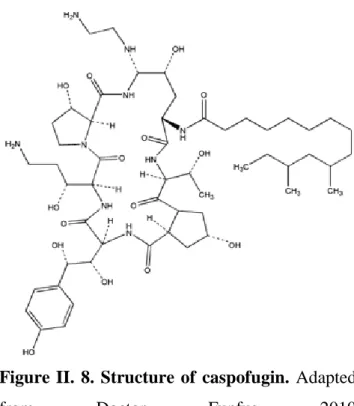
II.5.1.4. Echinocandins
The first echinocandin isolated was anidulafungin in 1974. Later, in 1989, caspofugin (Figure II.8) was discovered and micafugin was the last to be synthesized in 1990 (Cortés and Russi 2011).
Figure II. 8. Structure of caspofugin. Adapted
from Doctor Funfus 2010
[http://www.doctorfungus.org/Thedrugs/Caspofu ngin.htm].
Chapter II - Introduction
25
The echinocandins – anidulafungin, caspofugin and micafungin – are lipopeptides that bind to 1,3-β-D-glucan synthase, enzyme responsible for the biosynthesis of 1,3--D-glucan (a component of the cell wall), causing the formation of a defective cell wall associated with cellular instability and lysis in yeasts and aberrant hyphal growth in molds (Figure II.9) (Cortés and Russi 2011; Pfaller 2012).
Reduced susceptibility or resistance of Candida to echinocandins has been linked with point mutations in two “hot-spot” regions (HS1 and HS2) of FKS1, the gene encoding for the major and presumed catalytic subunit of 1,3- -D-glucan synthase. This resistance mechanism has been demonstrated in C. albicans and NCAC species as
C. glabrata, C. krusei, C. tropicalis, and C. dubliniensis. In C. glabrata, echinocandin
resistance has also been associated with mutations in the FKS2 gene (Pfaller 2012). Figure II. 9. Mechanism of action of echinocandins. Adapted from Doctor Fungus 2010 [http://www.doctorfungus.org/thedrugs/antif_pharm.htm].
Chapter II - Introduction
26
II.5.2. Biofilms
The biofilm state is the preferred mode of growth of microorganisms in natural environments. In the past years, several reports have associated biofilms with over 65% of hospital-acquired infections. It has also been suggested that Candida strains with a high ability to form biofilms are generally more virulent than others (Williams, Kuriyama et al. 2011).
The exact mechanism of biofilm resistance to antifungals remains unclear, but it is probably multifactorial. There are three possible mechanisms of biofilm resistance: restricted penetration of drugs through the biofilm matrix; phenotypic changes resulting from a decreased growth rate or nutrient limitation; expression of resistance genes induced by contact with a surface. It has also been suggested that a small number of “persister” cells are responsible for resistance (Douglas 2003).
Regarding the restricted penetration, it has long been supposed that the matrix of extracellular polymeric material might exclude or limit the access of drugs to organisms in the deeper part of the biofilm. To investigate if the matrix plays a role in the resistance of biofilms to antifungal agents, the susceptibility profiles of biofilms where compared between biofilms of C. albicans which have relatively little matrix and biofilms of C. albicans which produce much more matrix. No significant differences in susceptibility to any of the drugs tested were found, indicating that drug resistance is unrelated to the extent of matrix formation (Baillie and Douglas 2000). However, it had been shown, in another study (Baillie and Douglas 1999), that resuspended cells (which presumably had lost most of their matrix) were some 20% less resistant to amphotericin B than intact biofilms, suggesting that the matrix might play a minor role in drug resistance.
Biofilm cells are known to grow slowly because of the limited availability of nutrients, particularly at the base of the biofilm. A slow growth rate is often accompanied by changes in cell surface composition, which could affect the susceptibility of the microorganisms to antifungal agents. To investigate if growth rate is an important modulator of drug activity in biofilms, the susceptibility of C. albicans biofilms to ampB was compared with that of planktonic cells, the both cases with several growth rates. It has been demonstrated that biofilms were resistant to the drug at all growth rates tested whereas planktonic cells were resistant only at low growth rates (Baillie and Douglas 1998). Another study (Baillie and Douglas 1998) demonstrated
Chapter II - Introduction
27
that not only the low growth rates, but also other conditions of growth, like glucose and irion availability, can interfere with drug susceptibility. Glucose-limited and iron-limited biofilms, grown at the same low rate, were equally resistant to amphotericin B. Iron-limited biofilms probably resemble the most to biofilms growing in vivo, as to the fact there is an abundance of iron in the human body, most of it is located intracellularly or tightly complexed to iron-binding glycoproteins, thus being relatively inaccessible to microorganisms.
Microorganisms that form biofilms express an altered phenotype. To investigate the surface-induced expression of resistance genes, it had been identified genes that are activated or repressed in Candida biofilms compared with planktonic cells. Genes coding for multidrug efflux pumps are of particular interest, since the upregulation of these genes results in a multidrug-resistant phenotype. Candida albicans possesses two different types of efflux pump, ATP-binding cassette (ABC) transporters and major facilitators, which are encoded by CDR and MDR genes, respectively (Douglas 2003). A study (Ramage, Bachmann et al. 2002) has demonstrated that genes encoding both types of efflux pump are upregulated during biofilm formation and development. However, mutants carrying single or double deletion mutations in some of these genes were highly susceptible to fluconazole when growing planktonically but still retained the resistant phenotype during biofilm growth. These results strongly suggest that drug resistance in C. albicans biofilms is a complex process that cannot be explained by a single molecular mechanism.
However, it has been demonstrated in vitro that caspofungin is effective against
C. albicans and C. glabrata biofilms (Cateau, Berjeaud et al. 2001). Caspofungin
inhibits the synthesis of 1,3--D-glucan, the major structural component of Candida cell walls, suggesting that glucan synthesis might be a particularly effective target for biofilms if the biofilm matrix also contains this polysaccharide (Kuhn, George et al. 2002).
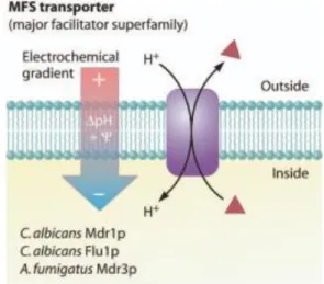
II.6. Drug Efflux pumps
There are two main drug efflux pumps classes, the ATP-binding cassette (ABC) transporters and the Major-Facilitator Superfamily (MFS) transporters that are involved
Chapter II - Introduction
28
in the resistance to antifungal agents, particularly azoles (Richard D. Cannon, Erwin Lamping et al. 2009).
II.6.1. Major-Facilitator Superfamily (MFS)
The MFS transporters are proteins with transmembrane domains (TMD) subtract specific. These transporters uses an electronic gradient as driving force to efflux the drugs out of the cell (Figure II.10) (Richard D. Cannon, Erwin Lamping et al. 2009).
However, there is a more evident relation with the resistance to azoles and the ABC transporters than with the MFS transporters.
II.6.2. ATP-Binding Cassette (ABC)
The ABC transporters are proteins localized in the cellular membrane and in the organelles membranes that contain TMD substrate-specific and nucleotide-binding domains (NBD). These transporters use the ATP hydrolyses to efflux the drug out of the cell (Figure II.11) (Richard D. Cannon, Erwin Lamping et al. 2009).
Figure II. 10. Representation of a MFS transporter: efflux of drugs with influx of protons into the cell. Adapted from (Richard D. Cannon, Erwin Lamping et al. 2009).
Chapter II - Introduction
29
Regarding the resistance to FLU acquired by C. glabrata cells, the transcriptional induction and upregulation of genes encoding ABC transporters (CDR1,
CDR2 and SNQ2) have been reported (Tscherner, Schwarzmüller et al. 2011;
Samaranayake, Cheung et al. 2013). The Figure II.12 illustrates the CDR1 transporter: two identical halves, each with a hydrophilic N-terminal domain which contains units of the ATP-binding (Walker A and Walker B), followed by a C-terminal hydrophobic domain with six transmembrane segments (Richard D. Cannon, Erwin Lamping et al. 2009).
Figure II. 11. Representation of an ABC
transporter: efflux of drugs with ATP
hydrolyses. Adapted from (Richard D. Cannon, Erwin Lamping et al. 2009).
Figure II. 12. Representation of the hydrophilic N-terminal and the hydrophobic C-terminal domains. Adapted from (Richard D. Cannon, Erwin Lamping et al. 2009).
Chapter II - Introduction
30
On other hand, a mutation in the gene that encodes a regulator of multidrug transporter genes, PDR1, was associated with its upregulation. This fact contributes to upregulation of CDR1 and SNQ2 genes (Vermitsky and Edlind 2004). However, little is known about the mechanisms of C. glabrata biofilms resistance.
II.7. Ergosterol
Ergosterol is a biomolecule that is one of the main components of fungus cellular membrane. Candida grown in presence of azoles has a reduction in the ergosterol content of membranes and also an accumulation of toxic ergosterol precursors, such as 14-α-methylergosta-8,24(28)-dien-3β,6α-diol (Richard D. Cannon, Erwin Lamping et al. 2009).
Figure II. 13. Ergosterol biosynthesis pathway. Adapted from Wikipathways 2010 [http://www.wikipathways.org/index.php/Pathway:WP343].
Chapter II - Introduction
31
It was been reported an overexpression and mutation of several genes involved in the ergosterol biosynthesis pathway (Figure II.13) as ERG1, ERG3, ERG6, ERG9 and
ERG11 (Antonia Geber, Hitchcock et al. 1995; Patrick Vandeputte, Guy Tronchin et al.
Chapter III
Chapter III – Materials and Methodology
35
III.1. Organisms and growth conditions
A total of four different Candida glabrata strains were used in this work. The reference strain C. glabrata 2001 from the American Type Culture Collection (ATCC), two oral isolate (AE2 and D1) from the biofilm group of the Centre of Biological Engineering, originally isolated from Clinic of Dentistry, Congregados, Portugal, two urinary (562123 and 513100) and two vaginal (534784 and 585626) tract isolates, both isolated from patients of the Hospital of S. Marcos, Braga, Portugal. The identity of all isolates was confirmed using CHROMagar Candida (CHROMagar, France) and by PCR-based sequencing using specific primers (ITS1 and ITS4) against the 5.8S subunit gene reference. Genomic DNA was extracted following previously described procedures (Williams, Wilson et al. 1995). The PCRs products were sequenced using the ABI-PRISM Big Dye terminator cycle sequencing kit (Perkin Elmer, Applied Biosystems, Warrington, UK). All Candida strains were subcultured on Sabouraud dextrose agar medium (SDA; Merck, Germany) at 37 oC for 48 h.
III.2. Antifungal susceptibility tests
Minimum inhibitory concentrations (MICs) for fluconazole (FLU; Sigma-Aldrich, USA) were determined using the microdilution method, in accordance with the guidelines of the Clinical Laboratory Standards Institute (CLSI) (M27-A2).
The FLU concentrations tested were of 5, 50, 312.5, 625 and 1250 mg ml-1 and were prepared in RPMI 1640 (Sigma-Aldrich, USA). Thus, a small colony of each strain cultured on SDA was suspended in 5 ml of saline solution (NaCl 0.85%) and the cellular density adjusted to turbidity equivalent to a 0.5 McFarland standard in saline buffer. The yeasts suspensions were diluted (1:100) in saline solution and afterward diluted (1:20) in RPMI 1640, according to the standard.
Each Candida suspension (100 l) was added to the respective well of microtiter plates (Orange Scientific, Braine-l’Alleud, Belgium) containing 100 l of each specific concentration of FLU solutions. Controls without antifungal agents were also performed. The microtiter plates were incubated at 37 oC, and the MICs values
Chapter III – Materials and Methodology
36
determined visually as the lowest concentration of FLU showing no yeast growth after 48 h. Additionally, a volume (50 l) of each cell suspension treated with FLU was recovered to a new well and serial decimal dilutions (in phosphate-buffered saline; PBS 0.1 M pH 7.5: NaCl 0.8%, KCl 0.02%, K2HPO4 0.02%, NaHPO412H2O 0.285%) were
plated onto SDA. Agar plates were incubated for 24 h at 37 oC, and the total number of colony forming units (CFUs) was determined. The results were presented per Log10
CFU per milliliter (Log10 CFU ml-1). The assays were performed in triplicate and on
three separate occasions.
III.3. Biofilm formation
An inoculum of each yeast strain, obtained from SDA plates, was suspended in 20 ml of Sabouraud dextrose broth (SDB; Merck, Germany) and incubated at 37 oC for 18 h under agitation (120 rpm). Then, the cells were harvested by centrifugation at 3000 g for 10 min at 4 oC and washed twice with 15 ml of PBS pH 7.5. Pellets were suspended in SDB and the cellular density adjusted to 2x10⁷ or 1x10⁷ cells ml-1 using a Neubauer counting chamber, to use according with each experiment.
III.4. Fluconazole effect against pre-formed Candida glabrata biofilms
In order to test biofilms resistance to FLU, C. glabrata biofilms were pre-formed during 24 h in SDB. For that, 200 l of each Candida suspension containing 1x10⁷ cells ml-1 was added to the respective well of microtiter plates (Orange Scientific, Braine-l’Alleud, Belgium) and incubated at 37 o
C under agitation (120 rpm). After this time, the medium was totally aspired and the biofilm washed once with 200 l of PBS to remove non-adherent cells. At this time, the solutions of FLU (at 50, 625 and 1250 mg ml-1) were added to the specific wells and incubated at 37 oC for extra 24 h. Controls devoid of FLU were also incubated. The assays were repeated in triplicate on three different occasions.
Chapter III – Materials and Methodology
37
III.5. Fluconazole effect on Candida glabrata biofilm formation
In order to study the effect of FLU in the biofilm formation, FLU was added in the beginning of the formation process. For that 96-wells microtiter plates (Orange Scientific, Braine-l’Alleud, Belgium) were filled with increased concentrations of FLU (at 50, 625 and 1250 mg ml-1) diluted in SDB. At each well containing 100 l of each specific concentration of FLU was added 100 l of Candida suspension containing 2x10⁷ cells ml-1. The microtiter plates were incubated at 37o C under agitation (120 rpm). Controls with Candida cells and without FLU were also performed. The assays were repeated in triplicate on three different occasions.
III.6. Biofilm analysis
III.6.1. Biofilm biomass determination
Total biofilm biomass was quantified by crystal violet staining methodology (Silva, Henriques et al. 2009). For that, the medium was totally aspirated and the biofilms washed once with 200 l of PBS to remove non-adherent cells. The biofilms were fixed with 200 μl of methanol and removed after 15 min. The microtiter plates were allowed to dry at room temperature. Then, 200 μl of crystal violet (CV; 1%, v/v) were added to each well. After 5 min, the excess of CV was removed and for that, the biofilms were gently washed twice with water. Lastly, 200 μl of acetic acid (33%, v/v) were added to each well to release and dissolve the CV stain. The absorbance of suspensions was measured at 570 nm and the results were presented as absorbance per unit area (Abs/cm2). The assays were performed fivefold and on three separate occasions.
III.6.2. Biofilm cultivable cells determination
The number of cultivable cells on biofilms was determined by the enumeration of colony forming units (CFUs). For both cases, the medium was aspired and the biofilms washed once with 200 l of PBS to remove non-adherent cells. Then, biofilms


![Figure II. 3. Mechanism of action of amphotericin B. Adapted from Doctor Fungus 2010 [http://www.doctorfungus.org/thedrugs/antif_pharm.htm]](https://thumb-eu.123doks.com/thumbv2/123dok_br/17750910.834413/37.892.185.720.734.1031/figure-mechanism-amphotericin-adapted-doctor-fungus-doctorfungus-thedrugs.webp)
![Figure II. 4. Structure of 5-fluorocytosine (5-FC). Adapted from Doctor Fungus 2010 [http://www.doctorfungus.org/thedrugs/Flucytosine.htm].](https://thumb-eu.123doks.com/thumbv2/123dok_br/17750910.834413/38.892.347.539.578.756/figure-structure-fluorocytosine-adapted-doctor-doctorfungus-thedrugs-flucytosine.webp)

![Figure II. 7. Mechanism of action of azoles. Adapted from Doctor Fungus 2010 [ http://www.doctorfungus.org/thedrugs/antif_pharm.htm]](https://thumb-eu.123doks.com/thumbv2/123dok_br/17750910.834413/40.892.182.716.372.750/figure-mechanism-action-adapted-doctor-fungus-doctorfungus-thedrugs.webp)
![Figure II. 9. Mechanism of action of echinocandins. Adapted from Doctor Fungus 2010 [http://www.doctorfungus.org/thedrugs/antif_pharm.htm]](https://thumb-eu.123doks.com/thumbv2/123dok_br/17750910.834413/42.892.174.710.303.754/figure-mechanism-echinocandins-adapted-doctor-fungus-doctorfungus-thedrugs.webp)