CYSTEINE METABOLISM AND PANCREATIC NEUROENDOCRINE
TUMOURS (PNETs) CHEMORESISTANCE
RAKHI CHANDA ROY
A dissertation submitted in partial fulfillment of the requirements for the Degree of Masters in Biomedical Research
Dissertação para obtenção do grau de Mestre emInvestigação Biomédica
at Faculdade de Ciências Médicas | NOVA Medical School of NOVA University Lisbon
CYSTEINE METABOLISM AND PANCREATIC NEUROENDOCRINE
TUMOURS (PNETs) CHEMORESISTANCE
Rakhi Chanda Roy
Supervisor: Jacinta Serpa, PhD; Assistant Professor; Faculdade de Ciências Médicas | NOVA Medical
School of NOVA University Lisbon
A dissertation submitted in partial fulfillment of the requirements for the Degree of Masters in Biomedical Research
Dissertação para obtenção do grau de Mestre em Investigação Biomédica
The work was approved by the Ethical Committee of NMS|FCM-UNL (76/2019/CEFCM) and by the IPOLFG (UIC-1082).
i
Acknowledgements
Acknowledgement is a pathway, which is allowing me to thank all the person who has helped me to complete the thesis project.
First, I would like to thank Professor Dr Jacinta Serpa , my supervisor, for letting me do this project and completion of the work and writing, would not have been possible without thoughtful advice of my caring mentor. Jacinta is guiding me not only in the research project but also giving me suggestion like an adviser throughout my working period. Thanks for your autonomy, trust and dedication, which helps me a lot to complete my thesis.
I would like to thank Dr Joana Simões Pereira, for her valuable suggestion and kind advice that helped me to complete the thesis writing.
My grateful also extended to Cristiano Ramos for helping me to learn all the research techniques, patient guidance and encouragement in this project. Thanks to all of my lab member for helping me all the time, whenever I asked help to them. Special thanks to Filipa Coelho for the sympathy, motivation, especially for her research experiences helped me a lot to gain knowledge in this research sector. I would also like to thank Ana Rita Hipólito, Filipa Martins and Cindy Mendes, Donato Urso for being the best laboratory companion, supporting me, advise me and making me always happy. All of my laboratory member giving me mental support during the working time, in this way I have completed my lab work smoothly and I never feel that I am far away from my family member. Thank you all for giving me the wonderful working environment. Thanks also to Professor Vasco Bonifácio and Adriana Cruz who, although not from our group but always helping me in the experimental procedure, advise me and always supporting me.
Thanks, all members from UIPM who always supported me so well all the time.
Finally, I thank my family, especially my mother and my husband for their unconditional mental and financial support, sympathy, inspiration, believing in me and making every effort to fulfil my goal.
The project was funded by IPOLFG, EPE, by iNOVA4Health (UID/Multi/04462/2019) a program financially supported by Fundação para a Ciência e Tecnologia/Ministério da Educação e Ciência, through national funds and co‐funded by FEDER under the PT2020 Partnership Agreement.
ii
LIST OF ABBREVIATIONS, ACRONYMS AND SYMBOLS
A
AA - Antibiotic-Antimycotic
ABC - ATP-Binding Cassette transporters
Akt – Protein Kinase B - serine/threonine-protein kinase ARE - Antioxidant-Responsive Element
APUD- amine precursor uptake and decarboxylation
ATRX- α-thalassemia/mental retardation syndrome x-linked ASCT - alanine -serine-cysteine transport system
ATP - Adenosine Triphosphate B
BCRP - Breast Cancer Resistant Proteins
BRAF - Murine Sarcoma Viral Oncogene Homolog B BSA - Bovine Serum Albumin
BSO - Buthionine Sulfoximine C
CAFS - Cancer Associated-Fibroblasts CAT - Cysteine Aminotransferase CBS - Cystathionine-β-synthase CDO - Cysteine Deoxygenase
CSE - Cystathionine-Ƴ-lyase CYP 450 - Cytochromes P450
iii D
DAPI - 4′-6-Diamidino-2-Phenylindole
DMEM - Dulbecco’s Modified Essential Medium DNA - Acid Deoxyribonucleic
DAXX- Death -domain associated protein E
EAAT3 - Excitatory Amino Acid Transporter, member 3 EDTA - Trypsin- Ethylenediamine Tetra-acetic Acid EGFR - Epidermal Growth Factor Receptor
ERA – Erastin F
FA - Fatty Acids
FBS - Fetal Bovine Serum
FITC - Fluorescein Isothiocyanate G
GCL - Glutamate Cysteine Ligase
GCLC - Glutamate-Cysteine Ligase Catalytic subunit GCLM - Glutamate-Cysteine Ligase Modifier subunit γ -Glu-Cys - gamma-L-Glutamyl-l-Cysteine GPx - Glutathione Peroxidases GR - GSH Reductase GSH - Glutathione GSS - GSH Synthetase GSSG - GSH disulphide GST - Glutathione S-Transferase H H H2O2 - Hydrogen Peroxide
iv H2S - Hydrogen Sulphide
HRP - Horse Raddish Peroxidase I
IF – Immunofluorescence K
KRAS - Kirsten Rat Sarcoma virus M
MAPK - Mitogen-Activated Protein Kinase MDR - Multidrug Resistance
MDR1 - Multidrug Resistance protein (MRP) 1 MpST - 3-Mercapto-pyruvate Sulphur Transferase MVP - Major Vault Protein
N
NADH - Nicotinamide Adenine Dinucleotide
NADPH - Nicotinamide Adenine Dinucleotide Phosphate NaOH - Sodium Hydroxide
NFκB - Nuclear Factor Kappa B
NRF2 - Nuclear Factor Erythroid 2- P45 -Related Factor2 O
OXPHOS-oxidative phosphorylation P
PNETs-Pancreatic Neuroendocrine Tumours PBS - Phosphate-Buffered Saline
P-gp - P-glycoprotein PI - Propidium Iodide
PPP - Pentose Phosphate Pathway PS - Phosphatidyl Serine
v PTEN - Phosphatase and Tensin homolog gene
R
RNA - Ribonucleic Acid
RIPA - Radio-Immunoprecipitation Assay ROS - Reactive Oxygen Species
RT - room temperature S
SDS-PAGE - Sodium Dodecyl Sulfate-Polyacrylamide Gel Electrophoresis Se - Selenium
SeChry/SeC - Selenium containing Chrysin SLC1A1 - Solute Carrier Family 1 member 1 SLC7A11 - Solute Carrier Family 7 member 11 SSZ – Sulfasalazine
T
TBS - Tris Buffered Saline TCA - Trichloroacetic acid X
TCA cycle - Tricarboxylic Acid cycle TMZ - Temozolomide
U
UGT - UDP-glycosyltransferases
UGT1A1 - UDP-glycosyltransferase family 1, member 1 V
V - volume
VDACs - Voltage-Dependent Anion Channel W
vi
TABLE OF CONTENTS
ACKNOWLEDGEMENT ... i
LIST OF ABBREVIATIONS, ACRONYMS ... ii
TABLE OF CONTENTS ... vi ABSTRACT ... ix RESUMO ... xi INTRODUCTION 1.1. CANCER BIOLOGY ... 1 1.2. NEUROENDOCRINE TUMOUR ... 1
1.2.1. PANCREATIC NEUROENDOCRINE TUMOUR ... 2
1.3. CHEMOTHERAPEUTIC AGENTS ... 4 1.4. CHEMORESISTANCE ... 4 1.5. CANCER METABOLISM ... 6 1.5.1 GSH and CYSTEINE ... 7 1.5.1.1 CYSTEINE METABOLISM ... 8 1.5.1.2. CYSTEINE/GLUTAMATE TRANSPORTER ... 9 1.5.1.2.1 SYSTEM xC- (xCT) ... 9
1.6 NEW STRATEGIES TO DISTURB xCT AND CYSTEINE/ GLUTATHIONE METABOLISM BY USING SeChry AND BUTHIONINE SULFOXIMINE (BSO) ... . 11
1.7. METABOLIC PROFILE OF PNETs ... 12
1.7.1 THIOLS (INCLUDING GSH AND CYSTEINE) METABOLISM IN PNETs ... 12
HYPOTHESIS AND AIMS ... 14
EXPERIMENTAL PROCEDURE 3.1. CELL CULTURE ... 15
3.2. FLOW CYTOMETRY ... 15
vii
3.4 TRANSFECTION ... 17
3.6. STATISTICAL ANALYSIS ... 18
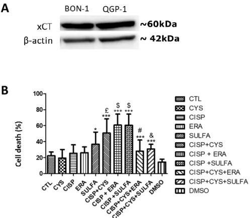
4. RESULTS 4.1.CISPLATIN INDUCES CELL DEATH IN BON-1 BUT NOT IN QGP-1 CELL LINE ... 19
4.2. INHIBITION OF xCT TOGETHER WITH CYSTEINE CONTRIBUTES FOR CISPLATIN RESISTANCE OF BON-1 CELL LINE ... 19
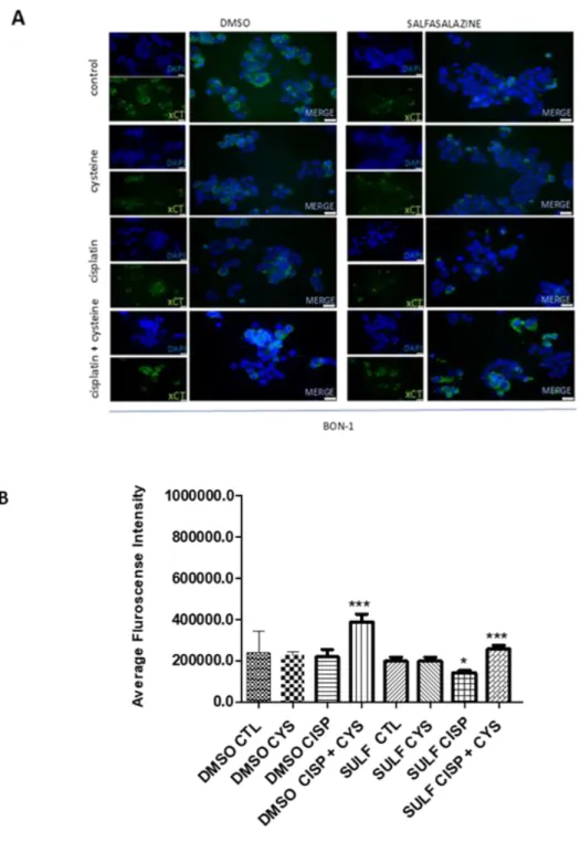
4.3. SULFASALAZINE COMBINE WITH CYSTEINE AND CISPLATIN STIMULATES THE EXPRESSIONOF xCT ... 21
4.4. SeChry BUT NOT SeChry ENCAPSULATED AND FUNCTIONALISED WITH FOLATE (SeChry@PUREG4-FA), INDUCES CELL DEATH IN PNETs CELL LINES ... 23
4.5 BUTHIONINE SULFOXIMINE ENCAPSULATED AND AND FUNCTIONALISED WITH FOLATE (BSO@PUREG4-FA), INDUCES CELL DEATH IN PNETs CELL LINES ... 23
4.6 FAILURE IN THE TRANSFECTION OF BON-1 CELL LINE WITH NRAS, KRAS AND BRAF MUTATED VARIANTS (BRAF- V600E, KRAS-G112V, NRAS-C161R) ... 24
4.7 FAILURE IN THE OVEREXPRESSION OF XCT ... 25
5. DISCUSSION ... 26 6. CONCLUSIONS... 32 7. FUTURE PERSPECTIVES... 33 8. BIBLIOGRAPHY ... 34 9 APPENDICES ... 47 INDEX OF FIGURES Figure 4.1. - The effect of cysteine, cisplatin and cysteine plus cisplatin, in BON-1 and QGP-1 cell lines ... 18
Figure 4.2. -- The effect of erastin, sulfasalazine and cysteine in the response of BON-1 cell line to cisplatin ... 19
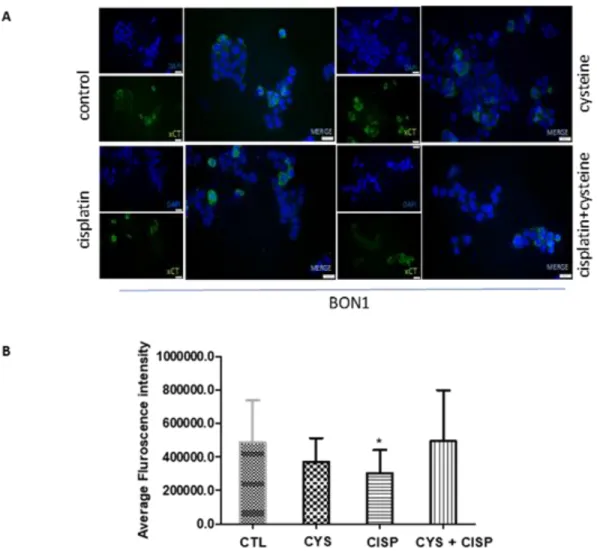
Figure 4.3. Cysteine, cisplatin, cysteine combined with cisplatin do not affect xCT protein level, in BON-1 cell lines ... 20
Figure 4.4. The effect of cysteine, cisplatin, cysteine combined with cisplatin and sulfasalazine in xCT protein level, in BON-1 cell lines ... 21
viii Figure 4.5 SeChry, but not SeChry encapsulated and functionalised with folate (SeChry@PUREG4-FA), induces cell death in PNETs cell lines ... 22 Figure 4.6 Buthionine sulfoximine encapsulated and functionalised with folate (BSO@PUREG4-FA) induces cell death in PNETs cell lines ... 23 Figure 4.7. p-Erk levels indicate that MAPK pathway has a lower, but non-significant, flow in transfected cells ... 24 Figure 5. Cooperation between different cystine/cysteine transporters ensures chemoresistance ... 29
ix
ABSTRACT
Cancer is characterised as a set of diseases that is involved in uncontrolled cell growth with the ability to invade or spread to the other part of the body. Carcinogenesis is recognized as a process through aggregation of genetic and epigenetic changes in normal cell that ultimately leading to unlimited growth proliferation and invasion. Pancreatic neuroendocrine tumour (PNET) is a rare tumour that arise from neuroendocrine gland, occurs in various part of the body. The prevalence rate of PNETs is near about 25–30 per 100,000 population in the United States and according to Surveillance Epidemiology and End Results (SEER), the incidence rate of PNETs increased five-fold from 1973 to 2011. PNETs comprises approximately 7% of all types of cancer in the pancreas. Normally, 5 years survival rate of PNETs near about 42%.
PNETs is a heterogenous group of disorder with less 5 years survival rate due to lack of effective therapeutic options for patients with advanced stages, absence of symptomatology specially in case of non-functional PNETs and also to the phenomenon of chemoresistance, dependent on multiple mechanisms. Recent data shows that incidence rate of this tumour increases as a result of germline genetic mutations. Concerning to genetic change, it is very important to explain the differences that occurs at the level of chemoresistance. The treatment plan of the PNETs varies on type, location and aggressiveness of the tumour. Surgery is the only curative treatment in early stage but in advanced stages chemotherapy and radiotherapy are the most palliative treatment option of PNETs. Chemotherapy which is mainly based cisplatin combined with capecitabine and the response rate of treatment is near about 30%. Cisplatin is responsible for the formation of DNA adducts, leading to DNA damage, and induces generation of ROS, that consequently leads to oxidative stress, cell damage and death. Glutathione (GSH) plays an important role in the maintenance of intercellular redox balance and detoxification. Chemoresistance can be based on the alteration of the detoxification mechanisms and GSH system has been pointed as one of the most important. Cysteine is a rate limitant substrate for GSH synthesis, and xCT cyst(e)ine transporter is implicated in cancer severity and chemoresistance.
The Hypothesis of the project is: the disruption of xCT and uptake of cysteine leads to the reversal of resistance to alkylating agents in pancreatic neuroendocrine tumours (PNETs).
To accomplish the hypothesis we defined 3 aims: 1st aim will be to address the expression of xCT
in PNETs cell lines, and the modulation of xCT expression by cysteine and cisplatin; 2nd aim will
be focused on the effect of xCT inhibition in PNETs cell death, using erastin and sulfasalazine, and
3rd aim will be focused on the effect of new nanoformulations in order to disturb cysteine uptake
x
Our work allowed to reveal the role of xCT transporter and the role of cysteine in PNETs cell line resistance. This cell line showing different response patterns in cysteine transporters activity helped to reveal the differences of the transporter in chemoresistance mechanism. It also showed that besides xCT transporter other cysteine transporter such as EAAT3 also appeared to be involved in the dynamics of chemoresistance mechanism.
This work was also important to undercover the effect of new nanoformulations in order to disturb cysteine uptake by using SeChry and GSH synthesis by using BSO in PNETs cell lines. SeChry, but not SeChry@PUREG4-FA, induced cell death in BON-1 cell lines. SeChry cytotoxicity
can be selective for cancer cells and this was taken in consideration in our new strategy by using SeChry@PUREG4-FA, however the assay was not successful and new markers for targeted delivery
must be investigated in PNETs.
BSO@PUREG4-FA induced cell death in combination with platinum salts in PNETs cell lines.
Possibly, the use of folate functionalised particles will help to bypass the critical step in the non-specific delivery of BSO to non-cancer cell. The targeted BSO delivery to cancer cells can be explored as a novel strategy in cancer therapeutics.
Moreover, more assays with cancer and non-cancer cells must be done in order to determine if folate receptor is in fact a suitable target to delivery drugs to PNETS cells, and find new and more specific targets.
Key-Words: Pancreatic neuroendocrine tumours (PNETs), platinum drugs, GSH, cysteine, xCT, ,
xi RESUMO
O cancro é caracterizado como um conjunto de doenças envolvidas no crescimento descontrolado das células, com capacidade de invadir ou se espalhar para a outras partes do corpo. A carcinogénese é reconhecida como um processo de agregação de alterações genéticas e epigenéticas nas células normais levando ao aumento da proliferação e invasão. O tumor neuroendócrino pancreático (PNET) é um tumor raro que surge da glândula neuroendócrina e ocorre em várias partes do corpo. A taxa de prevalência de PNETs é de cerca de 25 a 30 por 100.000 habitantes nos Estados Unidos e, de acordo com a Surveillance Epidemiology and End Results (SEER), a taxa de incidência de PNETs aumentou cinco vezes entre 1973 e 2011. Os PNETs representam aproximadamente 7% de todos os tipos de cancro no pâncreas. Normalmente, a taxa de sobrevida dos PNETs em 5 anos é de cerca de 42%.
Os PNETs são um grupo heterogéneo de distúrbios com sobrevida inferior a 5 anos, devido à falta de opções terapêuticas efetivas para doentes com estágios avançados, ausência de sintomatologia especialmente no caso de PNETs não funcionais e também ao fenómeno de quimiorresistência, dependente de múltiplos mecanismos. Dados recentes mostram que a taxa de incidência deste tumor aumenta como resultado de mutações genéticas na linha germinativa. No que diz respeito às alterações genéticas, é muito importante explicar as diferenças que ocorrem no nível de quimiorresistência. O plano de tratamento dos PNETs varia de acordo com o tipo, localização e agressividade do tumor. A cirurgia é o único tratamento em estágio inicial, mas em estágios avançados a quimioterapia e a radioterapia são a opção de tratamento mais paliativo dos PNETs. A quimioterapia que é principalmente baseada em cisplatina combinada com capecitabina tem uma taxa de resposta de tratamento aproximadamente de 30%. A cisplatina é responsável pela formação de adutos de DNA, levando a danos no DNA, e induz a geração de ROS (espécies reativas de oxigénio), que consequentemente leva ao stresse oxidativo, danos celulares e morte. A glutationa (GSH) desempenha um papel importante na manutenção do equilíbrio redox intercelular e na desintoxicação. A quimiorresistência pode ser baseada na alteração dos mecanismos de desintoxicação e o sistema GSH tem sido apontado como um dos mais importantes. A cisteína é um substrato limitante da taxa para a síntese de GSH, e o transportador de cisteína xCT está implicado na gravidade do cancro e na quimiorresistência A hipótese do projeto é: a interrupção do xCT e a captação de cisteína levam à reversão da resistência aos agentes alquilantes nos tumores neuroendócrinos pancreáticos (PNETs).
Para testar a hipótese, definimos três objetivos: o primeiro objetivo será abordar a expressão de xCT nas linhas celulares de PNETs e a modulação da expressão de xCT por cisteína e cisplatina; O
xii segundo objetivo será focado no efeito da inibição do xCT na morte celular de PNETs, usando
erastina e sulfassalazina, e o terceiro objetivo será focado no efeito de novas nanoformulações para perturbar a captação de cisteína (Sechry e Sechry @ PUREG4-FA) e asíntese de glutationa (BSO @ PUREG4-FA).
Este trabalho permitiu revelar o papel do transportador xCT e o papel da cisteína na resistência das linhas celulares de PNETs. Estas linhas celulares mostrando diferentes padrões de resposta na atividade dos transportadores de cisteína revelaram as diferença no mecanismo de quimiorresistência e mostraram que, além do transportador xCT, outro transportador de cisteína, como o EAAT3, também poderá estar envolvido na dinâmica do mecanismo de quimiorresistência.
Este trabalho também foi importante para desvendar o efeito de novas nanoformulações, a fim de perturbar a captação de cisteína usando a síntese de SeChry e GSH usando BSO nas linhas celulares de PNETs. SeChry, mas não SeChry @ PUREG4-FA, induziu a morte celular em linhas celulares BON-1. A citotoxicidade de SeChry pode ser seletiva para células cancerígenas e isso foi levado em consideração na nossa nova estratégia usando o SeChry @ PUREG4-FA; no entanto, o ensaio não foi bem-sucedido e novos marcadores para entrega direcionada devem ser investigados nos PNETs.
BSO @ PUREG4-FA em combinação com sais de platina nas linhas celulares de PNETs induziu morte celular. Possivelmente, o uso de partículas funcionalizadas com folato ajudará a contornar a etapa crítica na entrega inespecífica de BSO às células não cancerígenas. A entrega direcionada de BSO às células cancerígenas pode ser explorada como uma nova estratégia na terapêutica do cancro.
Além disso, mais ensaios com células cancerígenas e não cancerígenas devem ser feitos para determinar se o receptor de folato é de facto um alvo adequado para administrar drogas às células de PNETs e encontrar novos e mais específicos alvos.
Palavras-chave: Tumores neuroendócrinos pancreáticos (PNETs), drogas de platina, GSH,
1
1.INTRODUCTION
1.1 Cancer Biology:
The word cancer came from Greek, karkinos discovered by physician Hippocrates (460–370 B.C) to elucidate the carcinogenesis.1 Cancer can be defined as a group of diseases characterised by
abnormal cell division and alteration of normal cell behaviour leading to limitless growth of cell and proliferation.1 In the developed countries, cancer remains the second leading cause of death,
underlying approximately 10,000,000 deaths around the world.2
Cancer has some specific biological capabilities, justifying the complexity of neoplastic disease, called “Hallmarks” of cancer. The hallmarks of cancer are: sustained proliferative growth signals; evasion of antigrowth signalling; resisting programmed cell death; immense replicative capacity; induced angiogenesis, and capacity of cancer cell spreading - tissue invasion and metastasis.3
Reprogramming of energy metabolism and evading immune destruction were added as emerging hallmarks of cancer in the last decade .3
In 1956, Otto Warburg established for the first time a connection between alterations in metabolism and cancer cells. He stated that a higher aerobic glycolysis was a distinctive feature of non-cancer and cancer cells, known as the Warburg effect.4,5,6,7 Other evidence revealed that
continuous aerobic glycolysis helps the cancer cell to activate oncogenes.6,7,8 Additionally, the
cancer cells grow like a mass with low amounts of nutrient and oxygen that prompt the growth of new vessels that helps the tumour to proliferate rapidly.9
The cancer cell microenvironment gives signals that lay into the activation of signalling pathways, which ultimately activate transcription factors. Besides cancer cells, stromal or non-malignant cells, permitting the mesenchymal phenotype that is necessary to invasion and metastasis distance; also contribute for a specifically required cancer cells transcriptional program.10 Upon
metastasis, cancer cells turn off this program and rescue an epithelial phenotype. It is thought that tumour microenvironment is playing a crucial role in this process.
1.2 Neuroendocrine tumours:
In 1969, Pearse identified APUD (amine precursor uptake and decarboxylation) peptide hormone secreting cells in the body that share common morphological features for balancing the embryological differences.11 APUD cells have autocrine, paracrine and neuromodulator function,
they are located throughout the body and mostly originated from neural crest.12
The main function of the neuroendocrine cells is to synthetize hormone in the cytoplasm then transport it to axons and from the nerve ending ultimately it is secreted into the vessels.13
Neuroendocrine tumours (NETs), characterized as rare tumours that arise from neuroendocrine cells, can occur in various organs of the body, including pancreas, gastrointestinal tract and lung.14 The main origin of neuroendocrine cells is not well known. These cells have the behaviour
2
that come from nervous system.15,16 NETs which stereotypically ascend in the pancreas are
called islet cell tumours.15,16 NETs can be benign or malignant and they are usually slow growing,
but sometimes they grow abruptly.
Symptoms and signs of the NET depend on some factors such as the types of the tumour, the size of the tumour, location of the tumour, the pattern in situ or spread and functioning state of the tumour such as hormone or non-hormone producing cells.15,16 Detection and progression of
NETs depend also on the traits of the neuroendocrine cells, because functional NETs may overproduce certain hormones inducing a constellation of symptoms related to the hormone that is being oversecreted, whereas non-functional NETs are not associated to a specific clinical syndrome .17
Most of the NETs are sporadic whereas some of these tumours are diagnosed in the context of to autosomal dominant hereditary cancer syndromes, such as: multiple endocrine neoplasia type 1 (MEN-1); Von-Hippel–Lindau; tuberous sclerosis and neurofibromatosis type 1.18
Treatment of NETs varies, as it depends on type, stage and aggressiveness of the tumour.19
Therapeutic approaches, such as surgery and medical therapies may differ between each type of NET.
- Neuroendocrine tumour cells can be divided into aggregations of cells (glands) and diffusely distributed dispersed cells (disseminated system) and both differ from one another embryologically. 20
Data from the Surveillance Epidemiology and End Results programme (SEER) including patients with NETs from 1937 to 2012 shows that the incidence of neuroendocrine tumours has markedly increased over past few years.21
1.2.1 Pancreatic Neuroendocrine Tumours
Epidemiology:
Pancreatic neuroendocrine tumours (PNETs) are a subtype of neuroendocrine tumour derived from the islet of Langerhans cells with various morphologies and behaviours, including malignant potential.22
In the United States, the prevalence of PNETs is approximately 25-30 per 100,000 population.30
It is reported that the incidence rate of PNETs according to the population based report from Europe and Asia is near about <1 per 100 000 persons per year.23,24,25 In addition, their incidence
in autopsy studies is higher compared to diagnosed cases, ranging from 0.8% to 10%.26 PNETS
can be functional or non-functional. Functional PNETs are characterised by a hormonal hypersecretion syndrome such as hypoglycaemia, hyperglycaemia, glucose intolerance, peptic ulceration, watery diarrhoea, hypokalaemia etc. Non- functional PNETs do not produce any symptoms, but they cause illness and death by occupying the normal tissue and metastasizing.27,28,29 Epidemiology of the PNETs is not well known and there is no difference of
3 Biology and Genetics:
In 2000, the World Health Organization (WHO) histologically classified GEP-NENs as: well differentiated endocrine carcinoma and poorly differentiated endocrine carcinoma/small cell carcinoma. However, in 2010 they categorized the Classification based on Ki-67 proliferation index and mitotic count, as: neuroendocrine tumour grade 1 (NET-G1); neuroendocrine tumour grade 2 (NET-G2), and neuroendocrine carcinoma.31 Mitotic count ranges from <2 to >29 per HPF
(high power field) and Ki-67 proliferation index <3% to >20% per HPF according to the grade of tumour.31
Genetic information provides a better knowledge for the development of new treatment agent in the field of research. Genetic differentiation between the different grades of tumour in PNETs also gives us useful information to better understand the biology of PNETs. In 2009 Capelli et al. reported MEN1 gene mutation as the most common hereditary irregularity in the germline PNETs causing biallelic inactivation due to the fact that a mutation in one allele induces the loss of the second allele. MEN1 gene is a tumour suppressor gene and its germline mutation influences the development of multiple endocrine neoplasia syndrome type 1.32 Mutations in ATRX
(α-thalassemia/mental retardation syndrome x-linked) and DAXX (Death -domain associated protein) genes are reported as the most common somatic events involved in PNETs. Both genes are associated with chromatin remodelling in the telomeres and the encoded proteins of the genes are networked with one another.33 In addition, based on the molecular pathway of poorly
differentiated PNETs, cancer cells can be derived from the normal neuroendocrine cells which acquired mutations of TP53 and SMAD4 concomitant with KRAS mutations.34
Therapy:
The therapeutic approach for the PNETs patients differs, depending on the type, location, stages and aggressiveness of the tumour. Generally, it is divided into three levels, such as surgery, anti-proliferative and symptomatic therapies.34
Imaging studies are used to evaluate the extent and the location of the tumour, so that proper management can be taken.35 From past to present day, surgery with regional lymph node
resection is the only curative treatment recommended for the early stage PNETs and is specially used to prevent complications in PNET patients.36 However, in advanced stages, surgery is usually
not feasible and systemic therapy becomes the main treatment option. However, in patients with advanced PNETs and liver metastasis, partial hepatectomy may be recommended, depending on the patient response, size and location of primary tumour.36 Chemotherapy and radiotherapy are
the most common palliative approaches to advanced stage patients, when the liver is not resected.37
4 1.3 Chemotherapeutic agents:
Platinum based chemotherapy is the most frequently used chemotherapeutic strategy. Platinum drugs such as cisplatin, carboplatin or oxaliplatin are commonly combined with taxanes (paclitaxel or docetaxel). Cisplatin is a first line chemotherapeutic agent with the highest side effects. Carboplatin is the second-generation antineoplastic agent containing platinum. The main differences between carboplatin and cisplatin are that carboplatin has a cyclobutanedicarboxylate group, being more stable but to achieve the same effect as cisplatin this drug need to be applied in a higher concentration. Within this group of drugs, carboplatin is the best option as a neoadjuvant, adjuvant and palliative treatment because of its less side effects.38,39
Chemotherapy is the treatment for the advanced stage with unresectable pancreas tumour, resulting in an increase in overall survival of patients.40 When a patient shows symptomatic
features and/or has a tumour with a ki-67 value more than 10%, cytotoxic chemotherapy is well-thought-out as a first line of treatment. Two different groups of medications are utilized for the treatment purpose of well differentiated PNETs. They are alkylating agents such as platinum salts, Streptozocin (STZ), temozolomide (TEM), dacarbazine (DTIC) and antimetabolite agents such as 5-flurouracil (5FU), capecitabine (CAP) and doxorubicin (DOX). Nowadays, combination of STZ and 5FU is prescribed commonly instead of STZ and DOX combination. The limitation of use of DOX is due to cardiotoxicity with a dose of 50 mg/m2, as reported.41 STZ is a diabetogenic
substance, which is selectively incorporated into the beta cells of the pancreas via GLUT2 transporters that leads to cytotoxicity and the generation of reactive oxygen species (ROS). On the other hand, another combination of drugs, TEM combined with CAP, is recommended as an alternative treatment in well differentiated PNETs patients. In advanced cases of neuroendocrine tumours / neuroendocrine carcinomas, cisplatin-based chemotherapy is standard (generally, cisplatin and etoposide).41
1.4 Chemoresistance:
Chemoresistance is one of the most challenging issues in the field of cancer treatment. Consequently, to find out novel therapeutic approaches it is vital to know the molecular mechanisms of the chemoresistance, because it causes recurrence, relapse and death.42
Chemoresistance can be divided into two categories, one is intrinsic (de novo or innate) and another is acquired resistance.43
Intrinsic resistance is characterized as a circumstance when chemotherapy is not enough from the beginning of the treatment because of the patient endogenous component, on the other hand, when the tumour grows slowly after exposure of anticancer medications, because of epigenetic or molecular adjustment of malignant cells, it is known as acquired resistance.44 There
are many diverse mechanisms involved in chemoresistance such as alterations of drug transport pump, modification of drug target interaction, enhanced DNA repair activity, augmented drug damage tolerance, effect on protein associated with detoxification process and imperfection in
5
apoptotic pathway.45 In advanced stages of pancreatic cancer, chemotherapy is the most
effective treatment. Nevertheless, many patients lean towards the relapse of disease and chemoresistance.46
To understand the chemoresistance mechanism related to transport pumps, it is important to focus on p-glycoprotein. P-glycoproteins are membrane proteins, including Multidrug resistance protein 1 (MDR1) or ATP binding Cassette (ABC) protein, acting as an ATP -dependent efflux pump. These proteins cause decreased drug intracellular accumulation by altering the intracellular pH.38 There are some other transporters such as Breast Cancer Resistance Protein
(BCRP) and Major Vault Protein (MVP). BCRP bearing circulating microvesicles contribute to the mechanism of chemoresistance in breast cancer.47 MVP possibly will facilitate chemoresistance
by controlling the nucleo-cytoplasmic transport (hormones, ribosomal RNAs, drugs).38
Modification of drug target expression or mutation causes alterations in drug targeting that
ultimately leads to drug resistance in cancer. For example, a mutation in the topoisomerase II gene leads to reduced efficacy of topoisomerase inhibitors, affecting DNA synthesis, DNA damage and halting of mitotic processes. Some anticancer drugs target signalling kinases such as epidermal growth factor receptor (EGFR), Ras, Src, Raf, and MEK. In addition, some of these kinases are constantly active in certain types of cancer, leading to uncontrolled cell growth.48
In the field of cancer drug resistance, enhanced DNA repair and tolerance of damaged DNA are thought to be one of the possible mechanisms of chemoresistance. The role of chemotherapeutic agents is to damage the DNA of cancer cells indirectly or directly and DNA repair mechanisms fix the damage of the DNA49. It is known that platinum based chemotherapeutic agents cause
apoptosis of cancer cell through damaging the DNA.50
It is thought that polymerase enzymes play an important role in the process of drug tolerance. The normal DNA replication process requires the action of error free DNA polymerase. Moreover, when DNA injury cannot be repaired, in order to sustain the cell survival, the injury should be tolerated, and this can be accomplished by translational synthesis pathway catalysed by DNA Polymerase.51
Drug damage tolerance is associated with reduced susceptibility to apoptosis. Catalytic caspases,
which are associated to a group of cysteine-dependent aspartate-directed proteases, have their activity dependent on the formation of tetrahedral intermediates via promoting a cysteine residue that is linked with apoptotic activity and signalling. In addition, genes that regulate apoptosis are altered in this mechanism, accounting for apoptosis inhibition.52,53
Effect on proteins associated with the detoxification process, as glutathione and its related
enzymes, is another chemoresistance mechanism. Glutathione plays an important role in the process of detoxification. Glutathione-s-transferase (GST) superfamily network is an important example of drug activation and inactivation process. GST contributes for the progress of drug resistance mechanism by direct detoxification and through inhibiting the pathway of mitogen activated protein kinase (MAPK).48 The higher GSH level with over expression of
glutathione-s-6
transferase (GST) will increase the proportion of conjugation and detoxification reduces the effectiveness of therapeutic agents.54 Cytochrome P450 (CYP) is also involved in the process of
drug activation and inactivation. CYP is divided into two classes: Class I includes CYP1A1, CYP1A2, CYP2E1, CYP3A4 and Class II includes CYP2B6, CYP2C9, CYP2C19, CYP2D6. Class II has a characteristic feature of high polymorphism rate that is involved in the metabolism of cancer drugs and in drug resistance mechanism.48 Uridine diphosphate- glucuronosyltransferase (UGT) superfamily, which is a group of transferase enzymes involved in glucuronidation catalysis, regulating the formation of inactive hydrophilic glucuronides with substrates together with environmental carcinogens and cytotoxic. The expression of UDP glycosyltransferase family -1, member 1 (UGT1A1) thought to be negatively regulated by DNA methylation meanwhile due to epigenetic changes the overexpression of UGT1A1 is common in cancerous state, that ultimately leads to drug detoxification.41
Programmed cell death is markedly influenced by a variety of genes, some of them are mutated
or difunctionally regulated. Defective cell death can occur as a result of aberrant regulation of TP53 and PTEN, inhibitors of the PI3K/AKT pathway, which might impair the affiliation between DNA damage and the activation of programmed cell death, increasing chemoresistance and leading to drug induced apoptosis failure.54 TP53 inhibits PI3K ultimately abolishing the
mitochondrial p53 -dependent apoptosis. PTEN is an inhibitor of PI3K for the synthesis of PIP3 by phosphorylation, which will activate AKT. When PTEN in downregulated or inhibited, PI3K will be activated, PIP3 will be synthesized and AKT activated.55,56 Apoptosis is activated by the cleavage
of executioner caspases (eg. 3 or 7). There are two routes to apoptosis: extrinsic (or death receptors) and intrinsic or (mitochondrial).57,58 Extrinsic pathway is regulated by the action of FAS
and TNF- on the death receptors and it is mainly dependent on caspase 8 activation that will cleave the executioner caspases. Within the external pathway mutations, epigenetic or post-translational alterations may affect proteins function, which ultimately lead to the inhibition of cell death in cancer cells.57,58,59 Within the intrinsic pathway the up regulation of the
anti-apoptotic genes (eg. Bcl2) and down regulation of pro anti-apoptotic genes (eg. Bax, Bclxl) in tumour
cells lead to resistance to chemotherapeutic agents.60 XIAP is also an inhibitor of caspases 3, 7
and 9, affecting both apoptosis pathways.57TP53 is the master regulator of apoptosis, being an
activator of both intrinsic and extrinsic pathways, in cancer deregulation and mutation of TP53 affects apoptosis through the inhibition of mitochondrial and death receptor signalling. 57,61
1.5 Cancer metabolism.
The basic principle of cancer metabolism is that metabolic activities are changed in the cancer cells compared to normal cells. In 2011, Weinberg et. al. added a new hallmark of cancer that is reprogramming of cancer metabolism. The metabolic reprogramming has a reflective effect on gene expression, cellular proliferation and tumour microenvironment.54 Otto Warburg in 1923,
first made the connection between cancer and metabolism in the basis of the concept of Warburg effect, assuming that cancer cells would preferentially fulfil glycolysis instead of oxidative
7
phosphorylation (OXPHOS).62 Metabolism not only is related to intracellular network, but is also
related with extracellular organic and communication molecules, which control the complete metabolic function of the cell. Fibroblasts are the most important type of tumour stromal cells, playing a vital role in the process of cancer cell progression through their molecular cooperation.63 CAFs (cancer associated fibroblasts) and neoplasm cells permit an enhanced
microenvironment, vital for neoplasm survival, proliferation, migration and chemoresistance.64,65
CAFs are crucial in the metabolic process to organize the inner source of nutrients providing the Krebs cycle with metabolic intermediates.66 Fatty acids are major contributors for tricarboxylic
acid (TCA) cycle, that is required for the generation of nicotinamide adenine dinucleotide (NADH), vital for OXPHOS.63 Reactive oxygen species (ROS) are short lived molecules with unpaired
electrons that are constantly generated, modified and eliminated during the cellular processes together with metabolism, proliferation, differentiation, processes of immune system regulation and remodelling of vascular system; but the excess amount of ROS causes oxidative stress and altered cancer metabolism.67 Excess production of ROS causes alterations of sulfhydryl groups
of private kinase M2 (PKM2) leading to shunting of glucose away from glycolysis towards the pentose phosphate pathway (PPP), which produces NADPH and nucleotides, favouring cell proliferation.72 The NADPH reduces glutathione into an active antioxidant, functioning as a cell
protector and maintain the redox homeostasis.68,69
1.5.1 GSH and Cysteine:
Glutathione (GSH) and cysteine are both thiols, having a sulfhydryl or thiol group, made of sulphur and hydrogen atoms attached to a carbon. GSH is a low molecular weight thiol composed of glutamic acid, cysteine and glycine. GSH is very reactive and can conjugate with another molecule via sulfhydryl group. The bulk of GSH found intracellularly within the cytoplasm is approximately 90%, about 10% is found in mitochondria and a little amount is found in the endoplasmic reticulum.70 GSH is synthesized by two ATP requiring enzymes glutamate cysteine
ligase (GCL) and GSH synthetase (GSS). The synthesis of GSH is catalysed by GCL which have two subunits, one is GCLC (glutamate cysteine ligase catalytic subunit) that catalyses the linkage between the amine group of cysteine and the γ-carboxyl group of glutamic acid; and another is GCLM (glutamate cysteine ligase modifier) that increases the catalytic activity by interacting with GCLC. GSH synthetase catalyses the reduction of gamma-glutamylcystein and glycine to create GSH.71 Then this gamma-glutamylcystein can be transported back into the cells and again
metabolized into 5-oxoproline, ultimately converted to glutamate and used in GSH synthesis. The rate of GSH synthesis largely depends on cysteine availability and the activity of GCL. Cysteine plays a key role in the maintenance of protein structure and synthesis. The proper concentration of intracellular cysteine also helps to maintain the redox homeostasis.72
GSH is the most reduced form, which is being oxidized in the form of GSSG via its direct reaction with ROS or substitute as a co-enzyme for antioxidant like glutathione peroxidase (GPX) and recycled by glutathione reductase (GR).73,74,75 GPX is the member of the family of selenium
-8
dependent enzymes, which can differ from transferases because of its activation with hydrogen peroxide (H2O2). GPX enzymes use glutathione like a ROS scavenger, that convert H2O2 to water
and lipid peroxides.75,76 Glutathione plays an important role in the maintenance of redox balance
and protein status. The glutathione reaction with the protein varies with the concentration of GSH and GSSG. But the reversible thiolation reaction of proteins is responsible for the regulation of numerous metabolic processes such as enzymatic activity, signal transduction and gene expression with the help of redox sensitive nuclear transcription factor like AP-1, NF- kappa B and TP53.77,78
Another important function of GSH is the storage of cysteine because it is unstable extracellularly and rapidly oxidized to cystine through a process that produces toxic ROS. The -glutamyl cycle helps GSH in this procedure of releasing cysteine. In this cycle, GGT transfers the -glutamyl to an amino acid to form γ-glutamyl-aa and cysteinyl-glycine. GSH plays an important role not only in the protection of cells against apoptosis but it also has an extreme key role in detoxification process, and its disruption will activate diverse transcription factors like activator protein -1 (AP-1), activator protein -2 (AP-2), stress activated protein kinase (SAPK) and c-Jun-N -terminal kinase (JNK). Besides, GSH acts as an antioxidant .71,79, it is also involved in DNA repair and synthesis and
prostaglandin synthesis.80 Increased levels of GSH are considered as a causative factor of drug
resistance through binding to or interacting with the drug or ROS, preventing harm to protein or DNA or by taking part in the DNA repair process.68 However, specific GSTs overexpression can
even influence chemoresistance, while polymorphisms which reduce GST activity are related to a high risk of developing cancer.73 In combination, it will also increase the percentage of
conjugation and detoxification of chemotherapeutic agents, that ultimately reduces their effectiveness .81 Higher expression of GST can switch the balance of kinases that will cause a
potential benefit for cancer development.73
1.5.1.1 Cysteine metabolism
Cysteine is a semi essential amino acid which is mainly provided through diet or through the trans-sulphuration pathway. Serine and methionine-derived homocysteine reaction are catalysed through cystathionine-β-synthase (CBS) to form cystathionine, then the pathway is facilitated by cystathionine--lyase (CSE) and catalyses its final step through the conversion of L-cystathionine to cysteine and α-ketobutyrate. 82
Cysteine degradation is also a crucial metabolic reaction. Multienzymes are involved in this procedure. The process of sulfane sulphur generation from cysteine, homocysteine and their disulphides is carried out through three enzymes that are CBS, CSE and 3-mercapto-pyruvate sulphur transferase (MpST) along with cysteine aminotransferase (CAT). CSE is responsible for catalysing the conversion of L-cystine to thiocysteine, pyruvate, and ammonia. After that, thiocysteine in the presence of thiol forms cysteine and inorganic sulphur from hydrogen sulphide (H2S) and cystine.83 MpST enzyme is responsible for the relocation of sulphur ion from
9
and are responsible for mitochondrial ATP production dependent on H2S, while CBS and CSE are
repositioned in mitochondria under cellular stress.85 L-cysteine is also degraded by the help of
cysteine dioxygenase (CDO). Cysteine dioxygenase is accountable for adding an oxygen molecule to sulphur of cysteine, which is converted to sulfinic acid, known as cysteine-sulfinic acid. After that, cysteine-sulfinic acid is again metabolised into either one pyruvate and sulfate or to taurine and CO2.86
It has been reported through literature that cysteine is a tumorigenic promoter. Pancreatic cancer cells need an exogenous source of cysteine because of its crucial role in redox homeostasis contributing for tumour growth and maintenance. Cancer cells depend on oxidized cysteine, which detoxifies the lipid ROS and prevents ferroptosis. 87
1.5.1.2 Cyst(e)ine /Glutamate transporter: Cysteine transportation is mediated by alanine
-serine-cysteine transport system (ASCT), excitatory amino acid transporters (EAATs) system and the Xc- (xCT) system. ASCT system includes ASCT1 and 2, in which Na+ dependent mediated
transport exchanges small neutral amino acids like alanine, serine, threonine and cysteine and structure of the transport. EAATs mediate the transport of amino acids along with the co -transport of K+ via producing a net flux of charges. xC- system depends on xCT transporter, a
glutamate and cystine (the oxidized form of cysteine) antiporter that relies on proton electrochemical gradient.88,89
1.5.1.2.1 System xC-(xCT): System xC- was first explained by Bannai and Kitamura in 1980 in
culture of human fetal lung fibroblasts, which work as a sodium independent and chloride dependent antiporter system, being capable of transporting an anionic form of cysteine and glutamate in both directions.90,91,92 Cystine is the oxidised form of cysteine, which is rapidly
reduced and therefore the intracellular glutamate concentration is mostly elevated comparing with the extracellular space, this mechanism basically relies on the concomitant cysteine import and glutamate export.93
The xCT transporter is encoded by solute carrier family 7-member 11 (SLC7A11) gene, located on chromosome 4 (4q28.3). xCT/SLC7A11 gene has an antioxidant responsive element region (ARE) in its promoter that is controlled by the regulation of nuclear factor erythroid 2-related factor 2 (NRF2), which plays an important role in cellular protection against oxidative stress. In 2018, Carpi-Santos et al. found that alterations in xCT expression affected the retina of diabetic rats via regulation of NRF2.94,95 Meizi et.al made an experiment with the aim of making a link between
sepsis illness and protein interaction with C-kinase 1 (PICK1). Through this experiment they found that deficiency of PICK1 not only causes the inhibition of xCT expression, but also reduces the GSH synthesis, which ultimately leads to severe oxidative stress.96 xCT activity also has a role in
cancer. In invasive breast cancer and oesophageal squamous cell carcinomas, expression of xCT is negatively correlated with survival.97 Besides overall function, it is also reported that system
10
xCT also plays an important role in the immune system. xCT overall acts as an anti-oxidant, it contributes for cysteine uptake and GSH synthesis, ultimately solving the oxidative stress.88,98
xCT transporter is made of a light chain (xCT) and a heavy chain (4F2hc) and both chains are connected through a disulphide bridge. However, the heavy chain is responsible to link with specific amino acids of the light chain to form a heterodimer.74,88,99 In the cell membrane, xCT is
necessary to uptake the enough cysteine that is required for the GSH synthesis, which acts as an antioxidant for the maintenance of intracellular redox balance.93,100 xCT is not only highly
expressed in a variety of cancer cells like breast cancer, prostate cancer, lymphoma and glioma, but it is also engaged in various cellular functions in cancer cells like chemoresistance.
101,102,103,104,105,106 It is known that neurotransmitter glutamate depends on xCT, so the role of xCT
is well known in brain or central nervous system (CNS). It has been found that CNS dysfunction, accompanied by neural oxidative stress and accumulation of glutamate in the extracellular space, causes the reduction of cysteine import in the cell and increases the production of ROS which ultimately leads to excitotoxicity.87 It is also observed that glioblastoma cells show higher
expression of xCT.88,107
xCT transport system has a role in cancer context since it causes increased levels of GSH, involved in the process of chemoresistance.108,109 But the inhibition of xCT function also interferes with its
expression. Sulfasalazine and Erastin are well known inhibitors of this transport system.108,110
Sulfasalazine is a drug which is used in inflammatory bowel disease and rheumatoid arthritis.
Approximately 90% of sulfasalazine reaches the colon and it is metabolized into sulfapyridine and mesalazine by the help of bacteria.111 Recently, it has been appointed as an inhibitor of xCT, but
the mechanism of action remains unclear.88 A recent study found that sulfasalazine along with
autophagy-inducing agent temozolomide produce higher efficacy of chemotherapy drug in glioma treatment.112 It is known that caveolin-1 pathway is involved in the process of tumour
metastasis. Some recent experimental data provided evidence that sulfasalazine by interfering with the pathway of caveolin-1 causes the reduction of GSH levels, which ultimately reduces tumour metastasis in oesophageal squamous cell carcinoma.113 Data collected from current
studies demonstrate that xCT has a major role in pancreatic cancer via enhancing the GSH synthesis, whereas sulfasalazine known as a nontoxic drug combined with gemcitabine initiates an effective treatment in pancreatic cancer. 114
Erastin is a small molecule binding with voltage-dependent anion channels (VDAC) which are
responsible for the initiation of iron dependent cell death known as ferroptosis. Erastin acts as an inducer of ferroptosis by reducing cysteine uptake and diminishing the synthesis of GSH, ultimately maintaining the redox homeostasis.115 It was proven by some studies that RNA
interference mediated knockdown of VDAC2 or VDAC3 causes resistance to Erastin.116 VDAC2 or
VDAC3, which are the two isoforms of VDAC are involved in inhibiting the cysteine/glutamate transporter system. In addition, Erastin can inhibit the activity of GPX-4, which is a GSH related enzyme giving rise to oxidative damage to cells.117 It has a robust inhibitory effect on xCT activity
11
tumour cell death with harbouring mutations in RAS-RAF-MEK genes. They also found that this drug changed the permeability of the outer mitochondrial membrane, indicating that maybe Erastin induces apoptosis in cancer cells.116
1.6 New strategies to disturb xCT and cysteine /GSH metabolism by using selenium-chrysin (SeChry) and buthionine sulfoximine (BSO)
Selenium (Se), acts as a cofactor of mammalian enzymes, such as glutathione peroxidase, functioning as a great antioxidant.119 It is known that cancer cells are more sensitive to Se induced
cytotoxicity than non-cancer cells.120,121 Se produces ROS and instigates DNA strand breaks with
subsequent cell cycle arrest and apoptosis, leading to tumour growth suppression.122,123
Pro-oxidative effects of inorganic Se compounds were attributed to the antitumor mechanism, instead of antioxidative effects of organic selenium compound.124 Se is incorporated into
selenoproteins and the link between selenoproteins and cancer depends on the anti-oxidant properties of selenoproteins including the enzymes of glutathione peroxidase (Gpx) family, responsible for antioxidant protection against ROS.125 High ROS generation induces continuous
oxidative stress that provides expression of malignant phenotype of cancer cells.126 Se metabolite
conjugates to two glutathione (GSH) moieties, known as selenodiglutathione (SDG), increasing intracellular accumulation of Se. Evidences suggest that SDG in Se incorporation proceeds at cell surface by gamma-glutamyl transpeptidase (GGT), prompting the creation of selenocysteine, which is probably going to be exported through xCT. This mechanism could be arbitrated by xCT and may cause the Se induced cancer specific toxicity.119 As it is known, xCT transporter is related
to cancer drug resistance, in an effort to reduce the detoxifying activity through increasing the uptake of cysteine, accounting for GSh synthesis. Hence, selenated-compounds were taking in thought as potential xCT inhibitors.127,87 Selenium-chrysin (SeChry) is an organoselenium
compound composed of a selenium group and chrysin, a flavone, having tumoral and anti-oxidant properties, by protecting cells from toxic compounds and on the other hand, by inducing apoptosis.128,129,130
Buthionine sulfoximine (BSO) is a specific γ-glutamylcysteine inhibitor, developed by Griffith and Meister that reduced cellular GSH levels biosynthesis. BSO is a chiral compound and consists of two isomers, one is L-buthionine-(R)-sulfoximine and the other one is L-buthionine-(S)-sulfoximine. 131 L-buthionine-(S)-sulfoximine has a greater role in the inhibition of γ-GCS in
relation to is L-buthionine-(R)-sulfoximine in cell culture and in animals.132,133 It was utilized in
numerous investigations to reveal a decrease in GSH, sensitizing both cell lines and animals.134,135,136 As above-mentioned, GSH is the most commonly found intracellular thiol, its
depletion by BSO prompts oxidative stress, ultimately leading to increased radiation sensitivity.137 In addition, BSO sensitizes cancer cells to chemotherapy.138,139 Preliminary studies
from our group and studies from other teams revealed that continuous infusion of BSO give rise to the depletion of tumour GSH in patients with advanced cancers, specially ovarian cancer,140,141
12
Cancer cells express higher levels of folate receptor than normal cells 142,143 and folate receptor
targeted therapy is a way of directing nanoparticles to cancer cells both in vitro and in vivo.143,144
So, in an attempt to improve the specificity of SeChry and BSO towards cancer cells, folate receptor targeted delivery of these compounds was performed using SeChry@PUREG4-FA
nanoparticles as a drug delivery system.145,146,147
1.7 Metabolic profiles of PNET
Metabolic alterations in neoplastic cells is nowadays considered as a hallmark of cancer.3 In
addition, metabolic reprogramming accelerates the proliferation of cancer cells.148 Most of the
cancer cells depend on the amino acid glutamine, instead of glucose, to fulfil their biosynthesis need.149 As it was previously explained that PNET can exhibit mutations of TP53 with activating
KRAS mutations, it is thought that KRAS driven metabolic alterations might have a prominent role
in PNET proliferation. In pancreatic ductal adenocarcinoma, mutated tumour suppressor genes such as TP53, SMAD4 have a significant role in the process of carcinogenesis.150 It is proven that
elevated glycolysis flux causes a boost up the downstream pathway of KRAS. In the context of cancer, mutated KRAS transcriptionally upregulates the GLUT1 that ultimately causes elevated glucose uptake.151,152In glycolysis, pyruvate is converted to lactate upon the action of lactate
dehydrogenase enzyme (LDH). Mutated KRAS also has a role in increasing LDHA expression.116
1.7.1 Thiols (including GSH and Cysteine) metabolism in PNET: Thiols especially GSH interfere
with complex tumorigenic process. GSH conjugation of electrophilic carcinogens may prevent tumour initiation and defend cells against oxidative stress. It is thought that enhancement of antioxidants or conjugating capacity by increasing the levels of GSH, via precursor application, synthesis stimulation or inducing the related enzymes, may constitute a chemo preventive cell strategy.153
GPX-1 (GPX-1), which is a member of GPX family, is known as an enzyme that is related to tumour initiation and progression via modulating intracellular ROS by eliminating hydroperoxides.154,155,156 Interestingly, the relation between this enzyme and pancreatic cancer
is not well known, even though it was already proven by some experimental work that GPX-1 expression is directly linked to the initiation and progression of several malignancies such as breast, bladder and prostate cancer.157,158,159 It is thought that GPX-1 might have some role in
pancreatic cancer, although showing some controversy, as it seems that the abrogation of GPX-1 is related to pro-metastatic pathways dependent on ROS.160 However, in PNET there are no
studies on the role of thiols and related enzymes in disease progression. ROS have double roles, as hydroperoxides they promote tumour proliferation, migration and the process of angiogenesis, whereas excessive production causes cell apoptosis.161
Dietary source is considered as an etiological factor for pancreatic cancer. Therefore, some studies already proved that deficiency of dietary sources of methyl group like choline, methionine, vitamin B-12 and folate have a direct role in pancreatic dysfunction, in addition these elements have a role in developing various types of cancer including pancreatic cancer. 162,163,164
13
Folate has a methyl group, which is required for the intracellular methylation reaction. In folate metabolism methylene tetrahydrofolate reductase (MTHFR) catalyses the conversion of 5,10-methylenetetrahydrofolate to 5-methyltetrahydrofolate, which is the principle form of folate in the blood and acts as a co-substrate to form methionine from homocysteine. 165 Low levels of
folate are associated with the incorporation of uracil instead of thymine into DNA, which ultimately elevate the risk of DNA mutations and DNA strand breakage.166 Data collected from a
case control study conducted by Shirisha et. al showed the that percentage of direct DNA damage was higher in pancreatic cancer patients comparing to healthy individuals. 167 In addition, folate
pathway has an important role in the process of DNA synthesis and repair.166 It is thought that
altered folate metabolic pathway, might have some role in pancreatic cancer, as well as in PNET. Nevertheless, as far as we know, the knowledge about metabolic alterations in PNET, contributing for cancer progression and chemoresistance is scarce.
14
2. Hypothesis and Aims:
The Hypothesis of the project is: the disruption of xCT and uptake of cysteine leads to the reversal of resistance to alkylating agents in pancreatic neuroendocrine tumours (PNETs).
This thesis project has three main objectives:
First aim will be to address the expression of xCT in PNETs cell lines, and the modulation of xCT
expression by cysteine and cisplatin.
Second aim will be focused on the effect of xCT inhibition in PNETs cell death, using erastin and
sulfasalazine.
Third aim will be focused on the effect of new nanoformulations in order to disturb cysteine
15
3. Experimental procedures
3.1 Cell Culture
Two different pancreatic neuroendocrine tumours (PNETs) cell lines were used: BON-1 (CVCL_3985; JCRB cell Bank) and QGP-1 (CVCL_3143; JCRB cell Bank). BON-1 cell line was cultured in Dulbecco’s Modified Essential Medium/Nutrient mixture F- 12 (Ham) 1x (DMEM F-12) (11330-032, Gibco, Life Technologies) and QGP -1 cell line was cultured in Roswell Park Memorial Institute (RPMI ) 1640 Medium 1x (12-167F, Lonza, Bioscience), L-glutamine supplemented. Both mediums were supplemented with Fetal Bovine Serum 10% (FBS) (S 0615, Merck), Antibiotic-Antimycotic (AA) 1% (P06-07300, PAN Biotech) and Gentamicin 1% (15750-060, Gibco, Life Technologies). Cells were preserved under a temperature of 370C, 5% CO
2 in a humified
environment. The cells were cultured until 75% -100% optimal confluence before they were detached with 0.05% Trypsin- Ethylenediamine Tetra-acetic Acid (EDTA) (25300-054, Invitrogen, Thermo Fisher Scientific) at room temperature for 5min. Cell number was determined using a Bürker counting chamber.
For cell viability analysis, 2 x 10⁵ cells/mL were plated in 24 well-plates (500 µL/well) and for
western blotting 2.5 x 10⁵ cells/mL were plated in 6 well-plates (2 mL/well).
After 8 h of starvation (FBS free culture medium), cells were exposed to L-cysteine (0.402 mM); cisplatin (0.025 mg/mL), sulfasalazine (0.25 mM) and erastin (5 µM) isolated or in combination, in 1% FBS culture medium for 16 h.
In BON-1 data, a homozygous (AF 100%) g.114713908TOC mutation (ENSP00000358548 p.Q61R) was found in codon 61 in NRAS. Furthermore, a heterozygous g.25245350COA mutation (ENSP00000 308495 p.G12V) with an allelic ratio of 0.71 was originate in the KRAS gene in QGP 1. Data collected from (Whole -exome characterization of pancreatic neuroendocrine tumour cell
lines BON- 1 and QGP -1).
3.2 Flow Cytometry -cell death analysis
To evaluate the effect of cysteine (0.402 mM), cisplatin (0.025 mg/mL), erastin (5 µM), sulfasalazine (0.25 mM) and BSO@PUREG4-FA (456 µM) under (8h) starvation with 16h of
experimental conditions and in SeChry@PUREG4-FA (19 µM) without starvation with 24 h of
experimental conditions in PNET cell lines, cell death analysis by flow cytometry was performed. Flow Cytometry is a most commonly used technical method to measure the physical and chemical characteristics of cells. The most common typical feature of apoptotic cells is characterized by fragmentation and loss of nuclear DNA. Upon apoptosis the plasma membrane undergoes some structural changes and phosphatidyl serine (PS) is translocated onto the cell surface. Moreover, when the apoptotic process is started PS is translocated in the outer leaflet of the plasma membrane surface. Annexin V helps to identify the apoptotic cells by binding to PS. Red
16
fluorescent propidium iodide (PI) is also used to stain the necrotic cells. When the cell membrane is disrupted, it binds to double strand DNA by intercalating between base pairs, thus allowing PI entering into the cell and staining necrotic cells
.
168,169,170Furthermore, cells were collected. Supernatant from each well was collected to an Eppendorf and adherent cells were harvested with 100 µL of 0.05 % trypsin -EDTA (25300-054, Invitrogen), after that cells were collected to the same Eppendorf, in which the supernatant was at first collected. This way, it was ensured that all the cells that were in suspension in culture medium and adherent were analysed. Cells were centrifuged at 255 × g at room temperature for 2 min. The supernatant was discarded and cells were washed with PBS 1X - 0.1% BSA and stained with 0.5 µL FITC- Annexin V (640906, Bio Legend) (100 µg/mL) in 100 µL annexin V binding buffer 1X for each sample and then incubated in a dark place at room temperature for 15 mins. After that samples were rinsed with 100 µL PBS (1X) -0.1% of BSA, and centrifuged at 255 × g for 2 min. Cells were resuspended in 200 µL of annexin V binding buffer 1X 10 mM Hepes (pH 7.4), 0.14 M sodium chloride (NaCl), 2.5 mM calcium chloride (CaCl2). 2.5 µL of propidium iodide (PI; 50 µL/mL)
were added to all the samples, 5 min prior the acquisition in a FACS calibur (Becton Dickinson). Flow cytometry data was analysed by Flow Jo software.
3.3 Western Blotting
Western blotting is a widely used technique in cellular and molecular biology to identify the specific proteins from a complex mixture extracted from cells. In western blotting, through a sodium dodecyl sulphate polyacrylamide gel electrophoresis (SDS-PAGE) a mixture of proteins is separated, according to the molecular weight. After the electrophoresis, proteins are transferred, under an electric field, onto a nitrocellulose or polyvinylidene fluoride (PVDF) membrane and further incubated with an antibody specific for the protein we intend to identify. After the recognition of this antibody with a secondary antibody peroxidase conjugated and the chemo-luminescent development with the peroxidase substrate, luminol; a band for the protein of interest will be revealed.171
For western blotting the cells were cultured in 6 well-plate, afterwards cells were collected by scraping or by incubation of PBS (1X) + 2% EDTA. Then cells were centrifuged at 255 × g, cell extracts were performed by lysing cells with Radio-Immunoprecipitation Assay (RIPA) buffer and stored at -200C.
Cell lysates were centrifuged (Eppendorf) at 10600 × g for 5 min at 40C and supernatant were
collected. Protein concentration was measured through the Bradford method, using Bio-Rad protein assay reagent (500-0006, Bio-Rad) via spectrophometric quantification (595 nm). After quantification of protein, 50 µg of total protein from each sample lysate were loaded to the gel in 5X loading buffer (10% SDS, 0.5% bromophenol blue in Tris-HCL (pH 6.8) and 10% β-mercaptoethanol; M3148, Sigma), after protein denaturation under boiling at 950C -1000C for 10
min. After that, samples were centrifuged at 10600 × g for 2 mins, placed on ice and then samples were loaded in 12% acrylamide:bis acrylamide (29:1; 161-0156 Bio-Rad) gel (Tris-glycine