www.cbpv.org.br/rbpv ISSN 0103-846X (Print) / ISSN 1984-2961 (Electronic)
Doi: http://dx.doi.org/10.1590/S1984-29612017027
Gut transcriptome analysis on females of
Ornithodoros mimon
(Acari: Argasidae) and phylogenetic inference of ticks
Transcriptoma do intestino de fêmeas de
Ornithodoros mimon
(Acari: Argasidae) e inferência
filogenética de carrapatos
Gabriel Alves Landulfo1,2; José Salvatore Leister Patané3; Dalton Giovanni Nogueira da Silva1; Inácio Loiola Meirelles Junqueira-de-Azevedo4; Ronaldo Zucatelli Mendonca1; Simone Michaela Simons1;
Eneas de Carvalho5; Darci Moraes Barros-Battesti1,2*
1 Laboratório de Parasitologia, Instituto Butantan, São Paulo, SP, Brasil
2 Programa de Pós-graduação em Ciências Veterinárias, Departamento de Parasitologia Animal, Universidade Federal Rural Rio de
Janeiro – UFRRJ, Seropédica, RJ, Brasil
3 Laboratório de Bioinformática, Departamento de Bioquímica, Instituto de Química, Universidade de São Paulo – USP, São Paulo, SP, Brasil 4 Laboratório Especial de Toxinologia Aplicada, Instituto Butantan, São Paulo, SP, Brasil
5 Laboratório de Biotecnologia, Instituto Butantan, São Paulo, SP, Brasil
Received March 1, 2017 Accepted April 17, 2017
Abstract
Ornithodoros mimon is an argasid tick that parasitizes bats, birds and opossums and is also harmful to humans. Knowledge of the transcripts present in the tick gut helps in understanding the role of vital molecules in the digestion process and parasite-host relationship, while also providing information about the evolution of arthropod hematophagy. Thus, the present study aimed to know and ascertain the main molecules expressed in the gut of argasid after their blood meal, through analysis on the gut transcriptome of engorged females of O. mimon using 454-based RNA sequencing. The gut transcriptome analysis reveals several transcripts associated with hemoglobin digestion, such as serine, cysteine, aspartic proteases and metalloenzymes. The phylogenetic analysis on the peptidases confirmed that most of them are clustered with other tick genes. We recorded the presence a cathepsin O peptidase-coding transcript in ticks. The topology of the phylogenetic inferences, based on transcripts of inferred families of homologues, was similar to that of previous reports based on mitochondrial genome and nuclear rRNA sequences. We deposited 2,213 sequence of O. mimon to the public databases. Our findings may help towards better understanding of important argasid metabolic processes, such as digestion, nutrition and immunity.
Keywords: Argasidae, transcriptome, gut, phylogeny, Ornithodoros mimon.
Resumo
Ornithodoros mimon é um carrapato argasídeo parasita de morcegos, aves e marsupiais, além de ser bastante agressivo aos humanos. O conhecimento dos transcritos presentes no intestino dos carrapatos auxilia no entendimento do papel de moléculas vitais no processo de digestão e na relação parasito-hospedeiro, além de fornecer também informações sobre a evolução dos artrópodes hematófagos. Desta maneira, o presente estudo teve como objetivo conhecer e identificar as principais moléculas expressas no intestino de uma espécie de carrapato argasídeo após o repasto sanguíneo, através de uma análise transcritômica descritiva do intestino de fêmeas ingurgitadas de O. mimon, utilizando um sequenciamento
de RNA de nova geração da plataforma 454. Além de inferir a relação filogenética de carrapatos através de um conjunto de dados transcritômicos. O transcriptoma do intestino revelou diversos transcritos associados com a digestão da hemoglobina, como proteinases das classes serino, cisteína, aspártica e metalo. Registramos a presença de um transcrito de uma cisteína peptidase do tipo catepsina O em carrapatos. A inferência filogenética baseada em conjunto de dados transcritos homólogos tem uma resolução topológica similar a de outros conjuntos de dados moleculares. Foram depositados no banco de dados gênico público 2213 transcritos de O. mimon. Os achados obtidos no presente estudo podem contribuir para compreensão dos importantes processos, como digestão, nutrição e imunidade dos carrapatos da família Argasidae, além de fornecer informações sobre a filogenia da ordem Ixodida.
Palavras-chave: Argasidae, transcriptoma, intestino, filogenia, Ornithodoros mimon.
Introduction
Family Argasidae comprises ticks popularly known as soft ticks. This family differs from the Ixodidae in relation to some morphological features (lack of a dorsal scutum and the ventral position of the capitulum in the nymphal and adult stages), biological features (several nymphal instars and fast feeding) and ecological characteristics (living in nests or caves, as well as buried in sand) (VIAL, 2009). Argasid ticks are hematophagous and their hosts comprise a wide variety of vertebrates (amphibians, reptiles, birds and mammals), including humans (HOOGSTRAAL, 1985; BARROS-BATTESTI et al., 2013, 2015).
Considering the uncertainty of the classification of the family Argasidae by the phylogenetic analysis of ticks (LABRUNA et al., 2008, 2011; NAVA et al., 2009; ESTRADA-PEÑA et al., 2006, 2010; VENZAL et al., 2012; BURGER et al., 2014), we have adopted the classical systematic proposed by Hoogstraal (1985). This classification, adopted by Guglielmone et al. (2010), considers the following genera as valid to family Argasidae: Antricola, Argas, Nothoaspis, Ornithodoros and Otobius.
Ticks present different strategies and mechanisms for feeding on host’s blood, such as presence of anticoagulants, antiplatelet and modulators of the immune system in saliva and/or presence of peptidases, carrier molecules and proteins of the innate immunity in the gut (CHMELAŘ et al., 2016). Thus, knowledge about the vital molecular constituents of ticks, such as genes and/or proteins, have become the target of several studies (CASTRO et al., 2016; CHMELAŘ et al., 2016; DÍAZ-MARTÍN et al., 2015; HEEKIN et al., 2013; KOTSYFAKIS et al., 2015; PERNER et al., 2016; RIBEIRO et al., 2017; XU et al., 2016). These studies have sought to ascertain and understand the roles of the molecules involved in the tick-host or tick-pathogen relationship. In addition, there is great interest in discovering new molecules with potential for use within biotechnology and pharmacology, which might help in developing new control methods (CHMELAŘ et al., 2016; DÍAZ-MARTÍN et al., 2015; RIBEIRO et al., 2017). The evolutionary context of bloodsucking organisms is another prominent approach that has been studied, through making inferences about the evolution of genes and proteins, thereby helping to improve knowledge about the hematophagic behavior of the arthropods (MANS et al., 2008).
DNA sequencing technologies, together with bioinformatics tools, allow a broad view of the probable protein constituents of a tissue, by isolating the messenger RNA repertoire and sequencing of the cDNA (complementary DNA), leading to an increasing knowledge of the putative molecules expressed by a tissue, such as, salivary gland and midgut, from different species (CASTRO et al., 2016; CHMELAŘ et al., 2016; KOTSYFAKIS et al., 2015; PERNER et al., 2016; RIBEIRO et al. 2017; SCHWARZ et al., 2014; VALENZUELA, 2004; XU et al., 2016).
The gut, especially the midgut, is an essential organ for tick survival since it is the main responsible for digesting the blood meal and absorbing the nutrients produced through digestion (SONENSHINE, 1991). Furthermore, the midgut is the first tissue that microorganisms colonize, and these need to become established there before they can migrate to other organs and tissues
(ANDERSON et al., 2008; KONGSUWAN et al., 2010). Unlike other blood-sucking arthropods, the digestion of blood feeding in ticks is intracellular, occurring within the digestive lysosomal vesicles (equipped with proteinases) of intestinal epithelial cells, resulting in the formation of oligopeptides, dipeptides and free amino acids, which are released to the cell cytoplasm and used for biological processes (SONENSHINE, 1991; SOJKA et al., 2013). Peptidases such as cathepsins, legumain, aspartic, metallo and serine proteinase are cited as the main proteolytic enzymes actives in the digestive machinery of hemoglobin (SOJKA et al., 2013). All these peptidases have been identified and characterized by biochemical, proteomic and transcriptomic studies in different tick species, such as Dermacentor variabilis (Say, 1821) (ANDERSON et al., 2008),
Rhipicephalus microplus (Canestrini, 1888) (MENDIOLA et al., 1996; KONGSUWAN et al., 2010; HEEKIN et al., 2013),
Ixodes ricinus Linneaus, 1758 (SOJKA et al., 2007, 2008, 2013; HORN et al., 2009; FRANTA et al., 2010; KOTSYFAKIS et al., 2015; PERNER et al., 2016), Haemaphysalis longicornis Neumann,
1901 (KOH et al., 1990; BOLDBAATAR et al., 2006), Haemaphysalis flava Neumann 1897 (XU et al., 2016), Ornithodoros tholozani
(Laboulbène and Mégnin, 1882) (AKOV et al., 1976), Ornithodoros moubata (Murray, 1877) (GRANDJEAN, 1984) and Ornithodoros erraticus (Lucas, 1849) (OLEAGA et al., 2015).
Recently, Sojka et al. (2016) compared the proteolytic degradation of serum albumin and hemoglobin within the intestinal cells of
I. ricinus and reported differences and similarities between the processes of digestion. Both protein compounds of the blood are degraded under acidic conditions by a set of cysteine and aspartic proteinases, being albumin predominantly digested by the cathepsins.
Nevertheless even with all the current knowledge about the constituents of the gut, mainly the midgut, little is known about the true role and activity of intestinal proteins from ticks (DÍAZ-MARTÍN et al., 2015; OLEAGA et al., 2015). This is evidenced by the fact that there are scarce transcriptomic studies of gut tissue of ticks, especially with soft ticks. The gut transcriptome of the species D. variabilis has been explored, thus resulting in identification of 82 transcripts directly involved in the blood meal digestion process (ANDERSON et al., 2008). A gut transcriptome study on females of R. microplus, in which the expression profiles of some genes of infected and non-infected specimens with those of the protozoon Babesia bovis were compared, showed thirty-three genes with higher levels of expression in gut samples from female ticks feeding on a B. bovis-infected calf (HEEKIN et al., 2013). Schwarz et al. (2014) studied the transcriptome and proteome of the midgut and salivary gland of I. ricinus and identified 217 and 110 genes expressed exclusively in the midgut and in the salivary gland, respectively. Recently, midgut transcriptome analysis of nymphs and adult of I. ricinus revealed several enzymes associated with protein, carbohydrate and lipid digestion; transporters and channels that might be associated with nutrient uptake; and immunity-related transcripts including antimicrobial peptides (KOTSYFAKIS et al., 2015). Xu et al. (2016) analyzed the midgut transcriptome of H. flava and identified transcripts associated with
However, for argasid ticks, there are not currently transcriptomic studies about the gut. Therefore, more research is needed in this field, especially in relation to the members of the Argasidae family. Díaz-Martín et al. (2015) and Oleaga et al. (2015) warn of the need for more transcriptomic studies of gut tissue from soft ticks, for a better understanding of the proteins and genes of biological and pharmacological interest.
Thus, in the present study, we analyzed the gut transcriptome of engorged females of Ornithodoros mimon Kohls, Clifford & Jones (1969) (Acari: Argasidae) through construction of cDNA libraries and next-generation sequencing (NGS), in order to ascertain and describe the main molecules expressed in the gut after the blood meal. In addition, we inferred the phylogenetic relation of ticks through a set of transcriptomic data.
Materials and Methods
Ticks
The ticks used in this study came from a colony of O. mimon
that is maintained in the Parasitology Laboratory of the Butantan Institute. This colony was started in 2008, through collection of live specimens from a home in the city of Araraquara, whose residents had injuries resulting from bites. In order to obtain specimens for gut dissection and extraction, sixty females of O. mimon were fed on rabbits (Oryctolagus cuniculus) of New Zealand breed. The weight of engorged females was 3-4 times greater than before feeding (mean weights of unfed and engorged females were 7.5 ± 2 (4.7-10.6) and 21.6 ± 3.3 (16-25) mg, respectively).
The use of rabbits for artificial feeding of ticks was approved and permitted by the Ethics Committee for Animal Use of the Butantan Institute, under protocol no. 925/12. This study was also approved by the Law of Management of Genetic Heritage (CGEN-CNPq No. 010714/2013-8).
Tissue collection and cDNA library construction
After the engorged females had fed, they were dissected at two times (24 h and 48 h after the meal) in order to collect the gut tissue. However, the samples were considered as single pool of gut tissue from engorged females. During the dissection process, phosphate-buffered saline pH 7.4 (PBS) solution was used to clean, hydrate and view the organs. The gut samples were washed in PBS to remove all the luminal contents (vertebrate host blood) from the gut. After cleaned, samples were incubated in cryotubes containing RNA stabilization solution (RNAlater®; Ambion) at 4 °C overnight. After, the samples were removed from RNAlater before storage at -80 °C, and kept there until use. Gut sample mRNA extraction was performed directly from dissected and frozen tissues, using the Dynabeads mRNA DIRECT kit (Invitrogen), in accordance with the manufacturer’s instructions. The mRNA extracted from the intestine was quantified through use of fluorescent ligands of the Quant-iT RiboGreen RNA reagent kit (Invitrogen), in accordance with the manufacturer’s guidelines. This ribonucleic acid served as a template for double-stranded (ds) complementary DNA (cDNA) synthesis and unique library construction based in
the mixture of the gut tissue. The sequencing was carried out by means of pyrosequencing, using the GS-454 Junior system (Roche), and the whole process was performed using kits, in accordance with the manufacturer’s instructions for the equipment.
Bioinformatics
The sequences were assembled using the CLC genomics software (CLCBio). Initially, the sequence data was put through a filtration process to remove low-quality, small-sized (less than 40 bp) and ribosomal sequences (rRNA). For removal of rRNA, first we created a database composed of Acari rRNA sequences from data available in GenBank, and then used the CLC genomics software to compare our sequence data with the Acari rRNA database, removing those that showed similarity.
Small-sized (reads below length 40 bp) and low-quality sequences (quality scores below 0.05) were using CLC filtering analyses. De novo assembly of contiguous consensus sequences
(contigs) was performed using the filtered sequences and default assembly parameters (word size of 20 nucleotides and minimum contig length of 200 bp). The contigs represented the consensus obtained through grouping (clustering) sequences that related to overlapping regions of the transcriptome. The assembled transcripts were named using the designation Contig followed by an identification number based on the order of the assembly of the CLC genomics program. These contigs were analyzed and compared with the non-redundant (NR) GenBank database, using informatics tools for automated BLAST (BLASTX) searches (ALTSCHUL et al., 1990) and BLAST2GO (B2G) searches with default parameters (CONESA et al., 2005). Other databases, such as UniprotKB (UniProt Knowledgebase), MGI
(Mouse Genome Informatics), ZFIN (Zebrafish Model Organism Database), RGD (Rat Genome Database) and GO (Gene Ontology Consortium) also were used in the BLAST search. The automated functional annotation of the sequences was performed in three phases: search by homology (BLAST); mapping to collect the GO terms associated with the BLAST hits (e-value 10-6); and annotation to assign the information to sequences (CONESA et al., 2005). The annotated transcripts were grouped using the B2G software in accordance with their biological function assignment (CONESA et al., 2005). Predictive cell subcellular localization in the transcripts was also performed using the B2G software, which used the SignalP algorithm (NIELSEN et al., 1997) to identify presence of the signal peptide prediction (SignalP) and the TMHMM algorithm (KROGH et al., 2001) to verify of transmembrane regions (TMHMM).
After the automated search, we manually analyzed all contigs and regrouped them into categories according to the functions and biological process that had been assigned to them. We identified the putative conserved domain protein by means of the tool
Conserved domains within a protein or nucleotide coding sequence
The raw data was deposited to the Sequence Read Archives (SRA) of the National Center for Biotechnology Information (NCBI) under bioproject number PRJNA310605, biosample SAMN04452891 and run SRR3146437. The contigs of O. mimon
were deposited to the Transcriptome Shotgun Assembly Sequence Database (TSA) portal NCBI. This Transcriptome Shotgun Assembly project has been deposited at DDBJ/ENA/GenBank under the accession GEIB00000000. The version described in this paper is the first version, GEIB01000000. The contigs received the version accession number from GEIB1000001.1 to GEIB1002213.1.
Phylogeny
Phylogenetic analyses were performed in order to infer the relationships between the major cysteine peptidases deduced (cathepsins and midgut cysteine proteases) of the transcriptome with the sequences deposited in peptidase databases; and also to verify the usefulness of the whole dataset of transcripts for inferring evolutionary relationships among ticks.
For the first phylogenetic inference a set of metazoan peptidase sequences obtained from public databases and from our transcriptome (cysteine peptidases) was used for the phylogenetic analysis. Multiple alignment was conducted by means of the MAFFT algorithm (KATOH et al., 2002, 2005) with default parameters. IQTree v1.3.10 (NGUYEN et al., 2015) was used for maximum likelihood (ML) inferences, with the option of testing the best protein model on demand, and afterwards letting the program use this best model to infer the ML tree. The ultrafast bootstrap method (which was set at 1,000 cycles) was used as branch support. The tree was viewed and edited in the FigTree v.1.4.1 software. The group of the aspartic peptidase was used as an outgroup in the phylogenetic inference of cysteine peptidase.
In the second phylogenetic inference we searched in public databases (NCBI) for transcript sequences of tick species from different sources, such as proteins, EST and nucleotides. In total, we found and selected 267,488 sequences from 20 tick species and one mite species (Metaseiulus occidentalis). Coding sequences (CDS) for DNA subjects were extracted using an automated pipeline that searched and obtained CDS from the larger open reading frames of the nucleotide sequences and contigs. These multiple sequences were added to the transcripts of O. mimon and served as input
files to the Get Homologues software package (CONTRERAS-MOREIRA & VINUESA, 2013), which calculated the number of homologous families using the OMCL option (which uses the OrthoMCL algorithm, LI et al., 2003). For each gene tree, ML was estimated using IQTree (with automatic evolutionary model selection, as mentioned above), each with 1,000 ultrafast bootstraps. Branches with less than 70% support were collapsed using TreeCollapseCL4 (HODCROFT et al., 2014), in order to decrease the likelihood of false positives (i.e. clades that did not really exist).
The tick species tree was inferred using the MulRF software package, which is a tree that minimizes the modified Robinson-Foulds (RF) distance for all input gene trees (CHAUDHARY et al., 2013, 2015). MulRF infers species trees from multi-copy gene trees using a generalization of RF topological distance, and it works by minimizing the total RF distance from the input multi-labeled trees, being relatively accurate in spite of evolutionary processes
affecting single gene trees such as gene duplication and loss, deep coalescence, or lateral gene transfer (given our dataset, the most pervasive are gene duplication and loss). MulRF is able to analyze datasets in which some taxa are absent and/or others are duplicated per gene tree. Its power lies in not assuming any specific evolutionary process during the analysis (i.e. incomplete lineage sorting, gene duplication and/or gene loss).The MulRF method produces more accurate species trees than other gene tree parsimony approaches (CHAUDHARY et al., 2013). Nevertheless, because it works like a consensus tree, no nodal supports are available, so this issue must be overcome beforehand; we did this by collapsing branches with less than 70% support in each gene tree using TreeCollapseCL4 (HODCROFT et al., 2014), as mentioned above.
Results
cDNA sequencing
A total of 90,838 reads, with an average size of 420.76 bp, was obtained through pyrosequencing of the gut cDNA of O. mimon
from the unique library. Reads of small size and low quality, and those with sequences homologous to ribosomal genes were discarded, thus leaving 87,666 reads. A total of 5,457 contigs were assembled using 44,912 of the sequences (51.2% of the reads obtained); the remaining 42,754 reads were considered to be singlets or singletons. We discarded contigs that had been assembled from fewer than five reads, and thus the number of initial contigs assembled was reduced to 2,235. For the NCBI were deposited 2,213 assembled sequences (contigs), because 22 contigs not passed by the submission processing of TSA.
Automated annotation: function, gene ontology and cell
sublocalization
Out of the 2,235 sequences, 1,729 contigs (77.40%) presented BLASTX hits (Figure 1), while 506 (22.60%) did not present similarity with any database sequences. A total of 177 contigs were identified by means of BLASTX, but they did not present GO hits in relation to the functional mapping terms. The contigs that showed relevant similarity hits in BLAST and in GO totaled 1,552 annotated complete sequences. The contigs of O. mimon
presented similarity to sequences of mites, mammals, mollusks, insects and other organisms deposited in the databases, but mainly to other tick sequences, such as Ixodes scapularis Say 1821, I. ricinus,
R. microplus, D. variabilis, Amblyomma variegatum (Fabricius, 1794),
H. longicornis, Argas monolakensis Schwan, Corwin and Brown, 1992, O. moubata, Ornithodoros coriaceus Koch 1844, Ornithodoros capensis Neumann, 1901 and Ornithodoros parkeri Cooley, 1936. Most of the GO terms were retrieved from UniProtKB and MGI.
Functional assignment
Predictive cell sublocalization and categorization of
contigs
As a result of predictive cell sublocalization, 18.2% (406) of the contigs were categorized as belonging to the secretory pathway and 16% (359) as potential integral membrane proteins. Among the annotated contigs, the main GO terms for localization of cell components were cell, organelle and membrane with 718, 552 and 376 sequences, respectively.
All the contigs were grouped into 31 categories, according to the functions and biological processes that had been assigned to them (Table 1). Catalytic activity, transporter channels and unknown categories were the most representative of these, accounting for 17.45% (390), 11% (246) and 37% (825) of the sequences, respectively (Table 1). These categories also reported the highest accumulated levels of RPKM. A total of 825 contigs were grouped in the category of unknown function, because these sequences did not present BLASTX and GO hits with any sequence of databases, or because they showed similarity to hypothetical protein sequences. Other categories, such as energy metabolism
(2.8%), nuclear regulation (2.4%), oxidative metabolism (3%), peptidase (3%), cytoskeleton (2.3%), transcription mechanism (2.8%) and translation mechanism (2.7%) were also represented, although with smaller numbers of associated transcripts.
The distribution of the reads into each category was similar to the proportions of contig distribution and the accumulated RPKM levels in the categories, such that 27%, 14% and 12% of the reads belonged to the “unknown”, catalytic activity and transporter channel groups, respectively (Table 1).
Contigs with the highest RPKM level
Several gut transcripts of O. mimon with high RPKM are enzymes encoded by mitochondrial genes (cytochrome oxidase, ATP synthase and dehydrogenase), histones, Hsp90, β-tubulin, elongation factor, actin, beta 2c, ribosomal proteins and cyclophilin. The functions of these transcripts are required for cell activities that are essential for existence of a cell, such as replication, transcription, nuclear export, translation, mitosis, energy metabolism, nuclear regulation and others. Fifteen contigs with high RPKM levels were grouped in the unknown category and some these transcripts exhibited conserved domains or had hits relating to hypothetical proteins that lacked functional annotation, while others did not have hit any sequence databases.
Transcripts associated with digestion of the blood meal
and nutrient uptake
Transcripts of O. mimon potentially involved with gut functions (Table 2), such as digestion, uptake, detoxification and immunity, will be highlighted in the next two sections: Digestive enzymes; and other transcripts associated with the functional activities of the gut.
Digestive enzymes
We identified 103 digestive transcripts, which were divided into three categories: peptidases (67), lipid metabolism (17) and carbohydrate metabolism (19) (Table 1). Possible participation in the metabolic processes of lipids, carbohydrates and proteins was assigned to these enzymes.
Peptidases
We found cDNA peptidases belonging to the serine, cysteine, aspartic and metallo classes, which totaled 67 protease-coding genes. For all these contigs, the proteolytic function was assigned.
Cysteine peptidases were the most abundant category, with 32 transcripts (48% of the peptidases). Genes encoding for caspases, calpains, cathepsins and legumains constituted the cysteine peptidase group. Participation in mediated programmed cell death (apoptosis) was attributed to the O. mimon caspase and apoptotic cysteine genes, because these contigs exhibited conserved caspase and cysteine aspartase catalytic domains (cd00032, pfam00656 and smart00115). Transcripts of calpains exhibited the conserved
Figure 1. Summary results from BLAST (BLASTX hits) and annotation in the BLAST2GO (B2G). The sequences that presented BLASTX hit were represented in the With BLAST hit group. Annotated complete comprises the group of sequences that showed relevant similarity hits in BLAST and in Gene Ontology (GO). Without GO comprises the sequences that were identified by means of BLASTX, but they did not present GO hits in relation to the functional mapping terms. Without BLAST hit is the group of sequences that did not present similarity with any database sequences.
protein domain cd00044, which led us to characterize them as calcium-dependent cytoplasmic cysteine proteinase. These proteases have different functions, such as cytoskeletal remodeling, cell differentiation, apoptosis and signal transduction.
The cathepsins group was represented by cysteine peptidases B, C, L and O. The enzyme code (E.C) 3.4.22.1, which was assigned to Contig5313, and the presence of cathepsin B conserved catalytic domain (cd02620 and pfam00112) indicate that this contig is a protease that belongs to the papain cysteine endopeptidase family (Table 2). The transcriptome of engorged females of O. mimon
was enriched for five cathepsin B peptidase-coding transcripts, of which only one (Contig5218) showed a predictive signal for peptide secretion (SignalP). Two genes coding for cathepsin L (Contig4241 and Contig4886) also showed prediction of secretion (SignalP). The Contig4241 transcript presented the best hit in relation to cathepsin L of Strongylocentrotus purpuratus Brandt, 1835 (Echinoida: Strongylocentrotidae: sea urchin), and it did not have any similarity to tick cathepsin L. Differently, the Contig4886
transcript displayed hits mainly in relation to cathepsin L of ticks. We identified a single full-length cDNA of cathepsin C (Contig653) by means of BLASTX (Table 2).
We identified a single cathepsin O enzyme-coding transcript (Contig2830), which had higher similarity to a cysteine protease of
I. scapularis and to cathepsin O-like cysteine peptidase of scorpions (Tityus serrulatus) (Table 2). This contig displayed the papain-family peptidase C1A (cd002248, pfam00112 and COG4870) conserved domain, which classified it as a cysteine endopeptidase belonging to the C1 family and C1A subfamily. The protein coded by this contig (JAR87535.1) has a conserved motif of 12 amino acid residues (QLNCGACWAFST) that is characteristic of the active site of eukaryotic thiol (cysteine) proteases. In the alignment of cathepsin O sequences we can observe six identical residues in the conserved motif between the species (Figure 3).
The Contig957 contig showed 66% identification with the putative midgut cysteine proteinase of I. scapularis (Table 2) and exhibited papain-family cysteine protease conserved domain
Table 1. Categorization of the transcripts of female gut of O. mimon, according to functional and biological assignment. In bold are highlighted the categories of higher-level expression.
Categories Number of
contigs % Level of RPKM Total of Reads %
Amino acid metabolism 7 0.31 3,475.77 223 0.62
Apoptosis 12 0.54 4,000.15 136 0.38
Carbohydrate metabolism 19 0.85 6,578.08 399 1.10
Catalytic activity 390 17.45 115,983.49 5256 14.54
Cell cycle regulatory mechanism (mitosis) 23 1.03 10,003.50 395 1.09
Cytoskeleton 50 2.24 25,052.17 1137 3.15
Energy metabolism 63 2.82 58,631.91 3096 8.57
Enzyme inhibitor 3 0.13 1,322.80 88 0.24
Extracellular matrix and adhesion 25 1.12 8,284.17 478 1.32
Immunity 4 0.18 2,480.54 41 0.11
Lipid metabolism 17 0.76 8,966.18 395 1.09
Nuclear regulation 53 2.37 26,845.04 925 2.56
Oxidant metabolism/detox 70 3.13 27,302.06 1101 3.05
Peptidase inhibitor 17 0.76 11,049.58 395 1.09
Peptidases 67 3.00 42,802.16 1808 5.00
Proteasome machinery 19 0.85 4,869.57 157 0.43
Protein export machinery 3 0.13 1,037.33 26 0.07
Protein modification mechanism 42 1.88 19,357.70 1056 2.92
Protein synthesis 4 0.18 3,538.37 118 0.33
Replication machinery 20 0.89 9,499.16 344 0.95
Ribosomal proteins 27 1.21 10,587.92 374 1.03
Signal transduction 33 1.48 9,390.29 322 0.89
Signaling protein 8 0.36 2,034.22 83 0.23
Storage 6 0.27 5,618.01 175 0.48
Ticks Salivary proteins 12 0.54 4,729.74 141 0.39
Transcription factor 41 1.83 18,161.76 728 2.01
Transcription mechanism 64 2.86 22,813.75 793 2.19
Translation mechanism 60 2.68 31,904.46 1589 4.40
Transporter channel 246 11.01 90,811.27 4518 12.50
Transposable element 5 0.22 1,507.35 44 0.12
Unknown 825 36.91 331,830.85 9804 27.12
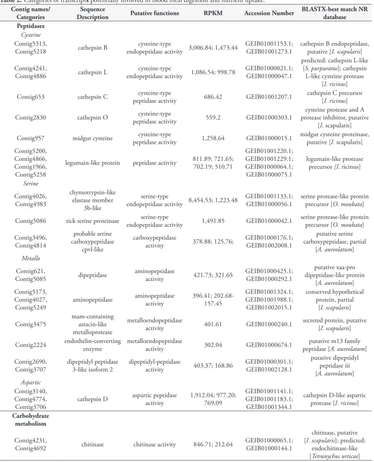
Table 2. Categories of transcripts potentially involved in blood meal digestion and nutrient uptake.
Contig names/ Categories
Sequence
Description Putative functions RPKM Accession Number
BLASTX-best match NR database Peptidases
Cysteine Contig5313,
Contig5218 cathepsin B
cysteine-type
endopeptidase activity 3,006.84; 1,473.44
GEIB01001153.1; GEIB01001273.1
cathepsin B endopeptidase, putative [I. scapularis]
Contig4241,
Contig4886 cathepsin L
cysteine-type
endopeptidase activity 1,086.54; 998.78
GEIB01000021.1; GEIB01000047.1
predicted: cathepsin L-like [S. purpuratus]; cathepsin
L-like cysteine protease [I. ricinus]
Contig653 cathepsin C peptidase activitycysteine-type 686.42 GEIB01001207.1 cathepsin C precursor [I. ricinus]
Contig2830 cathepsin O cysteine-type
peptidase activity 559.2 GEIB01000303.1
cysteine protease and A protease inhibitor, putative
[I. scapularis]
Contig957 midgut cysteine peptidase activitycysteine-type 1,258.64 GEIB01000015.1 midgut cysteine proteinase, putative [I. scapularis]
Contig5200, Contig4866, Contig1966, Contig5258
legumain-like protein peptidase activity 811.89; 721.65; 702.19; 510.71
GEIB01001220.1; GEIB01001229.1; GEIB01000064.1; GEIB01000075.1
legumain-like protease precursor [I. ricinus]
Serine
Contig4026, Contig4983
chymotrypsin-like elastase member
3b-like
serine-type
endopeptidase activity 8,454.53; 1,223.48
GEIB01001133.1; GEIB01000056.1
serine protease-like protein precursor [O. moubata]
Contig5086 tick serine proteinase serine-type
endopeptidase activity 1,491.85 GEIB01000042.1
serine protease-like protein precursor [O. moubata]
Contig3496, Contig4814
probable serine carboxypeptidase
cpvl-like
carboxypeptidase
activity 378.88; 125.76;
GEIB01000176.1; GEIB01002008.1
putative serine carboxypeptidase, partial
[A. aureolatum] Metallo
Contig621,
Contig5085 dipeptidase
aminopeptidase
activity 421.73; 321.65
GEIB01000425.1; GEIB01000292.1
putative xaa-pro dipeptidase-like protein
[A. aureolatum] Contig5173,
Contig4027, Contig5249
aminopeptidase aminopeptidase activity 396.41; 202.68- 157.45
GEIB01001324.1; GEIB01001988.1; GEIB01002015.1
conserved hypothetical protein, partial
[I. scapularis]
Contig3475
mam-containing astacin-like metalloprotease
metalloendopeptidase
activity 401.61 GEIB01000240.1
secreted protein, putative [I. scapularis]
Contig2224 endothelin-converting enzyme metalloendopeptidase activity 302.04 GEIB01000674.1 peptidase [A. aureolatum]putative m13 family
Contig2690, Contig3707
dipeptidyl peptidase 3-like isoform 2
dipeptidyl-peptidase
activity 403.37; 168.86
GEIB01000301.1; GEIB01002128.1
putative dipeptidyl peptidase iii [A. aureolatum] Aspartic
Contig3140, Contig4774, Contig3706
cathepsin D aspartic peptidase activity 1,912.04; 977.20; 769.09
GEIB01001141.1; GEIB01001183.1; GEIB01001344.1
cathepsin D-like aspartic protease [I. ricinus]
Carbohydrate metabolism
Contig4231,
Contig4692 chitinase chitinase activity 846.71; 212.64
GEIB01000065.1; GEIB01000144.1
chitinase, putative [I. scapularis]; predicted:
Contig names/ Categories
Sequence
Description Putative functions RPKM Accession Number
BLASTX-best match NR database
Contig571, Contig4163, Contig3170
maltase hydrolase activity 508.41; 506.43; 173.88
GEIB01000076.1; GEIB01001334.1; GEIB01002097.1
putative amino acid transporter, partial
[A. aureolatum]; PREDICTED: maltase-glucoamylase, intestinal-like, partial [Meleagris gallopavo]; putative
sucrase-isomaltase-like protein, partial [A. sculptum] Contig79,
Contig3343
fructose -bisphosphate aldolase
fructose-bisphosphate
aldolase activity 548.19; 286.33
GEIB01001240.1; GEIB01000239.1
fructose 1,6-bisphosphate aldolase [I. scapularis]
Contig4153,
Contig3020 α-glycosidase α-glucosidase activity 237.32; 150.55
GEIB01000542.1; GEIB01000699.1
glucosidase II, putative, partial [I. scapularis];
glucosidase catalytic [A. aureolatum]
Contig39 phosphorylaseglycogen phosphorylase activity 274.7 GEIB01001233.1 glycogen phosphorylase, putative [I. scapularis]
Contig103,
Contig5179 α -l-fucosidase hydrolase activity 138.33; 130.22
GEIB01002031.1; GEIB01000885.1
predicted: alpha-L-fucosidase-like [Octopus
bimaculoides]; putative alpha-l-fucosidase
[A. aureolatum]
Lipid metabolism
Contig1795 phospholipase b lipase activity 394.32 GEIB01001359.1
predicted: putative phospholipase B-like 2
[Limulus polyphemus]
Contig2581 lipase lipase activity 294.38 GEIB01000804.1
putative hormone-sensitive lipase hsl, partial
[A. aureolatum]
Peptidase inhibitor
Serine proteinase inhibitor
Contig4823 serine proteinase inhibitor serpin
serine-type endopeptidase inhibitor activity
990.82 GEIB01001172.1 serine protease inhibitor 5 RmS5 [R. microplus]
Contig3495, Contig3457
kunitz-type protease inhibitor
serine-type endopeptidase inhibitor activity
407.87; 204.22 GEIB01000363.1; GEIB01001015.1 full=Boophilin-G2 putative [R. microplus]
Contig3309 inhibitor precursorvenom protease
serine-type endopeptidase inhibitor activity
328.78 GEIB01001820.1
kunitz-type protease inhibitor 4 [Micrurus
fulvius]
Contig3184 alpha-2-macroglobulin precursor inhibitor activityendopeptidase 457.08 GEIB01001287.1
alpha-2-macroglobulin precursor splice variant 1
[O. moubata] Cysteine proteinase
inhibitor
Contig4119,
Contig3897 secreted cystatin
cysteine-type endopeptidase inhibitor activity
446.61; 271.95 GEIB01001544.1; GEIB01001844.1 cystatin precursor [O. moubata]
Contig5375,
Contig5044 thyropin precursor
cysteine-type endopeptidase inhibitor activity
3,454.82; 2,269.30 GEIB01000013.1; GEIB01001163.1 putative thyropin precursor [O. moubata]
Immunity
Contig5007 defensin a defense response 1,115.41 GEIB01000200.1 defensin a [O. moubata]
Contig2696 lysozyme precursor lysozyme activity 312.63 GEIB01000589.1 lysozyme precursor [O. moubata]
Contig names/ Categories
Sequence
Description Putative functions RPKM Accession Number
BLASTX-best match NR database Oxidant metabolism
Contig5141, Contig5040, Contig5426, Contig3924
phospholipid-hydroperoxide glutathione peroxidase
glutathione peroxidase activity
1,710.24; 991.96; 871.34; 259.29
GEIB01001198.1; GEIB01001224.1; GEIB01000051.1; GEIB01000721.1
phospholipid-hydroperoxide glutathione
peroxidase [R. microplus]
Contig5082, Contig5045, Contig5342, Contig3084
glutathione s-transferase
glutathione transferase activity
1,900.88; 1,153.5; 1,040.55; 341.27
GEIB01001162.1; GEIB01000083.1; GEIB01001199.1; GEIB01000333.1
glutationa-s-transferase, putative [I. scapularis]
Contig131
microsomal glutathione s-transferase 3
glutathione transferase
activity 166.75 GEIB01001909.1
microsomal glutathione S-transferase, putative
[I. scapularis]
Transporter channels
Contig5201 hemelipoglycoprotein lipid transporter
activity 3,078.88 GEIB01000004.1
vitellogenin-1 [H. longicornis]
Contig4288 fatty acid-binding protein lipid transporter activity 2,504.31 GEIB01000054.1
fatty acid-binding protein FABP, putative
[I. scapularis] Contig4030 beta 2c lipid binding 2,025.46 GEIB01001139.1 beta tubulin [I. scapularis]
Contig4250 adp atp translocase 1 transporter activity 1916.54 GEIB01000011.1
ADP/ATP translocase 1-like [Parasteatoda
tepidariorum].
Contig4329 vitellogenin-B lipid transporter activity 713.31 GEIB01001142.1 [H. longicornis]vitellogenin-B
Storage
Contig4611 ferritin ferroxidase activity 3,846.24 GEIB01001151.1 ferritin [O. moubata]
Table 2. Continued...
(cd02248 and pfam00112). We found four cDNA molecules for legumain-coding transcripts (Contig5200, Contig4866, Contig1966 and Contig5258). All of these legumains exhibited peptidase C13 family conserved domain (pfam01650), which defined them as asparaginyl peptidases (Table 2).
Thirteen transcripts were correlated with the functional activity of serine endopeptidase, according to sequence similarity, and included in the serine group. Among these, tick serine proteinase, chymotrypsin, lysosomal protein, carboxypeptidase and other serine proteinases can be highlighted (Table 2). The Contig5086 transcript is a tick serine proteinase, which presented signal peptide prediction and a conserved protein domain of trypsin. The Contig3496 and Contig4814 transcripts contained the serine carboxypeptidase conserved domain (pfam00450), and thus, the functional activity of carboxypeptidase was assigned to them (Table 2). Two well-expressed cDNA molecules coding for chymotrypsins (Contig4026 and Contig4983) were identified and had similarity to serine protease-like protein of O. moubata.
These chymotrypsin enzyme-coding transcripts exhibited a conserved domain of the catalytic site from serine protease-like trypsin (cd00190, pfam00089 and smart00020) and presented the enzymatic code (EC) 3.4.21.0, which classified it as a hydrolase that acts on peptide bonds.
The metallopeptidase genes were composed of endopeptidases and exopeptidases, such as oligopeptidase, dipeptidase, dipeptidyl peptidase and aminopeptidase, representing 28% (19 contigs) of the intestinal peptidases. Five cDNA molecules with aminopeptidase enzyme-coding (Contig621, Contig5173, Contig5085, Contig4027 and Contig5249) had EC 3.4.11.1 and a GO functional hit relating to exopeptidase, as well as exhibiting an aminopeptidase conserved domain (pfam05195) (Table 2). On the other hand, the Contig3475 and Contig2224 are metalloendopeptidases to which EC 3.4.24 was attributed (Table 2). Contig2690 and Contig3707 presented the type M19 metallopeptidase domain and were classified as dipeptidyl peptidase.
We identified three aspartic endopeptidase protease-coding transcripts (Contig3140, Contig4774 and Contig3706). The Contig3140 transcript was identified as cathepsin D and showed a conserved catalytic aspartic acid domain. This transcript presented signal peptide (SignalP) and transmembrane region (TMHMM) prediction. Contig4774 and Contig3706 were identified as lysosomal aspartic protease-like and aspartic protease, respectively. All these transcripts had similarity mainly to aspartic proteinases of ticks, such as I. ricinus and H. longicornis.
Carbohydrate metabolism
The carbohydrate metabolism category was composed of 19 contigs that represented genes for chitinase, galactosidase, maltase, fucosidase, phosphatase and glycosidase. Two contigs, Contig4231 and Contig4692, exhibited a chitinase conserved domain (cd02872 and COG3325) and EC 3.2.1.14, which classified them as genes coding for chitinase proteins. The Contig4231 presented SignalP, while Contig4692 showed TMHMM. Three cDNA molecules (Contig571, Contig4163 and Contig3170) with possible functions relating to intestinal maltase were identified in the transcriptome of O. mimon (Table 2). These contigs presented
the catalytic α-amylase conserved domain (cd11328), which is found in maltase proteins.
The transcripts Contig79 and Contig3343 are predicted to code for enzymes that metabolize fructose, because they exhibited a fructose-bisphosphate-aldolase conserved domain (cd00344 and cd00354). The E.C. 4.1.2.13 assigned to Contig79 and E.C. 3.1.3.11 to Contig3343, classified them as fructose-bisphosphate-aldolase and fructose-bisphosphate, respectively. The functional activity of α-glycosidase and the E.C. 3.2.1.20 were assigned to the Contig4153 and Contig3020, thus identifying them as glycosidase genes. The Contig39 transcript was identified as a glycogen phosphorylase exhibiting the E.C. 2.4.1.1. The conserved domain of α-l-fucosidase was identified in two cDNA molecules (Contig103 and Contig5179), representing the intestinal fucosidase of O. mimon. Glucosyltransferase, α-galactosidase, hexosaminidase and aminotransferase comprise the remaining carbohydrases.
Lipid metabolism
The lipid metabolism category was composed mainly of lipase, phospholipase, reductase, transferase, phosphodiesterase and ligase. All these transcripts were associated with the cell process of lipid metabolism. We identified a single phospholipase b enzyme-coding transcript (Contig1795), which showed best hit with a phospholipase of Limulus polyphemus (Xiphosura: Limulidae) (Lineu, 1758) (Table 2). Another transcript of lipase (Contig2581) had similarity to putative hormone-sensitive lipase hsl, partial of
Amblyomma aureolatum Pallas, 1772 (Table 2).
Other transcripts associated with the functional
activities of the gut
In this section, we highlight the transcripts of the categories of peptidase inhibitors, immunity, oxidant metabolism and transporter channels, which are associated with the functions of the gut.
Peptidase inhibitor
Seventeen transcripts were grouped in the peptidase inhibitor category (Table 1). Serine protease inhibitor (serpin), cystatins and Kunitz domain inhibitors were the most abundant. The putative conserved domain of serpin (cd00172 and pfam00079) was identified in 12 contigs. Contig4823 had best hit with serine protease inhibitor 5 RmS5 of R. microplus (Table 2). Five transcripts exhibited the Kunitz domain of serpin (cd0019, pfam00014 and smart00131). Of these, only Contig3309 and Contig3457 presented prediction of secretion. The Contig3309 transcript, identified as venom protease inhibitor precursor, displayed hits of 48 to 58% identification with Kunitz inhibitor of snakes. One of these peptidase inhibitors (Contig3184) exhibited macroglobulin conserved domains (cd02897, pfam07677 and pfam07678) and showed its best similarity hit with an α-2-macroglobulin splice variant of O. moubata (Table 2).
and Contig5044). Contig4119 and Contig3897 exhibited a cystatin-conserved domain (cd00042, smart00043 and pfam00031) and had best hits with the O. moubata precursor cystatin (Table 2). Two cDNA molecules coding for thyropin protein and presenting a thyroglobulin conserved protein domain (cd00191) were detected as highly expressed in the gut (Table 2).
Immunity
The immunity category was mainly represented by four contigs, three defensins and one lysozyme. These transcripts were associated with immunity because defensin A and lysozyme presented the GO terms of defense response against gram-negative and positive bacteria and lysosomal activity, respectively. One defensin transcript was highly expressed (Contig5007) and showed 80% identification with O. moubata defensin, while Contig2696 had its best hit with lysozyme of the same species (Table 2).
Oxidant metabolism
The oxidant metabolism category had 70 transcripts associated with it. These are enzymes-coding transcripts possibly related to the oxidation-reduction processes and to the oxidative stress response, such as phospholipid-hydroperoxide glutathione peroxidase (Contig5141, Contig5040, Contig5426 and Contig3924) and glutathione-s-transferase (GST) (Contig5082, Contig5045, Contig5342 and Contig3084). One of the glutathione peroxidase (GSH) transcripts had high levels of expression (Contig5141) and had best hit with the phospholipid-hydroperoxide glutathione peroxidase of R. microplus (Table 2). Other highly expressed transcripts associated with oxidant metabolism were Contig5082 and Contig5045 (Table 2), which exhibited conserved domains of the classes delta and epsilon (cd03045) and mu GST (cd03075), respectively. Contig5045 also exhibited signal peptide and transmembrane region prediction. These transcripts had similarity with GST putative, from I. scapularis and possibly belonged to the cytosolic GST category (Table 2). We identified a transcript of microsomal GST (Contig131), because they presented a Membrane Associated Proteins in Eicosanoid and Glutathione (MAPEG) metabolism conserved domain and signal peptide prediction.
Transporter channels and storage
The transporter channel category grouped 246 contigs. This category is characterized by transport of biological and chemical elements, such as proteins, carbohydrates, lipids, ions and anions. The most abundant transcripts in this category were vitellogenin (10 contigs) and hemelipoglycoprotein (6 contigs). Both transcripts were associated with lipid transporter function. The Contig4329 transcript, identified as vitellogenin-B, presented similarity hits with vitellogenin-B of H. longicornis (Table 2). This transcript exhibited a von Willebrand factor conserved domain (pfam00094 and smart00216) that was also found in other vitellogenins of O. mimon.
Contig4611 (ferritin) and Contig5201 (hemelipoglycoprotein) are probably associated with the nutritional process of ticks.
The Contig4611 transcript presented similarity with ferritins of
O. moubata, exhibited a ferroxidase-conserved protein domain (cd01056, COG1528, PRK1034 and pfam00210) and was classified in the storage category (Table 2). Contig5201, named as hemelipoglycoprotein, showed its reported best hit in relation to a vitellogenin of H. longicornis (Table 2). This transcript was included in the transporter channel category, because lipid transport function was assigned to it. Three other contigs (Contig4288, Contig4030 and Contig4250) with high RPKM were identified as transporters of lipid and chemical elements.
Phylogeny
In this section, we present the phylogenetic inference results relating to the digestive peptidases and transcript datasets of the ticks.
Phylogeny of digestive peptidases
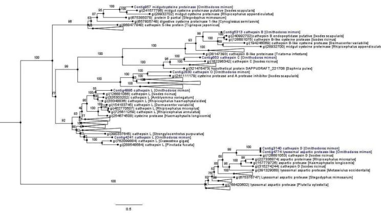
The monophyletic cathepsin cysteine clade was formed by cathepsins (B, C, L and O), midgut cysteine and dipeptidyl peptidase. Most of the cathepsin cysteines of O. mimon are closely related to tick cathepsins, thereby forming strongly supported monophyletic groups. The exception was for Contig4241, which grouped together with the cathepsin L branch of amphibians, birds and fish (Figure 4 and Supplementary Material Figure S1). Each cathepsin (B, C, L and O) formed a monophyletic group that was well supported in the cathepsin cysteine clade. Cathepsins B and C occurred in the same branch, while that cathepsin O formed a sister group with endopeptidases B and C. Cathepsin L was divided in two distinct clades with strong support. One of those clades was formed only by arthropod sequences, while the other clade was formed by bivalves, echinoderms, arthropods (spider, scorpions and ticks) and vertebrate proteinases. We demonstrated the presence of cathepsin O in ticks, for the first time. This proteinase is phylogenetically close to a protein of I. scapularis that was annotated as cysteine protease and a putative protease inhibitor. The cathepsin O group was also formed by two distinct clades in the inferred phylogeny: one group represented by arthropod peptidases and the other by bird and fish sequences.
Aspartic peptidases formed a well-supported monophyletic group represented by cathepsin D and lysosomal aspartic protease genes (A1A family; pepsin). Contig3140 (cathepsin D) and Contig4774 (lysosomal aspartic protease) were seen to be relatively close and they were included in the group of tick aspartic proteinases (Figure 4 and Supplementary Material Figure S1).
Phylogeny of ticks: species tree constructed using
transcript datasets
Figure 4. Phylogenetic tree of cathepsin peptidases. Phylogenetic comparison of O. mimon peptidases with selected proteinases that showed hits in the BLAST search. The tree was constructed using the maximum likelihood (ML) method and multiple alignment of peptidase amino acid sequences. Some branches were collapsed to reduce the size of the tree. The complete phylogenetic tree of the peptidases can be observed on the Supplementary Material (Figure S1). The O. mimon peptidase are highlighted in blue. Aspartic peptidases group was used as outgroup to rooting the phylogenetic tree.
Figure 5. Phylogenetic relationships of ticks, based on 425 ML trees from transcripts of tick transcriptomes, constructed in MulRF. The blue and red bars represent the Ornithodorinae and Argasinae subfamilies, respectively. Ornithodorinae is divided in two groups, Ornithodoros
(Figure 5). The mite M. occidentalis was used as outgroup. In the Argasidae clade, the subfamily Ornithodorinae was composed of species of Ornithodoros and Antricola. The genus Ornithodoros is paraphyletic, forming two groups: the Neotropical Ornithodorinae with the species O. mimon and Antricola delacruzi Estrada-Peña, Barros-Battesti and Venzal, 2004, and Ornithodoros sensu stricto (s.s), which comprises the species Ornithodoros parkeri Cooley, 1936, O. moubata, Ornithodoros rostratus Aragão, 1911 and
O. coriaceus. In the Neotropical Ornithodorinae group, O. mimon
and A. delacruzi are phylogenetically closely related species, forming a monophyletic group. The subfamily Argasinae is composed of
Argas monolakensis.
The Ixodidae clade is divided into two groups: Metastriata and Prostriata. The Metastriata group is formed by the subfamilies Rhipicephalinae (R. microplus, Rhipicephalus appendiculatus
Neumann 1901, Rhipicephalus sanguineus sensu lato (s.l.) Latreille, 1806, D. variabilis and Dermacentor andersoni Stiles, 1908), Amblyomminae (Amblyomma maculatum Koch, 1844,
A. variegatum, Amblyomma americanum (Linnaeus, 1758) and
Amblyomma cajennense s.l.) and Haemaphysalinae (H. longicornis). The species A. cajennense was split into 6 taxa (NAVA et al., 2014). The species H. longicornis forms an underlying group with other Metastriata ticks. The Prostriata group is represented by only one genus, Ixodes (Ixodinae subfamily).
Discussion
Gut transcriptome analysis on engorged females of O. mimon
identified several transcripts that may be associated with the functions of this organ. The transcripts obtained in this study were analyzed and functionally annotated according to the most significant similarity results (hits) in relation to sequences deposited in gene and protein databases such as GenBank and UniProt, using an automated search tool (CONESA et al., 2005). The BLAST search identifies sequences with potential evaluative and functional relationships to the contigs through comparison of similarity with sequences deposited in the databases available. BLAST2GO mapping is the step that retrieves the GO terms associated with each contig. The annotation step performed by this software is a process of assigning functional terms to query sequences from the pool of GO terms gathered in the mapping step (CONESA et al., 2005).
Most of the transcripts were completely annotated using BLAST2GO tool, with the exception of a percentage (22.6%) that did not have any homology with any nucleotide or amino acid sequence in current databases. In the literature, these complete sequences are referred to as “orphan genes”, and their proportion differs considerably from one genome to another (DUJON, 1996; FUKUCHI & NISHIKAWA, 2004). They are abundant among the transcriptomes of ticks (GIBSON et al., 2013). In the gut transcriptome of D. variabilis, 396 unknown transcripts were identified (47%) (ANDERSON et al., 2008); while in salivary transcriptomes of O. coriaceus and O. parkeri, respectively 59% and 49% of the sequence clusters were classified as unknown (FRANCISCHETTI et al., 2008a, b). In the sialome transcriptome of I. scapularis, 34% of its transcripts belonged to unknown
categories, while the sialome of A. variegatum showed that most of the genes (466) were of unknown function (RIBEIRO et al., 2006, 2011). Approximately 71% of the sequences of the transcriptome of A. americanum lack homology with sequences in databases (GIBSON et al., 2013). The reasons for this lack of homology may be the quality of the sequences, low assembly quality, absence of functional genes, taxonomic isolation of the species and emergence of new genes through duplication and/or transposition mechanisms (TAUTZ & DOMAZET-LOŠO, 2011). We can also suggest that other reasons for the existence of these unknown groups might include the small number of studies conducted on these ticks, or even some particular features of gene composition that are not found in other animals. The transcripts without associated GOs possibly represent genes that have functions that are not fully understood or have simply been poorly annotated.
Predictive analysis on subcellular localization identified that the percentage of secreted and transmembrane proteins was low, thus indicating that most of the gut transcripts of O. mimon are intracellular. Similar results were observed in the gut transcriptome of D. variabilis, for which the percentage of secreted transcripts among the genes expressed was small (ANDERSON et al., 2008). Fifty-three percentage of the transcripts differentially expressed in the gut transcriptome of R. microplus females were classified as intracellular and 18% as membranous (HEEKIN et al., 2013).
In the analysis on functional assignment, the predominant functional activities were binding and catalytic activity, thus indicating that a large proportion of gut genes are enzymes and interactive proteins (such as channels, transporters and receptors). The classification from the automated analysis showed that the category of catalytic activity category was prevalent, thus indicating that many of the digestive tract transcripts were enzymes, such as oxidoreductases, transferases, hydrolases, lyases, isomerases and ligases. In the gut transcriptome of R. microplus, binding and catalytic activity was also identified as the most common functional types of activity (HEEKIN et al., 2013). The same results were observed by Xu et al. (2016), who classified 47 and 42% of the transcripts of H. flava in the functional categories binding and catalytic activity, respectively. In the proteomic study of gut of
O. erraticus, Oleaga et al. (2015) also identified catalytic activity and binding as the most prevalent activities within their set of proteins, in accordance to our results. Transcripts associated with the transporter channel category were abundant, since several contigs with binding, carrier protein, receptor and channel functions were grouped into this category. The unknown category was the largest in terms of number of transcripts, because many transcripts without well-defined function, and which lacked homology and GO terms, were grouped into this category.
Blood meal digestion and nutrient uptake
degradation of hemoglobin in ticks is performed by cathepsin D, in association with the auxiliary enzymes cathepsin L and legumain, while cathepsins B and C cleave large hemoglobin fragments to produce dipeptides (HORN et al., 2009). Other peptidases, such as metallopeptidase and serine peptidase have roles in digestion that remain somewhat unclear, but it has been speculated that serine carboxypeptidase and aminopeptidase might participate in releasing free amino acids in the digestion process (HATTA et al., 2006; ANDERSON et al., 2008; HORN et al., 2009). Our results indicated that ticks’ digestive mechanisms are present in the transcriptome analysis on engorged females of O. mimon. All the digestive proteinases described above were identified at different levels of expression (RPKM) with the cathepsins (B, D and L), and they are well expressed in the gut of engorged females. These peptidases also were identified in gut transcriptome analyses on
I. ricinus, H. flava and D. variabilis, respectively, thus agreeing with our results (KOTSYFAKIS et al., 2015; PERNER et al., 2016; XU et al., 2016; ANDERSON et al., 2008). Oleaga et al. (2015) identified 15 proteins with peptidase activity belonging to four different groups (aspartic-type endopeptidase, metallopeptidase, cysteine-type endopeptidase and serine-type endopeptidase) in the midgut proteome of O. erraticus.
Adding a new member to the known tick digestive enzymes, we have recorded for the first time a sequence coding for a member of the cysteine proteinase family called cathepsin O. Although, this protease may already have been identified in the I. scapularis tick as a cysteine protease putative deposited in the Genbank database, but it was not labeled as cathepsin O. This cysteine peptidase was identified and characterized for the first time from a human breast carcinoma cDNA library, by using a polymerase chain reaction-based cloning strategy (VELASCO et al., 1994). Recently, Fuzita et al. (2015) identified two transcripts of cathepsin O in the transcriptomic and proteomic analyses of digestion in
T. serrulatus.
In addition to the digestive mechanisms, ticks have a heme detoxification mechanism based on accumulating heme in specialized organelles of the intestinal epithelium, called hemosomes (LARA et al., 2003). These organelles are formed by transport and delivery of carrier proteins such as hemelipoglycoproteins, which are associated with heme (MAYA-MONTEIRO et al., 2000; LARA et al., 2005; DUPEJOVA et al., 2011). Hemelipoglycoprotein in O. mimon constitute a transcript with high levels of RPKM, possibly indicating gene expression in response to digestion of the blood meal, in order to accumulate heme. Another transcript that may be associated with nutrient uptake is the ferritin gene. In response to exposure to iron, insect cells produce ferritin molecules of two types: cytoplasmic and secreted (GEISER et al., 2006). Ferritin was well-expressed in the engorged females of
O. mimon, possibly in response to the blood of the host, which is iron-rich. cDNA molecules of ferritin in two tick species (O. moubata and I. ricinus) were characterized and these molecules showed no evidence of being secreted proteins, thus indicating that the protein was cytoplasmic (KOPÁČEK et al., 2003). These findings are in agreement with our results, given that the
O. mimon ferritin did not exhibit any prediction of a signal peptide,
according to the SignalP algorithm. Iron is a substance derived from degradation of the hemoglobin present in the blood meal,
which results in compounds that serve as nutrients for ticks to feed on, develop and reproduce (KOPÁČEK et al., 2003). Ferritin is frequently found in the mialome and sialometranscriptome analysis of ticks (ANDERSON et al., 2008; RIBEIRO et al., 2012; KOTSYFAKIS et al., 2015; XU et al., 2016), as well as in the proteome studies (OLEAGA et al., 2015).
The gut of ticks also expresses enzymes that digest other blood meal constituents, such as carbohydrates and lipids (MORETI et al., 2013; KOTSYFAKIS et al., 2015; XU et al., 2016). Carbohydrase genes were identified in the digestive tract of O. mimon after the blood meal, among which chitinases and α-glycosidases were the most prevalent. The role of these enzymes within digestion has not been fully understood and there are few studies about it. However, on the other hand, the activity of carbohydrases such as N-acetyl glucosaminidase, chitinase and α-L-fucosidase on the intestinal content of Amblyomma sculptum (cited as cajennense) was recently identified and characterized, and it was suggested that digestion of carbohydrates occurs in the intestinal lumen, unlike the intracellular digestion of proteins (MORETI et al., 2013).
Protease inhibitors are expressed in both the intestine and the salivary glands of ticks (KOPÁČEK et al., 2010), but their function in ticks is not fully understood. However, other authors have suggested that inhibitors such as serpin can be used by ticks to regulate endopeptidase activity and hemostasis (MULENGA et al., 2001, 2003; PERNER et al., 2016; TIRLONI et al., 2014). Peptidase inhibitors have been mentioned as effective antigens for preparing vaccines, because these genes are essential for the survival of ticks, since they are associated with endogenous processes such as regulation of hemolymph coagulation through controlling proteinase activity (IMAMURA et al., 2005). Kunitz-like inhibitors of serine peptidase are known to act towards defending the intestine against rickettsial infections and protecting tick cells against acaricide (KOPÁČEK et al., 2010). In addition to Kunitz inhibitors in ticks, we also found a single transcript that coded for a protein similar to snake venom protease inhibitor, which showed similarity to of Kunitz inhibitors snakes sequences, and may also have a role relating to protease inhibition in ticks.
Tick cystatins act as regulators of blood meal digestion and as modulators of the host immune response (SCHWARZ et al., 2012). These inhibitors of cysteine peptidases also participate in the innate immunity of ticks by inhibiting specific enzymes of microorganisms (KOPÁČEK et al., 2010). Two types of cystatins have been found in argasid ticks (GRUNCLOVÁ et al., 2006). Cystatin type I is cytosolic, while type II is secreted, and both substantially diverge from other invertebrate, vertebrate and plant cystatins (GRUNCLOVÁ et al., 2006). The cystatins of O. mimon
did not show any prediction of secretion, thus suggesting that they may belong to type I. However, these transcripts showed similarity to sequences of O. moubata cystatins, which according to the literature (GRUNCLOVÁ et al., 2006), are type II (secreted). It is possible that the SignalP algorithm did not identify the signal peptide in the cDNA sequences of cystatins of O. mimon, or that
the assembled contig did not include the region where possible signal peptides could occur. The transcripts of thryporin are well expressed, but their function is not fully known.
In the transcriptome of O. mimon, we only found the