Diana Oliveira da Silva
Bachelor degree in Biochemistry
Chemical synthesis of new histone deacetylase
inhibitors and their evaluation as anticancer agents
Dissertation for the Master degree in Biochemistry for Health
Supervisor: Rita Ventura, PhD, ITQB-NOVA
Co-supervisor: Catarina Brito, PhD, IBET, ITQB-NOVA
Jury:
President: Doutor Pedro Manuel H. M. Matias, Investigador Principal ITQB-NOVA Arguer: Doutor Hélder João Ferreira Vila Real, Investigador IBET
Vowels: DoutoraMargarida Archer Franco Frazão, Investigadora Principal ITQB-NOVA Doutora Maria Rita Mendes Bordalo Ventura, Investigadora Auxiliar ITQB-NOVA
Instituto de Tecnologia Química e Biológica António Xavier da Universidade Nova de Lisboa
III
Chemical synthesis of new histone deacetylase inhibitors and their evaluation as anticancer agents
Copyright - Diana Oliveira da Silva, ITQB/NOVA
O Instituto de Tecnologia Química e Biológica António Xavier e a Universidade Nova de Lisboa têm o direito, perpétuo e sem limites geográficos, de arquivar e publicar esta dissertação através de exemplares impressos reproduzidos em papel ou de forma digital, ou por qualquer outro meio conhecido ou que venha a ser inventado, e de a divulgar através de repositórios científicos e de admitir a sua cópia e distribuição com objetivos educacionais ou de investigação, não comerciais, desde que seja dado crédito ao autor e editor.
V
(…). À parte isso, tenho em mim todos os sonhos do mundo. Em Tabacaria. Álvaro de Campos, Heterónimo de Fernando Pessoa
VII
Acknowledgements
I would like to acknowledge all the people directly or indirectly involved in this thesis.
To my supervisor, Dr. Rita Ventura, and my co-supervisor, Dr. Catarina Brito for the opportunity to work in such challenging project combining two very distinctive fields and for helping me to grow as a scientist in both areas.
To Dr. Rita Ventura for all the knowledge, guidance and for being so supportive since the beginning of this journey.
To Dr. Catarina Brito for all the scientific discussions and all guidance and knowledge provided. For all friendly conversations and motivation throughout this work.
To all colleagues in Bioorganic Chemistry and Organic Synthesis Units at ITQB-UNL, for the good working environment specially Vanessa Miranda, Filipa Almeida, Eva Lourenço, Saúl and Jessica Bevan.
To Osvaldo Ascenso, for all the guidance and help provided throughout the chemical synthesis. To Animal Cell Technology Unit at IBET, ITQB-UNL colleagues for the good working environment and including me in the group with such joy. To 3D Advanced cell models group specially Sofia Abreu, Daniel Simão, Tatiana Martins, Francisca Arez, Ana Paula and Catarina Pinto, for all scientific discussions and knowledge provided. For all patience to teach me and answer all my questions.
To Marta Estrada, for all the support and patience to teach me all the techniques of animal cell culture. For all the advices and help throughout all year. For always being there.
A todos os meus colegas de mestrado que me apoiaram e ajudaram durante os últimos dois anos. Aprendi sempre algo mais com cada um de vocês.
Aos meus amigos, aqueles que acreditam em mim e nas minhas capacidades e por todos os bons momentos. Pela vossa amizade.
Ao Nuno, por toda a paciência e carinho.
Aos meus pais, irmão e avó, que sempre me apoiaram, acreditaram em mim e ajudaram em todos os momentos de forma a conseguir atingir os meus objetivos. Por serem um exemplo de força. Sem vocês não era possível. Obrigada
IX
Abstract
Epigenetics and its key role in gene expression have become increasingly important in cancer research. Histone proteins play an important role in gene expression control through modifications like acetylation, phosphorylation and methylation. Histone deacetylases (HDAC) can regulate expression levels via acetylation of lysine residues of histone proteins.
Inhibitors of these HDAC have been associated with cancer and some are commercialized as anticancer drugs like SAHA (Vorinostat) and Belinostat (PXD101). These histone deacetylases inhibitors (HDACi) are known to have several effects in altered or malignant cells such as accumulation of acetylated proteins, proliferation arrest, cell cycle arrest, and induction of apoptosis.
The purpose of this work was to synthesize new HDACi with anticancer effects. For this, several analogues were synthesized inspired in the structures of SAHA and Belinostat where key aspects of the HDACi structures were altered. Breast cancer (MCF-7) and lung cancer (H460) cell lines were used in order to evaluate the newly synthesized compounds as antitumour agents.
Overall, 30 compounds were successfully synthesized and were used in further analysis in cancer cell lines.
Results showed that the synthesized compound
N-hydroxy-N'-(4-iodophenyl)octanediamide (16c) is a putative new HDACi with anticancer effects, exhibiting an cytotoxic effect in both cancer cell lines tested and preliminary data suggested that it induced the same cell death mechanisms as the previously described for SAHA.
XI
Resumo
A epigenética e o seu papel na regulação génica têm vindo a tornar-se muito importantes na investigação em oncologia. As histonas exercem uma função fundamental na regulação dos genes através de modificações como a acetilação, fosforilação e metilação. As deacetilases de histonas (HDAC) regulam os níveis de expressão através da acetilação de lisinas presentes em histonas.
Os inibidores destas enzimas têm sido usados na terapia do cancro e alguns são comercializados como fármacos anticancerígenos tais como o SAHA (Vorinostat) e o Belinostat (PXD101). Estes inibidores de deacetilases de histonas (HDACi) têm como efeitos conhecidos a acumulação de proteínas acetiladas, paragem da proliferação, paragem do ciclo celular e a indução da apoptose em células alteradas ou malignas.
O objetivo deste trabalho foi a síntese de novos HDACi com efeitos anticancerígenos. Para tal, vários compostos análogos foram sintetizados baseados nas estruturas dos fármacos SAHA e Belinostat onde aspetos importantes da estrutura foram alterados. Linhas celulares de cancro de mama (MCF-7) e de pulmão (H460) foram usadas para avaliar os compostos sintetizados como agentes anticancerígenos.
No total, foram sintetizados com sucesso e posteriormente testados em linhas celulares cancerígenas 30 compostos.
Os resultados obtidos mostraram que o composto sintetizado N-hidroxi-N'-(4-iodofenil)octanodiamida (16c), revelou um efeito anticancerígeno maior do que o provocado pelo SAHA em ambas as linhas celulares testadas e resultados preliminares sugerem que induz mecanismos de morte celular similares aos descritos previamente para o SAHA.
Palavras-chave: HDACi; Síntese Química; Ensaios farmacológicos; Agentes anticancerígenos;
XIII
Contents
I. INTRODUCTION ... 1
I.1 Cancer and Epigenetics ... 1
I.2 Histone Regulation and Acetylation ... 2
I.3 Histone Deacetylase (HDAC) ... 3
I.4 HDAC inhibitors (HDACi) ... 4
I.4.1 HDACi - multitargeted compounds ... 6
I.4.2 HDACi as anticancer agents ... 7
I.4.3 HDACi Action Mechanism ... 9
I.4.4 HDACi Synthesis ... 11
I.5 Thesis aim ... 13
II. MATERIALS AND METHODS ... 15
II.1 Synthesis of New HDACi ... 15
II.1.1 General conditions ... 15
II.1.2 General methods... 16
II.1.3 Experimental Procedures ... 17
II.2 Evaluation as anticancer agents ... 43
III. RESULTS AND DISCUSSION ... 49
III.1 Synthesis of new HDACi ... 49
III.1.2 Results from the 1st round of synthesis ... 53
III.1.3 Results from the 2nd round of synthesis ... 56
III.1.4 Discussion ... 59
III.2 New HDACi evaluation as anticancer agents ... 63
III.2.1 Determination of DMSO toxicity ... 64
III.2.2 Drug Testing ... 65
III.2.3 Evaluation of Compound 16c ... 73
IV. CONCLUSIONS ... 79
V. REFERENCES ... 81
XV
Figure Index
Figure I.1 - Most common causes of cancer death. Data obtained from the World Health
Organization1. ... 1
Figure I.2- Histone Acetylation - Acetylation of lysine residues (mediated by HATs) will neutralize the histone positive charge and weakening its interaction with DNA. Previously tightly wrapped DNA around the histone will be more loose and more accessible to several processes like transcription, replication, etc. By removing acetyl groups, HDACs will have the opposing action. Adapted from D.Pons et al44. ... 3
Figure I.3 - Structures of SAHA and Belinostat with their three important domains: capping group, linker and metal binding moiety. ... 8
Figure I.4 – Representation of the catalytic pocket in the crystal structure of HDAC protein with SAHA. In the catalytic pocket of HDAC enzyme is a Zinc molecule (in pink) where the hydroxamic part of SAHA binds. Adapted from Marks, P. A. et al 45. ... 8
Figure I.5 - Biological effects of HDACi in malignant cells. Adapted from K.Ververis et al10. ... 9
Figure I.6 - General scheme of HDAC inhibitors synthesis. ... 11
Figure I.7 - General structure of HDACi. ... 13
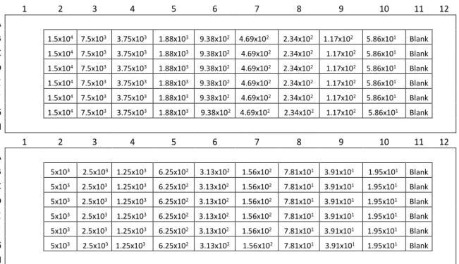
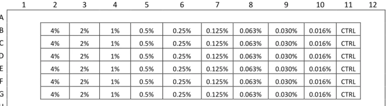
Figure II.8 – Plate schemes for implementation/optimization of PrestoBlue cell viability assay. 44 Figure II.9 – Plate scheme for determination of DMSO toxicity with % of DMSO tested. Control without DMSO (CTRL) ... 45
Figure II.10 – Plate scheme for dose-response curve of SAHA with concentrations of SAHA tested. ... 45
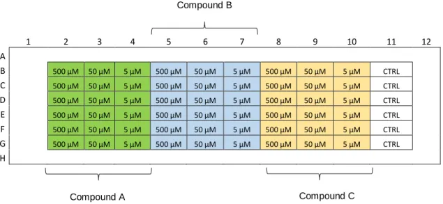
Figure II.11 - Plate scheme for drug testing of all compounds with concentrations tested. ... 46
Figure III.12 – 1H NMR spectra of compound 9c before (above) and after (below) the sodium bicarbonate (NaHCO3) washing step and of compound 9b... 59
Figure III.13 - 1H NMR spectra of compounds 14b (above) and 14c (below). In both spectra, the area important to distinguish between both derivatives is highlighted. At the top are the structures of compounds 14b and 14c, respectively. ... 60
Figure III.14 - 1H NMR spectra of compounds 4c, 4b, 2c and 2b, respectively. Structures of the compounds at the right of each spectrum. ... 61
Figure III.15 - Phase-contrast microscopy of H460 cells (left) and MCF-7 cells (right). Scale bars represents 100 µm. ... 63
Figure III.16 - Phase-contrast microscopy of Human Dermal Fibroblasts. Scale bar represents 100 µm. ... 63
Figure III.17 – Dose-response curves for DMSO for each cancer cell line (MCF-7 and H460). Dose-response curves were generated using Graph Pad Prism 6 software. ... 64
Figure III.18 - Effect of SAHA on MCF-7 and H460 cell viability, determined by PrestoBlue assay. Both cancer cells lines were treated with three different concentrations of SAHA: 500 50 and 5 µM, for 48h. Data are mean ± SD of three independent experiments; asterisks indicate significant difference to negative control (*p≤0.05, **p≤0.01, ***p≤0.001 and ****p≤0.0001) by one-way ANOVA analysis with Kruskal-Wallis comparison test. Graph constructed using Graph Pad Prism 6 software. ... 65
Figure III.19 - Effect of SAHA and the hydroxamic compounds synthesized in the 1st round of synthesis on MCF-7 and H460 cell viability, determined by PrestoBlue assay. Both cancer cells lines were treated with three different concentrations of SAHA: 500, 50 and 5 µM, for 48h. Data are mean ± SD of two independent experiments; asterisks indicate significant difference to negative control (*p≤0.05, **p≤0.01, ***p≤0.001 and ****p≤0.0001) by one-way ANOVA analysis with Kruskal-Wallis comparison test. Graph constructed using Graph Pad Prism 6 software. .... 67
XVI
Figure III.20 – Effect of SAHA and the carboxylic compounds synthesized in the 1st round of
synthesis on MCF-7 and H460 cell viability, determined by PrestoBlue assay. Both cancer cells lines were treated with three different concentrations of SAHA: 500, 50 and 5 µM, for 48h. Data are mean ± SD of two independent experiments; asterisks indicate significant difference to negative control (*p≤0.05, **p≤0.01, ***p≤0.001 and ****p≤0.0001) by one-way ANOVA analysis with Kruskal-Wallis comparison test. Graph constructed using Graph Pad Prism 6 software. .... 69
Figure III.21 – Effect of SAHA and all compounds synthesized in the 2nd round of synthesis on
MCF-7 and H460 cell viability, determined by PrestoBlue assay. Both cancer cells lines were treated with three different concentrations of SAHA: 500, 50 and 5 µM, for 48h. Data are mean ± SD of two independent experiments; asterisks indicate significant difference to negative control (*p≤0.05, **p≤0.01, ***p≤0.001 and ****p≤0.0001) by one-way ANOVA analysis with Kruskal-Wallis comparison test. Graph constructed using Graph Pad Prism 6 software... 71
Figure III.22- Effect of SAHA and compound 16c in MCF-7, H460 and HDF cell viability,
determined by PrestoBlue assay. All cell lines were treated with two different concentrations of SAHA and 16c: 50 and 5 µM, for 48h. Data are mean ± SD of two independent experiments; asterisks indicate significant difference (*p≤0.05 and **p≤0.01) by one-way ANOVA analysis with Kruskal-Wallis comparison test. Graph constructed using Graph Pad Prism 6 software. .... 73
Figure III.23 – Induction of apoptosis by SAHA and Compound 16c. MCF-7 cells were cultured
in the presence of compound 16c, SAHA or in culture medium with vehicle control (control) for 48h, collected and processed for apoptosis analysis using fluorescence labelling with a caspase probe (NucView) followed by flow cytometry. ... 75
Figure III.24 – Cell cycle analysis of MCF-7 cells. MCF-7 cells were cultured in the presence of
compound 16c, SAHA or in culture medium with vehicle control (control) for 48h, collected and processed for cell cycle analysis by flow cytometry. Gated region (left), doublet discrimination step (middle) and counted cells in each phase of the cell cycle (G1, S and G2/M). ... 76
Figure III.25 – Cell cycle analysis of MCF-7 cells. MCF-7 cells were cultured in the presence of compound 16c, SAHA or in culture medium with vehicle control (control) for 48h, collected and processed for cell cycle analysis by flow cytometry. ... 77
Figure III.26 - Standard curves obtained for each cells (MCF-7, H460 and HDF) for PrestoBlue
XVII
Table Index
Table I.1 - Classification of Zinc dependent family of HDAC enzymes. ... 4
Table I.2 - HDAC inhibitors categories, structures and information about approval status as
anticancer drugs. ... 5
Table III.3 - Structures of all synthesized compounds with respective numerations. ... 50
Table III.4 – 1st step of the synthesis using mono-ethyl fumarate as starting material and several
anilines. The table describes the compounds obtained (respective numbering) and
corresponding yields... 53
Table III.5 – Synthesis of carboxylic acid derivatives from compounds present in table III.4. The
table describes the compounds obtained (respective numbering) and corresponding yields. .... 54
Table III.6 - Synthesis of hydroxamic acid derivatives from compounds present in table III.4. The
table describes the compounds obtained (respective numbering) and related yields. ... 55
Table III.7 - 1st Step of the synthesis using adipic acid monomethyl ester as starting material
and several anilines. The table describes compounds obtained (respective numbering) and corresponding yields... 56
Table III.8 - Synthesis of carboxylic acid derivative 14b.... 56
Table III.9 - Synthesis of hydroxamic acid derivatives from compounds present in table III.7. The
table describes the compounds obtained (respective numbering) and corresponding yields. .... 57
Table III.10 - 1st Step of the synthesis using suberic acid monomethyl ester as starting material
and several anilines. The table describes the compounds obtained (respective numbering) and corresponding yields... 57
Table III.11 - Synthesis of hydroxamic acid derivatives from compounds present in table III.10.
The table describes the compounds obtained (respective numbering) and corresponding yields. ... 58
Table III.12 - 1st Step of the synthesis using azelaic acid monomethyl ester as starting material
and several anilines. The table describes compounds obtained (respective numbering) and corresponding yields... 58
Table III.13 - Synthesis of hydroxamic derivatives from compounds present in table III.12. The
XIX
Abbreviations and Symbols
13C-NMR - Carbon-13 nuclear magnetic resonance 1H-NMR - Proton nuclear magnetic resonance APT - Attached proton test
Ar - Aromatics
ATR-FTIR - Attenuated Total Reflectance-Fourier Transform Infra-red Spectroscopy CDCl3 - Deuterated chloroform
COSY - Correlation Spectroscopy d - Doublet
DMEM - Dulbecco's Modified Eagle Medium DMF - Dimethylformamide
DMSO - Dimethyl sulfoxide
DMSO-d6 -Deuterated dimethyl sulfoxide DNA - Deoxyribonucleic acid
EDC - 1-Ethyl-3-(3-dimethylaminopropyl)carbodiimide FBS - Fetal Bovine Serum
HAT - Histone acetyltransferase HDAC - Histone deacetylase
HDACi - Histone deacetylase inhibitors HDF - Human Dermal Fibroblasts
HMQC - Heteronuclear Multiple-Quantum Correlation IC50 - Half maximal inhibitory concentration
IMDM - Iscove's Modified Dulbecco's Medium IR - Infra-Red
m - Multiplet MeOH - Methanol
NaOH - Sodium hydroxide
NH2OH.HCl - Hydroxylamine hydrochloride PBS - Phosphate-buffered saline
XX
PTM - post-translational modification q - quartet
s - Singlet
SAHA - Suberoylanilide Hydroxamic acid or Vorinostat SIRT - sirtuin
t - Triplet
THF - Tetrahydrofuran
TLC - Thin Layer Chromatography UV - Ultraviolet
1
I.
INTRODUCTION
I.1 Cancer and Epigenetics
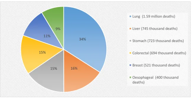
Cancer is one of the leading causes of mortality worldwide, with approximately 8.2 million cancer related deaths in 2012. In the next 2 decades, the number of new cases is expected to increase by approximately 70%. The most common causes of cancer death are lung, liver, stomach, colorectal, breast and oesophageal cancer1. (Figure I.1).
Treatment options against cancer comprise surgery, chemotherapy and radiotherapy and an early detection, accurate diagnosis, and effective treatment are crucial for increasing cancer survival1.
Figure I.1 - Most common causes of cancer death. Data obtained from the World Health Organization1.
Cancer is characterized by cells with rapid and abnormal growth which are able to invade adjacent parts of the body and spread to other organs, a process called metastasizing. Cancerous cells are the result of genetic and genomic alterations such as point mutations, deletions and translocations2. Genetic alterations can ultimately lead to the activation of oncogenes and
inactivation of tumor-suppressor genes. More recently, cancer development has been correlated not only with genetic alterations but also with epigenetic modifications that contribute to disease progression3. Genetics is concerned with the information transmitted on the basis of gene
sequence whereas epigenetics studies the inheritance of information based on gene-expression levels that do not involve changes in the underlying DNA sequence4.
34% 16% 15% 15% 11% 9%
Lung (1.59 million deaths) Liver (745 thousand deaths) Stomach (723 thousand deaths) Colorectal (694 thousand deaths) Breast (521 thousand deaths) Oesophageal (400 thousand deaths)
2
In recent years, numerous studies described extensive reprogramming of epigenetic machinery components in cancer, including DNA methylation and histone modifications. A first indication that epigenetic modifications such as histone acetylation may be strongly involved in cancer onset and progression came from a study demonstrating a global loss of monoacetylation and trimethylation on histone H4 in cancer cells5. Other similar studies have shown that epigenetic
enzymes are often dysregulated in human tumors4.
Identification of these enzymes has driven the rapid development of small-molecule inhibitors that target the cancer epigenome. Within this purpose, histone deacetylases inhibitors (HDACi) were studied as potential antitumor agents. There are now several HDACi commercialized as anticancer medicines such as suberoylanilide hydroxamic acid (SAHA) and Belinostat (PXD101), approved for the treatment of specific types of cancer (see section I.4.2).
I.2 Histone Regulation and Acetylation
Eukaryotic organisms have their genetic information packaged inside the cell nucleus. This arrangement is mediated by histones which are small basic proteins rich in lysine and arginine residues that strongly adhere to negatively-charged DNA and form complexes called nucleosomes6. Nucleosomes are the basic units of chromatin and are formed by an octamer of
the four core histones (H3, H4, H2A, H2B) around which DNA is wrapped 7.
The amino acid tails of core histones can be subjected to several post-translational modifications like acetylation, methylation and phosphorylation, and this facilitates processes including transcription, replication and repair7,8. While the base sequence of DNA provides the
fundamental code for proteins, post-translational modifications (PTM´s) of histone proteins play a major role in the control of gene transcription 9. These epigenetic modifications can lead to
changes in function and/or regulation of several proteins including histones, without altering their primary sequences.
Histone acetylation is the modification most widely studied. In general, high levels of acetylation (hyperacetylation) are correlated with higher transcriptional activity and a more open chromatin conformation, whereas low levels of acetylation (hypoacetylation) cause chromatin condensation leading mainly to transcriptional suppression.
Histone acetylation levels are mediated by the opposing actions of two different enzymes: Histone acetyl-transferases (HATs) that are responsible for transferring acetyl groups to lysine residues in histones N-terminal tails and histone deacetylases (HDACs) that remove the acetyl group 7,10. (Figure I.2)
3 I.3 Histone Deacetylase (HDAC)
To date, 18 mammalian HDAC enzymes have been identified and are classified based on their homology with yeast transcriptional regulators11. These enzymes are divided into four
different classes: classes I, II and IV, from the Zinc-dependent family (also called classical), and class III, which is a NAD + (Nicotinamide adenine dinucleotide) dependent family (also called SIRT-sirtuins) 10. Concerning the Zinc-dependent family of HDAC enzymes (Table I.1), class I
contains HDAC1, 2, 3, and 8 and shares homology with the yeast transcriptional regulator RDP3. The class II enzymes share homology with the yeast HDAC1 and are subdivided into class IIa, consisting of HDAC4, 5, 7, and 9, and class IIb, containing HDAC6 and 10. Class IIb differs structurally by containing two catalytic sites. HDAC11 shares characteristics with both class I and class II HDACs, so it is included in class IV12. Concerning the NAD + dependent family (Table
I.1), class III contains seven proteins (SIRT1 - 7) that differ in cellular localization, activity, and function13.
Figure I.2- Histone Acetylation - Acetylation of lysine residues (mediated by HATs) will neutralize the
histone positive charge and weakening its interaction with DNA. Previously tightly wrapped DNA around the histone will be more loose and more accessible to several processes like transcription, replication, etc. By removing acetyl groups, HDACs will have the opposing action. Adapted from D.Pons et al44.
4
Table I.1 - Classification of Zinc dependent family of HDAC enzymes.
CLASSES HDAC ENZYME LOCALIZATION
CLASS I HDAC1 Nucleus
HDAC2 Nucleus
HDAC3 Nucleus
HDAC8 Nucleus/Cytoplasm
CLASS IIa HDAC4 Nucleus/Cytoplasm
HDAC5 Nucleus/Cytoplasm
HDAC7 Nucleus/Cytoplasm
HDAC9 Nucleus/Cytoplasm
CLASS IIb HDAC6 Cytoplasm
HDAC10 Cytoplasm
CLASS III SIRT1 Nucleus
SIRT2 Cytoplasm SIRT3 Nucleus/Mitochondria SIRT4 Mitochondria SIRT5 Mitochondria SIRT6 Nucleus SIRT7 Nucleus
CLASS IV HDAC11 Nucleus/Cytoplasm
HDAC enzymes are predominantly in the cell nucleus in order to exert their function. However, some HDAC can shuttle between the nucleus and the cytoplasm11.
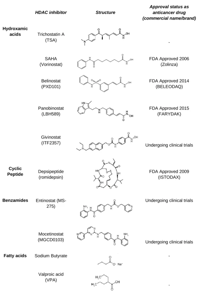
I.4 HDAC inhibitors (HDACi)
Over the years, many different types of HDAC inhibitors (HDACi) have been developed, ranging from simple structures like butyrate to more complex structures such as SAHA or romidepsin. HDACi can be categorised based on their structure: hydroxamic acids, cyclic peptides, benzamides, and short-chain fatty acids (Table I.2).
5
Table I.2 - HDAC inhibitors categories, structures and information about approval status as anticancer
drugs.
HDAC inhibitor Structure
Approval status as anticancer drug (commercial name/brand) Hydroxamic acids Trichostatin A (TSA) - SAHA (Vorinostat) FDA Approved 2006 (Zolinza) Belinostat (PXD101) FDA Approved 2014 (BELEODAQ) Panobinostat (LBH589) FDA Approved 2015 (FARYDAK) Givinostat (ITF2357)
Undergoing clinical trials
Cyclic Peptide Depsipeptide (romidepsin) FDA Approved 2009 (ISTODAX) Benzamides Entinostat (MS-275)
Undergoing clinical trials
Mocetinostat
(MGCD0103) Undergoing clinical trials
Fatty acids Sodium Butyrate -
Valproic acid (VPA)
6
The first proposed HDACi were short chain fatty acids, like sodium butyrate, but these were shown to be less effective in comparison with inhibitors of the other categories. Trichostatin A was the first natural product discovered to inhibit HDACs. After that, several molecules with similar structure started to be produced and tested as HDACi, such as SAHA or Belinostat.
I.4.1 HDACi - multitargeted compounds
HDACi have been studied as potential drugs for different final goals. The action of these inhibitors leads to a more open chromatin conformation and DNA becomes more accessible to several processes. One of those processes can be transcription, so HDACi have been correlated with higher transcription activity. Therefore, these compounds and analogues have been studied as potential additives capable of enhancing the expression of recombinant proteins in mammalian cell cultures (SME, from Small Molecule Enhancers)14. Recently, SME are being studied also in
plant cell cultures within the same purpose.
In the last decade, neurodegenerative diseases have also been proposed as targets for HDACi therapy. Brain disorders are often associated with imbalances in protein acetylation levels and transcriptional dysfunctions15. Treatment with various HDACi can potentially lead to the
correction of these variations. In several animal models of neurodegenerative diseases (such as Parkinson´s disease and Alzheimer´s disease among others), treatments with HDACi revealed effectiveness against neuronal cell death and improved neurological outcome16,17.
Cancer disease is another target of this type of compounds. HDACi have been studied as potential antitumor agents and there are now several approved as anti-cancer agents. Not only these compounds can act as radio-sensitizers in the treatment of cancer but also they show several effects against cancerous cells with an apparent selectivity, such as growth arrest, induction of apoptosis and cell cycle arrest. (See sections I.4.2 and I.4.3).
7 I.4.2 HDACi as anticancer agents
As mentioned before, the first evidences that epigenetics alterations, and especially histone acetylation could be involved in cancer came from studies showing different acetylation levels in cancerous and non-cancerous cells.Fraga et al showed global loss of monoacetylation
and trimethylation of lysine residues in histone H4 in cancer cells5. In related studies, authors
analyzed the expression of class I HDAC enzymes (HDAC1, 2, 3, and 8) in several cell lines and cancer tissues. Stomach, oesophagus, colon, prostate, breast, ovary, lung, pancreas and thyroid cancers were analysed and the results showed that over 75% of human cancer tissues and their corresponding non-cancerous epithelium showed high expression of these class I HDACs. Also, 5-40% of cancer tissues overexpressed class I HDACs, compared with corresponding normal epithelium18.
Due to these and similar findings, inhibitors of HDAC enzymes emerged as a new class of targeted therapeutics for a variety of human cancers. Currently, there are several clinical trials of this type of drugs alone or in combination with drugs already used in cancer therapy. To date, four HDAC inhibitors were approved by the U.S Food and Drug Administration (FDA) for lymphoma and myeloma: Belinostat, Romidepsin, Panobinostat and SAHA (Table I.2).
Belinostat (BELEODAQ, Spectrum Pharmaceuticals, Inc.) was approved by the FDA in July, 2014, for the treatment of patients with relapsed or refractory peripheral T-cell lymphoma (PTCL)19.
Romidepsin (ISTODAX, Gloucester Pharmaceuticals Inc.) was approved by the FDA in November, 2009, for the treatment of cutaneous T-cell lymphoma (CTCL) in patients that have received at least one prior systemic therapy19.
Panobinostat (FARYDAK capsules, Novartis Pharmaceuticals) was approved by the FDA in February, 2015, in combination with bortezomib and dexamethasone for the treatment of patients with multiple myeloma20.
SAHA or Vorinostat (Zolinza, Merck & Co., Inc.) acts as a strong chelator of Zinc ions and is an effective HDAC inhibitor. SAHA was approved by the FDA for the treatment of cutaneous T-cell lymphoma (CTCL) in October of 2006.21,22 SAHA is capable of inhibiting all HDAC in classes
I and II that are metal dependent (in nanomolar range)23 so it is considered a pan-inhibitor of
8
Structurally, hydroxamic acids HDACi, such as SAHA, include three important domains: a capping group, a metal binding moiety and a linker. (Figure I.3)
Figure I.3 - Structures of SAHA and Belinostat with their three important domains: capping group, linker and
metal binding moiety.
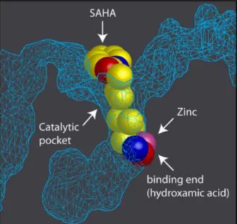
The capping group is important in the interaction with the amino acid residues near the entrance of the active site. The metal binding moiety is crucial for the interaction with the zinc atom present in the catalytic pocket of HDAC enzyme (Figure I.4). The linker connects the two groups10. In case of SAHA, the linker is only a carbon chain with 8 carbons without double bonds,
but in Belinostat the linker is much shorter with a double bond. Optimization of these key motifs is crucial for the discovery of a molecule better fitted to the catalytic pocket of the enzyme and consequently with higher potency, as HDAC inhibitor.
Figure I.4 – Representation of the catalytic pocket
in the crystal structure of HDAC protein with SAHA. In the catalytic pocket of HDAC enzyme is a Zinc molecule (in pink) where the hydroxamic part of SAHA binds. Adapted from Marks, P. A. et al 45.
9 I.4.3 HDACi Action Mechanism
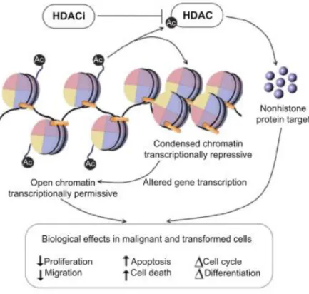
As previous mentioned in section I.1, cancer has been correlated in recent years with extensive reprogramming of epigenetic machinery components including DNA methylation and histone modifications. Treatment with HDACi can potentially lead to the correction of the epigenetic reprogramming found in cancer. HDACi have an important role in histone regulation, preventing the removal of acetyl groups from lysines by HDAC (see section I.2). Due to their effect in histone regulation, HDACi are responsible for an overall increase in acetylated histones causing altered gene transcription. HDACi can also act in other substrates leading to an increased acetylation level and altered gene transcription of non-histone proteins that are important regulators in several processes, resulting in numerous side effects that can also contribute to their anti-cancer action. Several studies have shown that HDACi can inhibit proliferation, stimulate apoptosis24, and induce cell cycle arrest25 in malignant cells10. (Figure I.5)
Figure I.5 - Biological effects of HDACi in malignant cells. Adapted from K.Ververis et al10.
Moreover, the SAHA compound produces several effects in cancerous cells such as alteration in gene transcription, an increase of acetylated proteins, growth arrest in vitro and in
vivo26, induction of apoptosis24 and cell cycle arrest25 with little toxicity to normal cells27. The
mechanisms behind these anticancer effects are not fully understood but several efforts have been made to correlate the increase of acetylation in histone and non-histone proteins with the effects in malignant cells.
10
Some mechanisms normally dysregulated in cancer are apoptosis/cell death and cell cycle progression, enabling cancer cells to growth in an abnormal and very rapid manner and enabling them to overcome checkpoints that usually lead to cell death. Therefore, these type of dysregulations are hallmarks for ongoing tumor progression and good targets for anticancer drugs.
Apoptosis is the process of programmed cell death and is mediated by intrinsic (mitochondrial) and extrinsic (death receptor) pathways. HDAC inhibitors induce apoptosis via both pathways by up-regulating pro-apoptotic and down-regulating anti-apoptotic proteins28,29.
The p53 tumor suppressor gene encodes for a transcriptional factor whose activity is modulated by post-translational modifications including acetylation and protein stability. Studies showed that through acetylation of p53, HDACi were able to up-regulate pro-apoptotic proteins such as Bim, Bak, and Bax, which function as sensors of cellular stress and initiate the intrinsic pathway29. At
a molecular level, apoptosis is caused by the activation of caspases29.
Several HDAC inhibitors induce cell cycle arrest at G1 via induction of the cyclin-dependent kinase inhibitor p21WAF/CIP1. HDAC inhibitors also induce G2/M arrest in normal and
transformed cells, but the latter lack a functional G2 checkpoint and frequently undergo apoptosis28.
As mentioned in section I.2, a higher acetylation of lysine residues of histones is correlated with a more open chromatin conformation. Therefore, DNA can be more accessible for DNA damage agents and reactive oxygen species (ROS) increasing the potential of causing DNA damage. Valproic acid has been reported to enhance the radiosensitivity of human tumour cells
in vivo and in vitro. In mice, irradiation of brain tumour cells after treatment with valproic acid
resulted in radiation-induced tumour growth delay30. Related studies with other HDACi showed
similar results. Therefore, these compounds can act as radio-sensitizers, being helpful in combined therapies with radiotherapy for cancer treatment.
One additional characteristic that makes HDACi appealing as anticancer agents is the fact that these inhibitors are normally correlated with a normal cell resistance phenomenon. They have antitumor action in cancerous cells at concentrations at which normal cells show little toxicity. Normal cells are up to tenfold more resistant to SAHA-induced cell death than cancer cells 27. The mechanism that leads to this selectivity of HDACi is not well understood and several
studies tried to answer this question. One study suggests that thioredoxin, a small redox protein, has a key role in the response of normal and cancer cells to HDACi 27. They show that higher
levels of thioredoxin in normal cells could explain, in part, the relative resistance of normal cells to these compounds effects. Low levels of thioredoxin in transformed cells increased their sensitivity to HDACi-induced cell death27.
Another study claims that HDACi are able to induce DNA damage in both cancerous and non-cancerous cells but only normal cells can repair this damage 31. The authors demonstrated
11
dermal fibroblasts) and cancer cells (LNCaP, A549). Using a marker of DNA DSB during continued culture with SAHA, the authors saw an increase in levels of that marker with time in cancer cells but a decrease in normal cells. Also, they found that SAHA was able to suppress DNA DSB repair proteins in cancer but not in normal cells31.
I.4.4 HDACi Synthesis
Based on previously published synthesis for HDAC inhibitors and derivatives, the synthesis of hydroxamic or carboxylic derivatives can be achieved in two steps 32–34 (See scheme in figure I.6).
Figure I.6 - General scheme of HDAC inhibitors synthesis.
The first step consists on the formation of an amide under an inert atmosphere (argon), using an aniline derivative and a carboxylic acid with a coupling agent such as 1-Ethyl-3-(3-dimethylaminopropyl)carbodiimide (EDC) in dry dimethylformamide (DMF) . The obtained ester is then converted into its corresponding hydroxamic derivative by using hydroxylamine hydrochloride (NH2OH.HCl) in dry methanol (MeOH) for 1-2h. The ester obtained in step 1 can
also be transformed to its carboxylic derivate using sodium hydroxide (NaOH). The ester is dissolved in tetrahydrofuran (THF), NaOH added to the mixture and the reaction is stirred for 1-2h. (See scheme in figure 6).
Other reagents can be used as coupling agents such as N,N'-dicyclohexylcarbodiimide (DCC) and N,N'-Diisopropylcarbodiimide (DIC).
1st STEP-AMIDE FORMATION CARBOXYLIC ACID HYDROXAMIC ACID
12
As mentioned before (See section I.4.2), HDACi such as SAHA and Belinostat structurally contain three important domains: a capping group, a metal binding moiety and a linker. Several analogues were synthesized where these key aspects were altered in order to assess which structures are more important to the effectiveness of these compounds.
Several analogues with different structures can be obtained changing the starting material and the aniline derivative used in the first step of synthesis. (See scheme in figure I.6). Different starting materials with different carbon lengths lead to analogues with different linkers. In this work, mono-ethyl fumarate, adipic acid monomethyl ester, suberic acid monomethyl ester and azelaic acid monomethyl ester were used as starting compounds. Also, different aniline derivatives used in the initial step (See scheme in figure I.6) will enable the synthesis of analogues containing different atoms in the benzenic ring, modifying the capping group.
Furthermore, a metal chelator moiety is crucial in the HDACi structure. Not only the hydroxamic acid moiety but also the carboxylic moiety are good chelators of metal, even though the carboxylic acid is less efficient in this role. Therefore, carboxylic acid analogues can also have activity as inhibitors of HDAC and were also synthesized and tested in cancer cell lines.
13 I.5 Thesis aim
The final goal of this work was to synthesize new HDAC inhibitors with anticancer effects. The first objective was to synthesize several compounds using the structures of SAHA and Belinostat as a starting point/model and evaluate them as anticancer agents in cancer cell lines. Within this purpose, the synthesized compounds were altered in key aspects of HDACi structure. All three important domains were changed: the capping group was altered with the addition of different atoms to the benzenic ring, the carbon length of linker was modified in different extents and both hydroxamic acid and carboxylic acid forms were synthesized. (Figure I.7).
Figure I.7 - General structure of HDACi.
These compounds were evaluated in cancer cell lines in parallel with SAHA as reference compound. The effect of the synthesized compounds on cell viability of a breast cancer cell line (MCF-7) and a lung cancer cell line (H460) was assessed in the presence and absence of the drugs to analyze which functional groups lead to a higher effect in cancer cells.
The second objective was to refine the compound structures taking to account the results obtained in the first round of drug testing by synthesizing new molecules combining the best groups in the same molecule, when possible.
The third objective was to evaluate these compounds in the same cancer cell lines and address the mechanisms of cell death of the compounds with higher impact on cancer cell viability. R1 – F, I, Br, CF3, OCH3 at different positions R2 – OH, NHOH n – 4,6, 7 R2 – OH, NHOH
15
II.
MATERIALS AND METHODS
II.1 Synthesis of New HDACi
II.1.1 General conditions
All reactions were carried out under an inert atmosphere (argon), except when the solvents were not dried. The synthesized compounds were purified by recrystallization or silica flash-column chromatography. Reactions were followed by Analytical TLC (thin-layer chromatography). The characterization of compounds was done by 1H-NMR, 13C-NMR, 13C-APT,
2D NMR techniques (COSY and HMQC), IR spectroscopy and melting point measurement. NMR peak assignments are supported by 2D correlation NMR studies and the peaks assigned when possible.
Analytical TLC was performed on aluminium-backed Merck 60 F254 silica gel plates. The spots corresponding to the products were identified by UV radiation (254 nm) and then immersed on a 5% phosphomolybdic acid solution in ethanol to be revealed. Compounds with the hydroxamic acid moiety were revealed using a solution containing 1-5% iron chloride (III) and 0.5 M HCl. After immersion in this solution, hydroxamic acids appear red instantly.
Silica flash-column chromatography in Silica gel Merck 60 was used, when applicable.
1H-NMR spectra were recorded on a Bruker 400 spectrometer and obtained at 400 MHz
in CDCl3 (deuterated chloroform) or DMSO-d6 (deuterated dimethyl sulfoxide). Chemical shifts
are given in ppm, downfield from tetramethylsilane, for solutions in CDCl3. 1H-NMR spectra were
analyzed using BRUKER TOPSIN 2.1.
13C-NMR spectra were recorded on a Bruker 400 spectrometer at 100.61 MHz in CDCl
3
or DMSO-d6. 13C-NMR spectra were analyzed using Bruker Topsin 2.1 software.
IR spectra were measured on a Nicolet 6700 ATR-FTIR spectrometer with a diamond
crystal. IR spectra were analyzed using Omnic software.
Melting Points were measured twice for each compound using a BUCHI 530 apparatus
16
Solvent and Reagent Purification
All the used solvents were previously distilled in the laboratory.
Aniline – distilled under reduced pressure.
Dry DMF: to previously distilled DMF, calcium hydride (drying agent) was added and the
mixture was left overnight, followed by decantation from the drying agent and distillation under reduced pressure.
Dry THF: to previously distilled THF, sodium wire and benzophenone were added, and
the mixture was refluxed under argon for several hours until the solvent turned deep blue in colour. Then the mixture was kept at low reflux, being only distilled before its utilization.
Dry Methanol: to 50-70 mL of previously distilled methanol, 5g of magnesium turnings
and iodine (0.5 g) were added. The mixture was refluxed until all the magnesium had been consumed. More methanol (1L) was added and the reflux was maintained for 2h before distillation.
II.1.2 General methods
Experiment A - General methods for the 1st Step synthesis – Amide formation
The experimental procedure to obtain the amides started by dissolving the starting material (mono-ethyl fumarate (Aldrich), adipic acid monomethyl ester (Aldrich), suberic acid monomethyl ester (Carbosynth) or azelaic acid monomethyl ester (Aldrich)) in dry DMF. The respective aniline was added (1.5 equivalents) and EDC (Fluka), a coupling agent was added (1.5 equivalents) stepwise. Reactions were maintained overnight under argon at room temperature. TLC (5:5 hexane: ethyl acetate) confirmed that the initial product was totally consumed. Reactions were stopped with the addition of water followed by organic phase extraction using ethyl acetate. Later, ethyl acetate and also DMF were evaporated, the last step using high vacuum. After the work-up procedure, purifications of the compounds were done by recrystallization using hexane and ethyl acetate or by silica flash column chromatography.
Experiment B - General methods for carboxylic acid synthesis
Compounds previously synthesized in the amide formation step were dissolved in THF and then NaOH 1M (1 equivalent) solution was added to the mixture. Reactions were stirred between 1h and 2h with strong agitation at room temperature. TLC (5:5 hexane: ethyl acetate) was performed to monitor the reactions. The reactions were stopped when the starting material was totally consumed with the addition of water followed by a washing step using ethyl ether. Subsequently, the aqueous phase was acidified using a solution of HCl 10% until it reached a pH
17
3-4. Precipitation of product occurred and was followed by organic phase extraction using ethyl acetate. Then, ethyl acetate was evaporated. After the work-up procedure, the purification of the compounds was done by recrystallization using hexane and ethyl acetate.
Experiment C - General methods for hydroxamic acid synthesis
Compounds previously synthesized in the amide formation step were dissolved in dry MeOH and then NH2OH.HCl (Merck) was added (0.8 equivalents) to the mixture. The reactions
were then placed on an ice bath (0ºC) and NaOH 4M (8 equivalents) was also added to the flask. The reactions were maintained for 1-2h under argon at 0ºC. TLC (5:5 hexane:ethyl acetate) was performed to monitor the reactions and the reactions were stopped when the starting material was totally consumed, by adding water followed by a washing step using ethyl ether. Next, the aqueous phase was acidified using an aqueous solution of HCl 10% until pH 3-4. Precipitation of the product occurred and was followed by organic phase extraction using ethyl acetate. A washing step using sodium bicarbonate (NaHCO3) was then performed and ethyl acetate phase was
evaporated. After the work-up procedure, purification of the compounds was done by recrystallization using hexane and ethyl acetate.
II.1.3 Experimental Procedures
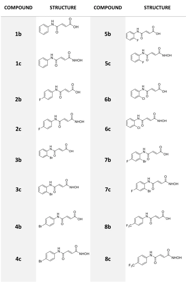
Experiment 1: Synthesis of ethyl (2E)-3-(phenylcarbamoyl)prop-2-enoate (1a)
The procedure in experiment A was applied to the starting material mono-ethyl fumarate (0.5 g, 3.5 mmol). Previously distilled aniline (5.2 mmol) was used in this experiment. Purification of the reaction crude performed by recrystallization afforded compound 1a (0.555 g, 73%) as a white solid. This reaction was repeated and compound 1a was also obtained (0.463 g, 61%).
1H-NMR (CDCl
3): δ 7.86 (1H, broad s, NH), 7.61 (2H, d, J= 7.8 Hz, Ar), 7.35 (2H, t, J=8.0 Hz, Ar),
7.18 – 7.14 (1H, m, Ar), 7.12 (1H, d. J= 15.2 Hz, CH=CH), 6.96 (1H, d, J=15.3 Hz, CH=CH) ,4.28 (2H, q, J=7.3 Hz, OCH2CH3), 1.33 (3H, t. J= 7.2 Hz, OCH2CH3)
13C-NMR (CDCl
3): δ 165.7 (C=O), 161.5 (C=O), 137.3 (Ar, quaternary C), 136.8 (C=C), 131.4
(C=C), 129.1 (Ar), 125.0 (Ar), 120.0 (Ar), 61.4 (CH2CH3), 14.1 (CH2CH3)
Melting Point – 105 °C
18
Experiment 2: Synthesis of (2E) -3-(phenylcarbamoyl) prop-2-enoic acid (1b)
The procedure in experiment B was applied to compound 1a (0.3 g, 1.4 mmol). Purification of the reaction crude performed by recrystallization afforded compound 1b (69 mg, 26 %) as a white solid.
1H-NMR (DMSO-d
6): δ 12.90 (1H, broad s, OH), 10.49 (1H, s, NH), 7.68 (2H, d, J= 7.7 Hz, Ar),
7.35(2H, t, J= 7.8 Hz, Ar), 7.15 (1H, d, J=15.3 Hz, Ar), 7.10 (1H, d, J= 7.4 Hz, CH=CH), 6.66 (1H, d, J= 15.3 Hz, (CH=CH).
13C-NMR (DMSO-d
6): δ 166.8 (C=O), 162.1 (C=O), 139.0 (Ar, quaternary C), 137.6 (C=C), 131.3
(C=C), 129.4 (Ar), 124.5 (Ar), 119.8 (Ar).
Melting Point – 228 °C
FT-IR (ATR): 3305 (O-H), 1659 (C=O), 1644 (C=O) (vmax/cm-1)
Experiment 3: Synthesis of (2E)-N-hydroxy-N'-phenylbut-2-enediamide (1c)
The procedure in experiment C was applied to compound 1a (0.5 g, 2.3 mmol). Purification of the reaction crude performed by recrystallization afforded compound 1c (203 mg, 43 %) as a white solid.
1H-NMR (DMSO-d
6): δ 11.12 (1H, broad s, OH), 10.41 (1H, s, NH), 9.20 (1H, broad s, NH-OH),
7.67 (2H, d, J= 7.8 Hz, Ar), 7.34 (2H, t, J= 7.8 Hz, Ar), 7.10 (1H, s, Ar), 7.08 (1H, d, J= 15.2 Hz, CH=CH), 6.82 (1H, d, J= 15.2 Hz, CH=CH).
13C-NMR (DMSO-d
6): δ 166.7 (C=O), 161.6 (C=O), 139.2 (Ar, quaternary C), 131.6 (C=C), 134.5
(C=C), 129.3 (Ar), 124.3 (Ar), 119.8 (Ar).
Melting Point – 197 °C
19
Experiment 4: Synthesis of ethyl (2E)-3-[(4-fluorophenyl)carbamoyl]prop-2-enoate (2a)
The procedure in experiment A was applied to mono-ethyl fumarate (0.8 g, 5.55 mmol). 2-Fluoroaniline (8.3 mmol) was used in this experiment. Purification of the reaction crude performed by recrystallization afforded compound 2a (0.974 g, 74 %) as a white solid.
1H-NMR (CDCl
3): δ 7.86 (1H, broad s, NH), 7.59 – 7.56 (2H, m, Ar), 7.09 (1H, d, J= 15.3 Hz,
CH=CH), 7.07 – 7.02 (2H, m, Ar), 6.95 (1H, d, J= 15.24 Hz, CH=CH), 4.27 (2H, q, J=7.2 Hz, OCH2CH3), 1.33 (3H, t. J= 7.2 Hz, OCH2CH3)
13C-NMR (CDCl
3): δ 165.6 (C=O), 161.5 (C=O), 160.1 (quaternary C, F-C), 136.4 (C=C), 133.4
(Ar, quaternary C), 131.5 (C=C), 121.9 (Ar), 121.8 (Ar), 115.9 (Ar), 115.7 (Ar), 61.5 (CH2CH3),
14.1 (CH2CH3)
Melting Point – 149 °C
FT-IR (ATR): 3302 (N-H), 1716 (C=O), 1670 (C=O) (vmax/cm-1)
Experiment 5: Synthesis of (2E)-2-[(4-fluorophenyl)carbamoyl]prop-2-enoic acid (2b)
The procedure in experiment B was applied to compound 2a (0.3 g, 1.3 mmol). Purification of the reaction crude performed by recrystallization afforded compound 2b (70 mg, 27 %) as a white solid.
1H-NMR (DMSO-d
6): δ 13.01 (1H, broad s, OH), 10.55 (1H, broad s, NH), 7.72 – 7.68 (2H, m, Ar),
7.19 (2H, t, J= 8.8 Hz, Ar), 7.10 (1H, d, J= 15.4 Hz, CH=CH), 6.66 (1H, d, J= 15.4 Hz, CH=CH)
13C-NMR (DMSO-d
6):
δ
166.7 (C=O), 162.0 (C=O), 160.1 (quaternary C, F-C), 137.3 (C=C), 135.4(Ar, quaternary C), 131.4 (C=C), 121.7 (Ar), 121.6 (Ar), 116.1 (Ar), 115.9 (Ar)
Melting Point – 235 °C
20
Experiment 6: Synthesis of (2E)-N'-(4-fluorophenyl)-N-hydroxybut-2-enediamide (2c)
The procedure in experiment C was applied to compound 2a (0.4 g, 1.7 mmol). Purification of the reaction crude performed by recrystallization afforded compound 2c (147 mg, 39 %) as a white solid.
1H-NMR (DMSO-d
6): δ 11.10 (1H, broad s, OH), 10.49 (1H, s, NH), 9.32 (1H, broad s, NH-OH),
7.71 – 7.66 (2H, m, Ar), 7.21 – 7.15 (2H, m, Ar), 7.06 (1H, d, J= 15.1 Hz, CH=CH), 6.82 (1H, d, J= 15.1 Hz, CH=CH)
13C-NMR (DMSO-d
6): δ 162.5 (C=O), 159.9 (C=O), 157.6 (quaternary C, F-C), 135.6 (Ar,
quaternary C), 131.6 (C=C), 121.6 (Ar), 121.5 (Ar), 116.0 (Ar), 115.8 (Ar)
Melting Point – 216 °C
FT-IR (ATR): 3257 (O-H), 1687 (C=O), 1647 (C=O) (vmax/cm-1)
Experiment 7: Synthesis of ethyl (2E)-3-[(2-bromophenyl)carbamoyl]prop-2-enoate (3a)
The procedure in experiment A was applied to mono-ethyl fumarate (0.8 g, 5.55 mmol). 2-Bromoaniline (8.3 mmol) was used in this experiment. Purification of the reaction crude performed by recrystallization afforded compound 3a (0.671 g, 41 %) as a brown solid.
1H-NMR (CDCl
3): δ 8.45 (1H, d, J= 8.1 Hz, Ar), 7.89 (1H, broad s, NH), 7.57 (1H, d, J=8.0 HZ,
Ar), 7.35 (1H, t, J= 8.0 Hz, Ar), 7.09 (1H, d, J=15.3 Hz, CH=CH), 7.03 (1H, t, J= 8.0 Hz, Ar), 6.95 (1H, d, J= 15.3 Hz, CH=CH), 4.29 (2H, q, J=7.2, OCH2CH3), 1.35 (3H, t. J= 7.12 Hz, OCH2CH3)
13C-NMR (CDCl
3): δ 166.7 (C=O), 161.5 (C=O), 136.1 (Ar, quaternary C), 135.7 (C=C), 131.8
(Ar), 127.9 (Ar), 127.5 (C=C), 125.5 (Ar), 117.5 (quaternary C, Br-C), 61.4 (CH2CH3), 14.4
(CH2CH3)
Melting Point – 115 °C
21
Experiment 8: Synthesis of (2E)-3-[(2-bromophenyl)carbamoyl]prop-2-enoic acid (3b)
The procedure in experiment B was applied to compound 3a (0.3 g, 1 mmol). Purification of the reaction crude performed by recrystallization afforded compound 3b (133 mg, 49 %) as a brown solid.
1H-NMR (DMSO-d
6): δ 13.02 (1H, broad s, OH), 10.08 (1H, s, NH), 7.70 - 7.65 (2H, m, Ar), 7.41
(1H, t, J= 7.8 Hz), 7.29 (1H. d, J= 15.4 Hz, CH=CH), 7.19 (1H, t, J= 7.9 Hz), 6.67 (1H, d, J= 15.4 Hz, CH=CH)
13C-NMR (DMSO-d
6): δ 166.7 (C=O), 162.6 (C=O), 136.9 (C=C), 136.1 (Ar, quaternary C), 133.3
(Ar), 131.9 (C=C), 128.5 (Ar), 128.1 (Ar), 127.8 (Ar), 118.5 (quaternary C, Br-C)
Melting Point – 213 °C
FT-IR (ATR): 3271 (O-H), 1696 (C=O), 1662 (C=O) (vmax/cm-1)
Experiment 9: Synthesis of (2E)-N'-(2-bromophenyl)-N-hydroxybut-2-enediamide (3c)
The procedure in experiment C was applied to compound 3a (0.3 g, 1 mmol). Purification of the reaction crude performed by recrystallization afforded compound 3c (146 mg, 51 %) as a white solid.
1H-NMR (DMSO-d
6): δ 11.11 (1H, s, OH), 10.01 (1H, s, NH), 9.32 (1H, broad s, NH-OH), 7.70 –
7.65 (2H, m, Ar), 7.41 (1H, t, J= 7.4 Hz, Ar), 7.24 (1H, J= 15.2 Hz, CH=CH), 7.18 (2H, t, J= 7.4 Hz, Ar), 6.84 (1H, d, J= 15.2 Hz, CH=CH)
13C-NMR (DMSO-d
6): δ 166.7 (C=O), 161.6 (C=O), 136.2 (Ar, quaternary C), 133.2 (C=C), 132.2
(Ar), 128.5 (C=C), 128.0 (Ar), 127.8 (Ar), 117.5 (quaternary C, Br-C)
Melting Point – 207 °C
22
Experiment 10: Synthesis of ethyl (2E)-3-[(4-bromophenyl)carbamoyl]prop-2-enoate (4a)
The procedure in experiment A was applied to mono-ethyl fumarate (0.8 g, 5.55 mmol). 4-Bromoaniline (8.4 mmol) was used in this experiment. Purification of the reaction crude performed by recrystallization afforded compound 4a (1.484 g, 90 %) as a white solid.
1H-NMR (CDCl
3): δ 7.82 (1H, broad s, NH),7.52 (2H, d, J=8.8 Hz, Ar), 7.46 (2H, d, J= 8.8 Hz,
Ar), 7.07 (1H, d, J=15.3 Hz, CH=CH), 6.95 (1H, d, J= 15.3 Hz, CH=CH), 4.28 (2H, q, J=7.2 Hz, OCH2CH3), 1.33 (3H, t. J= 7.2 Hz, OCH2CH3)
13C-NMR (CDCl
3):
δ
165.5 (C=O), 161.5 (C=O), 136.5 (Ar, quaternary C), 136.2 (C=C), 132.1(Ar), 131.8 (C=C), 121.5 (Ar), 117.8 (quaternary C, Br-C), 61.5 (CH2CH3), 14.1 (CH2CH3)
Melting Point – 159 °C
FT-IR (ATR): 3295 (N-H), 1712 (C=O), 1669 (C=O) (vmax/cm-1)
Experiment 11: Synthesis of (2E)-3-[(4-bromophenyl)carbamoyl]prop-2-enoic acid (4b)
The procedure in experiment B was applied to compound 4a (0.3 g, 1 mmol). Purification of the reaction crude performed by recrystallization afforded compound 4b (202 mg, 74 %) as a white solid.
1H-NMR (DMSO-d
6): δ 13.03 (1H, broad s, OH), 10.62 (1H, s, NH), 7.64 (2H, d, J= 8.9 Hz, Ar),
7.53 (2H, d, J=8.8 Hz, Ar), 7.11 (1H, d, J=15.4 Hz, CH=CH), 6.66 (1H, d, J= 15.4 Hz, CH=CH)
13C-NMR (DMSO-d
6): δ 166.7 (C=O), 162.2 (C=O), 138.4 (Ar, quaternary C), 137.3 (C=C), 132.2
(Ar), 131.5 (C=C), 121.8 (Ar), 116.2 (quaternary C, Br-C)
Melting Point – >250 °C
23
Experiment 12: Synthesis of (2E)-N'-(4-bromophenyl)-N-hydroxybut-2-enediamide (4c)
The procedure in experiment C was applied to compound 4a (0.4 g, 1.3 mmol). Purification of the reaction crude performed by recrystallization afforded compound 4c (110 mg, 29 %) as a yellowish solid.
1H-NMR (DMSO-d
6): δ 11.15 (1H, broad s, OH), 10.59 (1H, s, NH), 9.36 (1H, broad s, NH-OH),
7.65 (2H, d, J=8.8 Hz, Ar), 7.53 (2H, d, J= 8.8 Hz, Ar), 7.07 (1H, d, J=15.3 Hz, CH=CH), 6.83 (1H, d, J= 15.3 Hz, CH=CH)
13C-NMR (DMSO-d
6): δ 164.5 (C=O), 162.8 (C=O), 138.6 (Ar, quaternary C), 132.2 (Ar), 131.9
(C=C), 121.7 (Ar), 115.9 (quaternary C, Br-C).
Melting Point – 247 °C
FT-IR (ATR): 3207 (O-H), 1648 (C=O), 1621 (C=O) (vmax/cm-1)
Experiment 13: Synthesis of ethyl (2E)-3-[(2-fluorophenyl)carbamoyl]prop-2-enoate (5a)
The procedure in experiment A was applied to mono-ethyl fumarate (0.8 g, 5.55 mmol). 2-Fluoroaniline (8.4 mmol) was used in this experiment. Purification of the reaction crude performed by recrystallization afforded compound 5a (0.671 g, 52 %) as a white solid.
1H-NMR (CDCl
3): δ 8.41 (1H, t, J=8.1 Hz, Ar), 7.66 (1H, broad s, NH), 7.19 – 7.12 (3H, m, Ar),
7.07 (1H, d, J=15.3 Hz, CH=CH), 6.95 (1H, d, J= 15.3 Hz, CH=CH), 4.29 (2H, q, J=7.2 Hz, OCH2CH3), 1.34 (3H, t, J= 7.2 Hz, OCH2CH3)
13C-NMR (CDCl
3): δ 165.2 (C=O), 161.4 (C=O), 158.3 (F-C, quaternary C), 144.1 (Ar, quaternary
C), 135.8(C=C), 132.1 (C=C), 125.2 (Ar), 124.7 (Ar), 121.8 (Ar), 114.9 (Ar), 61.4 (CH2CH3), 14.2
(CH2CH3)
Melting Point – 96 °C
24
Experiment 14: Synthesis of (2E)-3-[(2-fluorophenyl)carbamoyl]prop-2-enoic acid (5b)
The procedure in experiment B was applied to compound 5a (0.3 g, 1.3 mmol). Purification of the reaction crude performed by recrystallization afforded compound 5b (195 mg, 74 %) as a white solid. 1H-NMR (DMSO-d 6): δ 13.01 (1H, s, OH), 10.32 (1H, s, NH), 8.03 – 7.99 (1H, m, Ar), 7.33 (2H. d, J= 15.4 Hz, CH=CH), 7.30 – 7.26 (1H, m, Ar), 7.23 – 7.18 (2H, m, Ar), 6.68 (1H, d, J= 15.4 Hz, CH=CH) 13C-NMR (DMSO-d
6):
δ
166.7 (C=O), 162.6 (C=O), 155.3 (F-C, quaternary C), 152.8 (Ar,quaternary C), 136.9 (C=C), 131.8 (C=C), 126.4 (Ar), 124.9 (Ar), 124.5 (Ar), 115.9 (Ar)
Melting Point – 215 °C
FT-IR (ATR): 3290 (O-H), 1693 (C=O), 1665 (C=O) (vmax/cm-1)
Experiment 15: Synthesis of (2E)-N'-(2-fluorophenyl)-N-hydroxybut-2-enediamide (5c)
The procedure in experiment C was applied to compound 5a (0.282 g, 1.2 mmol). Purification of the reaction crude performed by recrystallization afforded compound 5c (98 mg, 37 %) as a white solid.
1H-NMR (DMSO-d
6): δ 11.10 (1H, broad s, OH), 10.23 (1H, s, NH), 9.33 (1H, broad s, NH-OH),
8.01 – 7.96 (1H, m, Ar), 7.31 – 7.17 (4H, m, Ar and CH=CH), 6.84 (1H, d, J= 15.16 Hz, CH=CH)
.
13C-NMR (DMSO-d
6):
δ
166.7 (C=O), 163.1 (C=O), 155.3 (F-C, quaternary C), 132.2 (C=C), 126.3(C=C), 126.2 (Ar, quaternary C), 124.9 (Ar), 124.6 (Ar), 116.1 (Ar), 115.9 (Ar)
Melting Point – 212 °C
25
Experiment 16: Synthesis of ethyl (2E)-3-[(2-chlorophenyl)carbamoyl]prop-2-enoate (6a)
The procedure in experiment A was applied to mono-ethyl fumarate (0.8 g, 5.55 mmol). 2-chloroaniline (8.3 mmol) was used in this experiment. Purification of the reaction crude performed by recrystallization afforded compound 6a (0.758 g, 54 %) as a white solid.
1H-NMR (CDCl
3): δ 8.52 (1H, t, J=8.1 Hz, Ar), 7.91 (1H, broad s, NH), 7.42 (1H, d, J= 8.1 Hz, Ar),
7.31 (1H, t, J=8.0 Hz, Ar) 7.12 – 7.07 (2H, m. Ar and CH=CH), 6.95 (1H, d, J= 15.3 Hz, CH=CH), 4.29 (2H, q, J=7.2 Hz, OCH2CH3), 1.35 (3H, t, J= 7.2 Hz, OCH2CH3)
13C-NMR (CDCl
3):
δ
165.2 (C=O), 160.6 (C=O), 134.1 (Ar, quaternary C), 136.1(C=C), 132.1(C=C), 129.1 (Ar),128.2 (Cl-C, quaternary C), 127.9 (Ar), 125.4 (Ar), 121.8 (Ar), 121.6 (Ar), 61.4 (CH2CH3), 14.2 (CH2CH3)
Melting Point – 100 °C
FT-IR (ATR): 3282 (N-H), 1712 (C=O), 1667 (C=O) (vmax/cm-1)
Experiment 17: Synthesis of (2E)-3-[(2-chlorophenyl)carbamoyl]prop-2-enoic acid (6b)
The procedure in experiment B was applied to compound 6a (0.3 g, 1.1 mmol). Purification of the reaction crude performed by recrystallization afforded compound 6b (233 mg, 88 %) as a white solid.
1H-NMR (DMSO-d
6): δ 12.96 (1H, broad s, OH), 10.13 (1H, s, NH), 7.79 (1H, d, J=7.9 Hz, Ar),
7.53 (1H, d, J= 7.9 Hz, Ar), 7.39 – 7.32 (2H, m, Ar and CH=CH), 7.25 (1H, t, J=7.8 Hz, Ar), 6.68 (1H, d, J= 15.4 Hz, CH=CH)
13C-NMR (DMSO-d
6): δ 166.8 (C=O), 162.6 (C=O), 137.0 (C=C), 134.7 (Ar, quaternary C), 131.8
(C=C), 130.1 (Ar), 127.9 (Ar), 127.4 (Ar), 127.1 (quaternary C, Cl-C), 126.8 (Ar)
Melting Point – 220 °C
26
Experiment 18: Synthesis of (2E)-N'-(2-chlorophenyl)-N-hydroxybut-2-enediamide (6c)
The procedure in experiment C was applied to compound 6a (0.3 g, 1.1 mmol). Purification of the reaction crude performed by recrystallization afforded compound 6c (70 mg, 25 %) as a white solid.
1H-NMR (DMSO-d
6): δ 11.13 (1H, broad s, OH), 10.06 (1H, s, NH), 9.35 (1H, broad s, NH-OH),
7.77 (1H, d, J=7.9 Hz, Ar), 7.5 (1H, d, J= 8.0 Hz, Ar), 7.36 (1H, d, J=7.9 Hz, Ar), 7.28 – 7.21 (2H, m, Ar and CH=CH), 6.84 (1H, d, J=15.2 Hz, CH=CH)
13C-NMR (DMSO-d
6): δ166.1 (C=O), 163.1 (C=O), 134.9 (Ar, quaternary C), 132.2 (C=C), 130.0
(C=C), 127.9 (Ar), 127.3 (quaternary C, Cl-C), 127.3 (Ar), 126.9 (Ar)
Melting Point – 202 °C
FT-IR (ATR): 3255 (O-H), 1638 (C=O), 1618 (C=O) (vmax/cm-1)
Experiment 19: Synthesis of ethyl (2E)-3-[(2-bromo-4-fluorophenyl)carbamoyl]prop-2-enoate (7a)
The procedure in experiment A was applied to mono-ethyl fumarate (0.8 g, 5.55 mmol). 2-Bromo-4-fluoroaniline (8.3 mmol) was used in this experiment. Purification of the reaction crude performed by recrystallization afforded compound 7a (1g, 38 %) as a white solid.
1H-NMR (CDCl
3): δ 8.44 – 8.41 (1H, m, Ar), 7.77 (1H, broad s, NH), 7.35 – 7.32 (1H, m, Ar), 7.12–
7.05 (2H, m, Ar and CH=CH), 6.95 (1H, d, J= 15.3 Hz, CH=CH), 4.30 (2H, q, J=7.2 Hz, OCH2CH3),
1.35 (3H, t, J= 7.2 Hz, OCH2CH3)
13C-NMR (CDCl
3): δ 165.2 (C=O), 160.2 (C=O), 151.4 (F-C, quaternary C), 135.9 (C=C), 132.2
(C=C), 131.6 (Ar, quaternary C), 123.1 (Ar), 120.5 (Br-C, quaternary C), 119.5 (Ar), 115.4 (Ar), 61.5 (CH2CH3), 14.2 (CH2CH3)
Melting Point – 130 °C
27
Experiment 20: Synthesis of (2E)-3-[(2-bromo-4-fluorophenyl)carbamoyl]prop-2-enoic acid (7b)
The procedure in experiment B was applied to compound 7a (0.3 g, 0.95 mmol). Purification of the reaction crude performed by recrystallization afforded compound 7b (243 mg, 89%) as a white solid.
1H-NMR (DMSO-d
6): δ 13.05 (1H, broad s, OH), 10.13 (1H, s, NH), 7.70 – 7.63 (2H, m, Ar), 7.34
– 7.28 (1H, m, Ar), 7.25 (1H, d, J=15.4 Hz, CH=CH), 6.68 (1H, d, J=15.3 Hz, CH=CH)
13C-NMR (DMSO-d
6):
δ
166.7 (C=O), 162.8 (C=O), 161.2 (quaternary C, F-C), 136.8 (C=C), 132.8(Ar, quaternary C), 131.9 (C=C), 129.4 (Ar), 120.1 (Ar), 119.5 (quaternary C, Br-C),115.5 (Ar)
Melting Point – 230 °C
FT-IR (ATR): 3258 (O-H), 1698 (C=O), 1660 (C=O) (vmax/cm-1)
Experiment 21: Synthesis of (2E)-N'-(2-bromo-4-fluorophenyl)-N-hydroxybut-2-enediamide (7c)
The procedure in experiment C was applied to compound 7a (0.3 g, 1.1 mmol). Purification of the reaction crude performed by recrystallization afforded compound 7c (79 mg, 27 %) as a white solid.
1H-NMR (DMSO-d
6): δ 11.10 (1H, broad s, OH), 10.05 (1H, s, NH), 9.33 (1H, broad s, NH-OH),
7.69 – 7.62 (2H, m, Ar), 7.30 (1H, t, J= 8.2 Hz, Ar), 7.20 (1H, d, J= 15.2 Hz, CH=CH), 6.83 (1H, d, J= 15.2 Hz, CH=CH)
13C-NMR (DMSO-d
6):
δ
166.7 (C=O), 161.6 (C=O), 160.7 (quaternary C, F-C), 134.5 (C=C), 131.6(Ar, quaternary C), 130.8 (C=C), 125.4 (Ar), 124.2 (quaternary C, Br-C), 120.9 (Ar), 114.7 (Ar)
Melting Point – 203 °C
28
Experiment 22: Synthesis of ethyl (2E)-3-{[4-(trifluoromethyl)phenyl]carbamoyl}prop-2-enoate (8a)
The procedure in experiment A was applied to mono-ethyl fumarate (0.8 g, 5.55 mmol. 4-(Trifluoromethyl)aniline (8.3 mmol) was used in this experiment. Purification of the reaction crude performed by recrystallization afforded compound 8a (1.136 g, 71 %) as a white solid.
1H-NMR (CDCl
3): δ 8.00 (1H, broad s, NH), 7.75 (1H, d, J= 8.3 Hz, Ar), 7.61 (1H, d, J= 8.4 Hz,
Ar), 7.11 (1H, d, J= 15.3 Hz, CH=CH),6.98 (1H, d, J= 15.4 Hz, CH=CH), 4.29 (2H, q, J=7.2 Hz, OCH2CH3), 1.34 (3H, t, J= 7.1 Hz, OCH2CH3)
13C-NMR (CDCl
3): δ 166.7 (C=O), 162.5 (C=O), 140.9 (Ar, quaternary C), 135.7 (C=C), 133.1
(C=C), 132.1 (C-CF3, quaternary C), 125.3 (Ar), 124.1 (C-CF3, quaternary C), 121.9 (Ar), 61.4
(CH2CH3), 14.2 (CH2CH3)
Melting Point – 180 °C
FT-IR (ATR): 3350 (N-H), 1702 (C=O), 1682 (C=O) (vmax/cm-1)
Experiment 23: Synthesis of (2E)-3-{[4-(trifluoromethyl)phenyl]carbamoyl}prop-2-enoic acid (8b)
The procedure in experiment B was applied to compound 8a (0.3 g, 1.2 mmol). Purification of the reaction crude performed by recrystallization afforded compound 8b (211 mg, 70%) as a white solid.
1H-NMR (DMSO-d
6): δ 13.08 (1H, broad s, OH), 10.84 (1H, s, NH), 7.88 (2H, d, J=8.4 Hz, Ar),
7.73 (2H, d, J= 8.4 Hz, Ar), 7.15 (1H, d, J= 15.4 Hz, CH=CH), 6.70 (1H, d, J= 15.4 Hz, CH=CH)
13C-NMR (DMSO-d
6):
δ
166.6 (C=O), 162.6 (C=O), 142.7 (Ar, quaternary C), 137.0 (C=C), 131.9(C=C), 126.6 (Ar), 126.1 (C-CF3, quaternary C), 124.4 (C-CF3, quaternary C), 119.9 (Ar)
Melting Point – >250 °C