ISSN 0100-879X
BIOMEDICAL SCIENCES
AND
CLINICAL INVESTIGATION
www.bjournal.com.br
www.bjournal.com.br
Volume 45 (2) 93-178 February 2012
Braz J Med Biol Res, February 2012, Volume 45(2) 125-130
doi: 10.1590/S0100-879X2011007500163
Viability of mesenchymal stem cells during electrospinning
G. Zanatta, D. Steffens, D.I. Braghirolli, R.A. Fernandes, C.A. Netto and P. Pranke
Institutional Sponsors
The Brazilian Journal of Medical and Biological Research is partially financed by
Faculdade de Medicina de Ribeirão Preto Campus
Ribeirão Preto
Ex plor e H igh - Pe r for m a n ce M S Or bit r a p Te ch n ology I n Pr ot e om ics & M e t a bolom ics
Viability of mesenchymal stem cells
during electrospinning
G. Zanatta
1,2*, D. Steffens
1*, D.I. Braghirolli
1, R.A. Fernandes
3,
C.A. Netto
2and P. Pranke
1,41Laboratório de Hematologia e Células-tronco, Faculdade de Farmácia,
Universidade Federal do Rio Grande do Sul, Porto Alegre, RS, Brasil
2Laboratório de Isquemia Cerebral, Departamento de Bioquímica,
Universidade Federal do Rio Grande do Sul, Porto Alegre, RS, Brasil
3Hospital Nossa Senhora da Conceição, Grupo Hospitalar Conceição, Porto Alegre, RS, Brasil 4Instituto de Pesquisa com Células-tronco, Porto Alegre, RS, Brasil
Abstract
Tissue engineering is a technique by which a live tissue can be re-constructed and one of its main goals is to associate cells
with biomaterials. Electrospinning is a technique that facilitates the production of nanofibers and is commonly used to develop fibrous scaffolds to be used in tissue engineering. In the present study, a different approach for cell incorporation into fibrous
scaffolds was tested. Mesenchymal stem cells were extracted from the wall of the umbilical cord and mononuclear cells from umbilical cord blood. Cells were re-suspended in a 10% polyvinyl alcohol solution and subjected to electrospinning for 30 min under a voltage of 21 kV. Cell viability was assessed before and after the procedure by exclusion of dead cells using trypan blue staining. Fiber diameter was observed by scanning electron microscopy and the presence of cells within the scaffolds was analyzed by confocal laser scanning microscopy. After electrospinning, the viability of mesenchymal stem cells was reduced from 88 to 19.6% and the viability of mononuclear cells from 99 to 8.38%. The loss of viability was possibly due to the high viscosity of the polymer solution, which reduced the access to nutrients associated with electric and mechanical stress during
electrospinning. These results suggest that the incorporation of cells during fiber formation by electrospinning is a viable process that needs more investigation in order to find ways to protect cells from damage.
Key words: Nanotechnology; Tissue engineering; Electrospinning; Stem cells
Introduction
Correspondence: P. Pranke, Laboratório de Hematologia e Células-tronco, Faculdade de Farmácia,
Universidade Federal do Rio Grande do Sul, Av. Ipiranga, 2752, Laboratório 304, 90610-000 Porto Alegre, RS, Brasil. E-mail: [email protected] or [email protected]
*These authors contributed equally to this study.
Received July 7, 2011. Accepted December 12, 2011. Available online December 23, 2011. Published February 17, 2012.
Tissue engineering is a growing field with the potential to regenerate damaged tissue (1-3). One of the most important goals in this area is to associate cells with biomaterials to provide a scaffold on which they can anchor, migrate and proliferate three-dimensionally. This approach has a great potential for many tissues such as vessels (4,5), bone (6,7), neural tissue (8,9), cartilage (10,11), and tendons/ ligaments (12,13).
Among the various types of materials used in tissue engineering, scaffolds based on nanofibers produced by electrospinning have been widely studied. This technique is employed to produce nanofibers with a diameter in the range of nanometers to micrometers by the use of electrostatic
forces. Many parameters are involved in the failure of the final fiber, such as the applied voltage, tip-target distance, feeding rate, concentration and viscosity of the polymer solution, and temperature, among others (14,15).
126 G. Zanatta et al.
Mononuclear cells (MNCs) including lymphocytes, monocytes and progenitor cells can be obtained from um-bilical cord blood (UCB), together with a limited number of stem cells such as hematopoietic stem cells and, in lower quantities, mesenchymal stem cells (MSCs) (17-19). An-other source of MSCs is the umbilical cord (UC) itself, which provides more cells than umbilical cord blood (20). Due to their ability for self-renewal and high proliferation capac-ity, MSCs are candidates for cell therapies to regenerate tissues and organs affected by various diseases, such as neurologic, rheumatic, cardiac, and autoimmune diseases, among others.
Although electrospun fibers of various polymers and dif -ferent cell types have been brought into contact numerous times, the fibers are typically electrospun first and cells are seeded onto the material in a separate step (21-23). Due to the limitations of cell mobility or scaffold properties, such as low porosity, sometimes the thee-dimensional structure cannot be completely filled by biological material, compro -mising the structure and function of tissue formation.
In the present study, taking into consideration the pro-cess of co-electrospinning, attention was focused on cell incorporation into the scaffolds during nanofiber formation. The polymer of choice was polyvinyl alcohol (PVA), due to its low toxicity and high solubility in an aqueous medium. The viability of MSCs and MNCs after electrospinning was determined.
Material and Methods
Isolation and characterization of MSC and MNC
The study was approved by the Ethics Committee of the Hospital Moinhos de Vento (Process No. CEP/IEP-AHMV 2006/42) and the Universidade Federal do Rio Grande do Sul (Process No. CEP/UFRGS 2006563) and three samples each of UCB and UC were obtained from the Centro Obstétrico do Hospital Moinhos de Vento after the mothers gave written informed consent.
UCB samples were obtained by puncture of the umbili-cal vein of the umbiliumbili-cal cords of neonates during normal or cesarean delivery and collected into bags containing citrate, phosphate, dextrose, and adenine (CPDA-1) as an anticoagulant. UC samples were collected by cutting the umbilical cord near the placenta following the procedure described by Secco et al. (20) and were kept in a growth medium (DMEM-low glucose, 10% fetal bovine serum, 2 U/ mL penicillin, 2 μg/mL streptomycin, and 44.8 μg/mL gen -tamycin). Both the UCB and UC samples were transported to the laboratory and processed within 24 h post-delivery.
Separation of the MNC fraction from UCB. UCB samples were diluted 1:1 with PBS and MNCs were isolated by density gradient centrifugation at 1200 g for 20 min at room temperature using Ficoll-Paque (Sigma, USA) (24). MNCs were then washed twice with PBS and counted in a Neubauer chamber using 4% Trypan blue
for cell death exclusion.
MSC separation from UC. UC samples were incubated in 0.1% collagenase type I (Sigma) at 37°C for 15-20 min. Detached cells were then harvested after massage of the UC with the aid of tweezers and centrifuged at 300 g for 10 min. Samples were re-suspended in a medium, seeded in 25-cm2 flasks and kept at 37°C in a humidified atmosphere
containing 5% CO2. Non-adherent cells were removed
after 24-h incubation; the culture medium was replaced every 3-4 days.
MSC culture and characterization. Adherent cells were cultured according to established norms (25,26) and standard procedure of the laboratory (27). Passages were performed when cells reached 80-90% confluence at a split rate of 1:3. Cells used in the experiments were cultured until the fourth passage.
Flow cytometry was performed using a FacsCalibur instrument (Becton Dickinson, USA) equipped with an argon-ion laser tuned at 488 nm and the data were ana-lyzed using the CELLQuest software (Becton Dickinson). The cells were incubated with antibodies for 30 min at 4°C and were then washed with PBS. 7-Aminoactinomycin D (7AAD; Molecular Probes, Inc., USA) at a final concentration of 1 µg/mL was used to identify dead cells. The following specific antibodies for the human antigens were purchased from PharMingen/Becton Dickinson, USA): CD29/PE, CD34/PE, CD44/FITC, CD45/FITC, CD90/FITC, CD117/ PE, CD133/PE, CD146/FITC, CD184/PE, HLA-DR/FITC, and Stro-1/FITC, as well as the isotype control antibodies mouse IgG1,k/FITC and IgG1,k/PE. The gating strategy was used in order to acquire 10,000 events corresponding to a live MNC cluster. In this gate, the analysis was performed to observe double labeling in this population using a com-bination of the other antibodies.
Mesenchymal differentiation capacity was determined as described by Meirelles and Nardi (28).
Adipogenic differentiation. MSCs (2.0 x 10⁴ cells/cm2)
were cultured in Iscove’s medium supplemented with 20% human plasma 0.1 µM dexamethasone, 0.44 mM insulin, 5 µM indomethacin, 5 µM rosiglitazone, and 10 µM/mL sodium heparin. The medium was changed every 3 days. Adipogenic potential was determined after 21 days by the formation of intracellular lipid droplets demonstrated by oil red O staining (Sigma-Aldrich Ltd., USA).
Osteogenic differentiation. Cells were seeded at a con-centration of 2.0 x 10⁴ cells/cm2 and cultured in a medium supplemented with 15 mM HEPES, 10% SFB, 10 mM ß-glycerophosphate, 10 nM dexamethasone, and 5 µg/ mL ascorbic acid 2-phosphate. The medium was changed every 3 days. To evaluate the potential of osteogenic dif-ferentiation, the cultures were washed with PBS and calcium deposition was assessed by alizarin red S staining.
Chondrogenic differentiation. Cells at concentrations of 2.0 x 10⁴ cells/cm2 were cultured in a medium consisting
HEPES, 1.09 mM insulin, 10 ng/mL transforming growth factor-β1 (TGF-β1), and 50 nM ascorbic acid-2 phosphate (AsAP). The medium was changed every 3-4 days and the cells were fixed on day 21 with 4% paraformaldehyde. Chondrogenesis was indicated by the deposition of a matrix, rich in glycosaminoglycan sulfate, stained by Alcian blue.
Preparation of PVA solution
An aqueous solution of 10% (w/w) was prepared by mixing PVA (molecular weight: 3.44 x 10⁶ g/mol ± 6.0%)
in Milli-Q water and stirred at room temperature for 16 h to obtain a homogeneous solution.
Preparation of the PVA-cell suspension
MSCs were trypsinized, washed and quantified us -ing a Neubauer chamber. Both MSCs and MNCs were re-suspended in a culture medium and an aliquot of 30 µL containing 1 x 10⁶ cells was mixed with the PVA
solu-tion at a volume ratio of 1:12. The cells from the control group were re-suspended in a growth medium in the same proportions.
Electrospinning
Electrospinning was performed as described by Greiner and Wendorff (3). The PVA-cell suspension was stored in a 1-mL syringe with a hypodermic needle with an inner diameter of 0.45 mm. The suspension was fed at a rate of 0.34 mL/h through a syringe placed 15 cm from the collector plate and a voltage of 21 kV was applied. Room temperature was 25°C and relative humidity 35-50%. Nanofibers were collected on round glass coverslips placed on the ground collector. Fiber production was carried out for 30 min and all experiments were performed in triplicate.
Characterization of nanofibers
The morphology and diameter of the electrospun nanofibers were determined using a scanning electron microscope (SEM; JEOL - JSM 6060, JEOL Ltd., Japan). Samples collected on glass coverslips were mounted on an aluminum stub and sputter-coated with 15 nm carbon for analysis. Three SEM micrographs of distinct areas from each sample were used for analysis. Diameter analysis was performed by measuring the diameter of 30 fibers in each micrograph. The software used for the measurements and statistical analysis was ImageJ 1.38X.
Cell viability analysis
Cell viability was analyzed in three experiments carried out in triplicate in three distinct groups: 1) control: cells in growth medium at room temperature; 2) PVA: cells in a PVA solution at room temperature, and 3) electrospinning: cells electrospun in PVA solution. Cell viability was evaluated in all groups at the beginning of the experiments and after the electrospinning procedure.
Scaffolds produced in the electrospinning group were
transferred to a flask containing culture medium in order to dissolve the fibers. The medium was added to the flasks of the control groups (cells in growth medium and cells in PVA solution) at the same proportion.
All samples were centrifuged at 300 g for 5 min at room temperature and the supernatant was removed. Cells were re-suspended in a medium and cell viability was determined using 4% Trypan blue (Gibco, USA), in a 1:1 (v:v) ratio for dead cell exclusion.
Cell incorporation into scaffolds during electrospinning
The capacity of electrospinning to deliver cells among the fibers was assessed by cell nucleus staining with 0.2 µg/mL 4,6-diamidino-2-phenylindole (DAPI) for 20 min. The suspension was then washed with PBS and centrifuged at 300 g for 10 min. Fibers were collected on glass coverslips for 30 min and images were obtained by confocal laser scanning microscopy (Olympus FV-1000, Japan).
Statistical analysis
Cell viability was analyzed statistically using analysis of variance (ANOVA). Statistical analyses of groups of different cell types were performed by two-way ANOVA. All analyses were followed by the post hoc Tukey test for multiple comparisons when necessary. Data are reported as means ± SD and the level of significance was set at P < 0.05. All statistical analyses were performed using the Statistica® software package running on a compatible
personal computer.
Results
MSC characterization
The characterization of MSCs follows criteria as estab-lished in the literature (25,26) and standard laboratory proce-dures (27): 1) the cells were isolated for adherence in plastic and then proliferated in vitro; 2) they showed differentiation in adipocytes, osteoblasts and chondrocytes when submitted to differentiation assays, and 3) the cells demonstrated posi-tive immunophenotypic profile for mesenchymal stem cells: CD90 (mesenchymal marker), CD29 and CD44 (adhesion markers), and negative for hematopoietic markers: CD45, CD34, CD117, and HLA-DR (data not shown).
Effects of PVA and electrospinning on cell viability
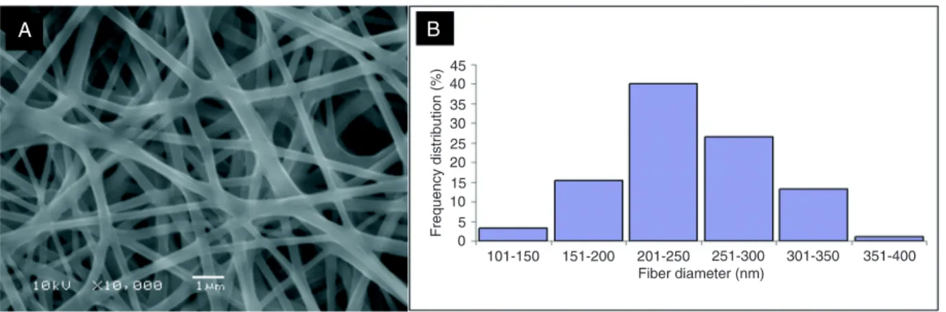
Fibers produced by electrospinning were smooth in appearance and free of beads, with an average diameter of 244 ± 49 nm (Figure 1).
128 G. Zanatta et al.
of viability from 88% in the control group to 64% when the cells were mixed with the PVA solution. Analyses of the cells subjected to electrospinning showed a significant reduction in viability in both types of cells: experiments using MSCs showed 19.6% cell viability and experiments using MNCs showed 8.38% cell viability (Figure 2).
Cell localization in scaffolds as the result of electrospinning
Analysis of the distribution of MSC and MNC on the nanofiber scaffolds was performed using confocal laser scanning microscopy. The images suggested that the cells were not encapsulated by the fibers, probably because of
the small fiber diameter, but rather that they were placed among the fibers, suggesting that they were distributed three-dimensionally in the scaffolds (Figure 3).
Discussion
In the present study, the influence of the electrospin -ning process and the solvent 10% PVA on MSC and MNC viability was analyzed. Electrospinning was performed at 21 kV with a suspension of cells:PVA at a ratio of 1:12 (v:v). Using cells:PVA solution at a higher rate, fibers were not formed and only drops were observed on the collector plate, probably due to the reduction in polymer chain interaction because of the low viscosity. When a higher proportion of PVA solution was used, fibers were continuously produced, but as the concentration of cells decreased, the analyses of viability became hard to implement. These data agree with a recent study performed by van Aalst et al. (2), in which they used electrospinning to perform cell incorpora-tion into nanofibers.
To assess cell viability after the electrospinning proce-dure, two different groups of cells from distinct sources were used. The first group was composed of MNCs, which are a heterogeneous population of cells obtained from UCB. These cells are of great interest for clinical procedures as they provoke less rejection in transplants and are easy to obtain without the need for a long-term process of cultivation or purification. Besides these advantages, it is known that there are a small number of stem cells among the fraction of MNCs (CD34+CD38- frequency = 0.05-0.08%) (29), which are the cells responsible for the regeneration of damaged areas in tissue engineering. The second group of cells used was MSCs, isolated directly from the wall of the UC. Although this population demands a long period for isola-tion and cultivaisola-tion, the use of a homogeneous populaisola-tion of MSCs seems to be more appropriate for the success of tissue engineering procedures. MSCs were collected from
Figure 1.A, Scanning electron micrographs of nanofibers produced by electrospinning using PVA solution at a concentration of 10 wt%
(w/v). B, Frequency of diameter distribution. Mean diameter 244 ± 49 nm.
Figure 2. Cell viability after 30 min of treatment. Control = cells in culture medium; PVA = cells in 10% polyvinyl alcohol solution; electrospinning = cells mixed with 10% PVA solution subjected to electrospinning. MNC = mononuclear cells; MSC = mesenchymal stem cells. Data are representative of three experiments carried out in triplicate and reported as mean cell viability ± SD. *P < 0.05 compared to control; #P< 0.05 compared to the PVA group
the wall of the UC instead of the UCB, as the number of these cells in the UC is greater than in UCB (20,30).
Cells from the second group were analyzed for the pres-ence of surface markers by flow cytometry and showed a classical profile of MSC markers (20,24,25). The antigens CD146 (or Mel CAM) and Stro-1 are generally present in a small subset of MSCs (31). Only a small number of MSCs were positive for CD146 and these cells were negative for Stro-1.
Analyses performed after the cells underwent electro-spinning showed a significant reduction in viability in both groups of cells, with 19.6% viability for MSCs and 8.38% viability for MNCs. In order to separate the toxic effect of the PVA from the cell damage caused by the electrospin-ning process, an experiment was performed to compare cells exposed to normal nutrient conditions (control group) and cells mixed in a solution of 10% PVA (PVA group). The results showed a statistical difference between the group of cells in the culture medium and the group of cells in the PVA solution, but no differences related to the type of cell used. The reduction of viability observed between the control and PVA groups suggests that the PVA solution has
a deleterious effect on cells and that the decrease in cell viability during electrospinning could be caused, at least in part, by the lack of nutrients since as viscosity of the solution can prevent cells from coming into contact with the nutritive medium. In a recent study, van Aalst et al. (2), observed a reduction of viability of fibroblasts (30-65%) and stem cells (22%) after the electrospinning process and suggested that this could be explained by the high viscosity of the PVA solution, which changes the shearing effect on cells as they pass through the equipment (2).
Data suggest that PVA has potential as a candidate as a polymer vehicle during electrospinning as it is able to trap cells on the fibers and is easily soluble in an aqueous me -dium, promoting the fast release of the cells. Nevertheless, our results reinforce the importance of the fine control of the viscosity of the polymer solution to avoid the reduction in cell viability due to the lack of nutrients and the shearing stress during the electrospinning procedure.
Acknowledgments
The authors thank CNPq for financial support.
References
1. Iwasa J, Engebretsen L, Shima Y, Ochi M. Clinical applica-tion of scaffolds for cartilage tissue engineering. Knee Surg
Sports Traumatol Arthrosc 2009; 17: 561-577.
2. van Aalst JA, Reed CR, Han L, Andrady T, Hromadka M,
Bernacki S, et al. Cellular incorporation into electrospun
nanofibers: retained viability, proliferation, and function in fibroblasts. Ann Plast Surg 2008; 60: 577-583.
3. Greiner A, Wendorff JH. Electrospinning: a fascinating
130 G. Zanatta et al.
method for the preparation of ultrathin fibers. Angew Chem
Int Ed Engl 2007; 46: 5670-5703.
4. Stitzel J, Liu J, Lee SJ, Komura M, Berry J, Soker S, et al. Controlled fabrication of a biological vascular substitute.
Biomaterials 2006; 27: 1088-1094.
5. Teebken O, Puschmann C, Rohde B, et al. Human iliac vein
replacement with a tissue-engineered graft. Bern: Huber;
2009.
6. Thomas V, Jose MV, Chowdhury S, Sullivan JF, Dean DR, Vohra YK. Mechano-morphological studies of aligned
nano-fibrous scaffolds of polycaprolactone fabricated by electro -spinning. J Biomater Sci Polym Ed 2006; 17: 969-984. 7. Sui G, Yang X, Mei F, Hu X, Chen G, Deng X, et al.
Poly-L-lactic acid/hydroxyapatite hybrid membrane for bone tissue regeneration. J Biomed Mater Res A 2007; 82: 445-454. 8. Schnell E, Klinkhammer K, Balzer S, Brook G, Klee D, Dalton
P, et al. Guidance of glial cell migration and axonal growth
on electrospun nanofibers of poly-epsilon-caprolactone and
a collagen/poly-epsilon-caprolactone blend. Biomaterials
2007; 28: 3012-3025.
9. Yang F, Murugan R, Wang S, Ramakrishna S.
Electrospin-ning of nano/micro scale poly(L-lactic acid) aligned fibers
and their potential in neural tissue engineering. Biomaterials
2005; 26: 2603-2610.
10. Zheng L, Sun J, Chen X, Wang G, Jiang B, Fan H, et al.
In vivo cartilage engineering with collagen hydrogel and
allogenous chondrocytes after diffusion chamber implanta-tion in immunocompetent host. Tissue Eng Part A 2009; 15: 2145-2153.
11. Zhao G, Yin S, Liu G, Cen L, Sun J, Zhou H, et al. In vitro
engineering of fibrocartilage using CDMP1 induced dermal fibroblasts and polyglycolide. Biomaterials 2009; 30: 3241-3250.
12. Sahoo S, Ouyang H, Goh JC, Tay TE, Toh SL. Characteriza-tion of a novel polymeric scaffold for potential applicaCharacteriza-tion in tendon/ligament tissue engineering. Tissue Eng 2006; 12: 91-99.
13. Lee CH, Shin HJ, Cho IH, Kang YM, Kim IA, Park KD, et al.
Nanofiber alignment and direction of mechanical strain affect the ECM production of human ACL fibroblast. Biomaterials
2005; 26: 1261-1270.
14. Subbiah T, Bhat GS, Tock RW, Parameswaran S, Ramkumar
SS. Electrospinning of nanofibers. J Appl Polym Sci 2005; 96: 557-569.
15. Hromadka M, Collins JB, Reed C, Han L, Kolappa KK,
Cairns BA, et al. Nanofiber applications for burn care. J Burn
Care Res 2008; 29: 695-703.
16. Hohman MM, Shin M, Rutledge G, Brenner MP. Electrospin-ning and electrically forced jets. I. Stability theory. Physics of
Fluids 2001; 13: 2201-2220.
17. Gluckman E, Broxmeyer HA, Auerbach AD, Friedman HS, Douglas GW, Devergie A, et al. Hematopoietic reconstitution in a patient with Fanconi’s anemia by means of umbilical-cord blood from an HLA-identical sibling. N Engl J Med 1989; 321: 1174-1178.
18. Lee OK, Kuo TK, Chen WM, Lee KD, Hsieh SL, Chen TH.
Isolation of multipotent mesenchymal stem cells from umbili-cal cord blood. Blood 2004; 103: 1669-1675.
19. Bieback K, Kern S, Kluter H, Eichler H. Critical parameters for the isolation of mesenchymal stem cells from umbilical cord blood. Stem Cells 2004; 22: 625-634.
20. Secco M, Zucconi E, Vieira NM, Fogaca LL, Cerqueira A, Carvalho MD, et al. Multipotent stem cells from umbilical cord: cord is richer than blood! Stem Cells 2008; 26: 146-150.
21. Li X, Xie J, Yuan X, Xia Y. Coating electrospun
poly(epsilon-caprolactone) fibers with gelatin and calcium phosphate and
their use as biomimetic scaffolds for bone tissue engineer-ing. Langmuir 2008; 24: 14145-14150.
22. Kazemnejad S, Allameh A, Soleimani M, Gharehbaghian A, Mohammadi Y, Amirizadeh N, et al. Biochemical and molecular characterization of hepatocyte-like cells derived from human bone marrow mesenchymal stem cells on a
novel three-dimensional biocompatible nanofibrous scaffold.
J Gastroenterol Hepatol 2009; 24: 278-287.
23. Boudriot U, Goetz B, Dersch R, Greiner A, Wendorff JH.
Role of electrospun nanofibers in stem cell technologies and
tissue engineering. Macromolecular Symposia 2005; 225: 9-16.
24. Pranke P, Hendrikx J, Debnath G, Alespeiti G, Rubinstein P, Nardi N, et al. Immunophenotype of hematopoietic stem cells from placental/umbilical cord blood after culture. Braz
J Med Biol Res 2005; 38: 1775-1789.
25. Dominici M, Le Blanc K, Mueller I, Slaper-Cortenbach I,
Marini F, Krause D, et al. Minimal criteria for defining multi -potent mesenchymal stromal cells. The International Society for Cellular Therapy position statement. Cytotherapy 2006; 8: 315-317.
26. Carrancio S, López-Holgado N, Sánchez-Guijo FM, Villarón E, Barbado V, Tabera S, et al. Optimization of mesenchy-mal stem cell expansion procedures by cell separation and
culture conditions modification. Exper Hematol 2008; 36: 1014-1021.
27. Bernardi L, Luisi SB, Fernandes R, Dalberto TP, Valentim L, Bogo Chies JA, et al. The isolation of stem cells from human deciduous teeth pulp is related to the physiological process of resorption. J Endod 2011; 37: 973-979.
28. Meirelles Lda S, Nardi NB. Murine marrow-derived mesen-chymal stem cell: isolation, in vitro expansion, and charac-terization. Br J Haematol 2003; 123: 702-711.
29. Pranke P, Failace RR, Allebrandt WF, Steibel G, Schmidt F, Nardi NB. Hematologic and immunophenotypic character-ization of human umbilical cord blood. Acta Haematol 2001; 105: 71-76.
30. Panepucci RA, Siufi JL, Silva WA Jr, Proto-Siquiera R, Neder
L, Orellana M, et al. Comparison of gene expression of umbilical cord vein and bone marrow-derived mesenchymal stem cells. Stem Cells 2004; 22: 1263-1278.